

The DOI matches its Crossref record; 67 of the 67 references it cites were found in a registry or an index; 22 of the 62 sources its claims cite were checked for support.
Zebrafish eda and edar Mutants Reveal Conserved and Ancestral Roles of Ectodysplasin Signaling in Vertebrates
Open accessAbstract
The genetic basis of the development and variation of adult form of vertebrates is not well understood. To address this problem, we performed a mutant screen to identify genes essential for the formation of adult skeletal structures of the zebrafish. Here, we describe the phenotypic and molecular characterization of a set of mutants showing loss of adult structures of the dermal skeleton, such as the rays of the fins and the scales, as well as the pharyngeal teeth. The mutations represent adult-viable, loss of function alleles in the ectodysplasin (eda) and ectodysplasin receptor (edar) genes. These genes are frequently mutated in the human hereditary disease hypohidrotic ectodermal dysplasia (HED; OMIM 224900, 305100) that affects the development of integumentary appendages such as hair and teeth. We find mutations in zebrafish edar that affect similar residues as mutated in human cases of HED and show similar phenotypic consequences. eda and edar are not required for early zebrafish development, but are rather specific for the development of adult skeletal and dental structures. We find that the defects of the fins and scales are due to the role of Eda signaling in organizing epidermal cells into discrete signaling centers of the scale epidermal placode and fin fold. Our genetic analysis demonstrates dose-sensitive and organ-specific response to alteration in levels of Eda signaling. In addition, we show substantial buffering of the effect of loss of edar function in different genetic backgrounds, suggesting canalization of this developmental system. We uncover a previously unknown role of Eda signaling in teleosts and show conservation of the developmental mechanisms involved in the formation and variation of both integumentary appendages and limbs. Lastly, our findings point to the utility of adult genetic screens in the zebrafish in identifying essential developmental processes involved in human disease and in morphological evolution.
Overview
AI snapshot
Written from the paper’s full text.
TL;DR
Adult-viable zebrafish mutants in *eda* and *edar* show that Eda signaling is required post-embryonically for dermal skeleton, scales, fin rays, teeth, and gill rakers, with conserved roles in organizing epithelial placodes/signaling centers. The mutants model human HED and reveal dose- and organ-specific sensitivity plus genetic-background buffering.
Key findings
- *fls* mutants are *edar* alleles and *Nkt* is an *eda* allele: *fls te370f* is a splice-acceptor A-to-T lesion predicted to be a molecular null; ENU alleles *fls t3R367W* and *fls dt3Tpl* alter death-domain residues matching human HED positions; *Nkt* is S243X truncating the TNF domain. *edar* showed no recombination with *fls* in 238 meioses, and *eda* was tightly linked to *Nkt* on LG5.
- Mutants are adult viable and visibly unaffected as larvae, but adults lose dermal skeletal structures: fin rays/lepidotrichia, scales, pharyngeal teeth, and gill rakers; skull bones are present but altered in size/shape/proportion. *Nkt* homozygotes are more severe than strong *fls* alleles, while *Nkt* heterozygotes show dominant scale, tooth, and gill-raker defects without fin effects.
- Eda signaling is dose- and organ-sensitive: the *fang* hypomorph produces a novel *edar* transcript representing 74% of total *edar* transcripts; homozygotes lack fin effects but have reduced scales/teeth, while *fang/te370f* shows intermediate fin, tooth, and scale defects. Background modifiers buffer *fls dt3Tpl* expressivity; scale counts include wild type 6.8±0.18 scales/stl versus *fls dt3Tpl*/Tü 3.0±0.20 and *fls te370f/te370f* 0.41±0.39.
- Eda signaling organizes epidermal signaling centers: *edar* and *eda* are expressed in early scale primordia, and *shh*/*bmp2b* expression is lost in *fls te370f*. Wild-type scale development includes a basal epidermal placode-like structure that is disorganized with cell death in *fls*; in fins, *edar* expression overlaps *shh*/*bmp2b*, and *fls* shows anterior expression loss, anterior-distal cell death, *msxa* misexpression, and distal epidermal degeneration.
Methods
- Forward ENU mutagenesis screen in zebrafish for adult-viable mutants affecting adult structures; five treatments with 3.3–3.5 mM ethylnitrosourea; strains included Tü, TLF, and WIK; complementation and non-complementation screens isolated *fls* and *Nkt* alleles.
- Positional cloning and linkage mapping with SSLP markers and radiation hybrid mapping; cDNA cloning by RT-PCR/RACE; quantitative real-time PCR; whole-mount in situ hybridization; acridine orange cell-death assay; alizarin red/alcian blue skeletal staining; light and transmission electron microscopy.
- Quantitative analyses used scale counts and shape measurements normalized to standard length, with t-tests and Hotelling’s T-squared test; Table 1 reports small genotype groups (n = 2–9 fish), and expression/histology used 7–10 mm juveniles around 30 dpf.
Limitations
- The paper states that *eda* expression could not be detected in sections due to weak hybridization signal, that a clear *eda* signal could not be resolved in forming fins, and that tooth and skull dermal-bone patterning defects were not characterized in detail.
- Evident from the text: Table 1’s scale-count comparisons rely on very small samples (n = 2–9 fish per genotype), limiting the strength of quantitative modifier and expressivity conclusions.
- Evident from the text: several mechanistic inferences about placode function, cell death, and mesenchymal effects are based on expression patterns, histology, and acridine orange retention rather than direct functional rescue or lineage tracing.
Model-written summary. Check it against the paper before relying on it.
Integrity signals
Registry
Cites
Statistics
Figures
Verification
Claims the paper makes
93 Own result · 55 Own interpretation · 21 No full text of the sources · 12 Partly supported · 12 No basis given · 4 Supported so far · 3 Supported · 1 Not supported · accuracy not yet measured
At least one cited source doesn’t support this claim or points to a different paper, and none confirmed it — see the verdicts below.
In the paper
“As the phenotype of the rs-3 mutant is similar to the fls dfang allele in the zebrafish, it is likely that the more subtle phenotypes observed in the medaka mutant is due to partial loss of function of edar caused by a hypomorphic rs-3 allele [47].”
§ Zebrafish eda and edar Mutants Reveal Conserved and Ancestral Roles of Ectodysplasin Signaling in Vertebrates > Discussion > Eda Signaling and the Development and Variation of Adult Form
What the sources say
- Disputedcites [47] S Kondo, 2001, The medaka rs-3 locus required for scale development encodes ectodysplasin -A receptor., Curr Bio sourceExperimental — accuracy not yet measured
The Results and discussion identify rs-3 as an EDAR mutation and report no detectable defects beyond scale loss. But the paper says the mutant transcript likely prevents EDAR protein synthesis, contrary to the claim that the allele retains partial function; it does not make the zebrafish comparison. (LLM, conf=0.86)
Checked by a reasoning model — may be wrong.
hedgedconditionalSingle case- Asserted: passes — The authors put it forward as true.
- Checkable: passes — It can be checked against evidence.
- Load-bearing: unclear — It is unclear whether the paper’s conclusions depend on it.
- Based: passes — The paper gives a reason why it holds.
- Disputed
The cited sources support only part of this claim — read each source’s verdict below.
In the paper
“These mutations were found at identical positions as seen in familial cases of HED in humans (Figure 3B, E; [11],[12].”
§ Zebrafish eda and edar Mutants Reveal Conserved and Ancestral Roles of Ectodysplasin Signaling in Vertebrates > Results > Mutations in fls Disrupt the Ectodysplasin Receptor in Zebrafish
What the sources say
- Not checked — no full text of the sourcecites [11] AW Monreal, 1999, Mutations in the human homologue of mouse dl cause autosomal recessive and dominant hypohidrotic ectodermal dysplasia., Nat Genet source
- Partly supportedcites [12] LK Lind, 2006, EDAR mutation in autosomal dominant hypohidrotic ectodermal dysplasia in two Swedish families., BMC Med Genet sourceExperimental — accuracy not yet measured
The Discussion reports that the EDAR p.Arg358X mutation in two Swedish families is identical to one previously described in an American family with HED. It does not establish that the unspecified multiple mutations referred to as “these mutations” occur at identical positions in human familial cases. (LLM, conf=0.78)
plaincorrelational- Asserted: passes — The authors put it forward as true.
- Checkable: passes — It can be checked against evidence.
- Load-bearing: passes — The paper’s conclusions depend on it.
- Based: passes — The paper gives a reason why it holds.
The cited sources support only part of this claim — read each source’s verdict below.
In the paper
“Eda signaling is necessary for the development and patterning of epithelial placodes of many integumentary organs in both the mouse and chick [19]–[22].”
§ Zebrafish eda and edar Mutants Reveal Conserved and Ancestral Roles of Ectodysplasin Signaling in Vertebrates > Results > Role of Ectodysplasin Signaling in Regulating Epithelial Signaling Centers: Scale Placode Formation
What the sources say
- Not checked — no full text of the sourcecites [19] G Barsh, 1999, Of ancient tales and hairless tails., Nat Genet source
- Partly supportedcites [20] C Mou, 2006, Generation of the primary hair follicle pattern., Proc Natl Acad Sci U S A sourceExperimental — accuracy not yet measured
The introduction (p. 1) says the Eda–Edar–Edaradd pathway is required for a specific subset of mouse hair follicles, and the Discussion (p. 5) says loss of Edar signaling blocks primary follicle formation. The paper does not establish necessity for placodes of many organs in both mouse and chick: its discussion of scales, feathers, and teeth is explicitly speculative. (LLM, conf=0.88)
- Not checked — no full text of the sourcecites [21] L Houghton, 2005, The ectodysplasin pathway in feather tract development., Development source
- Partly supportedcites [22] CF Drew, 2007, The Edar subfamily in feather placode formation., Dev Biol sourceExperimental — accuracy not yet measured
The Introduction says the Eda pathway is required to initiate primary hair follicles in mice, and the Discussion reports that suppressing related signaling in chick skin impairs feather placode formation and changes bud density. But the Discussion also says receptor activity is not required for the initial assumption of placodal fate. The paper does not establish necessity for the placodes of many organ types in both species; these passages have no page number in our copy. (LLM, conf=0.87)
plaindirect- Asserted: passes — The authors put it forward as true.
- Checkable: passes — It can be checked against evidence.
- Load-bearing: passes — The paper’s conclusions depend on it.
- Based: passes — The paper gives a reason why it holds.
The cited sources support only part of this claim — read each source’s verdict below.
In the paper
“For example, the I418T mutation in human, which affects the same amino acid as fls dt3Tpl (I327F), shows distinct phenotypic variability depending on genetic background [18].”
§ Zebrafish eda and edar Mutants Reveal Conserved and Ancestral Roles of Ectodysplasin Signaling in Vertebrates > Discussion > Genetic Commonalities of Eda Signaling: From Fish to Man
What the sources say
- Partly supportedcites [18] N Chassaing, 2006, Mutations in EDAR account for one-quarter of non-ED1-related hypohidrotic ectodermal dysplasia., Hum Mutat sourceExperimental — accuracy not yet measured
The Results report p.Ile418Thr in an affected girl whose father carried the variant but had only minor abnormalities. They describe intrafamilial phenotypic variability as a possibility, not an established effect, and say the variant’s role needs confirmation. The paper does not attribute the difference to genetic background or identify the mouse I327F mutation as affecting the corresponding amino acid. Page numbers are unavailable in our copy. (LLM, conf=0.86)
plaincorrelational- Asserted: passes — The authors put it forward as true.
- Checkable: passes — It can be checked against evidence.
- Load-bearing: unclear — It is unclear whether the paper’s conclusions depend on it.
- Based: passes — The paper gives a reason why it holds.
- Partly supported
The cited sources support only part of this claim — read each source’s verdict below.
In the paper
“Autosomal dominant HED in humans caused by mutation of EDAR within the death domain displays a large degree of phenotypic variability [12].”
§ Zebrafish eda and edar Mutants Reveal Conserved and Ancestral Roles of Ectodysplasin Signaling in Vertebrates > Discussion > Genetic Commonalities of Eda Signaling: From Fish to Man
What the sources say
- Partly supportedcites [12] LK Lind, 2006, EDAR mutation in autosomal dominant hypohidrotic ectodermal dysplasia in two Swedish families., BMC Med Genet sourceExperimental — accuracy not yet measured
The Discussion reports variable symptoms, including differences among people carrying the same EDAR mutation. It also specifies that the p.Arg358X mutation truncates EDAR at amino acid 358, abolishing the death domain at amino acids 367–431. Thus the variability is supported, but the mutation is not located within the death domain as claimed. (LLM, conf=0.88)
plaindirect- Asserted: passes — The authors put it forward as true.
- Checkable: passes — It can be checked against evidence.
- Load-bearing: unclear — It is unclear whether the paper’s conclusions depend on it.
- Based: passes — The paper gives a reason why it holds.
- Partly supported
The cited sources support only part of this claim — read each source’s verdict below.
In the paper
“both the expression of Edar and related receptor, Troy, have been detected in the AER of mice [40],[41].”
§ Zebrafish eda and edar Mutants Reveal Conserved and Ancestral Roles of Ectodysplasin Signaling in Vertebrates > Discussion > Conserved and Ancestral Role of Eda Signaling in Vertebrate Development
What the sources say
- Not checked — no full text of the sourcecites [40] J Pispa, 2003, Ectodysplasin, Edar and TNFRSF19 are expressed in complementary and overlapping patterns during mouse embryogenesis., Gene Expr Patterns source
- Partly supportedcites [41] AS Tucker, 2000, Edar/Eda interactions regulate enamel knot formation in tooth morphogenesis., Development sourceExperimental — accuracy not yet measured
The section “Expression of Dl and Ta in tooth development” reports strong expression of Dl (Edar) in the apical ectodermal ridge of a mouse limb at E10; the Fig. 1 caption also identifies AER expression. Our copy does not report Troy expression in the AER, so it supports only the Edar part of the claim. (LLM, conf=0.90)
plain- Asserted: passes — The authors put it forward as true.
- Checkable: passes — It can be checked against evidence.
- Load-bearing: passes — The paper’s conclusions depend on it.
- Based: passes — The paper gives a reason why it holds.
The sources with a verdict support this claim; the rest weren’t checked yet, have no full text we could read, or their verdicts aren’t rated yet.
In the paper
“There are several reports of hypodontia in humans resulting from altered EDA function that do not show other phenotypes such as hypothrichosis or nail defects [50]–[52].”
§ Zebrafish eda and edar Mutants Reveal Conserved and Ancestral Roles of Ectodysplasin Signaling in Vertebrates > Discussion > Eda Signaling and the Development and Variation of Adult Form
What the sources say
- Supportedcites [50] S Li, 2008, Non-syndromic tooth agenesis in two Chinese families associated with novel missense mutations in the TNF domain of EDA (ectodysplasin A)., PLoS ONE source
The Results (Clinical Examination; page not available) report tooth agenesis in two families whose affected members had normal hair, skin, nails, and sweating. The Abstract and Discussion link their isolated hypodontia to EDA mutations and describe two earlier reports of similar EDA-associated hypodontia. (LLM, conf=0.94)
- Not checked — no full text of the sourcecites [51] R Tao, 2006, A novel missense mutation of the EDA gene in a Mongolian family with congenital hypodontia., J Hum Genet source
- Not checked — no full text of the sourcecites [52] P Tarpey, 2007, A novel Gln358Glu mutation in ectodysplasin A associated with X-linked dominant incisor hypodontia., Am J Med Genet A source
plaindirect- Asserted: passes — The authors put it forward as true.
- Checkable: passes — It can be checked against evidence.
- Load-bearing: unclear — It is unclear whether the paper’s conclusions depend on it.
- Based: passes — The paper gives a reason why it holds.
- Supported
The cited sources support only part of this claim — read each source’s verdict below.
In the paper
“A quantitative trait analysis (QTL) of lateral plate formation in a low-plated form of the stickleback indicates a significant modification of the reduced plate phenotype (eda locus) with modifying effects within and between loci affecting plate number and size [53],[54].”
§ Zebrafish eda and edar Mutants Reveal Conserved and Ancestral Roles of Ectodysplasin Signaling in Vertebrates > Discussion > Eda Signaling and the Development and Variation of Adult Form
What the sources say
- Not checked — no full text of the sourcecites [53] CL Piechel, 2001, The genetic architecture of divergence between threespine stickleback species., Nature source
- Partly supportedcites [54] PF Colosimo, 2004, The genetic architecture of parallel armor plate reduction in threespine sticklebacks., PLoS Biol sourceExperimental — accuracy not yet measured
The Results and Discussion (QTL Architecture; page unavailable) identify a major LG 4 locus affecting plate morph, additional unlinked QTL modifying plate number, and QTL affecting plate size. They do not identify the major locus as Eda or establish modifying effects within that locus; the paper says further molecular work is needed to determine whether the LG 4 region contains one or multiple mutations. (LLM, conf=0.84)
hedgeddirect- Asserted: passes — The authors put it forward as true.
- Checkable: passes — It can be checked against evidence.
- Load-bearing: unclear — It is unclear whether the paper’s conclusions depend on it.
- Based: passes — The paper gives a reason why it holds.
The sources with a verdict support this claim; the rest weren’t checked yet, have no full text we could read, or their verdicts aren’t rated yet.
In the paper
“Similar dose sensitive responses to levels of EDA signaling are seen in tooth development of the mouse regulating the number and shape of teeth [48],[49].”
§ Zebrafish eda and edar Mutants Reveal Conserved and Ancestral Roles of Ectodysplasin Signaling in Vertebrates > Discussion > Eda Signaling and the Development and Variation of Adult Form
What the sources say
- Supportedcites [48] AS Tucker, 2004, The activation level of the TNF family receptor, Edar, determines cusp number and tooth number during tooth development., Dev Biol source
The Abstract reports that the level of Edar activation determines cusp number and tooth number in mice. The Discussion further describes different tooth-number and cusp outcomes at low, intermediate, and high levels of Edar signalling, supporting the claimed dose-sensitive effect on tooth number and shape. (LLM, conf=0.92)
- Not checked — no full text of the sourcecites [49] AT Kangas, 2004, Nonindependence of mammalian dental characters., Nature source
plaindirect- Asserted: passes — The authors put it forward as true.
- Checkable: passes — It can be checked against evidence.
- Load-bearing: unclear — It is unclear whether the paper’s conclusions depend on it.
- Based: passes — The paper gives a reason why it holds.
- Supported
A result the paper reports from its own data — it cites no other source for it, so there is none to check. The quote shows it is in the paper.
In the paper
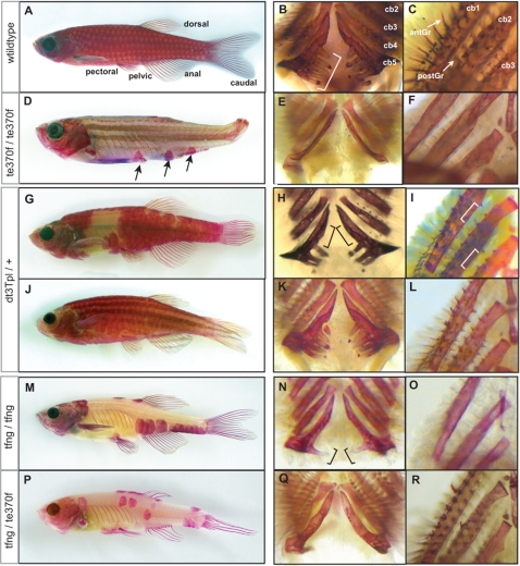
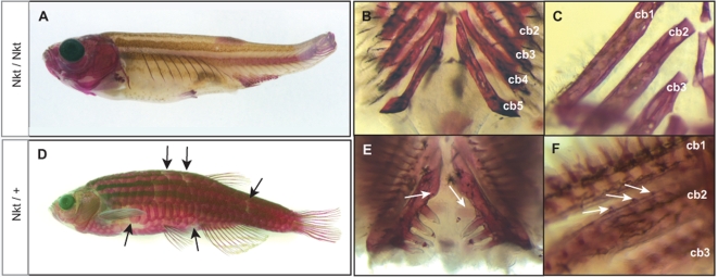
“With the exception of the fls tfng allele, the lepidotrichia that form during juvenile metamorphosis are defective, leading to fin loss in the adult (Figure 1D, 2A).”
§ Zebrafish eda and edar Mutants Reveal Conserved and Ancestral Roles of Ectodysplasin Signaling in Vertebrates > Results > finless and Nackt Mutants Exhibit Defects in the Development of the Dermal Skeleton
plaindirect- Asserted: passes — The authors put it forward as true.
- Checkable: passes — It can be checked against evidence.
- Load-bearing: passes — The paper’s conclusions depend on it.
- Based: passes — The paper gives a reason why it holds.
A result the paper reports from its own data — it cites no other source for it, so there is none to check. The quote shows it is in the paper.
In the paper
“scales are largely absent with infrequent formation of inappropriately shaped scales near the dorsal, anal and pectoral fins (Figure 1D, 2A).”
§ Zebrafish eda and edar Mutants Reveal Conserved and Ancestral Roles of Ectodysplasin Signaling in Vertebrates > Results > finless and Nackt Mutants Exhibit Defects in the Development of the Dermal Skeleton
plain- Asserted: passes — The authors put it forward as true.
- Checkable: passes — It can be checked against evidence.
- Load-bearing: passes — The paper’s conclusions depend on it.
- Based: passes — The paper gives a reason why it holds.
A result the paper reports from its own data — it cites no other source for it, so there is none to check. The quote shows it is in the paper.
In the paper
“Mutant fls dt3Tpl fish exhibit the strong fls phenotype when homozygous or heterozygous with other fls alleles.”
§ Zebrafish eda and edar Mutants Reveal Conserved and Ancestral Roles of Ectodysplasin Signaling in Vertebrates > Results > The Topless Allele of fls Uncovers Background Specific Modulation of fls Expressivity
plaindirect- Asserted: passes — The authors put it forward as true.
- Checkable: passes — It can be checked against evidence.
- Load-bearing: unclear — It is unclear whether the paper’s conclusions depend on it.
- Based: passes — The paper gives a reason why it holds.
A result the paper reports from its own data — it cites no other source for it, so there is none to check. The quote shows it is in the paper.
In the paper
“we identified three mutants that showed nearly identical defects in the formation of scales, lepidotrichia, and shape of the skull of homozygous fish.”
§ Zebrafish eda and edar Mutants Reveal Conserved and Ancestral Roles of Ectodysplasin Signaling in Vertebrates > Results > finless and Nackt Mutants Exhibit Defects in the Development of the Dermal Skeleton
plain- Asserted: passes — The authors put it forward as true.
- Checkable: passes — It can be checked against evidence.
- Load-bearing: passes — The paper’s conclusions depend on it.
- Based: passes — The paper gives a reason why it holds.
A result the paper reports from its own data — it cites no other source for it, so there is none to check. The quote shows it is in the paper.
In the paper
“Similar to Nkt, fls dt3Tpl exhibits a dominant effect on skull shape as well (Figure S1).”
§ Zebrafish eda and edar Mutants Reveal Conserved and Ancestral Roles of Ectodysplasin Signaling in Vertebrates > Results > The Topless Allele of fls Uncovers Background Specific Modulation of fls Expressivity
plaindirect- Asserted: passes — The authors put it forward as true.
- Checkable: passes — It can be checked against evidence.
- Load-bearing: unclear — It is unclear whether the paper’s conclusions depend on it.
- Based: passes — The paper gives a reason why it holds.
A result the paper reports from its own data — it cites no other source for it, so there is none to check. The quote shows it is in the paper.
In the paper
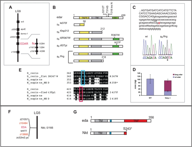
“The fls te370f mutation was linked to SSLP markers on linkage group 9 (LG9).”
§ Zebrafish eda and edar Mutants Reveal Conserved and Ancestral Roles of Ectodysplasin Signaling in Vertebrates > Results > Mutations in fls Disrupt the Ectodysplasin Receptor in Zebrafish
plaincorrelational- Asserted: passes — The authors put it forward as true.
- Checkable: passes — It can be checked against evidence.
- Load-bearing: passes — The paper’s conclusions depend on it.
- Based: passes — The paper gives a reason why it holds.
The paper states this without a source or data behind it here, so there is nothing to check it against. The quote shows it is in the paper.
In the paper
“the expressivity of dominant alleles is sensitive to background modifiers that buffer the phenotypic consequences of loss of Eda signaling.”
§ Zebrafish eda and edar Mutants Reveal Conserved and Ancestral Roles of Ectodysplasin Signaling in Vertebrates > Introduction
plaindirect- Asserted: passes — The authors put it forward as true.
- Checkable: passes — It can be checked against evidence.
- Load-bearing: unclear — It is unclear whether the paper’s conclusions depend on it.
- Based: unclear — It is unclear why the paper says it holds.
A result the paper reports from its own data — it cites no other source for it, so there is none to check. The quote shows it is in the paper.
In the paper
“The eda transcript from the Nkt mutant shows a precocious stop codon predicting a truncation of the protein at the beginning of the TNF domain, which is involved in ligand-receptor binding (S243X(C-A)); Figure 3G and Figure S3).”
§ Zebrafish eda and edar Mutants Reveal Conserved and Ancestral Roles of Ectodysplasin Signaling in Vertebrates > Results > Ectodysplasin Is Mutated in the Nackt Mutant
plaindirectSingle case- Asserted: passes — The authors put it forward as true.
- Checkable: passes — It can be checked against evidence.
- Load-bearing: passes — The paper’s conclusions depend on it.
- Based: passes — The paper gives a reason why it holds.
A result the paper reports from its own data — it cites no other source for it, so there is none to check. The quote shows it is in the paper.
In the paper
“The expressivity of the dominant fls dt3Tpl phenotype depends on the genetic background.”
§ Zebrafish eda and edar Mutants Reveal Conserved and Ancestral Roles of Ectodysplasin Signaling in Vertebrates > Results > The Topless Allele of fls Uncovers Background Specific Modulation of fls Expressivity
plaindirect- Asserted: passes — The authors put it forward as true.
- Checkable: passes — It can be checked against evidence.
- Load-bearing: passes — The paper’s conclusions depend on it.
- Based: passes — The paper gives a reason why it holds.
A result the paper reports from its own data — it cites no other source for it, so there is none to check. The quote shows it is in the paper.
In the paper
“The shape and number of the scales in fang is similar to the other homozygous fls alleles (Table 1).”
§ Zebrafish eda and edar Mutants Reveal Conserved and Ancestral Roles of Ectodysplasin Signaling in Vertebrates > Results > The fang Allele Uncovers Dose and Organ Specific Sensitivity to Levels of Eda Signaling
plain- Asserted: passes — The authors put it forward as true.
- Checkable: passes — It can be checked against evidence.
- Load-bearing: unclear — It is unclear whether the paper’s conclusions depend on it.
- Based: passes — The paper gives a reason why it holds.
A result the paper reports from its own data — it cites no other source for it, so there is none to check. The quote shows it is in the paper.
In the paper
“Fish heterozygous for fls dt3Tpl exhibited either a “strong” or a “weak” phenotype in the Tü background (Figure 1G and J, respectively).”
§ Zebrafish eda and edar Mutants Reveal Conserved and Ancestral Roles of Ectodysplasin Signaling in Vertebrates > Results > The Topless Allele of fls Uncovers Background Specific Modulation of fls Expressivity
plain- Asserted: passes — The authors put it forward as true.
- Checkable: passes — It can be checked against evidence.
- Load-bearing: passes — The paper’s conclusions depend on it.
- Based: passes — The paper gives a reason why it holds.
A result the paper reports from its own data — it cites no other source for it, so there is none to check. The quote shows it is in the paper.
In the paper
“Analysis of the sequence of the novel isoform showed the addition of intronic sequence leading to a premature termination codon (Figure 3C).”
§ Zebrafish eda and edar Mutants Reveal Conserved and Ancestral Roles of Ectodysplasin Signaling in Vertebrates > Results > The fang Allele Uncovers Dose and Organ Specific Sensitivity to Levels of Eda Signaling
plaindirectSingle case- Asserted: passes — The authors put it forward as true.
- Checkable: passes — It can be checked against evidence.
- Load-bearing: unclear — It is unclear whether the paper’s conclusions depend on it.
- Based: passes — The paper gives a reason why it holds.
A result the paper reports from its own data — it cites no other source for it, so there is none to check. The quote shows it is in the paper.
In the paper
“Nkt homozygous fish exhibit more severe defects in the formation of the dermal skeleton than fls alleles in the extent of lepidotrichial growth and number of residual scales formed (compare Figure 1D and Figure 2A).”
§ Zebrafish eda and edar Mutants Reveal Conserved and Ancestral Roles of Ectodysplasin Signaling in Vertebrates > Results > finless and Nackt Mutants Exhibit Defects in the Development of the Dermal Skeleton
plaincorrelational- Asserted: passes — The authors put it forward as true.
- Checkable: passes — It can be checked against evidence.
- Load-bearing: unclear — It is unclear whether the paper’s conclusions depend on it.
- Based: passes — The paper gives a reason why it holds.
A result the paper reports from its own data — it cites no other source for it, so there is none to check. The quote shows it is in the paper.
In the paper
“Analysis of the genomic sequence in the mutant revealed that the altered splicing is due to an A to G transition leading to the creation of a new splice donor site in the intron (Figure 3C).”
§ Zebrafish eda and edar Mutants Reveal Conserved and Ancestral Roles of Ectodysplasin Signaling in Vertebrates > Results > The fang Allele Uncovers Dose and Organ Specific Sensitivity to Levels of Eda Signaling
plaindirectSingle case- Asserted: passes — The authors put it forward as true.
- Checkable: passes — It can be checked against evidence.
- Load-bearing: unclear — It is unclear whether the paper’s conclusions depend on it.
- Based: passes — The paper gives a reason why it holds.
The paper states this without a source or data behind it here, so there is nothing to check it against. The quote shows it is in the paper.
In the paper
“None of the other fls alleles showed any dominance in the Tü, TLF, or WIK strains.”
§ Zebrafish eda and edar Mutants Reveal Conserved and Ancestral Roles of Ectodysplasin Signaling in Vertebrates > Results > The Topless Allele of fls Uncovers Background Specific Modulation of fls Expressivity
plain- Asserted: passes — The authors put it forward as true.
- Checkable: passes — It can be checked against evidence.
- Load-bearing: unclear — It is unclear whether the paper’s conclusions depend on it.
- Based: passes — The paper gives a reason why it holds.
A result the paper reports from its own data — it cites no other source for it, so there is none to check. The quote shows it is in the paper.
In the paper
“flstfng heterozygotes do not show any differences compared to wild type.”
§ Zebrafish eda and edar Mutants Reveal Conserved and Ancestral Roles of Ectodysplasin Signaling in Vertebrates > Results > The fang Allele Uncovers Dose and Organ Specific Sensitivity to Levels of Eda Signaling
plain- Asserted: passes — The authors put it forward as true.
- Checkable: passes — It can be checked against evidence.
- Load-bearing: unclear — It is unclear whether the paper’s conclusions depend on it.
- Based: passes — The paper gives a reason why it holds.
In the paper
“The first is allelic to the finless (fls te370f ) mutant that was previously isolated in the background of the Tübingen wild type stock (Tü) on the basis of the loss of fins in adults [10].”
§ Zebrafish eda and edar Mutants Reveal Conserved and Ancestral Roles of Ectodysplasin Signaling in Vertebrates > Results > finless and Nackt Mutants Exhibit Defects in the Development of the Dermal Skeleton
What the sources say
- Not checked — no full text of the sourcecites [10] P Haffter, 1996, Mutations affecting pigmentation and shape of the adult zebrafish., Dev Genes Evol source
plaincorrelational- Asserted: passes — The authors put it forward as true.
- Checkable: passes — It can be checked against evidence.
- Load-bearing: unclear — It is unclear whether the paper’s conclusions depend on it.
- Based: passes — The paper gives a reason why it holds.
A result the paper reports from its own data — it cites no other source for it, so there is none to check. The quote shows it is in the paper.
In the paper
“The edar gene is located on LG9 within the determined linkage interval for fls (see Methods).”
§ Zebrafish eda and edar Mutants Reveal Conserved and Ancestral Roles of Ectodysplasin Signaling in Vertebrates > Results > Mutations in fls Disrupt the Ectodysplasin Receptor in Zebrafish
plaincorrelational- Asserted: passes — The authors put it forward as true.
- Checkable: passes — It can be checked against evidence.
- Load-bearing: passes — The paper’s conclusions depend on it.
- Based: passes — The paper gives a reason why it holds.
A result the paper reports from its own data — it cites no other source for it, so there is none to check. The quote shows it is in the paper.
In the paper
“The two alleles generated by the ENU mutagen both have missense mutations resulting in amino acid changes in the death domain (fls t3R367W, R367W(C-T); fls dt3Tpl, I428F (A-T)).”
§ Zebrafish eda and edar Mutants Reveal Conserved and Ancestral Roles of Ectodysplasin Signaling in Vertebrates > Results > Mutations in fls Disrupt the Ectodysplasin Receptor in Zebrafish
plain- Asserted: passes — The authors put it forward as true.
- Checkable: passes — It can be checked against evidence.
- Load-bearing: passes — The paper’s conclusions depend on it.
- Based: passes — The paper gives a reason why it holds.
A result the paper reports from its own data — it cites no other source for it, so there is none to check. The quote shows it is in the paper.
In the paper
“These mutants fell into two complementation groups.”
§ Zebrafish eda and edar Mutants Reveal Conserved and Ancestral Roles of Ectodysplasin Signaling in Vertebrates > Results > finless and Nackt Mutants Exhibit Defects in the Development of the Dermal Skeleton
plain- Asserted: passes — The authors put it forward as true.
- Checkable: passes — It can be checked against evidence.
- Load-bearing: unclear — It is unclear whether the paper’s conclusions depend on it.
- Based: passes — The paper gives a reason why it holds.
A result the paper reports from its own data — it cites no other source for it, so there is none to check. The quote shows it is in the paper.
In the paper
“We named this allele fang (fls tfng) after its unique dental phenotype in homozygotes of having only one tooth on the fifth ceratobranchial (Figure 1N).”
§ Zebrafish eda and edar Mutants Reveal Conserved and Ancestral Roles of Ectodysplasin Signaling in Vertebrates > Results > finless and Nackt Mutants Exhibit Defects in the Development of the Dermal Skeleton
plain- Asserted: passes — The authors put it forward as true.
- Checkable: passes — It can be checked against evidence.
- Load-bearing: unclear — It is unclear whether the paper’s conclusions depend on it.
- Based: passes — The paper gives a reason why it holds.
A result the paper reports from its own data — it cites no other source for it, so there is none to check. The quote shows it is in the paper.
In the paper
“Analysis of edar RNA from homozygous fls tfng showed the presence of two distinct transcripts with an additional larger isoform than seen in wildtype.”
§ Zebrafish eda and edar Mutants Reveal Conserved and Ancestral Roles of Ectodysplasin Signaling in Vertebrates > Results > The fang Allele Uncovers Dose and Organ Specific Sensitivity to Levels of Eda Signaling
plainSingle case- Asserted: passes — The authors put it forward as true.
- Checkable: passes — It can be checked against evidence.
- Load-bearing: unclear — It is unclear whether the paper’s conclusions depend on it.
- Based: passes — The paper gives a reason why it holds.
A result the paper reports from its own data — it cites no other source for it, so there is none to check. The quote shows it is in the paper.
In the paper
“The homozygous phenotype is more severe than that of strong fls alleles (Figure 2A).”
§ Zebrafish eda and edar Mutants Reveal Conserved and Ancestral Roles of Ectodysplasin Signaling in Vertebrates > Results > finless and Nackt Mutants Exhibit Defects in the Development of the Dermal Skeleton
plain- Asserted: passes — The authors put it forward as true.
- Checkable: passes — It can be checked against evidence.
- Load-bearing: unclear — It is unclear whether the paper’s conclusions depend on it.
- Based: passes — The paper gives a reason why it holds.
A result the paper reports from its own data — it cites no other source for it, so there is none to check. The quote shows it is in the paper.
In the paper
“Using quantitative real time PCR we found that the fang-specific edar transcript represents 74% of the total pool of edar transcripts in homozygous mutants (Figure 3D).”
§ Zebrafish eda and edar Mutants Reveal Conserved and Ancestral Roles of Ectodysplasin Signaling in Vertebrates > Results > The fang Allele Uncovers Dose and Organ Specific Sensitivity to Levels of Eda Signaling
plain- Asserted: passes — The authors put it forward as true.
- Checkable: passes — It can be checked against evidence.
- Load-bearing: passes — The paper’s conclusions depend on it.
- Based: passes — The paper gives a reason why it holds.
A result the paper reports from its own data — it cites no other source for it, so there is none to check. The quote shows it is in the paper.
In the paper
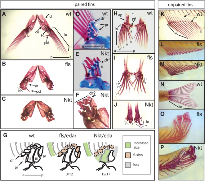
“The dermal bones of the pectoral girdle are present and patterned appropriately in both the fls and Nkt mutants.”
§ Zebrafish eda and edar Mutants Reveal Conserved and Ancestral Roles of Ectodysplasin Signaling in Vertebrates > Results > finless and Nackt Mutants Exhibit Defects in the Development of the Dermal Skeleton
plain- Asserted: passes — The authors put it forward as true.
- Checkable: passes — It can be checked against evidence.
- Load-bearing: unclear — It is unclear whether the paper’s conclusions depend on it.
- Based: passes — The paper gives a reason why it holds.
A result the paper reports from its own data — it cites no other source for it, so there is none to check. The quote shows it is in the paper.
In the paper
“An analysis of the location of eda in the zebrafish radiation hybrid map placed eda on LG5.”
§ Zebrafish eda and edar Mutants Reveal Conserved and Ancestral Roles of Ectodysplasin Signaling in Vertebrates > Results > Ectodysplasin Is Mutated in the Nackt Mutant
plain- Asserted: passes — The authors put it forward as true.
- Checkable: passes — It can be checked against evidence.
- Load-bearing: unclear — It is unclear whether the paper’s conclusions depend on it.
- Based: passes — The paper gives a reason why it holds.
A result the paper reports from its own data — it cites no other source for it, so there is none to check. The quote shows it is in the paper.
In the paper
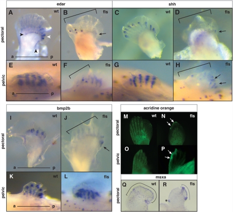
“In early fin fold stage of pectoral fin development (8 mm), we detected edar expression in both the distal margin of the endochondrial radials (black arrowhead, Figure 7A) as well as more distally in the forming lepidotrichial rays (Figure 7A).”
§ Zebrafish eda and edar Mutants Reveal Conserved and Ancestral Roles of Ectodysplasin Signaling in Vertebrates > Results > Role of Ectodysplasin Signaling in Regulating Epithelial Signaling Centers:Maintenance of the Fin Fold and Establishing Anterior-Posterior Polarity of the Developing Fin
plain- Asserted: passes — The authors put it forward as true.
- Checkable: passes — It can be checked against evidence.
- Load-bearing: unclear — It is unclear whether the paper’s conclusions depend on it.
- Based: passes — The paper gives a reason why it holds.
A result the paper reports from its own data — it cites no other source for it, so there is none to check. The quote shows it is in the paper.
In the paper
“However, residual expression of all three genes was found in the posterior margin of the fin (Figure 7B, D, J arrow).”
§ Zebrafish eda and edar Mutants Reveal Conserved and Ancestral Roles of Ectodysplasin Signaling in Vertebrates > Results > Role of Ectodysplasin Signaling in Regulating Epithelial Signaling Centers:Maintenance of the Fin Fold and Establishing Anterior-Posterior Polarity of the Developing Fin
plain- Asserted: passes — The authors put it forward as true.
- Checkable: passes — It can be checked against evidence.
- Load-bearing: unclear — It is unclear whether the paper’s conclusions depend on it.
- Based: passes — The paper gives a reason why it holds.
A result the paper reports from its own data — it cites no other source for it, so there is none to check. The quote shows it is in the paper.
In the paper
“In fls adults, fusions of the distal radials of the pectoral fin are seen at a low penetrance (Figure 6G, data not shown).”
§ Zebrafish eda and edar Mutants Reveal Conserved and Ancestral Roles of Ectodysplasin Signaling in Vertebrates > Results > Role of Ectodysplasin Signaling in Regulating Epithelial Signaling Centers:Maintenance of the Fin Fold and Establishing Anterior-Posterior Polarity of the Developing Fin
plain- Asserted: passes — The authors put it forward as true.
- Checkable: passes — It can be checked against evidence.
- Load-bearing: unclear — It is unclear whether the paper’s conclusions depend on it.
- Based: passes — The paper gives a reason why it holds.
A result the paper reports from its own data — it cites no other source for it, so there is none to check. The quote shows it is in the paper.
In the paper
“The pelvic fin showed similar expression of edar in forming lepidotrichial rays (Figure 7E).”
§ Zebrafish eda and edar Mutants Reveal Conserved and Ancestral Roles of Ectodysplasin Signaling in Vertebrates > Results > Role of Ectodysplasin Signaling in Regulating Epithelial Signaling Centers:Maintenance of the Fin Fold and Establishing Anterior-Posterior Polarity of the Developing Fin
plain- Asserted: passes — The authors put it forward as true.
- Checkable: passes — It can be checked against evidence.
- Load-bearing: unclear — It is unclear whether the paper’s conclusions depend on it.
- Based: passes — The paper gives a reason why it holds.
A result the paper reports from its own data — it cites no other source for it, so there is none to check. The quote shows it is in the paper.
In the paper
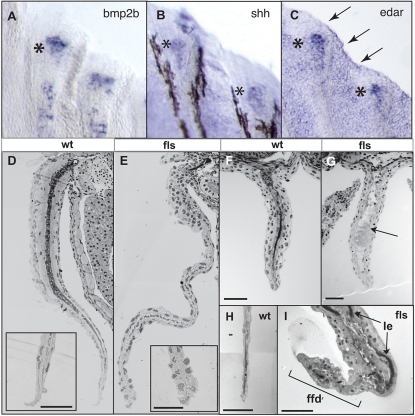
“The expression of edar during late fin development was observed in forming lepidotrichia of all fins with a distal bias in its expression (Figure 8C).”
§ Zebrafish eda and edar Mutants Reveal Conserved and Ancestral Roles of Ectodysplasin Signaling in Vertebrates > Results > Role of Ectodysplasin Signaling in Regulating Epithelial Signaling Centers:Maintenance of the Fin Fold and Establishing Anterior-Posterior Polarity of the Developing Fin
plain- Asserted: passes — The authors put it forward as true.
- Checkable: passes — It can be checked against evidence.
- Load-bearing: unclear — It is unclear whether the paper’s conclusions depend on it.
- Based: passes — The paper gives a reason why it holds.
A result the paper reports from its own data — it cites no other source for it, so there is none to check. The quote shows it is in the paper.
In the paper
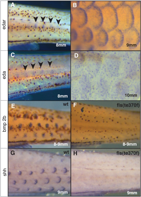
“Developmental genes shh and bone morphogenic protein 2b (bmp2b), whose orthologues are known to be essential for placode development in the mouse and chick, show similar placodal expression as seen with edar (Figure 4E and G).”
§ Zebrafish eda and edar Mutants Reveal Conserved and Ancestral Roles of Ectodysplasin Signaling in Vertebrates > Results > Role of Ectodysplasin Signaling in Regulating Epithelial Signaling Centers: Scale Placode Formation
plaincorrelational- Asserted: passes — The authors put it forward as true.
- Checkable: passes — It can be checked against evidence.
- Load-bearing: passes — The paper’s conclusions depend on it.
- Based: passes — The paper gives a reason why it holds.
A result the paper reports from its own data — it cites no other source for it, so there is none to check. The quote shows it is in the paper.
In the paper
“Consistent with these data suggesting asymmetrical loss of the fin fold epidermis, we find that msxa, a marker of the distal epithelium, is differentially expressed in the mutant (Figure 7Q and R).”
§ Zebrafish eda and edar Mutants Reveal Conserved and Ancestral Roles of Ectodysplasin Signaling in Vertebrates > Results > Role of Ectodysplasin Signaling in Regulating Epithelial Signaling Centers:Maintenance of the Fin Fold and Establishing Anterior-Posterior Polarity of the Developing Fin
plaincorrelational- Asserted: passes — The authors put it forward as true.
- Checkable: passes — It can be checked against evidence.
- Load-bearing: passes — The paper’s conclusions depend on it.
- Based: passes — The paper gives a reason why it holds.
- Rests on an assumption: msxa is a marker of the distal epithelium.
A result the paper reports from its own data — it cites no other source for it, so there is none to check. The quote shows it is in the paper.
In the paper
“shh and bmp2b expression was observed in the forming lepidotrichia of both the pectoral and pelvic fins of wild type juveniles, having a similar distal bias in the leading margin of all rays (Figure 7C, G and I, K, respectively).”
§ Zebrafish eda and edar Mutants Reveal Conserved and Ancestral Roles of Ectodysplasin Signaling in Vertebrates > Results > Role of Ectodysplasin Signaling in Regulating Epithelial Signaling Centers:Maintenance of the Fin Fold and Establishing Anterior-Posterior Polarity of the Developing Fin
plain- Asserted: passes — The authors put it forward as true.
- Checkable: passes — It can be checked against evidence.
- Load-bearing: unclear — It is unclear whether the paper’s conclusions depend on it.
- Based: passes — The paper gives a reason why it holds.
A result the paper reports from its own data — it cites no other source for it, so there is none to check. The quote shows it is in the paper.
In the paper
“Histological analysis of fls mutant fins at an early stage of lepidotrichial formation reveal a general deficiency of the development of the entire mesodermal component of the fin such as cartilage and muscle in both the paired (pectoral fin, Figure 8D, E) and unpaired fins (anal and caudal fins, Figure 8F–G; H–I, respectively).”
§ Zebrafish eda and edar Mutants Reveal Conserved and Ancestral Roles of Ectodysplasin Signaling in Vertebrates > Results > Role of Ectodysplasin Signaling in Regulating Epithelial Signaling Centers:Maintenance of the Fin Fold and Establishing Anterior-Posterior Polarity of the Developing Fin
plaindirect- Asserted: passes — The authors put it forward as true.
- Checkable: passes — It can be checked against evidence.
- Load-bearing: passes — The paper’s conclusions depend on it.
- Based: passes — The paper gives a reason why it holds.
A result the paper reports from its own data — it cites no other source for it, so there is none to check. The quote shows it is in the paper.
In the paper
“However, close inspection of the distal tip of the fins showed disorganization of the epidermis and degeneration of distal epidermal nuclei (insets Figure 8F, J).”
§ Zebrafish eda and edar Mutants Reveal Conserved and Ancestral Roles of Ectodysplasin Signaling in Vertebrates > Results > Role of Ectodysplasin Signaling in Regulating Epithelial Signaling Centers:Maintenance of the Fin Fold and Establishing Anterior-Posterior Polarity of the Developing Fin
plain- Asserted: passes — The authors put it forward as true.
- Checkable: passes — It can be checked against evidence.
- Load-bearing: passes — The paper’s conclusions depend on it.
- Based: passes — The paper gives a reason why it holds.
A result the paper reports from its own data — it cites no other source for it, so there is none to check. The quote shows it is in the paper.
In the paper
“Similarly, in the pelvic fin of 8–9 mm juveniles, retention of the label was seen in the anterior distal margin of the fin (Figure 7O–P).”
§ Zebrafish eda and edar Mutants Reveal Conserved and Ancestral Roles of Ectodysplasin Signaling in Vertebrates > Results > Role of Ectodysplasin Signaling in Regulating Epithelial Signaling Centers:Maintenance of the Fin Fold and Establishing Anterior-Posterior Polarity of the Developing Fin
plain- Asserted: passes — The authors put it forward as true.
- Checkable: passes — It can be checked against evidence.
- Load-bearing: passes — The paper’s conclusions depend on it.
- Based: passes — The paper gives a reason why it holds.
A result the paper reports from its own data — it cites no other source for it, so there is none to check. The quote shows it is in the paper.
In the paper
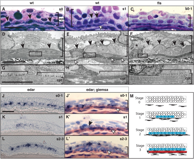
“In contrast, the adjacent lateral epidermal cells show high levels of both ER and hemidesmosomes (data not shown and Figure 5G brackets, respectively).”
§ Zebrafish eda and edar Mutants Reveal Conserved and Ancestral Roles of Ectodysplasin Signaling in Vertebrates > Results > Role of Ectodysplasin Signaling in Regulating Epithelial Signaling Centers: Scale Placode Formation
plain- Asserted: passes — The authors put it forward as true.
- Checkable: passes — It can be checked against evidence.
- Load-bearing: unclear — It is unclear whether the paper’s conclusions depend on it.
- Based: passes — The paper gives a reason why it holds.
A result the paper reports from its own data — it cites no other source for it, so there is none to check. The quote shows it is in the paper.
In the paper
“The expression of edar is seen in the basal cells of forming scale placodes (Figure 5J–L; stages 1–3 Figure 5M) arising during early specification of the scale placode (s1; arrowhead Figure 5K).”
§ Zebrafish eda and edar Mutants Reveal Conserved and Ancestral Roles of Ectodysplasin Signaling in Vertebrates > Results > Role of Ectodysplasin Signaling in Regulating Epithelial Signaling Centers: Scale Placode Formation
plaincorrelational- Asserted: passes — The authors put it forward as true.
- Checkable: passes — It can be checked against evidence.
- Load-bearing: unclear — It is unclear whether the paper’s conclusions depend on it.
- Based: passes — The paper gives a reason why it holds.
A result the paper reports from its own data — it cites no other source for it, so there is none to check. The quote shows it is in the paper.
In the paper
“In Nkt, there is an increase in the frequency of patterning and growth defects of the endochondrial components of the fin (Figure 6G).”
§ Zebrafish eda and edar Mutants Reveal Conserved and Ancestral Roles of Ectodysplasin Signaling in Vertebrates > Results > Role of Ectodysplasin Signaling in Regulating Epithelial Signaling Centers:Maintenance of the Fin Fold and Establishing Anterior-Posterior Polarity of the Developing Fin
plaindirect- Asserted: passes — The authors put it forward as true.
- Checkable: passes — It can be checked against evidence.
- Load-bearing: unclear — It is unclear whether the paper’s conclusions depend on it.
- Based: passes — The paper gives a reason why it holds.
A result the paper reports from its own data — it cites no other source for it, so there is none to check. The quote shows it is in the paper.
In the paper
“Nkt causes a strong effect on lepidotrichial growth of both the pectoral and pelvic fins (Figure 6E–F, J).”
§ Zebrafish eda and edar Mutants Reveal Conserved and Ancestral Roles of Ectodysplasin Signaling in Vertebrates > Results > Role of Ectodysplasin Signaling in Regulating Epithelial Signaling Centers:Maintenance of the Fin Fold and Establishing Anterior-Posterior Polarity of the Developing Fin
boosteddirect- Asserted: passes — The authors put it forward as true.
- Checkable: passes — It can be checked against evidence.
- Load-bearing: unclear — It is unclear whether the paper’s conclusions depend on it.
- Based: passes — The paper gives a reason why it holds.
A result the paper reports from its own data — it cites no other source for it, so there is none to check. The quote shows it is in the paper.
In the paper
“We find a specific alteration in the morphology of the basal epidermal cells in wild type juvenile fish that coincides with the initial accumulation of fibroblasts at the sites of scale development (Figure 5B, E).”
§ Zebrafish eda and edar Mutants Reveal Conserved and Ancestral Roles of Ectodysplasin Signaling in Vertebrates > Results > Role of Ectodysplasin Signaling in Regulating Epithelial Signaling Centers: Scale Placode Formation
plaincorrelational- Asserted: passes — The authors put it forward as true.
- Checkable: passes — It can be checked against evidence.
- Load-bearing: passes — The paper’s conclusions depend on it.
- Based: passes — The paper gives a reason why it holds.
A result the paper reports from its own data — it cites no other source for it, so there is none to check. The quote shows it is in the paper.
In the paper
“Expression of all three genes in fls was decreased in the anterior portion of the pectoral fins (Figure. 7B, D, J).”
§ Zebrafish eda and edar Mutants Reveal Conserved and Ancestral Roles of Ectodysplasin Signaling in Vertebrates > Results > Role of Ectodysplasin Signaling in Regulating Epithelial Signaling Centers:Maintenance of the Fin Fold and Establishing Anterior-Posterior Polarity of the Developing Fin
plaindirect- Asserted: passes — The authors put it forward as true.
- Checkable: passes — It can be checked against evidence.
- Load-bearing: passes — The paper’s conclusions depend on it.
- Based: passes — The paper gives a reason why it holds.
A result the paper reports from its own data — it cites no other source for it, so there is none to check. The quote shows it is in the paper.
In the paper
“We did not detect any difference in bmp2b expression in the pelvic fins even though obvious morphological differences in the developing rays of the samples could be seen (Figure 7L).”
§ Zebrafish eda and edar Mutants Reveal Conserved and Ancestral Roles of Ectodysplasin Signaling in Vertebrates > Results > Role of Ectodysplasin Signaling in Regulating Epithelial Signaling Centers:Maintenance of the Fin Fold and Establishing Anterior-Posterior Polarity of the Developing Fin
plain- Asserted: passes — The authors put it forward as true.
- Checkable: passes — It can be checked against evidence.
- Load-bearing: unclear — It is unclear whether the paper’s conclusions depend on it.
- Based: passes — The paper gives a reason why it holds.
The paper states this without a source or data behind it here, so there is nothing to check it against. The quote shows it is in the paper.
In the paper
“The expression of edar is again associated with that of shh and bmp2b.”
§ Zebrafish eda and edar Mutants Reveal Conserved and Ancestral Roles of Ectodysplasin Signaling in Vertebrates > Discussion > Conserved and Ancestral Role of Eda Signaling in Vertebrate Development
plaincorrelational- Asserted: passes — The authors put it forward as true.
- Checkable: passes — It can be checked against evidence.
- Load-bearing: passes — The paper’s conclusions depend on it.
- Based: passes — The paper gives a reason why it holds.
A result the paper reports from its own data — it cites no other source for it, so there is none to check. The quote shows it is in the paper.
In the paper
“In 7 mm juveniles, we detect a differential retention of acridine orange between fls and siblings in the anterior, and anterior-distal margin of the developing pectoral fin (Figure 7M–N).”
§ Zebrafish eda and edar Mutants Reveal Conserved and Ancestral Roles of Ectodysplasin Signaling in Vertebrates > Results > Role of Ectodysplasin Signaling in Regulating Epithelial Signaling Centers:Maintenance of the Fin Fold and Establishing Anterior-Posterior Polarity of the Developing Fin
plain- Asserted: passes — The authors put it forward as true.
- Checkable: passes — It can be checked against evidence.
- Load-bearing: passes — The paper’s conclusions depend on it.
- Based: passes — The paper gives a reason why it holds.
A result the paper reports from its own data — it cites no other source for it, so there is none to check. The quote shows it is in the paper.
In the paper
“The expression in the forming dermal ray was similar in both location and timing to that of bmp2b and shh (Figure 8A–B, asterisk).”
§ Zebrafish eda and edar Mutants Reveal Conserved and Ancestral Roles of Ectodysplasin Signaling in Vertebrates > Results > Role of Ectodysplasin Signaling in Regulating Epithelial Signaling Centers:Maintenance of the Fin Fold and Establishing Anterior-Posterior Polarity of the Developing Fin
plaincorrelational- Asserted: passes — The authors put it forward as true.
- Checkable: passes — It can be checked against evidence.
- Load-bearing: passes — The paper’s conclusions depend on it.
- Based: passes — The paper gives a reason why it holds.
A result the paper reports from its own data — it cites no other source for it, so there is none to check. The quote shows it is in the paper.
In the paper
“In fls te370f juvenile fish, at a corresponding site on the flank as in wild type, we detected the formation of similar aggregations of basal epidermal cells (black arrows. Figure 5C, F).”
§ Zebrafish eda and edar Mutants Reveal Conserved and Ancestral Roles of Ectodysplasin Signaling in Vertebrates > Results > Role of Ectodysplasin Signaling in Regulating Epithelial Signaling Centers: Scale Placode Formation
plain- Asserted: passes — The authors put it forward as true.
- Checkable: passes — It can be checked against evidence.
- Load-bearing: passes — The paper’s conclusions depend on it.
- Based: passes — The paper gives a reason why it holds.
A result the paper reports from its own data — it cites no other source for it, so there is none to check. The quote shows it is in the paper.
In the paper
“We were unable to resolve a clear signal for eda in the forming fins using WMISH.”
§ Zebrafish eda and edar Mutants Reveal Conserved and Ancestral Roles of Ectodysplasin Signaling in Vertebrates > Results > Role of Ectodysplasin Signaling in Regulating Epithelial Signaling Centers:Maintenance of the Fin Fold and Establishing Anterior-Posterior Polarity of the Developing Fin
plain- Asserted: passes — The authors put it forward as true.
- Checkable: passes — It can be checked against evidence.
- Load-bearing: unclear — It is unclear whether the paper’s conclusions depend on it.
- Based: passes — The paper gives a reason why it holds.
A result the paper reports from its own data — it cites no other source for it, so there is none to check. The quote shows it is in the paper.
In the paper
“In contrast, a specific effect on the growth of anterior lepidotrichia of the pelvic fin is seen in fls where the dermal rays of the anterior (e.g. 1,2) are significantly shorter than rays at equivalent positions in wild type (Figure 6I).”
§ Zebrafish eda and edar Mutants Reveal Conserved and Ancestral Roles of Ectodysplasin Signaling in Vertebrates > Results > Role of Ectodysplasin Signaling in Regulating Epithelial Signaling Centers:Maintenance of the Fin Fold and Establishing Anterior-Posterior Polarity of the Developing Fin
plaindirect- Asserted: passes — The authors put it forward as true.
- Checkable: passes — It can be checked against evidence.
- Load-bearing: unclear — It is unclear whether the paper’s conclusions depend on it.
- Based: passes — The paper gives a reason why it holds.
A result the paper reports from its own data — it cites no other source for it, so there is none to check. The quote shows it is in the paper.
In the paper
“The expression of edar in the fin fold had a posterior bias in wild type fins (Figure 7A, white arrow).”
§ Zebrafish eda and edar Mutants Reveal Conserved and Ancestral Roles of Ectodysplasin Signaling in Vertebrates > Results > Role of Ectodysplasin Signaling in Regulating Epithelial Signaling Centers:Maintenance of the Fin Fold and Establishing Anterior-Posterior Polarity of the Developing Fin
plain- Asserted: passes — The authors put it forward as true.
- Checkable: passes — It can be checked against evidence.
- Load-bearing: passes — The paper’s conclusions depend on it.
- Based: passes — The paper gives a reason why it holds.
In the paper
“The expression of both genes presaged the formation of the initial scale row along the flank just ventral from the midline mysoseptum in the caudal peduncle (arrowheads, Figure 4A and C; [24]).”
§ Zebrafish eda and edar Mutants Reveal Conserved and Ancestral Roles of Ectodysplasin Signaling in Vertebrates > Results > Role of Ectodysplasin Signaling in Regulating Epithelial Signaling Centers: Scale Placode Formation
What the sources say
- Not checked — no full text of the sourcecites [24] JY Sire, 1997, Scale development in zebrafish (Danio rerio)., J Anat source
plaincorrelational- Asserted: passes — The authors put it forward as true.
- Checkable: passes — It can be checked against evidence.
- Load-bearing: passes — The paper’s conclusions depend on it.
- Based: passes — The paper gives a reason why it holds.
A result the paper reports from its own data — it cites no other source for it, so there is none to check. The quote shows it is in the paper.
In the paper
“The growth and patterning of lepidotrichia are affected in all fins.”
§ Zebrafish eda and edar Mutants Reveal Conserved and Ancestral Roles of Ectodysplasin Signaling in Vertebrates > Results > Role of Ectodysplasin Signaling in Regulating Epithelial Signaling Centers:Maintenance of the Fin Fold and Establishing Anterior-Posterior Polarity of the Developing Fin
plaindirect- Asserted: passes — The authors put it forward as true.
- Checkable: passes — It can be checked against evidence.
- Load-bearing: passes — The paper’s conclusions depend on it.
- Based: passes — The paper gives a reason why it holds.
A result the paper reports from its own data — it cites no other source for it, so there is none to check. The quote shows it is in the paper.
In the paper
“Unpaired fins showed no defects in patterning of the endochondrial bones of the proximal and distal radials (Figure 6K–P);”
§ Zebrafish eda and edar Mutants Reveal Conserved and Ancestral Roles of Ectodysplasin Signaling in Vertebrates > Results > Role of Ectodysplasin Signaling in Regulating Epithelial Signaling Centers:Maintenance of the Fin Fold and Establishing Anterior-Posterior Polarity of the Developing Fin
plain- Asserted: passes — The authors put it forward as true.
- Checkable: passes — It can be checked against evidence.
- Load-bearing: unclear — It is unclear whether the paper’s conclusions depend on it.
- Based: passes — The paper gives a reason why it holds.
A result the paper reports from its own data — it cites no other source for it, so there is none to check. The quote shows it is in the paper.
In the paper
“This is observed above the localized accumulation of fibroblasts in the dermis (white arrows, Figure 5B, E).”
§ Zebrafish eda and edar Mutants Reveal Conserved and Ancestral Roles of Ectodysplasin Signaling in Vertebrates > Results > Role of Ectodysplasin Signaling in Regulating Epithelial Signaling Centers: Scale Placode Formation
plaincorrelational- Asserted: passes — The authors put it forward as true.
- Checkable: passes — It can be checked against evidence.
- Load-bearing: unclear — It is unclear whether the paper’s conclusions depend on it.
- Based: passes — The paper gives a reason why it holds.
A result the paper reports from its own data — it cites no other source for it, so there is none to check. The quote shows it is in the paper.
In the paper
“To our surprise, in wild type juvenile fish, we discovered the formation of histologically defined, localized assemblies of cells of the basal epidermis that resemble early stages of the formation of hair and feather placodes.”
§ Zebrafish eda and edar Mutants Reveal Conserved and Ancestral Roles of Ectodysplasin Signaling in Vertebrates > Results > Role of Ectodysplasin Signaling in Regulating Epithelial Signaling Centers: Scale Placode Formation
plain- Asserted: passes — The authors put it forward as true.
- Checkable: passes — It can be checked against evidence.
- Load-bearing: passes — The paper’s conclusions depend on it.
- Based: passes — The paper gives a reason why it holds.
A result the paper reports from its own data — it cites no other source for it, so there is none to check. The quote shows it is in the paper.
In the paper
“In the pelvic fins, similar loss of anterior expression of edar (Figure 7F) and shh (Figure 7H) was seen in the fls mutant.”
§ Zebrafish eda and edar Mutants Reveal Conserved and Ancestral Roles of Ectodysplasin Signaling in Vertebrates > Results > Role of Ectodysplasin Signaling in Regulating Epithelial Signaling Centers:Maintenance of the Fin Fold and Establishing Anterior-Posterior Polarity of the Developing Fin
plain- Asserted: passes — The authors put it forward as true.
- Checkable: passes — It can be checked against evidence.
- Load-bearing: passes — The paper’s conclusions depend on it.
- Based: passes — The paper gives a reason why it holds.
The paper states this without a source or data behind it here, so there is nothing to check it against. The quote shows it is in the paper.
In the paper
“Interestingly, the fls dt3Tpl zebrafish mutant displays similar dominance and variation as the human allele affecting the same residue.”
§ Zebrafish eda and edar Mutants Reveal Conserved and Ancestral Roles of Ectodysplasin Signaling in Vertebrates > Discussion > Genetic Commonalities of Eda Signaling: From Fish to Man
plaincorrelationalSingle case- Asserted: passes — The authors put it forward as true.
- Checkable: passes — It can be checked against evidence.
- Load-bearing: passes — The paper’s conclusions depend on it.
- Based: passes — The paper gives a reason why it holds.
A result the paper reports from its own data — it cites no other source for it, so there is none to check. The quote shows it is in the paper.
In the paper
“In addition, edar was found expressed proximally between forming rays and at the distal margin (Figure 8C arrow).”
§ Zebrafish eda and edar Mutants Reveal Conserved and Ancestral Roles of Ectodysplasin Signaling in Vertebrates > Results > Role of Ectodysplasin Signaling in Regulating Epithelial Signaling Centers:Maintenance of the Fin Fold and Establishing Anterior-Posterior Polarity of the Developing Fin
plain- Asserted: passes — The authors put it forward as true.
- Checkable: passes — It can be checked against evidence.
- Load-bearing: unclear — It is unclear whether the paper’s conclusions depend on it.
- Based: passes — The paper gives a reason why it holds.
A result the paper reports from its own data — it cites no other source for it, so there is none to check. The quote shows it is in the paper.
In the paper
“Similarly, the fang allele of fls clearly demonstrates this dose sensitivity as functional copies of Edar are titrated by the concomitant use of a new splice site in the mutant leading to the reduction in the amount of wild type transcript made (Figure 3D).”
§ Zebrafish eda and edar Mutants Reveal Conserved and Ancestral Roles of Ectodysplasin Signaling in Vertebrates > Discussion > Eda Signaling and the Development and Variation of Adult Form
boosteddirectSingle case- Asserted: passes — The authors put it forward as true.
- Checkable: passes — It can be checked against evidence.
- Load-bearing: passes — The paper’s conclusions depend on it.
- Based: passes — The paper gives a reason why it holds.
A result the paper reports from its own data — it cites no other source for it, so there is none to check. The quote shows it is in the paper.
In the paper
“Results from radiation hybrid screening indicated linkage of zebrafish edar to markers positioned on LG9 in the region predicted by initial mapping analysis.”
§ Zebrafish eda and edar Mutants Reveal Conserved and Ancestral Roles of Ectodysplasin Signaling in Vertebrates > Discussion > Mapping
hedgedcorrelational- Asserted: passes — The authors put it forward as true.
- Checkable: passes — It can be checked against evidence.
- Load-bearing: unclear — It is unclear whether the paper’s conclusions depend on it.
- Based: passes — The paper gives a reason why it holds.
In the paper
“Rough mapping of F2 progeny against a reference panel of SSLP markers [63] indicated that fls was located on linkage group (chromosome) 9 (LG9) with loose linkage to z20031 (61.3cR; Figure 3A).”
§ Zebrafish eda and edar Mutants Reveal Conserved and Ancestral Roles of Ectodysplasin Signaling in Vertebrates > Discussion > Mapping
What the sources say
- Not checked — no full text of the sourcecites [63] R Geisler, 2002, Mapping and Cloning. source
hedgedcorrelational- Asserted: passes — The authors put it forward as true.
- Checkable: passes — It can be checked against evidence.
- Load-bearing: unclear — It is unclear whether the paper’s conclusions depend on it.
- Based: passes — The paper gives a reason why it holds.
A result the paper reports from its own data — it cites no other source for it, so there is none to check. The quote shows it is in the paper.
In the paper
“In our analysis of the medaka rs-3/edar mutant, we find a high degree of variability in the extent of scale formation (Figure S4) suggesting the existence of background modifiers of Edar function in this species.”
§ Zebrafish eda and edar Mutants Reveal Conserved and Ancestral Roles of Ectodysplasin Signaling in Vertebrates > Discussion > Eda Signaling and the Development and Variation of Adult Form
hedgedcorrelational- Asserted: passes — The authors put it forward as true.
- Checkable: passes — It can be checked against evidence.
- Load-bearing: unclear — It is unclear whether the paper’s conclusions depend on it.
- Based: passes — The paper gives a reason why it holds.
A result the paper reports from its own data — it cites no other source for it, so there is none to check. The quote shows it is in the paper.
In the paper
“We found fls to be closely linked to markers z7001 and z11672.”
§ Zebrafish eda and edar Mutants Reveal Conserved and Ancestral Roles of Ectodysplasin Signaling in Vertebrates > Discussion > Mapping
plaincorrelational- Asserted: passes — The authors put it forward as true.
- Checkable: passes — It can be checked against evidence.
- Load-bearing: unclear — It is unclear whether the paper’s conclusions depend on it.
- Based: passes — The paper gives a reason why it holds.
A result the paper reports from its own data — it cites no other source for it, so there is none to check. The quote shows it is in the paper.
In the paper
“Supporting this finding, we observed significant modification of expressivity of fls dt3Tpl in different genetic backgrounds indicating the existence of genetic modifiers of Eda signaling.”
§ Zebrafish eda and edar Mutants Reveal Conserved and Ancestral Roles of Ectodysplasin Signaling in Vertebrates > Discussion > Eda Signaling and the Development and Variation of Adult Form
hedgeddirect- Asserted: passes — The authors put it forward as true.
- Checkable: passes — It can be checked against evidence.
- Load-bearing: passes — The paper’s conclusions depend on it.
- Based: passes — The paper gives a reason why it holds.
A result the paper reports from its own data — it cites no other source for it, so there is none to check. The quote shows it is in the paper.
In the paper
“Analysis of flanking markers and internal polymorphisms in edar showed tight linkage of the fls mutation to the edar gene.”
§ Zebrafish eda and edar Mutants Reveal Conserved and Ancestral Roles of Ectodysplasin Signaling in Vertebrates > Discussion > Mapping
plaincorrelational- Asserted: passes — The authors put it forward as true.
- Checkable: passes — It can be checked against evidence.
- Load-bearing: passes — The paper’s conclusions depend on it.
- Based: passes — The paper gives a reason why it holds.
A conclusion the paper draws from its own results — it cites no other source for it, so there is none to check. The quote shows it is in the paper.
In the paper
“The predicted protein would be similar to the fls t0sp212 allele having truncation just after the transmembrane domain at amino acid 218 (Figure 3B and Figure S2).”
§ Zebrafish eda and edar Mutants Reveal Conserved and Ancestral Roles of Ectodysplasin Signaling in Vertebrates > Results > The fang Allele Uncovers Dose and Organ Specific Sensitivity to Levels of Eda Signaling
hedgedSingle case- Asserted: passes — The authors put it forward as true.
- Checkable: passes — It can be checked against evidence.
- Load-bearing: unclear — It is unclear whether the paper’s conclusions depend on it.
- Based: passes — The paper gives a reason why it holds.
A conclusion the paper draws from its own results — it cites no other source for it, so there is none to check. The quote shows it is in the paper.
In the paper
“we show substantial buffering of the effect of loss of edar function in different genetic backgrounds, suggesting canalization of this developmental system.”
§ Zebrafish eda and edar Mutants Reveal Conserved and Ancestral Roles of Ectodysplasin Signaling in Vertebrates > Abstract
hedgedcorrelational- Asserted: passes — The authors put it forward as true.
- Checkable: passes — It can be checked against evidence.
- Load-bearing: passes — The paper’s conclusions depend on it.
- Based: passes — The paper gives a reason why it holds.
A conclusion the paper draws from its own results — it cites no other source for it, so there is none to check. The quote shows it is in the paper.
In the paper
“Our genetic analysis demonstrates dose-sensitive and organ-specific response to alteration in levels of Eda signaling.”
§ Zebrafish eda and edar Mutants Reveal Conserved and Ancestral Roles of Ectodysplasin Signaling in Vertebrates > Abstract
boosteddirect- Asserted: passes — The authors put it forward as true.
- Checkable: passes — It can be checked against evidence.
- Load-bearing: passes — The paper’s conclusions depend on it.
- Based: passes — The paper gives a reason why it holds.
A conclusion the paper draws from its own results — it cites no other source for it, so there is none to check. The quote shows it is in the paper.
In the paper
“show conservation of the developmental mechanisms involved in the formation and variation of both integumentary appendages and limbs.”
§ Zebrafish eda and edar Mutants Reveal Conserved and Ancestral Roles of Ectodysplasin Signaling in Vertebrates > Abstract
plaincorrelational- Asserted: passes — The authors put it forward as true.
- Checkable: passes — It can be checked against evidence.
- Load-bearing: passes — The paper’s conclusions depend on it.
- Based: passes — The paper gives a reason why it holds.
A conclusion the paper draws from its own results — it cites no other source for it, so there is none to check. The quote shows it is in the paper.
In the paper
“Given the presence of both isoforms in homozygous individuals, this novel splice site is used in addition to the normal splice junction.”
§ Zebrafish eda and edar Mutants Reveal Conserved and Ancestral Roles of Ectodysplasin Signaling in Vertebrates > Results > The fang Allele Uncovers Dose and Organ Specific Sensitivity to Levels of Eda Signaling
plaindirectSingle case- Asserted: passes — The authors put it forward as true.
- Checkable: passes — It can be checked against evidence.
- Load-bearing: unclear — It is unclear whether the paper’s conclusions depend on it.
- Based: passes — The paper gives a reason why it holds.
A conclusion the paper draws from its own results — it cites no other source for it, so there is none to check. The quote shows it is in the paper.
In the paper
“The dilution of wild type transcripts can explain the observed hypomorphic effect of the allele.”
§ Zebrafish eda and edar Mutants Reveal Conserved and Ancestral Roles of Ectodysplasin Signaling in Vertebrates > Results > The fang Allele Uncovers Dose and Organ Specific Sensitivity to Levels of Eda Signaling
plaindirectSingle case- Asserted: passes — The authors put it forward as true.
- Checkable: passes — It can be checked against evidence.
- Load-bearing: passes — The paper’s conclusions depend on it.
- Based: passes — The paper gives a reason why it holds.
A conclusion the paper draws from its own results — it cites no other source for it, so there is none to check. The quote shows it is in the paper.
In the paper
“We describe the requirement of Eda signaling in the zebrafish epidermis for the formation of a structure resembling an epidermal placode seen in the early development of other vertebrate integumentary appendages.”
§ Zebrafish eda and edar Mutants Reveal Conserved and Ancestral Roles of Ectodysplasin Signaling in Vertebrates > Introduction
plaindirect- Asserted: passes — The authors put it forward as true.
- Checkable: passes — It can be checked against evidence.
- Load-bearing: passes — The paper’s conclusions depend on it.
- Based: passes — The paper gives a reason why it holds.
A conclusion the paper draws from its own results — it cites no other source for it, so there is none to check. The quote shows it is in the paper.
In the paper
“Therefore, dominant modifier loci are present in the WIK strain that buffer the expressivity of the fls dt3Tpl dominant phenotype.”
§ Zebrafish eda and edar Mutants Reveal Conserved and Ancestral Roles of Ectodysplasin Signaling in Vertebrates > Results > The Topless Allele of fls Uncovers Background Specific Modulation of fls Expressivity
plaindirect- Asserted: passes — The authors put it forward as true.
- Checkable: passes — It can be checked against evidence.
- Load-bearing: passes — The paper’s conclusions depend on it.
- Based: passes — The paper gives a reason why it holds.
A conclusion the paper draws from its own results — it cites no other source for it, so there is none to check. The quote shows it is in the paper.
In the paper
“This allele is a likely molecular null mutation as only a fragment of the ligand-binding domain is present while the transmembrane and cytoplasmic death domains, which are essential for function of this protein, are both absent.”
§ Zebrafish eda and edar Mutants Reveal Conserved and Ancestral Roles of Ectodysplasin Signaling in Vertebrates > Results > Mutations in fls Disrupt the Ectodysplasin Receptor in Zebrafish
hedgedSingle case- Asserted: passes — The authors put it forward as true.
- Checkable: passes — It can be checked against evidence.
- Load-bearing: passes — The paper’s conclusions depend on it.
- Based: passes — The paper gives a reason why it holds.
- Rests on an assumption: The transmembrane and cytoplasmic death domains of the Edar protein are essential for its function.
A conclusion the paper draws from its own results — it cites no other source for it, so there is none to check. The quote shows it is in the paper.
In the paper
“we found that the fls dt3Tpl “strong” phenotype was partially suppressed when crossed with the polymorphic WIK mapping strain indicating the presence of dominant modifier(s) in the WIK line (Table 1).”
§ Zebrafish eda and edar Mutants Reveal Conserved and Ancestral Roles of Ectodysplasin Signaling in Vertebrates > Results > The Topless Allele of fls Uncovers Background Specific Modulation of fls Expressivity
hedgeddirect- Asserted: passes — The authors put it forward as true.
- Checkable: passes — It can be checked against evidence.
- Load-bearing: unclear — It is unclear whether the paper’s conclusions depend on it.
- Based: passes — The paper gives a reason why it holds.
A conclusion the paper draws from its own results — it cites no other source for it, so there is none to check. The quote shows it is in the paper.
In the paper
“Loss of Eda signaling in the zebrafish causes a spectrum of phenotypes corresponding to those described for HED in humans”
§ Zebrafish eda and edar Mutants Reveal Conserved and Ancestral Roles of Ectodysplasin Signaling in Vertebrates > Introduction
plaindirect- Asserted: passes — The authors put it forward as true.
- Checkable: passes — It can be checked against evidence.
- Load-bearing: passes — The paper’s conclusions depend on it.
- Based: passes — The paper gives a reason why it holds.
A conclusion the paper draws from its own results — it cites no other source for it, so there is none to check. The quote shows it is in the paper.
In the paper
“We uncover a previously unknown role of Eda signaling in teleosts”
§ Zebrafish eda and edar Mutants Reveal Conserved and Ancestral Roles of Ectodysplasin Signaling in Vertebrates > Abstract
plain- Asserted: passes — The authors put it forward as true.
- Checkable: passes — It can be checked against evidence.
- Load-bearing: passes — The paper’s conclusions depend on it.
- Based: passes — The paper gives a reason why it holds.
A conclusion the paper draws from its own results — it cites no other source for it, so there is none to check. The quote shows it is in the paper.
In the paper
“therefore the zebrafish mutants may serve as a genetic model of this disease.”
§ Zebrafish eda and edar Mutants Reveal Conserved and Ancestral Roles of Ectodysplasin Signaling in Vertebrates > Introduction
hedgedconditional- Asserted: passes — The authors put it forward as true.
- Checkable: passes — It can be checked against evidence.
- Load-bearing: passes — The paper’s conclusions depend on it.
- Based: passes — The paper gives a reason why it holds.
A conclusion the paper draws from its own results — it cites no other source for it, so there is none to check. The quote shows it is in the paper.
In the paper
“The segregation pattern of the two phenotypic classes of fls dt3Tpl suggests the presence of separate, unlinked, modifier loci in the Tü background affecting the number of scales (Table 1 and data not shown).”
§ Zebrafish eda and edar Mutants Reveal Conserved and Ancestral Roles of Ectodysplasin Signaling in Vertebrates > Results > The Topless Allele of fls Uncovers Background Specific Modulation of fls Expressivity
hedgedconditional- Asserted: passes — The authors put it forward as true.
- Checkable: passes — It can be checked against evidence.
- Load-bearing: unclear — It is unclear whether the paper’s conclusions depend on it.
- Based: passes — The paper gives a reason why it holds.
A conclusion the paper draws from its own results — it cites no other source for it, so there is none to check. The quote shows it is in the paper.
In the paper
“our findings point to the utility of adult genetic screens in the zebrafish in identifying essential developmental processes involved in human disease and in morphological evolution.”
§ Zebrafish eda and edar Mutants Reveal Conserved and Ancestral Roles of Ectodysplasin Signaling in Vertebrates > Abstract
hedgeddirect- Asserted: passes — The authors put it forward as true.
- Checkable: passes — It can be checked against evidence.
- Load-bearing: passes — The paper’s conclusions depend on it.
- Based: passes — The paper gives a reason why it holds.
A conclusion the paper draws from its own results — it cites no other source for it, so there is none to check. The quote shows it is in the paper.
In the paper
“We find that the defects of the fins and scales are due to the role of Eda signaling in organizing epidermal cells into discrete signaling centers of the scale epidermal placode and fin fold.”
§ Zebrafish eda and edar Mutants Reveal Conserved and Ancestral Roles of Ectodysplasin Signaling in Vertebrates > Abstract
plaindirect- Asserted: passes — The authors put it forward as true.
- Checkable: passes — It can be checked against evidence.
- Load-bearing: passes — The paper’s conclusions depend on it.
- Based: passes — The paper gives a reason why it holds.
A conclusion the paper draws from its own results — it cites no other source for it, so there is none to check. The quote shows it is in the paper.
In the paper
“Thus, Nkt exposes previously unknown dominant function of mutations in this gene since a true heterozygous condition is formed.”
§ Zebrafish eda and edar Mutants Reveal Conserved and Ancestral Roles of Ectodysplasin Signaling in Vertebrates > Discussion > Genetic Commonalities of Eda Signaling: From Fish to Man
plaindirectSingle case- Asserted: passes — The authors put it forward as true.
- Checkable: passes — It can be checked against evidence.
- Load-bearing: passes — The paper’s conclusions depend on it.
- Based: passes — The paper gives a reason why it holds.
A conclusion the paper draws from its own results — it cites no other source for it, so there is none to check. The quote shows it is in the paper.
In the paper
“This suggests that there is a conserved developmental role of Edar between dermal scales of fish and mammalian hairs.”
§ Zebrafish eda and edar Mutants Reveal Conserved and Ancestral Roles of Ectodysplasin Signaling in Vertebrates > Discussion > Conserved and Ancestral Role of Eda Signaling in Vertebrate Development
hedgedcorrelational- Asserted: passes — The authors put it forward as true.
- Checkable: passes — It can be checked against evidence.
- Load-bearing: passes — The paper’s conclusions depend on it.
- Based: passes — The paper gives a reason why it holds.
A conclusion the paper draws from its own results — it cites no other source for it, so there is none to check. The quote shows it is in the paper.
In the paper
“In the fin, Eda signaling directs late stages of fin development such as the formation and growth of the dermal rays.”
§ Zebrafish eda and edar Mutants Reveal Conserved and Ancestral Roles of Ectodysplasin Signaling in Vertebrates > Discussion > Conserved and Ancestral Role of Eda Signaling in Vertebrate Development
plaindirect- Asserted: passes — The authors put it forward as true.
- Checkable: passes — It can be checked against evidence.
- Load-bearing: passes — The paper’s conclusions depend on it.
- Based: passes — The paper gives a reason why it holds.
A conclusion the paper draws from its own results — it cites no other source for it, so there is none to check. The quote shows it is in the paper.
In the paper
“Given the expression of edar during fin development and the defects observed in the distal epidermis in the mutant, it is likely that the function of Eda signaling is to maintain the growth permissive function of the fin fold through its regulation of a distal signaling center of individual rays.”
§ Zebrafish eda and edar Mutants Reveal Conserved and Ancestral Roles of Ectodysplasin Signaling in Vertebrates > Discussion > Conserved and Ancestral Role of Eda Signaling in Vertebrate Development
hedgedconditional- Asserted: passes — The authors put it forward as true.
- Checkable: passes — It can be checked against evidence.
- Load-bearing: passes — The paper’s conclusions depend on it.
- Based: passes — The paper gives a reason why it holds.
A conclusion the paper draws from its own results — it cites no other source for it, so there is none to check. The quote shows it is in the paper.
In the paper
“These findings suggest that the molecular mechanisms of Edar function are similar between fish and humans.”
§ Zebrafish eda and edar Mutants Reveal Conserved and Ancestral Roles of Ectodysplasin Signaling in Vertebrates > Discussion > Genetic Commonalities of Eda Signaling: From Fish to Man
hedgedcorrelational- Asserted: passes — The authors put it forward as true.
- Checkable: passes — It can be checked against evidence.
- Load-bearing: passes — The paper’s conclusions depend on it.
- Based: passes — The paper gives a reason why it holds.
A conclusion the paper draws from its own results — it cites no other source for it, so there is none to check. The quote shows it is in the paper.
In the paper
“This stage closely resembles the stage of scale placode formation that is affected in fls shown here.”
§ Zebrafish eda and edar Mutants Reveal Conserved and Ancestral Roles of Ectodysplasin Signaling in Vertebrates > Discussion > Conserved and Ancestral Role of Eda Signaling in Vertebrate Development
plaincorrelational- Asserted: passes — The authors put it forward as true.
- Checkable: passes — It can be checked against evidence.
- Load-bearing: passes — The paper’s conclusions depend on it.
- Based: passes — The paper gives a reason why it holds.
A conclusion the paper draws from its own results — it cites no other source for it, so there is none to check. The quote shows it is in the paper.
In the paper
“Analysis of shh and bmp2b expression in fls te370f indicated the necessity of edar function for their expression (Figure 4F and H).”
§ Zebrafish eda and edar Mutants Reveal Conserved and Ancestral Roles of Ectodysplasin Signaling in Vertebrates > Results > Role of Ectodysplasin Signaling in Regulating Epithelial Signaling Centers: Scale Placode Formation
hedgeddirect- Asserted: passes — The authors put it forward as true.
- Checkable: passes — It can be checked against evidence.
- Load-bearing: passes — The paper’s conclusions depend on it.
- Based: passes — The paper gives a reason why it holds.
A conclusion the paper draws from its own results — it cites no other source for it, so there is none to check. The quote shows it is in the paper.
In the paper
“The correlated effect in these zebrafish structures is due to a developmental role of Eda signaling in organizing epithelial cells into signaling centers.”
§ Zebrafish eda and edar Mutants Reveal Conserved and Ancestral Roles of Ectodysplasin Signaling in Vertebrates > Discussion > Conserved and Ancestral Role of Eda Signaling in Vertebrate Development
plaindirect- Asserted: passes — The authors put it forward as true.
- Checkable: passes — It can be checked against evidence.
- Load-bearing: passes — The paper’s conclusions depend on it.
- Based: passes — The paper gives a reason why it holds.
In the paper
“The expression of shh and bmp2b has been shown to be within the basal epidermis overlying the forming lepidotrichia [31].”
§ Zebrafish eda and edar Mutants Reveal Conserved and Ancestral Roles of Ectodysplasin Signaling in Vertebrates > Discussion > Conserved and Ancestral Role of Eda Signaling in Vertebrate Development
What the sources say
- Not checked — no full text of the sourcecites [31] L Laforest, 1998, Involvement of the sonic hedgehog, patched 1 and bmp2 genes in patterning of the zebrafish dermal fin rays., Development source
plain- Asserted: passes — The authors put it forward as true.
- Checkable: passes — It can be checked against evidence.
- Load-bearing: passes — The paper’s conclusions depend on it.
- Based: passes — The paper gives a reason why it holds.
The paper states this without a source or data behind it here, so there is nothing to check it against. The quote shows it is in the paper.
In the paper
“The zebrafish eda gene is autosomal in the zebrafish.”
§ Zebrafish eda and edar Mutants Reveal Conserved and Ancestral Roles of Ectodysplasin Signaling in Vertebrates > Discussion > Genetic Commonalities of Eda Signaling: From Fish to Man
plain- Asserted: passes — The authors put it forward as true.
- Checkable: passes — It can be checked against evidence.
- Load-bearing: passes — The paper’s conclusions depend on it.
- Based: passes — The paper gives a reason why it holds.
- Rests on an assumption: The zebrafish eda gene is autosomal.
A conclusion the paper draws from its own results — it cites no other source for it, so there is none to check. The quote shows it is in the paper.
In the paper
“Thus, Eda signaling in mammals and teleosts is conserved in the early phases of placode formation in controlling the functional continuity and signaling of the epidermal placode to orchestrate appendage formation.”
§ Zebrafish eda and edar Mutants Reveal Conserved and Ancestral Roles of Ectodysplasin Signaling in Vertebrates > Discussion > Conserved and Ancestral Role of Eda Signaling in Vertebrate Development
plaindirect- Asserted: passes — The authors put it forward as true.
- Checkable: passes — It can be checked against evidence.
- Load-bearing: passes — The paper’s conclusions depend on it.
- Based: passes — The paper gives a reason why it holds.
A conclusion the paper draws from its own results — it cites no other source for it, so there is none to check. The quote shows it is in the paper.
In the paper
“Further we find that the epithelial organization and function of the developing scale placode is dependent on edar.”
§ Zebrafish eda and edar Mutants Reveal Conserved and Ancestral Roles of Ectodysplasin Signaling in Vertebrates > Results > Role of Ectodysplasin Signaling in Regulating Epithelial Signaling Centers: Scale Placode Formation
plaindirectSingle case- Asserted: passes — The authors put it forward as true.
- Checkable: passes — It can be checked against evidence.
- Load-bearing: passes — The paper’s conclusions depend on it.
- Based: passes — The paper gives a reason why it holds.
A conclusion the paper draws from its own results — it cites no other source for it, so there is none to check. The quote shows it is in the paper.
In the paper
“Hemizygous wildtype condition in humans indicates that the dominance we see in Eda is probably not due to haploinsufficiency.”
§ Zebrafish eda and edar Mutants Reveal Conserved and Ancestral Roles of Ectodysplasin Signaling in Vertebrates > Discussion > Genetic Commonalities of Eda Signaling: From Fish to Man
hedged- Asserted: passes — The authors put it forward as true.
- Checkable: passes — It can be checked against evidence.
- Load-bearing: passes — The paper’s conclusions depend on it.
- Based: passes — The paper gives a reason why it holds.
In the paper
“evidence for an early developmental role of the epidermis in induction or patterning of the teleost scale is lacking.”
§ Zebrafish eda and edar Mutants Reveal Conserved and Ancestral Roles of Ectodysplasin Signaling in Vertebrates > Results > Role of Ectodysplasin Signaling in Regulating Epithelial Signaling Centers: Scale Placode Formation
What the sources say
- Not checked — no full text of the sourcecites [24] JY Sire, 1997, Scale development in zebrafish (Danio rerio)., J Anat source
plain- Asserted: passes — The authors put it forward as true.
- Checkable: passes — It can be checked against evidence.
- Load-bearing: unclear — It is unclear whether the paper’s conclusions depend on it.
- Based: passes — The paper gives a reason why it holds.
A conclusion the paper draws from its own results — it cites no other source for it, so there is none to check. The quote shows it is in the paper.
In the paper
“In the case of scale development, Eda signaling is necessary for the basal epidermal cells to form a functional placode.”
§ Zebrafish eda and edar Mutants Reveal Conserved and Ancestral Roles of Ectodysplasin Signaling in Vertebrates > Discussion > Conserved and Ancestral Role of Eda Signaling in Vertebrate Development
plaindirect- Asserted: passes — The authors put it forward as true.
- Checkable: passes — It can be checked against evidence.
- Load-bearing: passes — The paper’s conclusions depend on it.
- Based: passes — The paper gives a reason why it holds.
A conclusion the paper draws from its own results — it cites no other source for it, so there is none to check. The quote shows it is in the paper.
In the paper
“The asymmetry of lepidotrichial development suggests that, like the proximal endochondrial fin skeleton, the fin rays have a specific regional identity to provide the shape and form of the fin.”
§ Zebrafish eda and edar Mutants Reveal Conserved and Ancestral Roles of Ectodysplasin Signaling in Vertebrates > Results > Role of Ectodysplasin Signaling in Regulating Epithelial Signaling Centers:Maintenance of the Fin Fold and Establishing Anterior-Posterior Polarity of the Developing Fin
hedgeddirect- Asserted: passes — The authors put it forward as true.
- Checkable: passes — It can be checked against evidence.
- Load-bearing: unclear — It is unclear whether the paper’s conclusions depend on it.
- Based: passes — The paper gives a reason why it holds.
A conclusion the paper draws from its own results — it cites no other source for it, so there is none to check. The quote shows it is in the paper.
In the paper
“The phenotype of both Nkt and fls demonstrate that Eda signaling is necessary for fin development.”
§ Zebrafish eda and edar Mutants Reveal Conserved and Ancestral Roles of Ectodysplasin Signaling in Vertebrates > Results > Role of Ectodysplasin Signaling in Regulating Epithelial Signaling Centers:Maintenance of the Fin Fold and Establishing Anterior-Posterior Polarity of the Developing Fin
boosteddirect- Asserted: passes — The authors put it forward as true.
- Checkable: passes — It can be checked against evidence.
- Load-bearing: passes — The paper’s conclusions depend on it.
- Based: passes — The paper gives a reason why it holds.
A conclusion the paper draws from its own results — it cites no other source for it, so there is none to check. The quote shows it is in the paper.
In the paper
“These data support the notion that an epidermal placode is involved in dermal scale formation.”
§ Zebrafish eda and edar Mutants Reveal Conserved and Ancestral Roles of Ectodysplasin Signaling in Vertebrates > Results > Role of Ectodysplasin Signaling in Regulating Epithelial Signaling Centers: Scale Placode Formation
hedgedcorrelational- Asserted: passes — The authors put it forward as true.
- Checkable: passes — It can be checked against evidence.
- Load-bearing: passes — The paper’s conclusions depend on it.
- Based: passes — The paper gives a reason why it holds.
A conclusion the paper draws from its own results — it cites no other source for it, so there is none to check. The quote shows it is in the paper.
In the paper
“The effect of loss of edar function on fin development uncovers an intrinsic developmental polarity of the late developing fish fin.”
§ Zebrafish eda and edar Mutants Reveal Conserved and Ancestral Roles of Ectodysplasin Signaling in Vertebrates > Discussion > Conserved and Ancestral Role of Eda Signaling in Vertebrate Development
plaindirect- Asserted: passes — The authors put it forward as true.
- Checkable: passes — It can be checked against evidence.
- Load-bearing: passes — The paper’s conclusions depend on it.
- Based: passes — The paper gives a reason why it holds.
A conclusion the paper draws from its own results — it cites no other source for it, so there is none to check. The quote shows it is in the paper.
In the paper
“These changes lead to viable changes in adult morphology.”
§ Zebrafish eda and edar Mutants Reveal Conserved and Ancestral Roles of Ectodysplasin Signaling in Vertebrates > Discussion > Eda Signaling and the Development and Variation of Adult Form
plaindirect- Asserted: passes — The authors put it forward as true.
- Checkable: passes — It can be checked against evidence.
- Load-bearing: passes — The paper’s conclusions depend on it.
- Based: passes — The paper gives a reason why it holds.
The paper states this without a source or data behind it here, so there is nothing to check it against. The quote shows it is in the paper.
In the paper
“Mutations affecting Eda signaling lead to impaired development of integumentary appendages of fish, birds, and man.”
§ Zebrafish eda and edar Mutants Reveal Conserved and Ancestral Roles of Ectodysplasin Signaling in Vertebrates > Discussion > Eda Signaling and the Development and Variation of Adult Form
plaindirect- Asserted: passes — The authors put it forward as true.
- Checkable: passes — It can be checked against evidence.
- Load-bearing: passes — The paper’s conclusions depend on it.
- Based: passes — The paper gives a reason why it holds.
A conclusion the paper draws from its own results — it cites no other source for it, so there is none to check. The quote shows it is in the paper.
In the paper
“The concomitant expression of edar, shh and bmp2b in both distal lepidotrichia development and during placode specification suggest that they work in concert to mediate the inductive and/or permissive effects of the epidermis – thus organizing signaling centers for the development of the dermal skeleton.”
§ Zebrafish eda and edar Mutants Reveal Conserved and Ancestral Roles of Ectodysplasin Signaling in Vertebrates > Discussion > Conserved and Ancestral Role of Eda Signaling in Vertebrate Development
hedgedconditional- Asserted: passes — The authors put it forward as true.
- Checkable: passes — It can be checked against evidence.
- Load-bearing: passes — The paper’s conclusions depend on it.
- Based: passes — The paper gives a reason why it holds.
A conclusion the paper draws from its own results — it cites no other source for it, so there is none to check. The quote shows it is in the paper.
In the paper
“A second developmental role of Eda signaling in the developing fin is observed in the outgrowth and patterning of the individual lepidotrichial rays evidenced by expression of edar in the distal tip of the forming rays and distal epidermis.”
§ Zebrafish eda and edar Mutants Reveal Conserved and Ancestral Roles of Ectodysplasin Signaling in Vertebrates > Discussion > Conserved and Ancestral Role of Eda Signaling in Vertebrate Development
plaincorrelational- Asserted: passes — The authors put it forward as true.
- Checkable: passes — It can be checked against evidence.
- Load-bearing: passes — The paper’s conclusions depend on it.
- Based: passes — The paper gives a reason why it holds.
In the paper
“They report that the downless mouse mutant, which has a mutation that disrupts the mouse Edar gene [36], causes arrest of placode formation at a pre-placode stage of development (P0–P1).”
§ Zebrafish eda and edar Mutants Reveal Conserved and Ancestral Roles of Ectodysplasin Signaling in Vertebrates > Discussion > Conserved and Ancestral Role of Eda Signaling in Vertebrate Development
What the sources say
- Not checked — no full text of the sourcecites [36] DJ Headon, 1999, Involvement of a novel Tnf receptor homologue in hair follicle induction., Nat Genet source
plaindirect- Asserted: passes — The authors put it forward as true.
- Checkable: passes — It can be checked against evidence.
- Load-bearing: passes — The paper’s conclusions depend on it.
- Based: passes — The paper gives a reason why it holds.
A conclusion the paper draws from its own results — it cites no other source for it, so there is none to check. The quote shows it is in the paper.
In the paper
“We show that Eda signaling is necessary for the development and patterning of the dermal bones of the skull, scales, fin rays as well as teeth of the adult zebrafish.”
§ Zebrafish eda and edar Mutants Reveal Conserved and Ancestral Roles of Ectodysplasin Signaling in Vertebrates > Discussion > Conserved and Ancestral Role of Eda Signaling in Vertebrate Development
plaindirect- Asserted: passes — The authors put it forward as true.
- Checkable: passes — It can be checked against evidence.
- Load-bearing: passes — The paper’s conclusions depend on it.
- Based: passes — The paper gives a reason why it holds.
A conclusion the paper draws from its own results — it cites no other source for it, so there is none to check. The quote shows it is in the paper.
In the paper
“the dorsal pitch of the caudal fin is an indirect effect of the mutation on swimming without fin rays (amputated fins that fail to regenerate show similar morphology).”
§ Zebrafish eda and edar Mutants Reveal Conserved and Ancestral Roles of Ectodysplasin Signaling in Vertebrates > Results > Role of Ectodysplasin Signaling in Regulating Epithelial Signaling Centers:Maintenance of the Fin Fold and Establishing Anterior-Posterior Polarity of the Developing Fin
plaindirect- Asserted: passes — The authors put it forward as true.
- Checkable: passes — It can be checked against evidence.
- Load-bearing: unclear — It is unclear whether the paper’s conclusions depend on it.
- Based: passes — The paper gives a reason why it holds.
A conclusion the paper draws from its own results — it cites no other source for it, so there is none to check. The quote shows it is in the paper.
In the paper
“The expression of edar in the distal lepidotrichial tips suggests a late developmental role of Eda signaling in regulating formation of the lepidotrichia in concert with shh and bmp2b.”
§ Zebrafish eda and edar Mutants Reveal Conserved and Ancestral Roles of Ectodysplasin Signaling in Vertebrates > Results > Role of Ectodysplasin Signaling in Regulating Epithelial Signaling Centers:Maintenance of the Fin Fold and Establishing Anterior-Posterior Polarity of the Developing Fin
hedgedconditional- Asserted: passes — The authors put it forward as true.
- Checkable: passes — It can be checked against evidence.
- Load-bearing: passes — The paper’s conclusions depend on it.
- Based: passes — The paper gives a reason why it holds.
In the paper
“Since EDA functions as a homotrimeric protein [46], a plausible mechanistic explanation for the observed dominance of Nkt is that the C-terminal truncation inhibits the function of the wild type protein in binding to Edar.”
§ Zebrafish eda and edar Mutants Reveal Conserved and Ancestral Roles of Ectodysplasin Signaling in Vertebrates > Discussion > Genetic Commonalities of Eda Signaling: From Fish to Man
What the sources say
- Not checked — no full text of the sourcecites [46] S Ezer, 1999, Ectodysplasin is a collagenous trimeric type II membrane protein with a tumor necrosis factor-like domain and co-localizes with cytoskeletal structures at lateral and apical surfaces of cells., Hum Mol Genet source
hedgedconditional- Asserted: passes — The authors put it forward as true.
- Checkable: passes — It can be checked against evidence.
- Load-bearing: unclear — It is unclear whether the paper’s conclusions depend on it.
- Based: passes — The paper gives a reason why it holds.
A conclusion the paper draws from its own results — it cites no other source for it, so there is none to check. The quote shows it is in the paper.
In the paper
“Eda signaling likely shares a common role in inductive signaling in each of these diverse organs.”
§ Zebrafish eda and edar Mutants Reveal Conserved and Ancestral Roles of Ectodysplasin Signaling in Vertebrates > Discussion > Conserved and Ancestral Role of Eda Signaling in Vertebrate Development
hedgedconditional- Asserted: passes — The authors put it forward as true.
- Checkable: passes — It can be checked against evidence.
- Load-bearing: passes — The paper’s conclusions depend on it.
- Based: passes — The paper gives a reason why it holds.
A conclusion the paper draws from its own results — it cites no other source for it, so there is none to check. The quote shows it is in the paper.
In the paper
“Thus variation at these gene loci may act in concert to regulate number of dermal plates/scales.”
§ Zebrafish eda and edar Mutants Reveal Conserved and Ancestral Roles of Ectodysplasin Signaling in Vertebrates > Discussion > Eda Signaling and the Development and Variation of Adult Form
hedgedconditional- Asserted: passes — The authors put it forward as true.
- Checkable: passes — It can be checked against evidence.
- Load-bearing: passes — The paper’s conclusions depend on it.
- Based: passes — The paper gives a reason why it holds.
A conclusion the paper draws from its own results — it cites no other source for it, so there is none to check. The quote shows it is in the paper.
In the paper
“Therefore, buffering of the phenotypic outcome that results from defective Eda signaling could be a common mechanism that permits viable and diverse phenotypes.”
§ Zebrafish eda and edar Mutants Reveal Conserved and Ancestral Roles of Ectodysplasin Signaling in Vertebrates > Discussion > Eda Signaling and the Development and Variation of Adult Form
hedgedconditional- Asserted: passes — The authors put it forward as true.
- Checkable: passes — It can be checked against evidence.
- Load-bearing: passes — The paper’s conclusions depend on it.
- Based: passes — The paper gives a reason why it holds.
A conclusion the paper draws from its own results — it cites no other source for it, so there is none to check. The quote shows it is in the paper.
In the paper
“These data indicate that control of the level of Eda signaling in post-embryonic development is an essential component for the determination of the number and form of many different organ systems of the adult.”
§ Zebrafish eda and edar Mutants Reveal Conserved and Ancestral Roles of Ectodysplasin Signaling in Vertebrates > Discussion > Eda Signaling and the Development and Variation of Adult Form
hedgeddirect- Asserted: passes — The authors put it forward as true.
- Checkable: passes — It can be checked against evidence.
- Load-bearing: passes — The paper’s conclusions depend on it.
- Based: passes — The paper gives a reason why it holds.
A conclusion the paper draws from its own results — it cites no other source for it, so there is none to check. The quote shows it is in the paper.
In the paper
“Our findings on the dose and organ specific sensitivity of Eda signaling in different structures of the zebrafish argues that evolution of this trait could result from a regulation of absolute levels of expression.”
§ Zebrafish eda and edar Mutants Reveal Conserved and Ancestral Roles of Ectodysplasin Signaling in Vertebrates > Discussion > Eda Signaling and the Development and Variation of Adult Form
hedgedconditional- Asserted: passes — The authors put it forward as true.
- Checkable: passes — It can be checked against evidence.
- Load-bearing: passes — The paper’s conclusions depend on it.
- Based: passes — The paper gives a reason why it holds.
In the paper
“Interestingly, recent evidence also shows a significant association between the edar locus and dermal plate number in sticklebacks in addition to the predominant eda locus [55].”
§ Zebrafish eda and edar Mutants Reveal Conserved and Ancestral Roles of Ectodysplasin Signaling in Vertebrates > Discussion > Eda Signaling and the Development and Variation of Adult Form
What the sources say
- Not checked — no full text of the sourcecites [55] AK Knecht, 2007, Constraints on utilization of the EDA-signaling pathway in threespine stickleback evolution., Evol Dev source
plaincorrelational- Asserted: passes — The authors put it forward as true.
- Checkable: passes — It can be checked against evidence.
- Load-bearing: unclear — It is unclear whether the paper’s conclusions depend on it.
- Based: passes — The paper gives a reason why it holds.
A conclusion the paper draws from its own results — it cites no other source for it, so there is none to check. The quote shows it is in the paper.
In the paper
“The graded effects seen in the expressivity of mutations affecting eda and edar points to a dose sensitive readout of the Eda signaling pathway that affects different organ systems with varied expressivity.”
§ Zebrafish eda and edar Mutants Reveal Conserved and Ancestral Roles of Ectodysplasin Signaling in Vertebrates > Discussion > Eda Signaling and the Development and Variation of Adult Form
plaindirect- Asserted: passes — The authors put it forward as true.
- Checkable: passes — It can be checked against evidence.
- Load-bearing: passes — The paper’s conclusions depend on it.
- Based: passes — The paper gives a reason why it holds.
In the paper
“Additionally, evidence from the stickleback, Gasterosteus aculeatus, suggests that genetic variance at the eda locus underlies differences in the extent of dermal plate formation in diverged populations of this species [33].”
§ Zebrafish eda and edar Mutants Reveal Conserved and Ancestral Roles of Ectodysplasin Signaling in Vertebrates > Discussion > Eda Signaling and the Development and Variation of Adult Form
What the sources say
- Not checked — no full text of the sourcecites [33] PF Colosimo, 2005, Widespread parallel evolution in sticklebacks by repeated fixation of Ectodysplasin alleles., Science source
hedgeddirect- Asserted: passes — The authors put it forward as true.
- Checkable: passes — It can be checked against evidence.
- Load-bearing: unclear — It is unclear whether the paper’s conclusions depend on it.
- Based: passes — The paper gives a reason why it holds.
- Probably not a substantial claim. 20
- It may state two separate claims as one. 60
The cited sources support only part of this claim — read each source’s verdict below.
In the paper
“Expression of Eda and Edar is found predominantly in the basal epidermal cells, but in the case of feathers Eda is detected in the subjacent mesenchyme as well [21],[22].”
§ Zebrafish eda and edar Mutants Reveal Conserved and Ancestral Roles of Ectodysplasin Signaling in Vertebrates > Results > Role of Ectodysplasin Signaling in Regulating Epithelial Signaling Centers: Scale Placode Formation
What the sources say
- Not checked — no full text of the sourcecites [21] L Houghton, 2005, The ectodysplasin pathway in feather tract development., Development source
- Partly supportedcites [22] CF Drew, 2007, The Edar subfamily in feather placode formation., Dev Biol sourceExperimental — accuracy not yet measured
In “Cloning and expression of the chicken Edar subfamily” (page unavailable), cEdar is detected specifically in the epidermis. “Eda splice variants and protein solubilization” (page unavailable) detects Eda in both epidermis and dermis, supporting the feather-specific dermal component. The paper does not locate either gene specifically in basal epidermal cells; it reports higher Eda transcript levels in the dermis than in the epidermis at E7 and E10, contrary to the claim that Eda is predominantly epidermal. (LLM, conf=0.88)
plain- Asserted: passes — The authors put it forward as true.
- Checkable: passes — It can be checked against evidence.
- Load-bearing: fails — The paper’s conclusions may not depend on it.
- Based: passes — The paper gives a reason why it holds.
Probably not a substantial claim. The paper’s conclusions may not depend on it. It may state two separate claims as one.
Every source the paper cites for this claim was checked and supports it.
In the paper
“In agreement with our findings, Schmidt-Ullrich et al. further note a reduction of cell-to-cell adhesion within the placode and find increased apoptosis in the absence of Edar function.”
§ Zebrafish eda and edar Mutants Reveal Conserved and Ancestral Roles of Ectodysplasin Signaling in Vertebrates > Discussion > Conserved and Ancestral Role of Eda Signaling in Vertebrate Development
What the sources say
- Supportedcites [35] R Schmidt-Ullrich, 2006, NF-{kappa}B transmits Eda A1/EdaR signalling to activate Shh and cyclin D1 expression, and controls post-initiation hair placode down growth., Development source
The Research Article’s account of Fig. 4 reports fewer and smaller desmosomal junctions and increased apoptosis at placode-formation sites in downless mice. The Fig. 4 legend also describes a lack of basal-cell cohesion and increased epidermal apoptosis in those mice. The paper locates much of the apoptosis in the suprabasal epidermis and underlying dermis, rather than specifically within placode cells. (LLM, conf=0.93)
plaindirect- Asserted: passes — The authors put it forward as true.
- Checkable: passes — It can be checked against evidence.
- Load-bearing: passes — The paper’s conclusions depend on it.
- Based: passes — The paper gives a reason why it holds.
It may state two separate claims as one.
- Supported
The sources with a verdict support this claim; the rest weren’t checked yet, have no full text we could read, or their verdicts aren’t rated yet.
In the paper
“there is evidence that inductive signaling from the pharyngeal epithelium or cranial epidermis is necessary for appropriate development of both tooth [42] and skull [6], respectively”
§ Zebrafish eda and edar Mutants Reveal Conserved and Ancestral Roles of Ectodysplasin Signaling in Vertebrates > Discussion > Conserved and Ancestral Role of Eda Signaling in Vertebrate Development
What the sources say
- Supportedcites [42] J Pispa, 2003, Mechanisms of ectodermal organogenesis., Dev Biol source
The ‘Specifying a dental field’ section says an early instructive signal for tooth development comes from the oral epithelium and that inactivating Fgf8 in first branchial arch ectoderm results in loss of tooth development. This supports the tooth-development point attributed to this source; the skull-development point is attached to a different citation. (LLM, conf=0.88)
- Not checked — no full text of the sourcecites [6] AK Sil, 2004, IkappaB kinase-alpha acts in the epidermis to control skeletal and craniofacial morphogenesis., Nature source
hedgeddirect- Asserted: passes — The authors put it forward as true.
- Checkable: passes — It can be checked against evidence.
- Load-bearing: passes — The paper’s conclusions depend on it.
- Based: passes — The paper gives a reason why it holds.
It may state two separate claims as one.
- Supported
The cited sources support only part of this claim — read each source’s verdict below.
In the paper
“X-linked HED caused by mutations in the human EDA gene represents the majority of cases of this disease [43],[45].”
§ Zebrafish eda and edar Mutants Reveal Conserved and Ancestral Roles of Ectodysplasin Signaling in Vertebrates > Discussion > Genetic Commonalities of Eda Signaling: From Fish to Man
What the sources say
- Not checked — no full text of the sourcecites [43] MC Vincent, 2001, Mutational spectrum of the ED1 gene in X-linked hypohidrotic ectodermal dysplasia., Eur J Hum Genet source
- Partly supportedcites [45] K Paakkonen, 2001, The mutation spectrum of the EDA gene in X-linked anhidrotic ectodermal dysplasia., Hum Mutat sourceExperimental — accuracy not yet measured
The Introduction (page unavailable) identifies X-linked HED as the most common form of ectodermal dysplasia and links it to the EDA gene. It does not establish that the X-linked form accounts for a majority of HED cases; 'most common' does not necessarily mean more than half. (LLM, conf=0.83)
plaindirect- Asserted: passes — The authors put it forward as true.
- Checkable: passes — It can be checked against evidence.
- Load-bearing: fails — The paper’s conclusions may not depend on it.
- Based: passes — The paper gives a reason why it holds.
Probably not a substantial claim. The paper’s conclusions may not depend on it.
Every source the paper cites for this claim was checked and supports it.
In the paper
“The spontaneous rs-3 mutant in medaka (Oryzias latipes), is shown to have a transposon insertion in the 5′ UTR of edar resulting in the reduction of scales but no effect on fin or teeth development [47].”
§ Zebrafish eda and edar Mutants Reveal Conserved and Ancestral Roles of Ectodysplasin Signaling in Vertebrates > Discussion > Eda Signaling and the Development and Variation of Adult Form
What the sources say
- Supportedcites [47] S Kondo, 2001, The medaka rs-3 locus required for scale development encodes ectodysplasin -A receptor., Curr Bio source
The Results and discussion (page not specified) locate a transposon-like insertion in EDAR’s first intron, which lies in the 5′ UTR, and report that rs-3 mutants retain only a few scales. The same section says the mutants have no detectable defects other than scale loss and reports no observed defects in tooth shape or size. (LLM, conf=0.90)
plaindirectSingle case- Asserted: passes — The authors put it forward as true.
- Checkable: passes — It can be checked against evidence.
- Load-bearing: passes — The paper’s conclusions depend on it.
- Based: passes — The paper gives a reason why it holds.
It may state two separate claims as one.
- Supported
The cited sources support only part of this claim — read each source’s verdict below.
In the paper
“This finding is similar to the effect of loss of AER function resulting in anterior-distal cell death and reduction of Shh activity in tetrapod limbs [37]–[39].”
§ Zebrafish eda and edar Mutants Reveal Conserved and Ancestral Roles of Ectodysplasin Signaling in Vertebrates > Discussion > Conserved and Ancestral Role of Eda Signaling in Vertebrate Development
What the sources say
- Not checked — no full text of the sourcecites [37] I Delgado, 2008, The incomplete inactivation of Fgf8 in the limb ectoderm affects the morphogenesis of the anterior autopod through BMP-mediated cell death., Dev Dyn source
- Not checked — no full text of the sourcecites [38] DA Rowe, 1982, Spatial and temporal patterns of cell death in limb bud mesoderm after apical ectodermal ridge removal., Dev Biol source
- Partly supportedcites [39] C Chiang, 2001, Manifestation of the limb prepattern: limb development in the absence of sonic hedgehog function., Dev Biol sourceExperimental — accuracy not yet measured
The Discussion (page unavailable) says that chick limbless buds lack an AER, do not express Shh, and are eliminated by cell death. But it describes death of the entire bud, not specifically anterior-distal cells. The Cell Death and Proliferation section (page unavailable) locates anterior cell death in Shh-deficient mouse limbs; it does not show that loss of AER function caused that death or reduced Shh activity. (LLM, conf=0.84)
plaindirect- Asserted: passes — The authors put it forward as true.
- Checkable: passes — It can be checked against evidence.
- Load-bearing: passes — The paper’s conclusions depend on it.
- Based: passes — The paper gives a reason why it holds.
Probably not a substantial claim. It may state two separate claims as one.
The sources with a verdict support this claim; the rest weren’t checked yet, have no full text we could read, or their verdicts aren’t rated yet.
In the paper
“Mutations in the EDAR and EDA genes underlie a large percentage of autosomal and X-linked HED in humans, respectively [18],[43].”
§ Zebrafish eda and edar Mutants Reveal Conserved and Ancestral Roles of Ectodysplasin Signaling in Vertebrates > Discussion > Genetic Commonalities of Eda Signaling: From Fish to Man
What the sources say
- Supportedcites [18] N Chassaing, 2006, Mutations in EDAR account for one-quarter of non-ED1-related hypohidrotic ectodermal dysplasia., Hum Mutat source
The Introduction associates EDAR mutations with autosomal-dominant and autosomal-recessive HED and says ED1 (EDA) mutations cause most X-linked HED. The Mutation Detection Rate section estimates that EDAR mutations account for about 25% of non-ED1 HED cases, supporting the claim's broad wording without implying that this percentage applies to all autosomal cases. (LLM, conf=0.88)
- Not checked — no full text of the sourcecites [43] MC Vincent, 2001, Mutational spectrum of the ED1 gene in X-linked hypohidrotic ectodermal dysplasia., Eur J Hum Genet source
plaindirect- Asserted: passes — The authors put it forward as true.
- Checkable: passes — It can be checked against evidence.
- Load-bearing: unclear — It is unclear whether the paper’s conclusions depend on it.
- Based: passes — The paper gives a reason why it holds.
It may state two separate claims as one.
- Supported
The cited sources support only part of this claim — read each source’s verdict below.
In the paper
“In zebrafish, the AER is involved in larval patterning of the paired fins, while the later stages of fin development are organized by an analogous epidermal formation of the fin fold in both paired and unpaired fins [26],[27].”
§ Zebrafish eda and edar Mutants Reveal Conserved and Ancestral Roles of Ectodysplasin Signaling in Vertebrates > Results > Role of Ectodysplasin Signaling in Regulating Epithelial Signaling Centers:Maintenance of the Fin Fold and Establishing Anterior-Posterior Polarity of the Developing Fin
What the sources say
- Partly supportedcites [26] H Grandel, 1998, The development of the paired fins in the zebrafish (Danio rerio)., Mech Dev sourceExperimental — accuracy not yet measured
The comparative-analysis section describes the paired-fin apical folds as structurally homologous to tetrapod AERs and says they form much like the embryonic median fin fold. But the Introduction says the organizing centers’ function in zebrafish had not been demonstrated, and the Conclusions say tests of apical-fold function were still under way. The paper therefore does not establish that the AER patterns larval paired fins or that the fin fold organizes later development in both paired and unpaired fins. Page numbers were not available in our copy. (LLM, conf=0.88)
- Not checked — no full text of the sourcecites [27] PJ Dane, 1985, Modulation of epidermal cell shaping and extracellular matrix during caudal fin morphogenesis in the zebra fish Brachydanio rerio., J Embryol Exp Morphol source
plaindirect- Asserted: passes — The authors put it forward as true.
- Checkable: passes — It can be checked against evidence.
- Load-bearing: fails — The paper’s conclusions may not depend on it.
- Based: passes — The paper gives a reason why it holds.
Probably not a substantial claim. The paper’s conclusions may not depend on it. It may state two separate claims as one.
- Partly supported
The cited sources support only part of this claim — read each source’s verdict below.
In the paper
“Analysis of SNP variation between diverse human populations shows evidence of selection of the EDAR locus in East Asian and American populations [56],[57].”
§ Zebrafish eda and edar Mutants Reveal Conserved and Ancestral Roles of Ectodysplasin Signaling in Vertebrates > Discussion > Eda Signaling and the Development and Variation of Adult Form
What the sources say
- Partly supportedcites [56] R Kimura, 2007, A Practical Genome Scan for Population-Specific Strong Selective Sweeps That Have Reached Fixation., PLoS ONE sourceExperimental — accuracy not yet measured
The Results section (page unavailable) identifies EDAR in a region with a strong signature of selection in East Asians compared with Yoruba and European-ancestry populations. The Results and Methods describe the populations analyzed but include no American population, so this source does not support that part of the claim. (LLM, conf=0.91)
- Could not verifycites [57] SH Williamson, 2007, Localizing Recent Adaptive Evolution in the Human Genome., PLoS Genet sourceExperimental — accuracy not yet measured
The Discussion reports selective sweeps in Chinese and European-American samples, but does not identify EDAR as one of the selected loci. Searches for EDAR and ectodysplasin found no passage in our copy addressing selection at that locus in East Asian or American populations. (LLM, conf=0.72)
plaincorrelational- Asserted: passes — The authors put it forward as true.
- Checkable: passes — It can be checked against evidence.
- Load-bearing: fails — The paper’s conclusions may not depend on it.
- Based: passes — The paper gives a reason why it holds.
Probably not a substantial claim. The paper’s conclusions may not depend on it.
- Partly supported
The cited sources support only part of this claim — read each source’s verdict below.
In the paper
“It is interesting to note that recent work has identified this allele of EDAR as having an enhanced effect on Eda signaling in mouse models containing the altered human residue [59].”
§ Zebrafish eda and edar Mutants Reveal Conserved and Ancestral Roles of Ectodysplasin Signaling in Vertebrates > Discussion > Eda Signaling and the Development and Variation of Adult Form
What the sources say
- Partly supportedcites [59] C Mou, 2008, Enhanced ectodysplasin-A receptor (EDAR) signaling alters multiple fiber characteristics to produce the East Asian hair form., Hum Mutat. sourceExperimental — accuracy not yet measured
The indexed title states that EDAR signaling is enhanced, and the metadata lists transgenic mice and an amino-acid substitution. However, the available passages do not establish that mouse models containing the altered human residue showed enhanced Eda signaling. (LLM, conf=0.72)
plaindirect- Asserted: passes — The authors put it forward as true.
- Checkable: passes — It can be checked against evidence.
- Load-bearing: fails — The paper’s conclusions may not depend on it.
- Based: passes — The paper gives a reason why it holds.
Probably not a substantial claim. The paper’s conclusions may not depend on it.
- Partly supported
Every source the paper cites for this claim was checked and supports it.
In the paper
“A defined allelic variant of EDAR within these populations leads to a coding change in the death domain of EDAR and is a candidate allele for altered gene function that could have lead to the region being fixed in these populations [34].”
§ Zebrafish eda and edar Mutants Reveal Conserved and Ancestral Roles of Ectodysplasin Signaling in Vertebrates > Discussion > Eda Signaling and the Development and Variation of Adult Form
What the sources say
- Supportedcites [34] PC Sabeti, 2007, Genome-wide detection and characterization of positive selection in human populations., Nature source
The passages labeled Abstract (page not provided) identify the EDAR V370A coding substitution as a proposed target of selection, place it in EDAR’s death domain, and report that it is near fixation in Asia. The claim presents altered function and a role in the region’s selection as possibilities, consistent with the paper’s qualified language. (LLM, conf=0.91)
hedgeddirect- Asserted: passes — The authors put it forward as true.
- Checkable: passes — It can be checked against evidence.
- Load-bearing: fails — The paper’s conclusions may not depend on it.
- Based: passes — The paper gives a reason why it holds.
Probably not a substantial claim. The paper’s conclusions may not depend on it. It may state two separate claims as one.
- Supported
A result the paper reports from its own data — it cites no other source for it, so there is none to check. The quote shows it is in the paper.
In the paper
“The fls tfng allele shows no effect on fin development and has a slight increase in the number of scales than the other fls alleles isolated.”
§ Zebrafish eda and edar Mutants Reveal Conserved and Ancestral Roles of Ectodysplasin Signaling in Vertebrates > Results > finless and Nackt Mutants Exhibit Defects in the Development of the Dermal Skeleton
plainSingle case- Asserted: passes — The authors put it forward as true.
- Checkable: passes — It can be checked against evidence.
- Load-bearing: unclear — It is unclear whether the paper’s conclusions depend on it.
- Based: passes — The paper gives a reason why it holds.
It may state two separate claims as one.
A result the paper reports from its own data — it cites no other source for it, so there is none to check. The quote shows it is in the paper.
In the paper
“The skull of mutants has a normal appearance with all the bones being present, although the size, shape and relative proportion of the various bones differ compared to wild type individuals (Figure S1); no change in cranial shape was apparent in larvae.”
§ Zebrafish eda and edar Mutants Reveal Conserved and Ancestral Roles of Ectodysplasin Signaling in Vertebrates > Results > finless and Nackt Mutants Exhibit Defects in the Development of the Dermal Skeleton
plain- Asserted: passes — The authors put it forward as true.
- Checkable: passes — It can be checked against evidence.
- Load-bearing: unclear — It is unclear whether the paper’s conclusions depend on it.
- Based: passes — The paper gives a reason why it holds.
It may state two separate claims as one.
A result the paper reports from its own data — it cites no other source for it, so there is none to check. The quote shows it is in the paper.
In the paper
“Subsequent linkage analysis of the Nkt mutation and markers indicated by radiation hybrid analysis demonstrated tight linkage of the mutant to this region (Figure 3F); the S243X mutation was always found in fish with the Nkt phenotype and served as a consistent genotypic marker.”
§ Zebrafish eda and edar Mutants Reveal Conserved and Ancestral Roles of Ectodysplasin Signaling in Vertebrates > Results > Ectodysplasin Is Mutated in the Nackt Mutant
boostedcorrelational- Asserted: passes — The authors put it forward as true.
- Checkable: passes — It can be checked against evidence.
- Load-bearing: passes — The paper’s conclusions depend on it.
- Based: passes — The paper gives a reason why it holds.
It may state two separate claims as one.
The paper states this without a source or data behind it here, so there is nothing to check it against. The quote shows it is in the paper.
In the paper
“we find that the response to reduction of Eda signaling is dose sensitive and organ specific.”
§ Zebrafish eda and edar Mutants Reveal Conserved and Ancestral Roles of Ectodysplasin Signaling in Vertebrates > Introduction
plaindirect- Asserted: passes — The authors put it forward as true.
- Checkable: passes — It can be checked against evidence.
- Load-bearing: passes — The paper’s conclusions depend on it.
- Based: passes — The paper gives a reason why it holds.
It may state two separate claims as one.
A result the paper reports from its own data — it cites no other source for it, so there is none to check. The quote shows it is in the paper.
In the paper
“The fls te370f mutation is an A to T transversion at a splice acceptor site, resulting in missplicing of the mRNA leading to a frame shift in translation and the generation of a premature stop codon (Figure 3B and Figure S2).”
§ Zebrafish eda and edar Mutants Reveal Conserved and Ancestral Roles of Ectodysplasin Signaling in Vertebrates > Results > Mutations in fls Disrupt the Ectodysplasin Receptor in Zebrafish
plaindirectSingle case- Asserted: passes — The authors put it forward as true.
- Checkable: passes — It can be checked against evidence.
- Load-bearing: passes — The paper’s conclusions depend on it.
- Based: passes — The paper gives a reason why it holds.
It may state two separate claims as one.
A result the paper reports from its own data — it cites no other source for it, so there is none to check. The quote shows it is in the paper.
In the paper
“Homozygous adults are viable, and of normal size.”
§ Zebrafish eda and edar Mutants Reveal Conserved and Ancestral Roles of Ectodysplasin Signaling in Vertebrates > Results > finless and Nackt Mutants Exhibit Defects in the Development of the Dermal Skeleton
plain- Asserted: passes — The authors put it forward as true.
- Checkable: passes — It can be checked against evidence.
- Load-bearing: unclear — It is unclear whether the paper’s conclusions depend on it.
- Based: passes — The paper gives a reason why it holds.
It may state two separate claims as one.
A result the paper reports from its own data — it cites no other source for it, so there is none to check. The quote shows it is in the paper.
In the paper
“We find mutations in zebrafish edar that affect similar residues as mutated in human cases of HED and show similar phenotypic consequences.”
§ Zebrafish eda and edar Mutants Reveal Conserved and Ancestral Roles of Ectodysplasin Signaling in Vertebrates > Abstract
plaincorrelational- Asserted: passes — The authors put it forward as true.
- Checkable: passes — It can be checked against evidence.
- Load-bearing: passes — The paper’s conclusions depend on it.
- Based: passes — The paper gives a reason why it holds.
It may state two separate claims as one.
The paper states this without a source or data behind it here, so there is nothing to check it against. The quote shows it is in the paper.
In the paper
“The mutations represent adult-viable, loss of function alleles in the ectodysplasin (eda) and ectodysplasin receptor (edar) genes.”
§ Zebrafish eda and edar Mutants Reveal Conserved and Ancestral Roles of Ectodysplasin Signaling in Vertebrates > Abstract
plain- Asserted: passes — The authors put it forward as true.
- Checkable: passes — It can be checked against evidence.
- Load-bearing: passes — The paper’s conclusions depend on it.
- Based: passes — The paper gives a reason why it holds.
It may state two separate claims as one.
A result the paper reports from its own data — it cites no other source for it, so there is none to check. The quote shows it is in the paper.
In the paper
“the fls dt3Tpl allele is dominant with a partial scale loss phenotype in heterozygotes (Figure 1G, J).”
§ Zebrafish eda and edar Mutants Reveal Conserved and Ancestral Roles of Ectodysplasin Signaling in Vertebrates > Results > finless and Nackt Mutants Exhibit Defects in the Development of the Dermal Skeleton
plaindirect- Asserted: passes — The authors put it forward as true.
- Checkable: passes — It can be checked against evidence.
- Load-bearing: unclear — It is unclear whether the paper’s conclusions depend on it.
- Based: passes — The paper gives a reason why it holds.
It may state two separate claims as one.
A result the paper reports from its own data — it cites no other source for it, so there is none to check. The quote shows it is in the paper.
In the paper
“the “weak” phenotype displays only subtle variation in scale patterning and no effect on the teeth or gill rakers could be detected (Figure 1J–L).”
§ Zebrafish eda and edar Mutants Reveal Conserved and Ancestral Roles of Ectodysplasin Signaling in Vertebrates > Results > The Topless Allele of fls Uncovers Background Specific Modulation of fls Expressivity
plain- Asserted: passes — The authors put it forward as true.
- Checkable: passes — It can be checked against evidence.
- Load-bearing: unclear — It is unclear whether the paper’s conclusions depend on it.
- Based: passes — The paper gives a reason why it holds.
It may state two separate claims as one.
A result the paper reports from its own data — it cites no other source for it, so there is none to check. The quote shows it is in the paper.
In the paper
“Heterozygous fls dt3Tpl have a reduction in the number of scales, teeth and gill rakers, but show little to no effect on lepidotrichia development (Figure 1G–I).”
§ Zebrafish eda and edar Mutants Reveal Conserved and Ancestral Roles of Ectodysplasin Signaling in Vertebrates > Results > The Topless Allele of fls Uncovers Background Specific Modulation of fls Expressivity
plaindirect- Asserted: passes — The authors put it forward as true.
- Checkable: passes — It can be checked against evidence.
- Load-bearing: unclear — It is unclear whether the paper’s conclusions depend on it.
- Based: passes — The paper gives a reason why it holds.
It may state two separate claims as one.
A result the paper reports from its own data — it cites no other source for it, so there is none to check. The quote shows it is in the paper.
In the paper
“Phenotypic defects of fls and Nkt mutants become apparent in juvenile fish; as larvae, homozygous fls and Nkt mutants are visibly unaffected.”
§ Zebrafish eda and edar Mutants Reveal Conserved and Ancestral Roles of Ectodysplasin Signaling in Vertebrates > Results > finless and Nackt Mutants Exhibit Defects in the Development of the Dermal Skeleton
plain- Asserted: passes — The authors put it forward as true.
- Checkable: passes — It can be checked against evidence.
- Load-bearing: unclear — It is unclear whether the paper’s conclusions depend on it.
- Based: passes — The paper gives a reason why it holds.
It may state two separate claims as one.
A result the paper reports from its own data — it cites no other source for it, so there is none to check. The quote shows it is in the paper.
In the paper
“The resulting heterozygous progeny showed reduced scale-loss compared to fls dt3Tpl heterozygotes in a Tü background, but had similar defects in scale shape (Table 1).”
§ Zebrafish eda and edar Mutants Reveal Conserved and Ancestral Roles of Ectodysplasin Signaling in Vertebrates > Results > The Topless Allele of fls Uncovers Background Specific Modulation of fls Expressivity
plaindirect- Asserted: passes — The authors put it forward as true.
- Checkable: passes — It can be checked against evidence.
- Load-bearing: unclear — It is unclear whether the paper’s conclusions depend on it.
- Based: passes — The paper gives a reason why it holds.
It may state two separate claims as one.
A result the paper reports from its own data — it cites no other source for it, so there is none to check. The quote shows it is in the paper.
In the paper
“The spontaneous mutation fls t0sp212 was found to have a splicing defect leading to the inclusion of intronic sequence. This is predicted to form a protein with incorrect amino acid sequence after residue 212, at the end of the transmembrane domain leading to a premature termination codon (Figure 3B, Figure S2).”
§ Zebrafish eda and edar Mutants Reveal Conserved and Ancestral Roles of Ectodysplasin Signaling in Vertebrates > Results > Mutations in fls Disrupt the Ectodysplasin Receptor in Zebrafish
hedgeddirectSingle case- Asserted: passes — The authors put it forward as true.
- Checkable: passes — It can be checked against evidence.
- Load-bearing: passes — The paper’s conclusions depend on it.
- Based: passes — The paper gives a reason why it holds.
It may state two separate claims as one.
A result the paper reports from its own data — it cites no other source for it, so there is none to check. The quote shows it is in the paper.
In the paper
“The “strong” phenotype shows loss of scales regionally in the midflank, loss of medial pharyngeal teeth along the fifth ceratobranchial and loss of posterior gill rakers of the anterior arches (Figure 1G–I).”
§ Zebrafish eda and edar Mutants Reveal Conserved and Ancestral Roles of Ectodysplasin Signaling in Vertebrates > Results > The Topless Allele of fls Uncovers Background Specific Modulation of fls Expressivity
plain- Asserted: passes — The authors put it forward as true.
- Checkable: passes — It can be checked against evidence.
- Load-bearing: unclear — It is unclear whether the paper’s conclusions depend on it.
- Based: passes — The paper gives a reason why it holds.
It may state two separate claims as one.
A result the paper reports from its own data — it cites no other source for it, so there is none to check. The quote shows it is in the paper.
In the paper
“eda and edar are not required for early zebrafish development, but are rather specific for the development of adult skeletal and dental structures.”
§ Zebrafish eda and edar Mutants Reveal Conserved and Ancestral Roles of Ectodysplasin Signaling in Vertebrates > Abstract
plain- Asserted: passes — The authors put it forward as true.
- Checkable: passes — It can be checked against evidence.
- Load-bearing: passes — The paper’s conclusions depend on it.
- Based: passes — The paper gives a reason why it holds.
It may state two separate claims as one.
A result the paper reports from its own data — it cites no other source for it, so there is none to check. The quote shows it is in the paper.
In the paper
“The second complementation group was comprised of a single gene, which we called Nackt (Nkt). This allele is dominant causing a slight defect in the patterning and shape of scales as heterozygotes (Figure 2D).”
§ Zebrafish eda and edar Mutants Reveal Conserved and Ancestral Roles of Ectodysplasin Signaling in Vertebrates > Results > finless and Nackt Mutants Exhibit Defects in the Development of the Dermal Skeleton
plaindirect- Asserted: passes — The authors put it forward as true.
- Checkable: passes — It can be checked against evidence.
- Load-bearing: unclear — It is unclear whether the paper’s conclusions depend on it.
- Based: passes — The paper gives a reason why it holds.
It may state two separate claims as one.
A result the paper reports from its own data — it cites no other source for it, so there is none to check. The quote shows it is in the paper.
In the paper
“Nkt heterozygous fish exhibit a dominant phenotype as they lack several scales on the flank and those present at the flank are elongated dorso-ventrally.”
§ Zebrafish eda and edar Mutants Reveal Conserved and Ancestral Roles of Ectodysplasin Signaling in Vertebrates > Results > finless and Nackt Mutants Exhibit Defects in the Development of the Dermal Skeleton
plaindirect- Asserted: passes — The authors put it forward as true.
- Checkable: passes — It can be checked against evidence.
- Load-bearing: passes — The paper’s conclusions depend on it.
- Based: passes — The paper gives a reason why it holds.
It may state two separate claims as one.
A result the paper reports from its own data — it cites no other source for it, so there is none to check. The quote shows it is in the paper.
In the paper
“The skulls of Nkt heterozygotes do not show increased size, but retain altered shape and proportion as seen in homozygotes (Figure S1).”
§ Zebrafish eda and edar Mutants Reveal Conserved and Ancestral Roles of Ectodysplasin Signaling in Vertebrates > Results > finless and Nackt Mutants Exhibit Defects in the Development of the Dermal Skeleton
plain- Asserted: passes — The authors put it forward as true.
- Checkable: passes — It can be checked against evidence.
- Load-bearing: unclear — It is unclear whether the paper’s conclusions depend on it.
- Based: passes — The paper gives a reason why it holds.
It may state two separate claims as one.
A result the paper reports from its own data — it cites no other source for it, so there is none to check. The quote shows it is in the paper.
In the paper
“The number of teeth and gill rakers is reduced, however lepidotrichia formation and growth of the fins are not affected (Figure 2D–F).”
§ Zebrafish eda and edar Mutants Reveal Conserved and Ancestral Roles of Ectodysplasin Signaling in Vertebrates > Results > finless and Nackt Mutants Exhibit Defects in the Development of the Dermal Skeleton
plaindirect- Asserted: passes — The authors put it forward as true.
- Checkable: passes — It can be checked against evidence.
- Load-bearing: passes — The paper’s conclusions depend on it.
- Based: passes — The paper gives a reason why it holds.
It may state two separate claims as one.
A result the paper reports from its own data — it cites no other source for it, so there is none to check. The quote shows it is in the paper.
In the paper
“neither the pharyngeal teeth, nor the bony substrates of the gills, the gill rakers, are formed (Figure 1E and F; Figure 2B, C).”
§ Zebrafish eda and edar Mutants Reveal Conserved and Ancestral Roles of Ectodysplasin Signaling in Vertebrates > Results > finless and Nackt Mutants Exhibit Defects in the Development of the Dermal Skeleton
plain- Asserted: passes — The authors put it forward as true.
- Checkable: passes — It can be checked against evidence.
- Load-bearing: passes — The paper’s conclusions depend on it.
- Based: passes — The paper gives a reason why it holds.
It may state two separate claims as one.
A result the paper reports from its own data — it cites no other source for it, so there is none to check. The quote shows it is in the paper.
In the paper
“In addition, in these placodal-like cells, the ER appears less prominent (data not shown), and hemidesmosomes, the cellular junctions involved in the attachment of the basal epidermal cells to the basal lamina, are almost completely absent (brackets, Figure 5H and I).”
§ Zebrafish eda and edar Mutants Reveal Conserved and Ancestral Roles of Ectodysplasin Signaling in Vertebrates > Results > Role of Ectodysplasin Signaling in Regulating Epithelial Signaling Centers: Scale Placode Formation
hedged- Asserted: passes — The authors put it forward as true.
- Checkable: passes — It can be checked against evidence.
- Load-bearing: unclear — It is unclear whether the paper’s conclusions depend on it.
- Based: passes — The paper gives a reason why it holds.
It may state two separate claims as one.
A result the paper reports from its own data — it cites no other source for it, so there is none to check. The quote shows it is in the paper.
In the paper
“We detected the expression of both edar and eda in the skin of juvenile fish by whole mount in situ analysis (WMISH).”
§ Zebrafish eda and edar Mutants Reveal Conserved and Ancestral Roles of Ectodysplasin Signaling in Vertebrates > Results > Role of Ectodysplasin Signaling in Regulating Epithelial Signaling Centers: Scale Placode Formation
plain- Asserted: passes — The authors put it forward as true.
- Checkable: passes — It can be checked against evidence.
- Load-bearing: passes — The paper’s conclusions depend on it.
- Based: passes — The paper gives a reason why it holds.
It may state two separate claims as one.
A result the paper reports from its own data — it cites no other source for it, so there is none to check. The quote shows it is in the paper.
In the paper
“As is the case in wild type, the epidermal basal cells in the placode of fls display a reduced ER, however hemidesmosomes are present in the same high numbers as in adjacent cells in wildtype (brackets, Figure 5H compared to brackets Fig. 5I).”
§ Zebrafish eda and edar Mutants Reveal Conserved and Ancestral Roles of Ectodysplasin Signaling in Vertebrates > Results > Role of Ectodysplasin Signaling in Regulating Epithelial Signaling Centers: Scale Placode Formation
plainSingle case- Asserted: passes — The authors put it forward as true.
- Checkable: passes — It can be checked against evidence.
- Load-bearing: unclear — It is unclear whether the paper’s conclusions depend on it.
- Based: passes — The paper gives a reason why it holds.
It may state two separate claims as one.
A result the paper reports from its own data — it cites no other source for it, so there is none to check. The quote shows it is in the paper.
In the paper
“The zebrafish Nkt mutation described here is affected in the TNF domain and shows a mild dominant phenotype (S243X).”
§ Zebrafish eda and edar Mutants Reveal Conserved and Ancestral Roles of Ectodysplasin Signaling in Vertebrates > Discussion > Genetic Commonalities of Eda Signaling: From Fish to Man
plaindirectSingle case- Asserted: passes — The authors put it forward as true.
- Checkable: passes — It can be checked against evidence.
- Load-bearing: passes — The paper’s conclusions depend on it.
- Based: passes — The paper gives a reason why it holds.
It may state two separate claims as one.
A result the paper reports from its own data — it cites no other source for it, so there is none to check. The quote shows it is in the paper.
In the paper
“During scale formation, the expression of edar becomes progressively restricted to the posterior margin (Figure 4B) while eda expression persists throughout the scale primordia (Figure 4D).”
§ Zebrafish eda and edar Mutants Reveal Conserved and Ancestral Roles of Ectodysplasin Signaling in Vertebrates > Results > Role of Ectodysplasin Signaling in Regulating Epithelial Signaling Centers: Scale Placode Formation
plaincorrelational- Asserted: passes — The authors put it forward as true.
- Checkable: passes — It can be checked against evidence.
- Load-bearing: passes — The paper’s conclusions depend on it.
- Based: passes — The paper gives a reason why it holds.
It may state two separate claims as one.
A result the paper reports from its own data — it cites no other source for it, so there is none to check. The quote shows it is in the paper.
In the paper
“We were unable to detect eda expression in sections due to the weak hybridization signal.”
§ Zebrafish eda and edar Mutants Reveal Conserved and Ancestral Roles of Ectodysplasin Signaling in Vertebrates > Results > Role of Ectodysplasin Signaling in Regulating Epithelial Signaling Centers: Scale Placode Formation
plain- Asserted: passes — The authors put it forward as true.
- Checkable: passes — It can be checked against evidence.
- Load-bearing: fails — The paper’s conclusions may not depend on it.
- Based: passes — The paper gives a reason why it holds.
Probably not a substantial claim. The paper’s conclusions may not depend on it.
In the paper
“At the initiation of scale development, there is an accumulation of fibroblasts subjacent to the basal epidermis, associated with a reworking of the collagen strata [24].”
§ Zebrafish eda and edar Mutants Reveal Conserved and Ancestral Roles of Ectodysplasin Signaling in Vertebrates > Results > Role of Ectodysplasin Signaling in Regulating Epithelial Signaling Centers: Scale Placode Formation
What the sources say
- Not checked — no full text of the sourcecites [24] JY Sire, 1997, Scale development in zebrafish (Danio rerio)., J Anat source
plaincorrelational- Asserted: passes — The authors put it forward as true.
- Checkable: passes — It can be checked against evidence.
- Load-bearing: unclear — It is unclear whether the paper’s conclusions depend on it.
- Based: passes — The paper gives a reason why it holds.
It may state two separate claims as one.
A result the paper reports from its own data — it cites no other source for it, so there is none to check. The quote shows it is in the paper.
In the paper
“However, unlike the structures found in the wild type zebrafish, the epidermal cells of the placode were disorganized and showed histological evidence of cell death (Figure 5F).”
§ Zebrafish eda and edar Mutants Reveal Conserved and Ancestral Roles of Ectodysplasin Signaling in Vertebrates > Results > Role of Ectodysplasin Signaling in Regulating Epithelial Signaling Centers: Scale Placode Formation
plaincorrelational- Asserted: passes — The authors put it forward as true.
- Checkable: passes — It can be checked against evidence.
- Load-bearing: passes — The paper’s conclusions depend on it.
- Based: passes — The paper gives a reason why it holds.
It may state two separate claims as one.
A result the paper reports from its own data — it cites no other source for it, so there is none to check. The quote shows it is in the paper.
In the paper
“Lateral basal epidermal cells in fls te370f /edar showed elongate morphology similar to those of their wild type siblings (data not shown).”
§ Zebrafish eda and edar Mutants Reveal Conserved and Ancestral Roles of Ectodysplasin Signaling in Vertebrates > Results > Role of Ectodysplasin Signaling in Regulating Epithelial Signaling Centers: Scale Placode Formation
plain- Asserted: passes — The authors put it forward as true.
- Checkable: passes — It can be checked against evidence.
- Load-bearing: fails — The paper’s conclusions may not depend on it.
- Based: passes — The paper gives a reason why it holds.
Probably not a substantial claim. The paper’s conclusions may not depend on it.
A result the paper reports from its own data — it cites no other source for it, so there is none to check. The quote shows it is in the paper.
In the paper
“We see similar dominance of a fls allele that affects the death domain of edar while all fls mutations outside this region do not show a dominant phenotype.”
§ Zebrafish eda and edar Mutants Reveal Conserved and Ancestral Roles of Ectodysplasin Signaling in Vertebrates > Discussion > Genetic Commonalities of Eda Signaling: From Fish to Man
plaincorrelationalSingle case- Asserted: passes — The authors put it forward as true.
- Checkable: passes — It can be checked against evidence.
- Load-bearing: passes — The paper’s conclusions depend on it.
- Based: passes — The paper gives a reason why it holds.
It may state two separate claims as one.
A result the paper reports from its own data — it cites no other source for it, so there is none to check. The quote shows it is in the paper.
In the paper
“These alterations include the loss of the fourth proximal radial, altered growth patterns of anterior proximal radials 1 and 2, as well as lack of articulation of the distal radials (Figure 6 D–F).”
§ Zebrafish eda and edar Mutants Reveal Conserved and Ancestral Roles of Ectodysplasin Signaling in Vertebrates > Results > Role of Ectodysplasin Signaling in Regulating Epithelial Signaling Centers:Maintenance of the Fin Fold and Establishing Anterior-Posterior Polarity of the Developing Fin
plain- Asserted: passes — The authors put it forward as true.
- Checkable: passes — It can be checked against evidence.
- Load-bearing: passes — The paper’s conclusions depend on it.
- Based: passes — The paper gives a reason why it holds.
It may state two separate claims as one.
A result the paper reports from its own data — it cites no other source for it, so there is none to check. The quote shows it is in the paper.
In the paper
“In contrast, the epidermis of the fin is formed and is similar to that of size matched siblings.”
§ Zebrafish eda and edar Mutants Reveal Conserved and Ancestral Roles of Ectodysplasin Signaling in Vertebrates > Results > Role of Ectodysplasin Signaling in Regulating Epithelial Signaling Centers:Maintenance of the Fin Fold and Establishing Anterior-Posterior Polarity of the Developing Fin
plain- Asserted: passes — The authors put it forward as true.
- Checkable: passes — It can be checked against evidence.
- Load-bearing: passes — The paper’s conclusions depend on it.
- Based: passes — The paper gives a reason why it holds.
It may state two separate claims as one.
A result the paper reports from its own data — it cites no other source for it, so there is none to check. The quote shows it is in the paper.
In the paper
“The epidermal basal cells have a uniform elongate morphology (black arrows, Figure 5A, D) with high levels of basally located intermediate fibrils (Figure 5G).”
§ Zebrafish eda and edar Mutants Reveal Conserved and Ancestral Roles of Ectodysplasin Signaling in Vertebrates > Results > Role of Ectodysplasin Signaling in Regulating Epithelial Signaling Centers: Scale Placode Formation
plain- Asserted: passes — The authors put it forward as true.
- Checkable: passes — It can be checked against evidence.
- Load-bearing: unclear — It is unclear whether the paper’s conclusions depend on it.
- Based: passes — The paper gives a reason why it holds.
It may state two separate claims as one.
A result the paper reports from its own data — it cites no other source for it, so there is none to check. The quote shows it is in the paper.
In the paper
“We find that the change in patterning in the mutants is correlated with asymmetrical cell death of the distal marginal fin fold as well as a reduction of shh expression.”
§ Zebrafish eda and edar Mutants Reveal Conserved and Ancestral Roles of Ectodysplasin Signaling in Vertebrates > Discussion > Conserved and Ancestral Role of Eda Signaling in Vertebrate Development
plaincorrelational- Asserted: passes — The authors put it forward as true.
- Checkable: passes — It can be checked against evidence.
- Load-bearing: passes — The paper’s conclusions depend on it.
- Based: passes — The paper gives a reason why it holds.
It may state two separate claims as one.
A result the paper reports from its own data — it cites no other source for it, so there is none to check. The quote shows it is in the paper.
In the paper
“These basal cells become cuboidal and have decreased width, such that they form a unit of closely packed cells (black arrows, Figure 5B, E).”
§ Zebrafish eda and edar Mutants Reveal Conserved and Ancestral Roles of Ectodysplasin Signaling in Vertebrates > Results > Role of Ectodysplasin Signaling in Regulating Epithelial Signaling Centers: Scale Placode Formation
plain- Asserted: passes — The authors put it forward as true.
- Checkable: passes — It can be checked against evidence.
- Load-bearing: passes — The paper’s conclusions depend on it.
- Based: passes — The paper gives a reason why it holds.
It may state two separate claims as one.
A result the paper reports from its own data — it cites no other source for it, so there is none to check. The quote shows it is in the paper.
In the paper
“Using the defined molecular differences between WIK and Tü strains, we did not find recombination in 238 meioses indicating that the mutation was located less than 0.4 cM away.”
§ Zebrafish eda and edar Mutants Reveal Conserved and Ancestral Roles of Ectodysplasin Signaling in Vertebrates > Discussion > Mapping
hedgedcorrelational- Asserted: passes — The authors put it forward as true.
- Checkable: passes — It can be checked against evidence.
- Load-bearing: unclear — It is unclear whether the paper’s conclusions depend on it.
- Based: passes — The paper gives a reason why it holds.
It may state two separate claims as one.
The paper states this without a source or data behind it here, so there is nothing to check it against. The quote shows it is in the paper.
In the paper
“In the dominant fls dt3Tpl or Nkt heterozygotes, the shape and number of scales and teeth as well as patterning of the skull are affected, however there is no change in fin ray development.”
§ Zebrafish eda and edar Mutants Reveal Conserved and Ancestral Roles of Ectodysplasin Signaling in Vertebrates > Discussion > Eda Signaling and the Development and Variation of Adult Form
plain- Asserted: passes — The authors put it forward as true.
- Checkable: passes — It can be checked against evidence.
- Load-bearing: passes — The paper’s conclusions depend on it.
- Based: passes — The paper gives a reason why it holds.
It may state two separate claims as one.
The paper states this without a source or data behind it here, so there is nothing to check it against. The quote shows it is in the paper.
In the paper
“fang/te370f, in which the fang allele is in trans to a presumed null, further reduces the relative levels of wild type edar transcripts leading to further reduction of both teeth as well as fin lepidotrichia.”
§ Zebrafish eda and edar Mutants Reveal Conserved and Ancestral Roles of Ectodysplasin Signaling in Vertebrates > Discussion > Eda Signaling and the Development and Variation of Adult Form
plaindirectSingle case- Asserted: passes — The authors put it forward as true.
- Checkable: passes — It can be checked against evidence.
- Load-bearing: passes — The paper’s conclusions depend on it.
- Based: passes — The paper gives a reason why it holds.
It may state two separate claims as one.
- The authors state this as an assumption: The fls te370f allele is a null.
A conclusion the paper draws from its own results — it cites no other source for it, so there is none to check. The quote shows it is in the paper.
In the paper
“We suggest that such alleles may provide a basis for morphological variation in evolution.”
§ Zebrafish eda and edar Mutants Reveal Conserved and Ancestral Roles of Ectodysplasin Signaling in Vertebrates > Introduction
hedgedconditional- Asserted: passes — The authors put it forward as true.
- Checkable: fails — It may be too vague to check against evidence.
- Load-bearing: fails — The paper’s conclusions may not depend on it.
- Based: passes — The paper gives a reason why it holds.
Probably not a substantial claim. It may be too vague to check against evidence. The paper’s conclusions may not depend on it.
A conclusion the paper draws from its own results — it cites no other source for it, so there is none to check. The quote shows it is in the paper.
In the paper
“From this unique allele of fls, it is clear that the phenotypic effect of loss of Eda signaling is dose dependent and that scales and teeth are more sensitive to alterations in the level of Eda signaling than fins.”
§ Zebrafish eda and edar Mutants Reveal Conserved and Ancestral Roles of Ectodysplasin Signaling in Vertebrates > Results > The fang Allele Uncovers Dose and Organ Specific Sensitivity to Levels of Eda Signaling
boosteddirectSingle case- Asserted: passes — The authors put it forward as true.
- Checkable: passes — It can be checked against evidence.
- Load-bearing: passes — The paper’s conclusions depend on it.
- Based: passes — The paper gives a reason why it holds.
It may state two separate claims as one.
A claim is checked only against the cited paper’s full text, never its abstract, and none is available for its sources.
In the paper
“The initiation and patterning of dermal elements are thought to be similar to epidermal appendages (e.g. hairs and feathers) and is controlled by reciprocal signaling between an epithelium and mesenchyme (see [5],[6]).”
§ Zebrafish eda and edar Mutants Reveal Conserved and Ancestral Roles of Ectodysplasin Signaling in Vertebrates > Introduction
What the sources say
- Not checked — no full text of the sourcecites [5] AJ Coulombre, 1962, The skeleton of the eye. I. Conjunctival papillae and scleral ossicles., Dev Biol source
- Not checked — no full text of the sourcecites [6] AK Sil, 2004, IkappaB kinase-alpha acts in the epidermis to control skeletal and craniofacial morphogenesis., Nature source
hedgeddirect- Asserted: passes — The authors put it forward as true.
- Checkable: passes — It can be checked against evidence.
- Load-bearing: passes — The paper’s conclusions depend on it.
- Based: passes — The paper gives a reason why it holds.
Probably not a substantial claim. It may state two separate claims as one.
In the paper
“In mammals the EDA signaling pathway is involved in hair and teeth formation [9] and mutations affecting this pathway cause the human hereditary disease hypohidrotic ectodermal dysplasia (HED).”
§ Zebrafish eda and edar Mutants Reveal Conserved and Ancestral Roles of Ectodysplasin Signaling in Vertebrates > Introduction
What the sources say
- Not checked — no full text of the sourcecites [9] ML Mikkola, 2003, Ectodysplasin signaling in development., Cytokine Growth Factor Rev source
plaindirect- Asserted: passes — The authors put it forward as true.
- Checkable: passes — It can be checked against evidence.
- Load-bearing: passes — The paper’s conclusions depend on it.
- Based: passes — The paper gives a reason why it holds.
It may state two separate claims as one.
A conclusion the paper draws from its own results — it cites no other source for it, so there is none to check. The quote shows it is in the paper.
In the paper
“The mutations also result in defects of skeletal elements unique to fish and suggest an ancestral role of Eda signaling in the formation and patterning of the dermal skeleton.”
§ Zebrafish eda and edar Mutants Reveal Conserved and Ancestral Roles of Ectodysplasin Signaling in Vertebrates > Introduction
hedgeddirect- Asserted: passes — The authors put it forward as true.
- Checkable: passes — It can be checked against evidence.
- Load-bearing: passes — The paper’s conclusions depend on it.
- Based: passes — The paper gives a reason why it holds.
It may state two separate claims as one.
A claim is checked only against the cited paper’s full text, never its abstract, and none is available for its sources.
In the paper
“while the genetic basis of embryonic development has been extensively studied by systematic mutagenesis screens, we know little of the genes involved in the development of adult morphology.”
§ Zebrafish eda and edar Mutants Reveal Conserved and Ancestral Roles of Ectodysplasin Signaling in Vertebrates > Introduction
What the sources say
- Not checked — no full text of the sourcecites [1] MK Richardson, 1999, Vertebrate evolution: the developmental origins of adult variation., Bioessays source
- Not checked — no full text of the sourcecites [2] DL Stern, 2000, Evolutionary developmental biology and the problem of variation., Evolution Int J Org Evolution source
plain- Asserted: passes — The authors put it forward as true.
- Checkable: passes — It can be checked against evidence.
- Load-bearing: passes — The paper’s conclusions depend on it.
- Based: passes — The paper gives a reason why it holds.
It may state two separate claims as one.
A conclusion the paper draws from its own results — it cites no other source for it, so there is none to check. The quote shows it is in the paper.
In the paper
“The fang allele in trans to the te370f putative null allele shows an intermediate phenotype affecting lepidotrichial growth and a further reduction of teeth and scales suggesting that the fang allele is a hypomorph (Figure 1P–R);”
§ Zebrafish eda and edar Mutants Reveal Conserved and Ancestral Roles of Ectodysplasin Signaling in Vertebrates > Results > The fang Allele Uncovers Dose and Organ Specific Sensitivity to Levels of Eda Signaling
hedgeddirect- Asserted: passes — The authors put it forward as true.
- Checkable: passes — It can be checked against evidence.
- Load-bearing: passes — The paper’s conclusions depend on it.
- Based: passes — The paper gives a reason why it holds.
It may state two separate claims as one.
In the paper
“Prior to the development of the scale, the dermis consists of compact collagen layers (stratum compactum) and scattered dermal fibroblasts [24].”
§ Zebrafish eda and edar Mutants Reveal Conserved and Ancestral Roles of Ectodysplasin Signaling in Vertebrates > Results > Role of Ectodysplasin Signaling in Regulating Epithelial Signaling Centers: Scale Placode Formation
What the sources say
- Not checked — no full text of the sourcecites [24] JY Sire, 1997, Scale development in zebrafish (Danio rerio)., J Anat source
plain- Asserted: passes — The authors put it forward as true.
- Checkable: passes — It can be checked against evidence.
- Load-bearing: fails — The paper’s conclusions may not depend on it.
- Based: passes — The paper gives a reason why it holds.
Probably not a substantial claim. The paper’s conclusions may not depend on it.
A conclusion the paper draws from its own results — it cites no other source for it, so there is none to check. The quote shows it is in the paper.
In the paper
“The zebrafish mutations described here show a previously undescribed role of Eda signaling in the development of the fins, teeth, as well as dermal bones of the skull – phenotypic traits observed in human alleles but not reported in the medaka mutant.”
§ Zebrafish eda and edar Mutants Reveal Conserved and Ancestral Roles of Ectodysplasin Signaling in Vertebrates > Discussion > Eda Signaling and the Development and Variation of Adult Form
plaindirect- Asserted: passes — The authors put it forward as true.
- Checkable: passes — It can be checked against evidence.
- Load-bearing: passes — The paper’s conclusions depend on it.
- Based: passes — The paper gives a reason why it holds.
It may state two separate claims as one.
In the paper
“The formation of an epithelial placode and signaling center in the development of amniote integumentary appendages is associated with histological changes in the basal cells of the epidermis; a similar structure has not been described in fish epidermis [24].”
§ Zebrafish eda and edar Mutants Reveal Conserved and Ancestral Roles of Ectodysplasin Signaling in Vertebrates > Results > Role of Ectodysplasin Signaling in Regulating Epithelial Signaling Centers: Scale Placode Formation
What the sources say
- Not checked — no full text of the sourcecites [24] JY Sire, 1997, Scale development in zebrafish (Danio rerio)., J Anat source
plaincorrelational- Asserted: passes — The authors put it forward as true.
- Checkable: passes — It can be checked against evidence.
- Load-bearing: passes — The paper’s conclusions depend on it.
- Based: passes — The paper gives a reason why it holds.
It may state two separate claims as one.
In the paper
“Early limb development is driven by a localized organization of epithelial cells at the distal tip of the forming limb, termed the apical ectodermal ridge (AER) [25].”
§ Zebrafish eda and edar Mutants Reveal Conserved and Ancestral Roles of Ectodysplasin Signaling in Vertebrates > Results > Role of Ectodysplasin Signaling in Regulating Epithelial Signaling Centers:Maintenance of the Fin Fold and Establishing Anterior-Posterior Polarity of the Developing Fin
What the sources say
- Not checked — no full text of the sourcecites [25] J Saunders, 1948, The proximodistal sequence of origin of the parts of the chick wing and the role of the ectoderm., J Exp Zool source
plaindirect- Asserted: passes — The authors put it forward as true.
- Checkable: passes — It can be checked against evidence.
- Load-bearing: fails — The paper’s conclusions may not depend on it.
- Based: passes — The paper gives a reason why it holds.
Probably not a substantial claim. The paper’s conclusions may not depend on it.
The paper states this without a source or data behind it here, so there is nothing to check it against. The quote shows it is in the paper.
In the paper
“In the case of EDAR, both recessive and dominant mutations are associated with the HED phenotype in humans, however dominant mutations are found only within the death domain of the protein.”
§ Zebrafish eda and edar Mutants Reveal Conserved and Ancestral Roles of Ectodysplasin Signaling in Vertebrates > Discussion > Genetic Commonalities of Eda Signaling: From Fish to Man
plaincorrelational- Asserted: passes — The authors put it forward as true.
- Checkable: passes — It can be checked against evidence.
- Load-bearing: passes — The paper’s conclusions depend on it.
- Based: passes — The paper gives a reason why it holds.
It may state two separate claims as one.
In the paper
“These mutations are believed to act in a dominant negative fashion, although by unknown mechanisms [44].”
§ Zebrafish eda and edar Mutants Reveal Conserved and Ancestral Roles of Ectodysplasin Signaling in Vertebrates > Discussion > Genetic Commonalities of Eda Signaling: From Fish to Man
What the sources say
- Not checked — no full text of the sourcecites [44] P Koppinen, 2001, Signaling and subcellular localization of the TNF receptor Edar., Exp Cell Res source
hedgedconditional- Asserted: passes — The authors put it forward as true.
- Checkable: passes — It can be checked against evidence.
- Load-bearing: fails — The paper’s conclusions may not depend on it.
- Based: passes — The paper gives a reason why it holds.
Probably not a substantial claim. The paper’s conclusions may not depend on it.
In the paper
“In addition, genes functioning early in fin development, such as shh, play important roles during late fin development regulating growth and branching of lepidotrichia growth [31].”
§ Zebrafish eda and edar Mutants Reveal Conserved and Ancestral Roles of Ectodysplasin Signaling in Vertebrates > Results > Role of Ectodysplasin Signaling in Regulating Epithelial Signaling Centers:Maintenance of the Fin Fold and Establishing Anterior-Posterior Polarity of the Developing Fin
What the sources say
- Not checked — no full text of the sourcecites [31] L Laforest, 1998, Involvement of the sonic hedgehog, patched 1 and bmp2 genes in patterning of the zebrafish dermal fin rays., Development source
plaindirect- Asserted: passes — The authors put it forward as true.
- Checkable: passes — It can be checked against evidence.
- Load-bearing: fails — The paper’s conclusions may not depend on it.
- Based: passes — The paper gives a reason why it holds.
Probably not a substantial claim. The paper’s conclusions may not depend on it.
A conclusion the paper draws from its own results — it cites no other source for it, so there is none to check. The quote shows it is in the paper.
In the paper
“Lepidotrichia are specified, however fail to maintain growth and elaboration of the fin rays (Figure 6A–C, H–P).”
§ Zebrafish eda and edar Mutants Reveal Conserved and Ancestral Roles of Ectodysplasin Signaling in Vertebrates > Results > Role of Ectodysplasin Signaling in Regulating Epithelial Signaling Centers:Maintenance of the Fin Fold and Establishing Anterior-Posterior Polarity of the Developing Fin
plaindirect- Asserted: passes — The authors put it forward as true.
- Checkable: passes — It can be checked against evidence.
- Load-bearing: passes — The paper’s conclusions depend on it.
- Based: passes — The paper gives a reason why it holds.
It may state two separate claims as one.
A conclusion the paper draws from its own results — it cites no other source for it, so there is none to check. The quote shows it is in the paper.
In the paper
“From these analyses, we hypothesize that loss of edar-mediated signaling leads to a defect in mesenchymal cell proliferation, muscle cell migration and defective lepidotrichial growth in the fin that correlates with degenerative defects seen in the distal epidermal fin fold.”
§ Zebrafish eda and edar Mutants Reveal Conserved and Ancestral Roles of Ectodysplasin Signaling in Vertebrates > Results > Role of Ectodysplasin Signaling in Regulating Epithelial Signaling Centers:Maintenance of the Fin Fold and Establishing Anterior-Posterior Polarity of the Developing Fin
hedgeddirect- Asserted: fails — The authors may not put it forward as true.
- Checkable: passes — It can be checked against evidence.
- Load-bearing: passes — The paper’s conclusions depend on it.
- Based: passes — The paper gives a reason why it holds.
Probably not a substantial claim. The authors may not put it forward as true. It may state two separate claims as one.
A conclusion the paper draws from its own results — it cites no other source for it, so there is none to check. The quote shows it is in the paper.
In the paper
“However, the downstream interpretation of the epithelial-mesenchymal signaling differs beyond this point leading to altered morphogenetic responses and histological differentiation to form diverse appendages such as scales and hair.”
§ Zebrafish eda and edar Mutants Reveal Conserved and Ancestral Roles of Ectodysplasin Signaling in Vertebrates > Discussion > Conserved and Ancestral Role of Eda Signaling in Vertebrate Development
plaindirect- Asserted: passes — The authors put it forward as true.
- Checkable: passes — It can be checked against evidence.
- Load-bearing: passes — The paper’s conclusions depend on it.
- Based: passes — The paper gives a reason why it holds.
It may state two separate claims as one.
- Rests on an assumption: Post-placodal events of scale formation in fish do not involve morphogenetic changes of the epidermis, rather the accumulation of mesenchymal cells subjacent to the epidermal placodal cells to form the scale pocket.
The paper states this without a source or data behind it here, so there is nothing to check it against. The quote shows it is in the paper.
In the paper
“As the EDA gene is sex linked in humans the molecular nature of different alleles can not be analyzed since the allele will be hemizygous in males and mosaic in female carriers.”
§ Zebrafish eda and edar Mutants Reveal Conserved and Ancestral Roles of Ectodysplasin Signaling in Vertebrates > Discussion > Genetic Commonalities of Eda Signaling: From Fish to Man
plaindirect- Asserted: passes — The authors put it forward as true.
- Checkable: passes — It can be checked against evidence.
- Load-bearing: passes — The paper’s conclusions depend on it.
- Based: passes — The paper gives a reason why it holds.
It may state two separate claims as one.
- Rests on an assumption: The EDA gene is sex linked in humans.
A claim is checked only against the cited paper’s full text, never its abstract, and none is available for its sources.
In the paper
“In the zebrafish, shh and signals from the AER also orchestrate patterning and outgrowth of the early fin buds [28]–[30].”
§ Zebrafish eda and edar Mutants Reveal Conserved and Ancestral Roles of Ectodysplasin Signaling in Vertebrates > Results > Role of Ectodysplasin Signaling in Regulating Epithelial Signaling Centers:Maintenance of the Fin Fold and Establishing Anterior-Posterior Polarity of the Developing Fin
What the sources say
- Not checked — no full text of the sourcecites [28] MA Akimenko, 1995, Anterior duplication of the Sonic hedgehog expression pattern in the pectoral fin buds of zebrafish treated with retinoic acid., Dev Biol source
- Not checked — no full text of the sourcecites [29] HE Schauerte, 1998, Sonic hedgehog is not required for the induction of medial floor plate cells in the zebrafish., Development source
- Not checked — no full text of the sourcecites [30] H Grandel, 2000, dackel acts in the ectoderm of the zebrafish pectoral fin bud to maintain AER signaling., Development source
plaindirect- Asserted: passes — The authors put it forward as true.
- Checkable: passes — It can be checked against evidence.
- Load-bearing: fails — The paper’s conclusions may not depend on it.
- Based: passes — The paper gives a reason why it holds.
Probably not a substantial claim. The paper’s conclusions may not depend on it.
In the paper
“There is evidence from association data that this allele is associated with thick hair in these populations [58]”
§ Zebrafish eda and edar Mutants Reveal Conserved and Ancestral Roles of Ectodysplasin Signaling in Vertebrates > Discussion > Eda Signaling and the Development and Variation of Adult Form
What the sources say
- Not checked — no full text of the sourcecites [58] A Fujimoto, 2008, A scan for genetic determinants of human hair morphology: EDAR is associated with Asian hair thickness., Hum Mol Genet source
plaincorrelational- Asserted: passes — The authors put it forward as true.
- Checkable: passes — It can be checked against evidence.
- Load-bearing: fails — The paper’s conclusions may not depend on it.
- Based: passes — The paper gives a reason why it holds.
Probably not a substantial claim. The paper’s conclusions may not depend on it.
A conclusion the paper draws from its own results — it cites no other source for it, so there is none to check. The quote shows it is in the paper.
In the paper
“This reduction in the amount of edar transcripts cause defects in scale and tooth development, however fins are normal.”
§ Zebrafish eda and edar Mutants Reveal Conserved and Ancestral Roles of Ectodysplasin Signaling in Vertebrates > Discussion > Eda Signaling and the Development and Variation of Adult Form
plaindirectSingle case- Asserted: passes — The authors put it forward as true.
- Checkable: passes — It can be checked against evidence.
- Load-bearing: passes — The paper’s conclusions depend on it.
- Based: passes — The paper gives a reason why it holds.
It may state two separate claims as one.
A conclusion the paper draws from its own results — it cites no other source for it, so there is none to check. The quote shows it is in the paper.
In the paper
“Thus, while loss of Eda signaling can lead to severe phenotypes, the phenotypic consequences of variation in Eda signaling are graded and canalization of Eda signaling is prevalent.”
§ Zebrafish eda and edar Mutants Reveal Conserved and Ancestral Roles of Ectodysplasin Signaling in Vertebrates > Discussion > Eda Signaling and the Development and Variation of Adult Form
plaindirect- Asserted: passes — The authors put it forward as true.
- Checkable: passes — It can be checked against evidence.
- Load-bearing: passes — The paper’s conclusions depend on it.
- Based: passes — The paper gives a reason why it holds.
It may state two separate claims as one.
A conclusion the paper draws from its own results — it cites no other source for it, so there is none to check. The quote shows it is in the paper.
In the paper
“These viable phenotypic variations then could serve as a basis for selection.”
§ Zebrafish eda and edar Mutants Reveal Conserved and Ancestral Roles of Ectodysplasin Signaling in Vertebrates > Discussion > Eda Signaling and the Development and Variation of Adult Form
hedgedconditional- Asserted: passes — The authors put it forward as true.
- Checkable: fails — It may be too vague to check against evidence.
- Load-bearing: fails — The paper’s conclusions may not depend on it.
- Based: passes — The paper gives a reason why it holds.
Probably not a substantial claim. It may be too vague to check against evidence. The paper’s conclusions may not depend on it.
In the paper
“The lack of a coding change at the eda locus in sticklebacks that is associated with the loss of dermal plates has lead to the argument that, in this case, evolution of this trait is due to changes at cis-regulatory elements controlling eda expression [33].”
§ Zebrafish eda and edar Mutants Reveal Conserved and Ancestral Roles of Ectodysplasin Signaling in Vertebrates > Discussion > Eda Signaling and the Development and Variation of Adult Form
What the sources say
- Not checked — no full text of the sourcecites [33] PF Colosimo, 2005, Widespread parallel evolution in sticklebacks by repeated fixation of Ectodysplasin alleles., Science source
plaindirect- Asserted: passes — The authors put it forward as true.
- Checkable: passes — It can be checked against evidence.
- Load-bearing: fails — The paper’s conclusions may not depend on it.
- Based: passes — The paper gives a reason why it holds.
Probably not a substantial claim. The paper’s conclusions may not depend on it.
The paper relies on this without support.
Heritable variation in adult form is what natural selection primarily acts on during evolution.
“it is the heritable variation in adult form that natural selection primarily acts on during evolution.”
The paper relies on this without support.
In zebrafish, as in most teleosts, the majority of dermal skeletal elements are not formed during larval development but through juvenile metamorphosis and development of the adult pattern.
“in zebrafish, as in most teleosts, the majority of dermal skeletal elements are not formed during larval development, rather through juvenile metamorphosis and development of the adult pattern.”
The paper relies on this without support.
The transmembrane and cytoplasmic death domains of the Edar protein are essential for its function.
“the transmembrane and cytoplasmic death domains, which are essential for function of this protein, are both absent.”
The paper relies on this without support.
In tetrapod limb development, anterior-posterior specification is controlled by posterior mesenchyme expressing Shh.
“In tetrapod limb development anterior-posterior specification is controlled by posterior mesenchyme expressing Shh.”
The paper relies on this without support.
The function of the ZPA expressing Shh is maintained by reciprocal signaling interactions between the ZPA and the AER, and this interaction is necessary for proper patterning and growth of the tetrapod limb.
“The function of this zone of polarizing activity (ZPA) expressing Shh is maintained by reciprocal signaling interactions between the ZPA and the AER. This interaction is necessary for proper patterning and growth of the tetrapod limb.”
The paper relies on this without support.
Identification of zebrafish genes homologous to human genes associated with disease that arise during postnatal development into adulthood is likely in this screen.
“Identification of zebrafish genes homologous to human genes associated with disease that arise during postnatal development into adulthood is also likely in this screen.”
The paper relies on this without support.
msxa is a marker of the distal epithelium.
“msxa, a marker of the distal epithelium”
The authors state this as an assumption.
The fls te370f allele is a null.
“fang/te370f, in which the fang allele is in trans to a presumed null”
The paper relies on this without support.
Post-placodal events of scale formation in fish do not involve morphogenetic changes of the epidermis, rather the accumulation of mesenchymal cells subjacent to the epidermal placodal cells to form the scale pocket.
“By contrast, the post-placodal events of scale formation in fish do not involve morphogenetic changes of the epidermis, rather the accumulation of mesenchymal cells subjacent to the epidermal placodal cells to form the scale pocket.”
The paper relies on this without support.
Epidermal placodes are involved in the formation of integumentary appendages of other vertebrates such as hair, glands, feathers and teeth.
“Epidermal placodes are involved in the formation of integumentary appendages of other vertebrates such as hair, glands, feathers and teeth.”
The paper relies on this without support.
These structures act as signaling centers to orchestrate appendage development.
“These structures have been shown to act as signaling centers to orchestrate appendage development.”
The paper relies on this without support.
The EDA gene is sex linked in humans.
“As the EDA gene is sex linked in humans”
The paper relies on this without support.
The zebrafish eda gene is autosomal.
“The zebrafish eda gene is autosomal in the zebrafish.”
Self-check
Percentages, sums and reported differences re-computed from the paper’s own numbers.
Each contradiction between two passages listed here was confirmed by an independent check.
Rated minor: it would not touch a result — a value no result rests on, a p-value that stays on the same side of the significance level, or a conflict that is clerical, resolved elsewhere in the paper, or outside its own findings.
In the paper
§ Zebrafish eda and edar Mutants Reveal Conserved and Ancestral Roles of Ectodysplasin Signaling in Vertebrates > Discussion
“the ENU generated alleles of fls, t3R357W and dt3Tpl, affect similar residues as those mutated in familial cases of HED”
§ Zebrafish eda and edar Mutants Reveal Conserved and Ancestral Roles of Ectodysplasin Signaling in Vertebrates > Discussion > Mutagenesis and Allele Designation
“fls t3R367W (#0621); fls dt3Tpl (#1248); Nkt dt3S243X (#1261).”
Citations
Cites
A new mathematical model for relative quantification in real-time RT-PCR
Pfaffl, M. W. (2001) · Nucleic Acids Research · doi:10.1093/nar/29.9.e45 · Cited by 34,610
Genome-wide detection and characterization of positive selection in human populations
The International HapMap Consortium, Sabeti, P. C., Varilly, P., Fry, B., Lohmueller, J., Hostetter, E., Cotsapas, C., Xie, X., Byrne, E. H., McCarroll, S. A., et al. (2007) · Nature · doi:10.1038/nature06250 · Cited by 2,131
Haffter, P., Granato, M., Brand, M., Mullins, M. C., Hammerschmidt, M., Kane, D. A., Odenthal, J., van Eeden, F. J. M., Jiang, Y., Heisenberg, C., et al. (1996) · Development · doi:10.1242/dev.123.1.1 · Cited by 1,691
Self-citationWidespread parallel evolution in sticklebacks by repeated fixation of Ectodysplasin alleles.
Colosimo, P. F., Hosemann, K. E., Balabhadra, S., Villarreal, G., Dickson, M., Grimwood, J., Schmutz, J., Myers, R. M., Schluter, D., & Kingsley, D. M. (2005) · Science · doi:10.1126/science.1107239 · Cited by 1,448
The proximo‐distal sequence of origin of the parts of the chick wing and the role of the ectoderm
Saunders, J. W. (1948) · Journal of Experimental Zoology · doi:10.1002/jez.1401080304 · Cited by 1,092
Kere, J., Srivastava, A. K., Montonen, O., Zonana, J., Thomas, N., Ferguson, B., Munoz, F., Morgan, D., Clarke, A., Baybayan, P., et al. (1996) · Nature Genetics · doi:10.1038/ng0895-409 · Cited by 749
The Evolutionary Biology of the Threespine Stickleback
Reynolds, J. D., Bell, M. A., & Foster, S. A. (1995) · Journal of Animal Ecology · doi:10.2307/5902 · Cited by 736
Programmed cell death during Drosophila embryogenesis
Abrams, J., White, K., Fessler, L. I., & Steller, H. (1993) · Development · doi:10.1242/dev.117.1.29 · Cited by 714
The genetic architecture of divergence between threespine stickleback species.
Peichel, C. L., Nereng, K. S., Ohgi, K. A., Cole, B. L. E., Colosimo, P. F., Buerkle, C. A., Schluter, D., & Kingsley, D. M. (2001) · Nature · doi:10.1038/414901a · Cited by 521
Localizing Recent Adaptive Evolution in the Human Genome.
Williamson, S. H., Hubisz, M. J., Clark, A. G., Payseur, B. A., Bustamante, C. D., & Nielsen, R. (2007) · PLoS Genetics · doi:10.1371/journal.pgen.0030090 · Cited by 507
Cited by
Sorted by citations
Parichy, D. M., Elizondo, M. R., Mills, M. G., Gordon, T. N., & Engeszer, R. E. (2009) · Developmental Dynamics · doi:10.1002/dvdy.22113 · Cited by 832
Skeletogenic Fate of Zebrafish Cranial and Trunk Neural Crest
Kague, É., Gallagher, M. D., Burke, S., Parsons, M. J., Franz‐Odendaal, T. A., & Fisher, S. (2012) · PLoS ONE · doi:10.1371/journal.pone.0047394 · Cited by 234
An ancient gene network is co-opted for teeth on old and new jaws
Fraser, G. J., Hulsey, C. D., Bloomquist, R. F., Uyesugi, K., Manley, N. R., & Streelman, J. T. (2009) · PLoS Biology · doi:10.1371/journal.pbio.1000031 · Cited by 228
A Common Variation in EDAR Is a Genetic Determinant of Shovel-Shaped Incisors
Kimura, R., Yamaguchi, T., Takeda, M., Kondo, O., Toma, T., Haneji, K., Hanihara, T., Matsukusa, H., Kawamura, S., Maki, K., et al. (2009) · The American Journal of Human Genetics · doi:10.1016/j.ajhg.2009.09.006 · Cited by 224
McMenamin, S. K., & Parichy, D. M. (2013) · Current Topics in Developmental Biology · doi:10.1016/b978-0-12-385979-2.00005-8 · Cited by 211
Metadata
- Authors
- Harris, M. P., Rohner, N., Schwarz, H., Perathoner, S., Konstantinidis, P., & Nüsslein-Volhard, C.
- Affiliations
- Max Planck Institute for Developmental Biology (DE)
- Corresponding author
- Harris, Matthew P.
- Venue
- PLoS Genetics
- Publisher
- Public Library of Science (PLoS)
- Published
- 2008-10-03
- Volume
- 4
- Issue
- 10
- Pages
- e1000206
- ISSN
- 1553-7390 (ISSN-L)
- 1553-7404
- PMC
- PMC2542418
- OpenAlex
- W2086253706
- Cited by
- 224
- References
- 67
- Citations by year
- 2026: 4 · 2025: 12 · 2024: 17 · 2023: 19 · 2022: 12 · 2021: 12 · 2020: 15 · 2019: 17 · 2018: 14 · 2017: 8 · 2016: 11 · 2015: 14 · 2014: 19 · 2013: 9 · 2012: 11
- FWCI
- 4.11
- Type
- journal article
- Language
- en
- Open access
- gold
- doi.org
- Locations
- PLoS Genetics (publishedVersion, cc-by)
- PubMed Central (publishedVersion, cc-by)
- PubMed (publishedVersion)
- DOAJ (DOAJ: Directory of Open Access Journals) (submittedVersion, cc-by-sa)
- Figshare (submittedVersion, cc-by)
- Topics
- Health Sciences › Dentistry › General Dentistry › dental development and anomalies
- Life Sciences › Biochemistry, Genetics and Molecular Biology › Molecular Biology › Developmental Biology and Gene Regulation
- Life Sciences › Biochemistry, Genetics and Molecular Biology › Cell Biology › Zebrafish Biomedical Research Applications
- Keywords
- ectodysplasin, EDA, EDAR, hypohidrotic ectodermal dysplasia, zebrafish mutants, dermal skeleton, fin rays, scale, pharyngeal teeth, limb development, developmental canalization, morphological evolution
- MeSH terms
- Animals, Ectodermal Dysplasia, Epidermis, Humans, Mutation, Skeleton, Vertebrates, Zebrafish, Signal Transduction, Evolution, Molecular, Body Patterning, Zebrafish Proteins, Ectodysplasins, Edar Receptor
About these checks
Registry, retraction, Cites and statistics results are computed, not judged, with one exception: a printed reference that no index lists is read once by a model before it counts as a citation error. Claims are extracted by a model and kept only when their quote appears verbatim in the text; each cited source is then checked, with a model’s help, for whether it says what the claim relies on. Those verdicts can be wrong — read the claim’s quote and the cited source before relying on one.
A language model drew the claims from the paper on 2026-11-02, under version 3 of the claim definition (model deepseek-v4.1-flash, prompt extract-v3).
A second model labelled each claim on 2026-11-02 (model jev-1.13-20260917, definition revision gold-guideline-v0.2).
Cited sources are checked with a language model’s help (gpt-6-sol).
Statistics and arithmetic are recomputed, not judged.
Confidence bands are shown only for fields calibrated on our annotated test set. None is calibrated yet, so no band is shown.
What counts as a claim ›How we measure confidence ›How we check this ›
Are you an author, or do you see a problem with this page? Request a review
