

The DOI matches its Crossref record; 61 of the 61 references it cites were found in a registry or an index.
Development Trends of White Matter Connectivity in the First Years of Life
Open accessAbstract
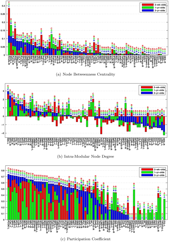
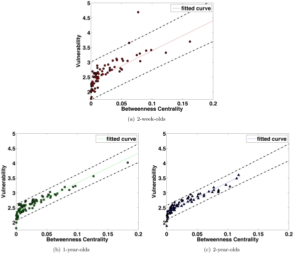
The human brain is organized into a collection of interacting networks with specialized functions to support various cognitive functions. Recent research has reached a consensus that the brain manifests small-world topology, which implicates both global and local efficiency at minimal wiring costs, and also modular organization, which indicates functional segregation and specialization. However, the important questions of how and when the small-world topology and modular organization come into existence remain largely unanswered. Taking a graph theoretic approach, we attempt to shed light on this matter by an in vivo study, using diffusion tensor imaging based fiber tractography, on 39 healthy pediatric subjects with longitudinal data collected at average ages of 2 weeks, 1 year, and 2 years. Our results indicate that the small-world architecture exists at birth with efficiency that increases in later stages of development. In addition, we found that the networks are broad scale in nature, signifying the existence of pivotal connection hubs and resilience of the brain network to random and targeted attacks. We also observed, with development, that the brain network seems to evolve progressively from a local, predominantly proximity based, connectivity pattern to a more distributed, predominantly functional based, connectivity pattern. These observations suggest that the brain in the early years of life has relatively efficient systems that may solve similar information processing problems, but in divergent ways.
Overview
The claims and statistics of this paper haven’t been checked yet.
Run the check — free, no account needed. It reads the open-access text and checks the sources it cites.
Integrity signals
Registry
Cites
Statistics
Figures
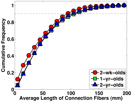
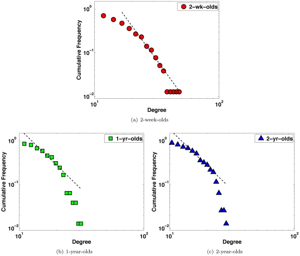
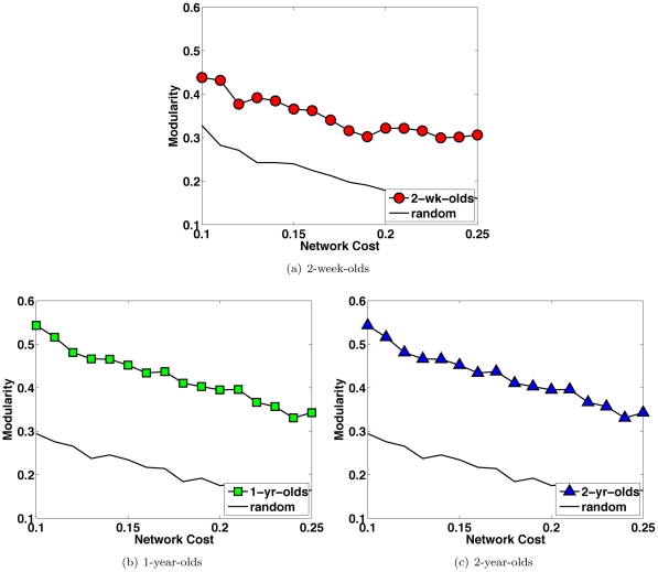
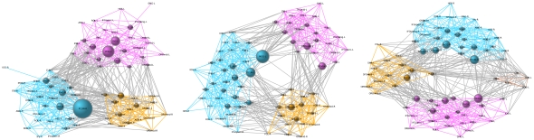
Verification
Claims the paper makes
The claims and statistics of this paper haven’t been checked yet.
Run the check — free, no account needed. It reads the open-access text and checks the sources it cites.
Claims other papers make about this one
Each is checked against this paper’s own text: does it say what the citing paper says it does?
The source backs part of the claim, not all of it — or backs it for a different population, condition or comparison.
In the citing paper
“Network maturation in the first 2 years after birth generally consists of increased network efficiency and network integration [G], decreased network segregation [G], and some changes in the modularity48, 51, with most major hubs and modules in place by age 2 years52.”
From Imaging structural and functional brain development in early childhood (2018) · cites this paper as [48]
Experimental — accuracy not yet measuredThe source backs part of the claim, not all of it — or backs it for a different population, condition or comparison.
In the citing paper
“The centrality of the pre-cuneus and cuneus still increases before pre-adolescence (Huang et al., 2015; Yap et al., 2011).”
From Quantitative mapping of the brain’s structural connectivity using diffusion MRI tractography: A review (2022) · cites this paper as Yap et al., 2011
Experimental — accuracy not yet measuredThe source backs part of the claim, not all of it — or backs it for a different population, condition or comparison.
In the citing paper
“although neonatal brains already show similar structural hub distribution as adult brains”
From Quantitative mapping of the brain’s structural connectivity using diffusion MRI tractography: A review (2022) · cites this paper as Yap et al., 2011
Experimental — accuracy not yet measuredThe cited source says what the claim attributes to it.
In the citing paper
“many refinements occur for the topological roles for each node (Yap et al., 2011).”
From Quantitative mapping of the brain’s structural connectivity using diffusion MRI tractography: A review (2022) · cites this paper as Yap et al., 2011
In the citing paper
“Overall, the white-matter connectome at birth is highly organized and includes hub regions and connections that are similar to those observed in adults48,49.”
From Imaging structural and functional brain development in early childhood (2018) · cites this paper as [48]
Self-check
Percentages, sums and reported differences re-computed from the paper’s own numbers.
The self-check needs the paper’s full text.
Citations
Cites
Collective dynamics of ‘small-world’ networks
Watts, D. J., & Strogatz, S. H. (1998) · Nature · doi:10.1038/30918 · Cited by 43,060
Tzourio-Mazoyer, N., Landeau, B., Papathanassiou, D., Crivello, F., Etard, O., Delcroix, N., Mazoyer, B., & Joliot, M. (2002) · NeuroImage · doi:10.1006/nimg.2001.0978 · Cited by 16,319
Finding and evaluating community structure in networks
Newman, M. E. J., & Girvan, M. (2004) · Physical Review E · doi:10.1103/physreve.69.026113 · Cited by 13,931
A set of measures of centrality based upon betweenness
Freeman, L. (1977) · Sociometry · doi:10.2307/3033543 · Cited by 10,557
Strogatz, S. H. (2001) · Nature · doi:10.1038/35065725 · Cited by 8,236
Error and attack tolerance of complex networks
Albert, R., Jeong, H., & Barabási, A.-L. (2000) · Nature · doi:10.1038/35019019 · Cited by 7,534
Finding community structure in very large networks
Clauset, A., Newman, M. E. J., & Moore, C. (2004) · Physical Review E · doi:10.1103/physreve.70.066111 · Cited by 7,390
Error and attack tolerance of complex networksPreprint
Albert, R., Jeong, H., & Barabási, A.-Ĺ. (2000) · Cited by 6,554
The precuneus: a review of its functional anatomy and behavioural correlates
Cavanna, A. E., & Trimble, M. R. (2006) · Brain · doi:10.1093/brain/awl004 · Cited by 5,128
Efficient behavior of small-world networks
Latora, V., & Marchiori, M. (2001) · Physical Review Letters · doi:10.1103/physrevlett.87.198701 · Cited by 5,119
Cited by
Sorted by citations
BrainNet Viewer: A Network Visualization Tool for Human Brain Connectomics
Xia, M., Wang, J., & He, Y. (2013) · PLoS ONE · doi:10.1371/journal.pone.0068910 · Cited by 4,106
Imaging structural and functional brain development in early childhood
Gilmore, J. H., Knickmeyer, R. C., & Gao, W. (2018) · Nature Reviews Neuroscience · doi:10.1038/nrn.2018.1 · Cited by 1,013
Self-citationDubois, J., Dehaene-Lambertz, G., Kulikova, S., Poupon, C., Hüppi, P. S., & Hertz-Pannier, L. (2014) · Neuroscience · doi:10.1016/j.neuroscience.2013.12.044 · Cited by 851
Deep convolutional neural networks for multi-modality isointense infant brain image segmentation
Zhang, W., Li, R., Deng, H., Wang, L., Lin, W., Ji, S., & Shen, D. (2015) · NeuroImage · doi:10.1016/j.neuroimage.2014.12.061 · Cited by 817
Self-citationBetzel, R. F., Byrge, L., He, Y., Goñi, J., Zuo, X.-N., & Sporns, O. (2014) · NeuroImage · doi:10.1016/j.neuroimage.2014.07.067 · Cited by 807
The organization of physiological brain networks
Stam, C. J., & van Straaten, E. C. W. (2012) · Clinical Neurophysiology · doi:10.1016/j.clinph.2012.01.011 · Cited by 591
Holzman, J. B., & Bridgett, D. J. (2017) · Neuroscience & Biobehavioral Reviews · doi:10.1016/j.neubiorev.2016.12.032 · Cited by 449
Small-world human brain networks: Perspectives and challenges
Liao, X., Vasilakos, A. V., & Yong He, A. (2017) · Neuroscience & Biobehavioral Reviews · doi:10.1016/j.neubiorev.2017.03.018 · Cited by 443
The Neonatal Connectome During Preterm Brain Development
van den Heuvel, M. P., Kersbergen, K. J., de Reus, M. A., Keunen, K., Kahn, R. S., Groenendaal, F., de Vries, L. S., & Benders, M. J. N. L. (2015) · Cerebral Cortex · doi:10.1093/cercor/bhu095 · Cited by 386
Gao, W., Alcauter, S., Elton, A., Hernandez-Castillo, C. R., Smith, J. K., Ramirez, J., & Lin, W. (2015) · Cerebral Cortex · doi:10.1093/cercor/bhu088 · Cited by 380
Self-citationLebel, C., Treit, S., & Beaulieu, C. (2019) · NMR in Biomedicine · doi:10.1002/nbm.3778 · Cited by 379
Rich-club organization of the newborn human brain
Ball, G., Aljabar, P., Zebari, S., Tusor, N., Arichi, T., Merchant, N., Robinson, E. C., Ogundipe, E., Rueckert, D., Edwards, A. D., et al. (2014) · Proceedings of the National Academy of Sciences · doi:10.1073/pnas.1324118111 · Cited by 360
Unraveling the Miswired Connectome: A Developmental Perspective
Martino, A. D., Fair, D. A., Kelly, C. A. M., Satterthwaite, T. D., Castellanos, F. X., Thomason, M. E., Craddock, R. C., Luna, B., LEVENTHAL, B. L., Zuo, X., et al. (2014) · Neuron · doi:10.1016/j.neuron.2014.08.050 · Cited by 357
Zhang, F., Daducci, A., He, Y., Schiavi, S., Seguin, C., Smith, R. E., Yeh, C.-H., Zhao, T., & O’Donnell, L. J. (2022) · NeuroImage · doi:10.1016/j.neuroimage.2021.118870 · Cited by 302
“The centrality of the precuneus and cuneus still increases before pre-adolescence [205, 509].”
Developmental connectomics from infancy through early childhood
Cao, M., Huang, H., & He, Y. (2017) · Trends in Neurosciences · doi:10.1016/j.tins.2017.06.003 · Cited by 284
Neural histology and neurogenesis of the human fetal and infant brain
Kostović, I., Sedmak, G., & Judáš, M. (2018) · NeuroImage · doi:10.1016/j.neuroimage.2018.12.043 · Cited by 272
Li, G., Wang, L., Shi, F., Lyall, A. E., Lin, W., Gilmore, J. H., & Shen, D. (2014) · The Journal of Neuroscience · doi:10.1523/jneurosci.3976-13.2014 · Cited by 251
Self-citationThe development of brain network hubs
Oldham, S., & Fornito, A. (2018) · Developmental Cognitive Neuroscience · doi:10.1016/j.dcn.2018.12.005 · Cited by 257
The influence of preterm birth on the developing thalamocortical connectome
Ball, G., Boardman, J. P., Aljabar, P., Pandit, A. S., Arichi, T., Merchant, N. M., Rueckert, D., Edwards, A. D., & Counsell, S. J. (2012) · Cortex · doi:10.1016/j.cortex.2012.07.006 · Cited by 249
Diffusion tensor imaging for understanding brain development in early life
Qiu, A., Mori, S., & Miller, M. I. (2015) · Annual Review of Psychology · doi:10.1146/annurev-psych-010814-015340 · Cited by 238
LINKS: Learning-based multi-source IntegratioN frameworK for Segmentation of infant brain images
Wang, L., Gao, Y., Shi, F., Li, G., Gilmore, J. H., Lin, W., & Shen, D. G. (2014) · NeuroImage · doi:10.1016/j.neuroimage.2014.12.042 · Cited by 232
Self-citationMRI of the Neonatal Brain: A Review of Methodological Challenges and Neuroscientific Advances
Dubois, J., Alison, M., Counsell, S. J., Hertz‐Pannier, L., Hüppi, P. S., & Benders, M. J. N. L. (2021) · Journal of Magnetic Resonance Imaging · doi:10.1002/jmri.27192 · Cited by 226
Development of Human Brain Structural Networks Through Infancy and Childhood
Huang, H., Shu, N., Mishra, V., Jeon, T., Chalak, L., Wang, Z. J., Rollins, N., Gong, G., Cheng, H., Peng, Y., et al. (2015) · Cerebral Cortex · doi:10.1093/cercor/bht335 · Cited by 223
Assessment of system dysfunction in the brain through MRI-based connectomics
Filippi, M., van den Heuvel, M. P., Fornito, A., Yong He, A., Pol, H. E. H., Agosta, F., Comı, G., & Rocca, M. A. (2013) · The Lancet Neurology · doi:10.1016/s1474-4422(13)70144-3 · Cited by 219
Tracking the Brain's Functional Coupling Dynamics over Development
Hutchison, R. M., & Morton, J. B. (2015) · Journal of Neuroscience · doi:10.1523/jneurosci.4638-14.2015 · Cited by 222
Collin, G., & van den Heuvel, M. P. (2013) · The Neuroscientist · doi:10.1177/1073858413503712 · Cited by 198
Vértes, P. E., & Bullmore, E. T. (2015) · Journal of Child Psychology and Psychiatry · doi:10.1111/jcpp.12365 · Cited by 197
Fischi-Gómez, E., Vasung, L., Meskaldji, D. E., Lazeyras, F., Borradori-Tolsa, C., Hagmann, P., Barisnikov, K., Thiran, J., & Hüppi, P. S. (2014) · Cerebral Cortex · doi:10.1093/cercor/bhu073 · Cited by 207
Wu, K., Taki, Y., Sato, K., Hashizume, H., Sassa, Y., Takeuchi, H., Thyreau, B., Yong He, A., Evans, A. C., Li, X., et al. (2013) · PLoS ONE · doi:10.1371/journal.pone.0055347 · Cited by 185
The emergence of functional architecture during early brain development
Keunen, K., Counsell, S. J., & Benders, M. J. N. L. (2017) · NeuroImage · doi:10.1016/j.neuroimage.2017.01.047 · Cited by 189
Batallé, D., Eixarch, E., Figueras, F., Muñoz‐Moreno, E., Bargalló, N., Illa, M., Acosta‐Rojas, R., Amat‐Roldan, I., & Gratacós, E. (2012) · NeuroImage · doi:10.1016/j.neuroimage.2012.01.059 · Cited by 162
Functional Connectivity in Islets of Langerhans from Mouse Pancreas Tissue Slices
Stožer, A., Gosak, M., Dolenšek, J., Perc, M., Marhl, M., Rupnik, M. S., & Korošak, D. (2013) · PLoS Computational Biology · doi:10.1371/journal.pcbi.1002923 · Cited by 162
Boersma, M., Smit, D. J. A., Boomsma, D. I., de Geus, E. J. C., de Waal, H. A. D., & Stam, C. J. (2012) · Brain Connectivity · doi:10.1089/brain.2012.0106 · Cited by 160
Toward Developmental Connectomics of the Human Brain
Cao, M., Huang, H., Peng, Y., Dong, Q., & Yong He, A. (2016) · Frontiers in Neuroanatomy · doi:10.3389/fnana.2016.00025 · Cited by 156
Kenett, Y. N., Beaty, R. E., Silvia, P. J., Anaki, D., & Faust, M. (2016) · Psychology of Aesthetics Creativity and the Arts · doi:10.1037/aca0000056 · Cited by 150
Lim, S. I., Han, C. E., Uhlhaas, P. J., & Kaiser, M. (2013) · Cerebral Cortex · doi:10.1093/cercor/bht333 · Cited by 154
Construction of 4D high-definition cortical surface atlases of infants: Methods and applications
Li, G., Wang, L., Shi, F., Gilmore, J. H., Lin, W., & Shen, D. G. (2015) · Medical Image Analysis · doi:10.1016/j.media.2015.04.005 · Cited by 139
Self-citationMultiscale Structure–Function Gradients in the Neonatal Connectome
Larivière, S., de Wael, R. V., Hong, S., Paquola, C., Tavakol, S., Lowe, A. J., Schrader, D. V., & Bernhardt, B. C. (2019) · Cerebral Cortex · doi:10.1093/cercor/bhz069 · Cited by 136
Semantic organization in children with cochlear implants: computational analysis of verbal fluency
Kenett, Y. N., Wechsler-Kashi, D., Kenett, D. Y., Schwartz, R. G., Ben‐Jacob, E., & Faust, M. (2013) · Frontiers in Psychology · doi:10.3389/fpsyg.2013.00543 · Cited by 137
Shi, F., Yap, P., Gao, W., Lin, W., Gilmore, J. H., & Shen, D. G. (2012) · NeuroImage · doi:10.1016/j.neuroimage.2012.05.026 · Cited by 137
Self-citationLi, G., Nie, J., Wang, L., Shi, F., Gilmore, J. H., Lin, W., & Shen, D. G. (2014) · NeuroImage · doi:10.1016/j.neuroimage.2013.12.038 · Cited by 127
Self-citationStructural connectivity asymmetry in the neonatal brain
Ratnarajah, N., Rifkin-Graboi, A., Fortier, M. V., Chong, Y. S., Kwek, K., Saw, S.-M., Godfrey, K. M., Gluckman, P. D., Meaney, M. J., & Qiu, A. (2013) · NeuroImage · doi:10.1016/j.neuroimage.2013.02.052 · Cited by 124
Early breast milk exposure modifies brain connectivity in preterm infants
Blesa, M., Sullivan, G., Anblagan, D., Telford, E. J., Quigley, A. J., Sparrow, S. A., Serag, A., Semple, S. I. K., Bastin, M. E., & Boardman, J. P. (2018) · NeuroImage · doi:10.1016/j.neuroimage.2018.09.045 · Cited by 124
Short-range connections in the developmental connectome during typical and atypical brain maturation
Ouyang, M., Kang, H., Detre, J. A., Roberts, T. P. L., & Huang, H. (2017) · Neuroscience & Biobehavioral Reviews · doi:10.1016/j.neubiorev.2017.10.007 · Cited by 120
Wang, L., Shi, F., Gao, Y., Li, G., Gilmore, J. H., Lin, W., & Shen, D. G. (2013) · NeuroImage · doi:10.1016/j.neuroimage.2013.11.040 · Cited by 104
Self-citationGraph theoretical modeling of baby brain networks
Zhao, T., Xu, Y., & Yong He, A. (2018) · NeuroImage · doi:10.1016/j.neuroimage.2018.06.038 · Cited by 108
Structural network analysis of brain development in young preterm neonates
Brown, C. J., Miller, S. P., Booth, B. G., Andrews, S., Chau, V., Poskitt, K. J., & Hamarneh, G. (2014) · NeuroImage · doi:10.1016/j.neuroimage.2014.07.030 · Cited by 103
Early human brain development: Starring the subplate
Hadders-Algra, M. (2018) · Neuroscience & Biobehavioral Reviews · doi:10.1016/j.neubiorev.2018.06.017 · Cited by 100
Topological turning points across the human lifespan
Mousley, A., Bethlehem, R. A. I., Yeh, F.-C. F., & Astle, D. E. (2025) · Nature Communications · doi:10.1038/s41467-025-65974-8 · Cited by 105
Diffusion properties of major white matter tracts in young, typically developing children
Johnson, R. T., Yeatman, J. D., Wandell, B. A., Buonocore, M. H., Amaral, D. G., & Nordahl, C. W. (2013) · NeuroImage · doi:10.1016/j.neuroimage.2013.11.025 · Cited by 93
Disrupted Functional Brain Networks in Autistic Toddlers
Boersma, M., Kemner, C., de Reus, M. A., Collin, G., Snijders, T. M., Hofman, D., Buitelaar, J. K., Stam, C. J., & van den Heuvel, M. P. (2012) · Brain Connectivity · doi:10.1089/brain.2012.0127 · Cited by 93
Brain anatomical networks in world class gymnasts: A DTI tractography study
Wang, B., Fan, Y., Lü, M., Li, S., Song, Z., Peng, X., Zhang, R., Lin, Q., Yong He, A., Wang, J., et al. (2012) · NeuroImage · doi:10.1016/j.neuroimage.2012.10.007 · Cited by 91
Self-citationA DTI-based template-free cortical connectome study of brain maturation
Tymofiyeva, O., Hess, C. P., Ziv, E., Lee, P. N., Glass, H. C., Ferriero, D. M., Barkovich, A. J., & Xu, D. (2013) · PLoS ONE · doi:10.1371/journal.pone.0063310 · Cited by 86
The structural connectome of the human brain in agenesis of the corpus callosum
Owen, J. P., Li, Y., Ziv, E., Strominger, Z. A., Gold, J., Bukhpun, P., Wakahiro, M. L. J., Friedman, E. J., Sherr, E. H., & Mukherjee, P. (2012) · NeuroImage · doi:10.1016/j.neuroimage.2012.12.031 · Cited by 87
Neonatal brain injury and aberrant connectivity
Smyser, C. D., Wheelock, M. D., Limbrick, D. D., & Neil, J. J. (2018) · NeuroImage · doi:10.1016/j.neuroimage.2018.07.057 · Cited by 87
Faust, M., & Kenett, Y. N. (2014) · Frontiers in Human Neuroscience · doi:10.3389/fnhum.2014.00511 · Cited by 81
Towards the “Baby Connectome”: Mapping the Structural Connectivity of the Newborn Brain
Tymofiyeva, O., Hess, C. P., Ziv, E., Tian, N., Bonifacio, S. L., McQuillen, P. S., Ferriero, D. M., Barkovich, A. J., & Xu, D. (2012) · PLoS ONE · doi:10.1371/journal.pone.0031029 · Cited by 80
Graph theoretical analysis of developmental patterns of the white matter network
Chen, Z., Liu, M., Gross, D. W., & Beaulieu, C. (2013) · Frontiers in Human Neuroscience · doi:10.3389/fnhum.2013.00716 · Cited by 77
Imaging Evidence of the Effect of Socio-Economic Status on Brain Structure and Development
Leijser, L. M., Siddiqi, A., & Miller, S. P. (2018) · Seminars in Pediatric Neurology · doi:10.1016/j.spen.2018.03.004 · Cited by 78
Owen, J. P., Ziv, E., Bukshpun, P., Pojman, N. J., Wakahiro, M. L. J., Berman, J., Roberts, T. P. L., Friedman, E. J., Sherr, E. H., & Mukherjee, P. (2013) · Brain Connectivity · doi:10.1089/brain.2012.0121 · Cited by 79
Cai, L., Dong, Q., & Niu, H. (2018) · Developmental Cognitive Neuroscience · doi:10.1016/j.dcn.2018.03.003 · Cited by 78
Batallé, D., Muñoz‐Moreno, E., Figueras, F., Bargalló, N., Eixarch, E., & Gratacós, E. (2013) · NeuroImage · doi:10.1016/j.neuroimage.2013.07.045 · Cited by 75
Human fetal brain connectome: structural network development from middle fetal stage to birth
Song, L., Mishra, V., Ouyang, M., Peng, Q., Slinger, M., Liu, S., & Huang, H. (2017) · Frontiers in Neuroscience · doi:10.3389/fnins.2017.00561 · Cited by 71
Richmond, S., Johnson, K. A., Seal, M. L., Allen, N. B., & Whittle, S. (2016) · Neuroscience & Biobehavioral Reviews · doi:10.1016/j.neubiorev.2016.08.024 · Cited by 70
Altered intrahemispheric structural connectivity in Gilles de la Tourette syndrome
Cheng, B., Braaß, H., Ganos, C., Treszl, A., Biermann‐Ruben, K., Hummel, F. C., Müller‐Vahl, K., Schnitzler, A., Gerloff, C. P., Münchau, A. M., et al. (2013) · NeuroImage Clinical · doi:10.1016/j.nicl.2013.11.011 · Cited by 70
Zhong, S., Yong He, A., Shu, H., & Gong, G. (2016) · Cerebral Cortex · doi:10.1093/cercor/bhw109 · Cited by 69
Structural network maturation of the preterm human brain
Zhao, T., Mishra, V. R., Jeon, T., Ouyang, M., Peng, Q., Chalak, L. F., Wisnowski, J. L., Heyne, R. J., Rollins, N. K., Shu, N., et al. (2018) · NeuroImage · doi:10.1016/j.neuroimage.2018.06.047 · Cited by 69
Brain Connectomics of Visual-Motor Deficits in Children with Developmental Coordination Disorder
Debrabant, J., Vingerhoets, G., Waelvelde, H. V., Leemans, A., Taymans, T., & Caeyenberghs, K. (2015) · The Journal of Pediatrics · doi:10.1016/j.jpeds.2015.09.069 · Cited by 74
Prenatal cocaine effects on brain structure in early infancy
Grewen, K., Burchinal, M., Vachet, C., Gouttard, S., Gilmore, J. H., Lin, W., Johns, J., Elam, M., & Gerig, G. (2014) · NeuroImage · doi:10.1016/j.neuroimage.2014.06.070 · Cited by 65
Self-citationGaldi, P., Blesa, M., Stoye, D. Q., Sullivan, G., Lamb, G. J., Quigley, A. J., Thrippleton, M. J., Bastin, M. E., & Boardman, J. P. (2020) · NeuroImage Clinical · doi:10.1016/j.nicl.2020.102195 · Cited by 68
A Network Neuroscience Approach to Typical and Atypical Brain Development
Morgan, S. E., White, S. R., Bullmore, E. T., & Vértes, P. E. (2018) · Biological Psychiatry Cognitive Neuroscience and Neuroimaging · doi:10.1016/j.bpsc.2018.03.003 · Cited by 69
White matter connectomes at birth accurately predict cognitive abilities at age 2
Girault, J. B., Munsell, B. C., Puechmaille, D., Goldman, B. D., Prieto, J. C., Styner, M. A., & Gilmore, J. H. (2019) · NeuroImage · doi:10.1016/j.neuroimage.2019.02.060 · Cited by 60
Self-citationCortical thickness and surface area in neonates at high risk for schizophrenia
Li, G., Wang, L., Shi, F., Lyall, A. E., Ahn, M., Peng, Z., Hongtu Zhu, B., Lin, W., Gilmore, J. H., & Shen, D. G. (2014) · Brain Structure and Function · doi:10.1007/s00429-014-0917-3 · Cited by 62
Self-citationde Almeida, J. S., Meskaldji, D. E., Loukas, S., Lordier, L., Gui, L., Lazeyras, F., & Hüppi, P. S. (2020) · NeuroImage · doi:10.1016/j.neuroimage.2020.117440 · Cited by 61
Age-specific gray and white matter DTI atlas for human brain at 33, 36 and 39 postmenstrual weeks
Feng, L., Li, H., Oishi, K., Mishra, V. R., Song, L., Peng, Q., Ouyang, M., Wang, J., Slinger, M. A., Jeon, T., et al. (2018) · NeuroImage · doi:10.1016/j.neuroimage.2018.06.069 · Cited by 59
Structural MRI connectome in development: challenges of the changing brain
Tymofiyeva, O., Hess, C. P., Xu, D., & Barkovich, A. J. (2014) · British Journal of Radiology · doi:10.1259/bjr.20140086 · Cited by 55
Zhong, S., Yong He, A., & Gong, G. (2015) · Human Brain Mapping · doi:10.1002/hbm.22751 · Cited by 52
Development of Structural and Functional Connectivity
Dubois, J., Kostović, I., & Judas, M. (2015) · Elsevier eBooks · doi:10.1016/b978-0-12-397025-1.00360-2 · Cited by 55
Fausto‐Sterling, A. (2021) · Frontiers in Human Neuroscience · doi:10.3389/fnhum.2021.613789 · Cited by 57
Denoising magnetic resonance images using collaborative non-local means
Chen, G., Zhang, P., Wu, Y., Shen, D. G., & Yap, P. (2015) · Neurocomputing · doi:10.1016/j.neucom.2015.11.031 · Cited by 48
Self-citationAltered white matter development in children born very preterm
Young, J. M., Vandewouw, M. M., Morgan, B. R., Smith, M. L., Sled, J. G., & Taylor, M. J. (2018) · Brain Structure and Function · doi:10.1007/s00429-018-1614-4 · Cited by 53
Development of cortical motor circuits between childhood and adulthood: A navigated TMS‐HdEEG study
Määttä, S., Könönen, M., Kallioniemi, E., Lakka, T. A., Lintu, N., Lindi, V., Ferreri, F., Ponzo, D., & Säïsänen, L. (2017) · Human Brain Mapping · doi:10.1002/hbm.23545 · Cited by 47
Cheng, J., Rachid Deriche, B., Jiang, T., Shen, D. G., & Yap, P. (2014) · NeuroImage · doi:10.1016/j.neuroimage.2014.07.062 · Cited by 42
Self-citationDubois, J., Adibpour, P., Poupon, C., Hertz-Pannier, L., & Dehaene-Lambertz, G. (2016) · Brain Plasticity · doi:10.3233/bpl-160031 · Cited by 40
Muñoz‐Moreno, E., Fischi-Gómez, E., Batallé, D., Borradori-Tolsa, C., Eixarch, E., Thiran, J., Gratacós, E., & Hüppi, P. S. (2016) · Frontiers in Neuroscience · doi:10.3389/fnins.2016.00560 · Cited by 40
Neonatal atlas construction using sparse representation
Shi, F., Wang, L., Guorong Wu, A., Li, G., Gilmore, J. H., Lin, W., & Shen, D. G. (2014) · Human Brain Mapping · doi:10.1002/hbm.22502 · Cited by 38
Self-citationThe YOUth cohort study: MRI protocol and test-retest reliability in adults
Buimer, E. E. L., Pas, P., Brouwer, R. M., Froeling, M., Hoogduin, H. M., Leemans, A., Luijten, P. R., van Nierop, B. J., Raemaekers, M. A. H., Schnack, H. G., et al. (2020) · Developmental Cognitive Neuroscience · doi:10.1016/j.dcn.2020.100816 · Cited by 38
Batallé, D., Muñoz‐Moreno, E., Arbat‐Plana, A., Illa, M., Figueras, F., Eixarch, E., & Gratacós, E. (2014) · NeuroImage · doi:10.1016/j.neuroimage.2014.05.065 · Cited by 38
Schmithorst, V. J., Votava‐Smith, J. K., Tran, N., Kim, R., Lee, V., Ceschin, R. C., Lai, H., Johnson, J. A., Sánchez-de-Toledo, J., Blüml, S., et al. (2018) · Human Brain Mapping · doi:10.1002/hbm.24308 · Cited by 36
Simultaneous and consistent labeling of longitudinal dynamic developing cortical surfaces in infants
Li, G., Wang, L., Shi, F., Lin, W., & Shen, D. G. (2014) · Medical Image Analysis · doi:10.1016/j.media.2014.06.007 · Cited by 33
Self-citationFields, C., & Glazebrook, J. F. (2016) · Cognitive Neurodynamics · doi:10.1007/s11571-016-9419-8 · Cited by 33
Graph theory in paediatric epilepsy: A systematic review
Falsaperla, R., Vitaliti, G., Marino, S. D., Praticò, A. D., Mailo, J. A., Spatuzza, M., Cilio, M. R., Foti, R., & Ruggieri, M. (2021) · Dialogues in Clinical Neuroscience · doi:10.1080/19585969.2022.2043128 · Cited by 33
Li, G., Liu, T., Ni, D., Lin, W., Gilmore, J. H., & Shen, D. G. (2015) · Human Brain Mapping · doi:10.1002/hbm.23003 · Cited by 33
Self-citationInvestigating the Microstructural Correlation of White Matter in Autism Spectrum Disorder
Dean, D., Travers, B. G., Adluru, N., Tromp, D., Destiche, D. J., Samsin, D., Prigge, M. B. D., Zielinski, B. A., Fletcher, P. T., Anderson, J. S., et al. (2016) · Brain Connectivity · doi:10.1089/brain.2015.0385 · Cited by 30
Woodburn, M., Bricken, C. L., Wu, Z., Li, G., Wang, L., Lin, W., Sheridan, M. A., & Cohen, J. R. (2021) · NeuroImage · doi:10.1016/j.neuroimage.2021.118232 · Cited by 32
Self-citationDenoising of Diffusion MRI Data via Graph Framelet Matching in x-q Space
Chen, G., Dong, B., Zhang, Y., Lin, W., Shen, D. G., & Yap, P. (2019) · IEEE Transactions on Medical Imaging · doi:10.1109/tmi.2019.2915629 · Cited by 29
Self-citationIncreased segregation of functional networks in developing brains
He, W., Sowman, P. F., Brock, J., Etchell, A. C., Stam, C. J., & Hillebrand, A. (2019) · NeuroImage · doi:10.1016/j.neuroimage.2019.06.055 · Cited by 31
The emergence of a theta social brain network during infancy
van der Velde, B., White, T. J. H., & Kemner, C. (2021) · NeuroImage · doi:10.1016/j.neuroimage.2021.118298 · Cited by 31
Multimodality evaluation of the pediatric brain: DTI and its competitors
Vasung, L., Fischi-Gómez, E., & Hüppi, P. S. (2013) · Pediatric Radiology · doi:10.1007/s00247-012-2515-y · Cited by 27
A Hybrid CPU-GPU Accelerated Framework for Fast Mapping of High-Resolution Human Brain Connectome
Wang, Y., Du, H., Xia, M., Ren, L., Xu, M., Xie, T., Gong, G., Xu, N., Yang, H., & Yong He, A. (2013) · PLoS ONE · doi:10.1371/journal.pone.0062789 · Cited by 27
Metadata
- Authors
- Yap, P.-T., Fan, Y., Chen, Y., Gilmore, J. H., Lin, W., & Shen, D.
- Affiliations
- University of North Carolina at Chapel Hill (US)
- Corresponding author
- Shen, Dinggang
- Venue
- PLoS ONE
- Publisher
- Public Library of Science (PLoS)
- Published
- 2011-09-23
- Volume
- 6
- Issue
- 9
- Pages
- e24678
- ISSN
- 1932-6203 (ISSN-L)
- PMC
- PMC3179462
- OpenAlex
- W2171775073
- Cited by
- 190
- References
- 60
- Citations by year
- 2026: 7 · 2025: 5 · 2024: 6 · 2023: 5 · 2022: 13 · 2021: 11 · 2020: 9 · 2019: 9 · 2018: 18 · 2017: 19 · 2016: 15 · 2015: 11 · 2014: 26 · 2013: 25 · 2012: 11
- FWCI
- 10.07
- Type
- journal article
- Language
- en
- Open access
- gold
- doi.org
- Locations
- PLoS ONE (publishedVersion, cc-by)
- PubMed Central (publishedVersion, cc-by)
- PubMed (publishedVersion)
- Carolina Digital Repository (University of North Carolina at Chapel Hill) (submittedVersion)
- DOAJ (DOAJ: Directory of Open Access Journals) (submittedVersion, cc-by-sa)
- UNC Libraries
- Funders
- National Institutes of Health (MH064065)
- National Institute of Mental Health (MH064065, P50MH064065, R03MH076970, MH076970)
- National Institute of Biomedical Imaging and Bioengineering (EB009634, R01EB009634)
- Eunice Kennedy Shriver National Institute of Child Health and Human Development (HD05300)
- Topics
- Life Sciences › Neuroscience › Cognitive Neuroscience › Functional Brain Connectivity Studies
- Health Sciences › Medicine › Radiology, Nuclear Medicine and Imaging › Advanced Neuroimaging Techniques and Applications
- Physical Sciences › Physics and Astronomy › Statistical and Nonlinear Physics › Complex Network Analysis Techniques
- Keywords
- white matter connectivity, brain network development, small-world networks, brain network efficiency, network modularity, network hubs, network resilience, early infancy, functional segregation, graph theory, diffusion tensor imaging, fiber tractography, child brain development
- MeSH terms
- Algorithms, Brain, Brain Mapping, Child, Preschool, Female, Humans, Infant, Infant, Newborn, Longitudinal Studies, Male, Models, Neurological, Nerve Net, Neural Pathways, Diffusion Tensor Imaging
About these checks
Registry, retraction, Cites and statistics results are computed, not judged, with one exception: a printed reference that no index lists is read once by a model before it counts as a citation error. Claims are extracted by a model and kept only when their quote appears verbatim in the text; each cited source is then checked, with a model’s help, for whether it says what the claim relies on. Those verdicts can be wrong — read the claim’s quote and the cited source before relying on one.
Statistics and arithmetic are recomputed, not judged.
Confidence bands are shown only for fields calibrated on our annotated test set. None is calibrated yet, so no band is shown.
What counts as a claim ›How we measure confidence ›How we check this ›
Are you an author, or do you see a problem with this page? Request a review
