

The DOI matches its Crossref record; 83 of the 84 references it cites were found in a registry or an index.
Protein 3D Structure Computed from Evolutionary Sequence Variation
Open accessAbstract
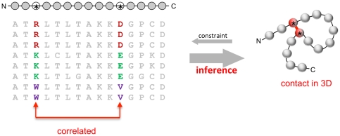
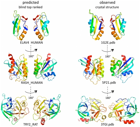
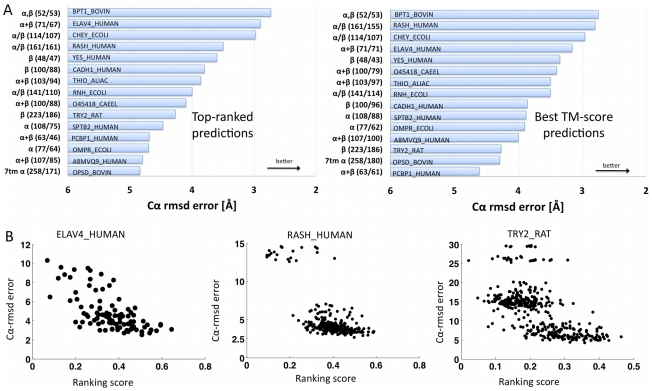
The evolutionary trajectory of a protein through sequence space is constrained by its function. Collections of sequence homologs record the outcomes of millions of evolutionary experiments in which the protein evolves according to these constraints. Deciphering the evolutionary record held in these sequences and exploiting it for predictive and engineering purposes presents a formidable challenge. The potential benefit of solving this challenge is amplified by the advent of inexpensive high-throughput genomic sequencing.In this paper we ask whether we can infer evolutionary constraints from a set of sequence homologs of a protein. The challenge is to distinguish true co-evolution couplings from the noisy set of observed correlations. We address this challenge using a maximum entropy model of the protein sequence, constrained by the statistics of the multiple sequence alignment, to infer residue pair couplings. Surprisingly, we find that the strength of these inferred couplings is an excellent predictor of residue-residue proximity in folded structures. Indeed, the top-scoring residue couplings are sufficiently accurate and well-distributed to define the 3D protein fold with remarkable accuracy.We quantify this observation by computing, from sequence alone, all-atom 3D structures of fifteen test proteins from different fold classes, ranging in size from 50 to 260 residues, including a G-protein coupled receptor. These blinded inferences are de novo, i.e., they do not use homology modeling or sequence-similar fragments from known structures. The co-evolution signals provide sufficient information to determine accurate 3D protein structure to 2.7-4.8 Å C(α)-RMSD error relative to the observed structure, over at least two-thirds of the protein (method called EVfold, details at http://EVfold.org). This discovery provides insight into essential interactions constraining protein evolution and will facilitate a comprehensive survey of the universe of protein structures, new strategies in protein and drug design, and the identification of functional genetic variants in normal and disease genomes.
Overview
The claims and statistics of this paper haven’t been checked yet.
Run the check — free, no account needed. It reads the open-access text and checks the sources it cites.
Integrity signals
Registry
Cites
Statistics
Figures
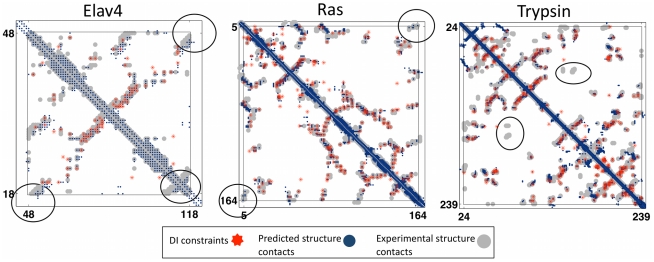
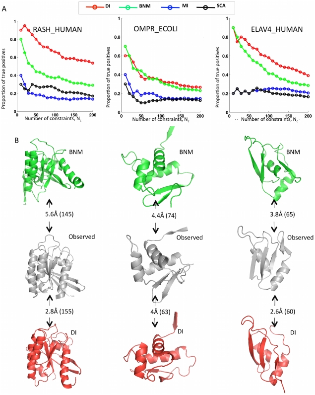
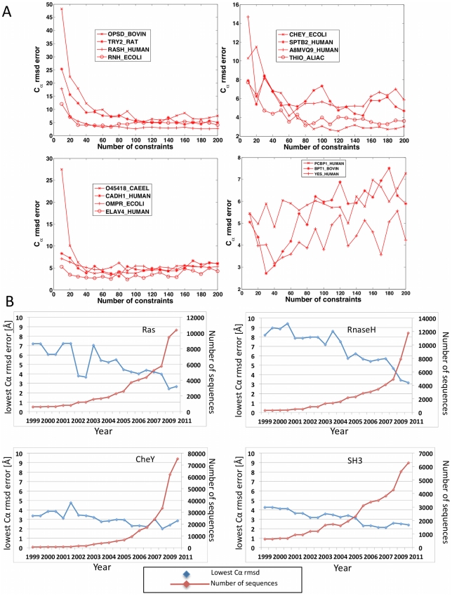
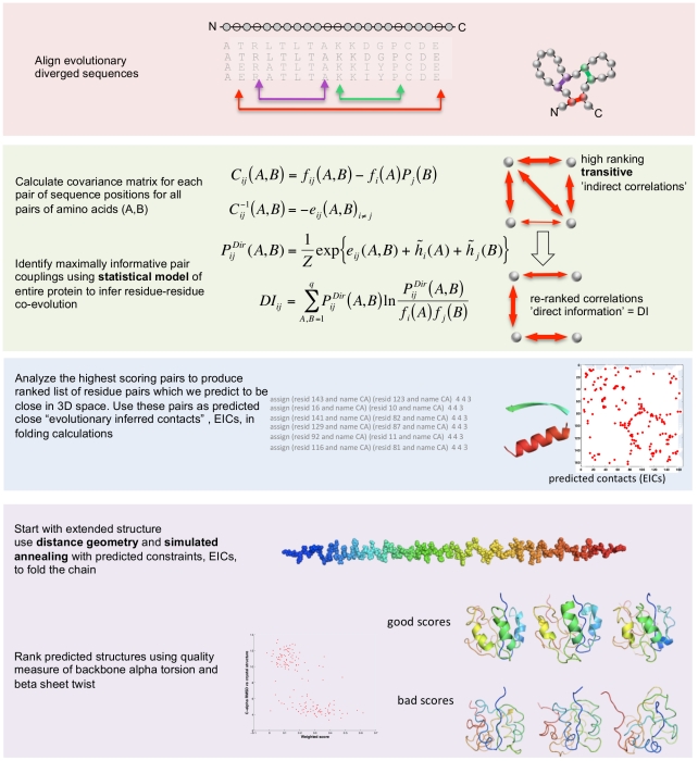
![Figure 3 Progress in contact prediction using the maximum entropy method. Extraction of evolutionary information about residue coupling and predicted contacts from multiple sequence alignments works much better using the global statistical model (right, Direct Information, DI, Equation 3) than the local statistical model (left, Mutual Information, MI, Equation 1). Predicted contacts for DI (red lines connecting the residues predicted to be coupled from sequence information) are better positioned in the experimentally observed structure (grey ribbon diagram), than those for MI (left, blue lines), shown here for the RAS protein (upper) and ELAV4 protein (lower). The DI residue pairs are also more evenly distributed along the chain and overlap more accurately with the contacts in the observed structure (red stars [predicted, grey circles [observed] in contact map; center, upper right triangle) than those using MI (blue [predicted], grey circles [observed]; center, lower left triangle). Details of contact maps for all proteins comparing predicted and observed contacts are in Figures S1 and S2, Text S1.](/paper-figures/ad0edff5338c41479018a65a69fdb4cd/protein-3d-structure-computed-from-evolutionary-sequence-variation-figure-3.jpg)
Verification
Claims the paper makes
The claims and statistics of this paper haven’t been checked yet.
Run the check — free, no account needed. It reads the open-access text and checks the sources it cites.
Claims other papers make about this one
Each is checked against this paper’s own text: does it say what the citing paper says it does?
In the citing paper
“The premise of methods to infer structure based on evolutionary couplings is that, because amino acids exist and evolve in the context of 3D structure, they are not free to evolve independently, but instead co-evolve in patterns reflective of the underlying structure.”
From Predicting multiple conformations via sequence clustering and AlphaFold2 (2024) · cites this paper as [9]
In the citing paper
“One of the major insights leveraged by AlphaFold 1,2 comes from the use of evolutionary covariations that can be extracted from large-scale multiple sequence alignments.[49][50][51][52][53][54]”
From AlphaFold and Implications for Intrinsically Disordered Proteins (2021) · cites this paper as [50]
Self-check
Percentages, sums and reported differences re-computed from the paper’s own numbers.
The self-check needs the paper’s full text.
Citations
Cites
The PyMOL Molecular Graphics System
DeLano, W. L. (2002) · Medical Entomology and Zoology · Cited by 18,147
Crystallography & NMR System: A New Software Suite for Macromolecular Structure Determination
Brünger, A. T., Adams, P. D., Clore, G. M., DeLano, W. L., Gros, P., Grosse‐Kunstleve, R. W., Jiang, J., Kuszewski, J. J., Nilges, M., Pannu, N., et al. (1998) · Acta Crystallographica Section D Biological Crystallography · doi:10.1107/s0907444998003254 · Cited by 15,723
The Pfam Protein Families Database
Bateman, A. G. (2002) · Nucleic Acids Research · doi:10.1093/nar/30.1.276 · Cited by 14,075
I-TASSER: a unified platform for automated protein structure and function prediction
Roy, A., Küçükural, A., & Zhang, Y. (2010) · Nature Protocols · doi:10.1038/nprot.2010.5 · Cited by 6,212
The Pfam protein families database
Finn, R. D., Mistry, J., Tate, J., Coggill, P., Heger, A., Pollington, J. E., Gavin, O. L., Gunasekaran, P., Ceric, G., Forslund, K., et al. (2010) · Nucleic Acids Research · doi:10.1093/nar/gkp985 · Cited by 2,694
Scoring function for automated assessment of protein structure template quality
Zhang, Y., & Skolnick, J. (2004) · Proteins: Structure, Function, and Bioinformatics · doi:10.1002/prot.20264 · Cited by 2,392
Atomic-level characterization of the structural dynamics of proteins
Shaw, D. E., Maragakis, P., Lindorff-Larsen, K., Piana, S., Dror, R. O., Eastwood, M. P., Bank, J. A., Jumper, J. M., Salmon, J. K., Shan, Y., et al. (2010) · Science · doi:10.1126/science.1187409 · Cited by 1,923
Weak pairwise correlations imply strongly correlated network states in a neural population
Schneidman, E., Berry, M. J., Segev, R., & Bialek, W. (2006) · Nature · doi:10.1038/nature04701 · Cited by 1,745
Holm, L., & Sander, C. (1996) · Science · doi:10.1126/science.273.5275.595 · Cited by 1,497
Self-citationMorcos, F., Pagnani, A., Lunt, B., Bertolino, A., Marks, D. S., Sander, C., Zecchina, R., Onuchic, J. N., Hwa, T., & Weigt, M. (2011) · Proceedings of the National Academy of Sciences · doi:10.1073/pnas.1111471108 · Cited by 1,434
Self-citation
Research works printed but not linked: 3
- KT Simons, 1999, Ab initio protein structure prediction of CASP III targets using ROSETTA., Proteins, Suppl 3, 171, 10.1002/(SICI)1097-0134(1999)37:3+<171::AID-PROT21>3.0.CO;2-Z · DOI
- MMT Mezard, 2009, Constraint satisfaction problems and neural networks: A statistical physics perspective., Journal of Physiology, 103, 107
- TR Lezon, 2006, Using the principle of entropy maximization to infer genetic interaction networks from gene expression patterns., Proceedings of the National Academy of Sciences of the United States of America, 103, 19033, 10.1073/pnas.0609152103 · DOI
Cited by
Sorted by citations
Items 901–1,000 of 1,005
Jia, K., & Jernigan, R. L. (2018) · bioRxiv (Cold Spring Harbor Laboratory) · doi:10.1101/268904 · Cited by 2
Size and structure of the sequence space of repeat proteinsPreprint
Marchi, J., Galpern, E. A., Espada, R., Ferreiro, D., Walczak, A., & Mora, T. (2019) · bioRxiv · doi:10.1101/635581 · Cited by 20
Improving protein structure prediction using amino acid contact & distance prediction
Ji, S. (2019) · University of Birmingham Institutional Research Archive (University of Birmingham) · Cited by 2
Correlated evolution: models and methodsPreprint
Achaz, G., & Dutheil, J. Y. (2021) · arXiv (Cornell University) · doi:10.48550/arxiv.2103.11809 · Cited by 2
Liu, X., Jin, L., Gao, S., & Zhao, S. (2021) · bioRxiv (Cold Spring Harbor Laboratory) · doi:10.1101/2021.05.12.443740 · Cited by 2
Posani, L., Rizzato, F., Monasson, R., & Cocco, S. (2022) · bioRxiv (Cold Spring Harbor Laboratory) · doi:10.1101/2022.12.12.520004 · Cited by 2
Modeling the structure-conditioned sequence landscape for large-scale protein design with TriFlowPreprint
Srinivasan, H., Yuan, R., Zhang, J., Cong, Q., & Zhou, J. (2025) · bioRxiv (Cold Spring Harbor Laboratory) · doi:10.64898/2025.11.30.691458 · Cited by 2
GROQ-seq Enables Cross-site Reproducibility for High-Throughput Measurement of Protein FunctionPreprint
Spinner, A., Ross, D., Cortade, D. L., Ikonomova, S. P., Baranowski, C., Dhroso, A., Apel, A. R., Sheldon, K. N., Duquette, C., Densmore, D. M., et al. (2026) · bioRxiv (Cold Spring Harbor Laboratory) · doi:10.64898/2026.04.07.716961 · Cited by 2
Improving AlphaFold3 by Engineering MSA and Template InputsPreprint
Neupane, P., Liu, J., & Cheng, J. (2026) · bioRxiv (Cold Spring Harbor Laboratory) · doi:10.64898/2026.04.22.720119 · Cited by 4
Coevolutionary mining of prokaryotic non-coding elements with a genome language modelPreprint
Li, D., Brixi, G., Kim, A., Fiamenghi, M. B., Driscoll, C. L., Evans, S. A., Gao, L., Ivanova, N., Kyrpides, N. C., Deisseroth, K. A., et al. (2026) · bioRxiv (Cold Spring Harbor Laboratory) · doi:10.64898/2026.09.22.753630 · Cited by 5
Molecular Evolution Bioinformatics Toward Structural Biology of TRPV1-4 Channels
Doñate‐Macian, P., Crespi-Boixader, A., & Perálvarez‐Marín, A. (2019) · Methods in molecular biology · doi:10.1007/978-1-4939-9446-5_1 · Cited by 1
Exploring generality of experimental conformational changes with AlphaFold predictions
Castellví, A., Medina, A., Petrillo, G., Sagmeister, T., Pavkov‐Keller, T., Govantes, F., Diederichs, K., Sammito, M. D., & Usón, I. (2022) · Acta Crystallographica Section A Foundations and Advances · doi:10.1107/s2053273322096462 · Cited by 2
Impact of phylogeny on the inference of functional sectors from protein sequence data
Dietler, N., Abbara, A., Choudhury, S., & Bitbol, A.-F. (2024) · PLOS Computational Biology · doi:10.1371/journal.pcbi.1012091 · Cited by 1
Hidden Data Structures and Statistics of Natural Images
Fleig, P., & Nemenman, I. M. (2025) · PRX Life · doi:10.1103/8xd4-6hsr · Cited by 1
Shukla, A., Kashyap, S., Patil, S. D., Varma, A. K., Ghosh, S., & Kondabagil, K. R. (2025) · The FASEB Journal · doi:10.1096/fj.202402627r · Cited by 1
Amino acid exchangeability and surface accessibility underpin the effects of single substitutions
Alpay, B. A., Nanda, P., Nagy, E., & Desai, M. M. (2026) · Genetics · doi:10.1093/genetics/iyag104 · Cited by 1
Optimizing Engineering Change Management Through ECR Data Analysis and Mining Techniques
Xinyu, Z., & Xin, H. (2025) · Journal of Enterprise and Business Intelligence · doi:10.53759/5181/jebi202505022 · Cited by 1
Quercia, C. A., Robeson, L., & Brauchi, S. (2025) · Computational and Structural Biotechnology Journal · doi:10.1016/j.csbj.2025.11.003 · Cited by 1
Sequence-based calculation of local energetic frustration in proteins
Kuhn, A. M., Contessoto, V. G., Onuchic, J. N., & Phillips, G. N., Jr. (2025) · Structural Dynamics · doi:10.1063/4.0000781 · Cited by 1
Energy landscapes in protein folding
Contessoto, V. G., & Onuchic, J. N. (2025) · Elsevier eBooks · doi:10.1016/b978-0-323-99507-8.00025-1 · Cited by 1
Global genomic diversity of temperate P2-like viruses
Liu, Y., Liu, R., Zheng, K., Wang, Z., Shao, H., McMinn, A., Wang, M., & Liang, Y. (2026) · Communications Biology · doi:10.1038/s42003-026-09823-4 · Cited by 1
Engineering and Application of a Thermostable MHETase for PET Depolymerization
Murphy, N. P., Gado, J. E., Neumann, T., Pérez-García, P., Komp, E., Avilán, L., Mathews, I. I., Bell, E. L., Norton‐Baker, B., Clark, M., et al. (2026) · ACS Sustainable Chemistry & Engineering · doi:10.1021/acssuschemeng.6c01404 · Cited by 1
Singh, S., Singh, R., & Sharma, S. (2026) · BioData Mining · doi:10.1186/s13040-026-00577-7 · Cited by 2
AI-Driven Protein Research: From Prediction to Design
Min, G., & Peng, H. (2026) · Methods in molecular biology · doi:10.1007/978-1-0716-5249-7_13 · Cited by 1
Hao, K., Liu, J. C., Tang, H., Zhang, Y., Sun, Y., Zhang, H., Wang, J., Liu, P., Luo, J., & Zhao, J. B. (2026) · Bioresources and Bioprocessing · doi:10.1186/s40643-026-01096-3 · Cited by 1
Mechanism of activation of an ancestral TEC kinase by PIP3
Krötenheerdt, E., Piëch, L., von Raußendorf, F., Reinhardt, R., Nataraj, R., Wedige, N., Nyvall, H. G., Walsh, E. E., Burke, J. E., & Leonard, T. A. (2026) · npj Structural Biology · doi:10.1038/s44511-026-00001-z · Cited by 1
Membrane Protein Structure Predictions for Exploration
Grishin, N. V. (2012) · Cell · doi:10.1016/j.cell.2012.06.004 · Cited by 1
Predicting residue contacts for protein-protein interactions by integration of multiple information
Le, T. K. T., Hirose, O., Vu, T. A., Saethang, T., Nguyen, L. A., Dang, X. T., Ngo, D. L., Kubo, M., Yamada, Y. M. A., & Satou, K. (2014) · Journal of Biomedical Science and Engineering · doi:10.4236/jbise.2014.71005 · Cited by 1
The observation of evolutionary interaction pattern pairs in membrane proteins
Grunert, S., & Labudde, D. (2015) · BMC Structural Biology · doi:10.1186/s12900-015-0033-5 · Cited by 1
Correlated rigid modes in protein families
Striegel, D. A., Wójtowicz, D., Przytycka, T. M., & Periwal, V. (2016) · Physical Biology · doi:10.1088/1478-3975/13/2/025003 · Cited by 1
Folding Proteins One Loop at a Time
Frenkel, D. (2016) · Biophysical Journal · doi:10.1016/j.bpj.2016.07.032 · Cited by 1
Assessing Predicted Contacts for Building Protein Three-Dimensional Models
Adhikari, B., Bhattacharya, D., Cao, R., & Cheng, J. J. (2016) · Methods in molecular biology · doi:10.1007/978-1-4939-6406-2_9 · Cited by 1
Prediction of protein contact map by fully convolutional dilated residual network
Xu, J., Feng, S., Yang, J., Yang, Y., & Shen, H. (2018) · Chinese Automation Congress · doi:10.1109/cac.2018.8623068 · Cited by 1
Szurmant, H. (2019) · Molecular Microbiology · doi:10.1111/mmi.14282 · Cited by 1
Miyazawa, S. (2020) · IEEE Transactions on Computational Biology and Bioinformatics · doi:10.1109/tcbb.2020.2993232 · Cited by 2
eCOMPASS: evaluative comparison of multiple protein alignments by statistical score
Neuwald, A. F., Kolaczkowski, B. D., & Altschul, S. F. (2021) · Bioinformatics · doi:10.1093/bioinformatics/btab374 · Cited by 1
Ward, K. M., Pickett, B. D., Ebbert, M., Kauwe, J. S. K., & Miller, J. B. (2022) · Genes · doi:10.3390/genes13081346 · Cited by 1
Artificial Intelligence-Assisted Production of Biomolecules
Wani, A. W., Khalangre, A. B., Rajan, R., Mirza, A. A., Yaseen, I., Abdi, G., Rahim, A., Tarighat, M. A., & Ingle, K. P. (2023) · Springer eBooks · doi:10.1007/978-981-19-7911-8_6 · Cited by 1
CovET: A covariation-evolutionary trace method that identifies protein structure–function modules
Konecki, D. M., Hamrick, S., Wang, C., Agosto, M. A., Wensel, T. G., & Lichtarge, O. (2023) · Journal of Biological Chemistry · doi:10.1016/j.jbc.2023.104896 · Cited by 1
Inferring local structure from pairwise correlations
Rahman, M., & Nemenman, I. M. (2023) · Physical review. E · doi:10.1103/physreve.108.034410 · Cited by 1
Improving ramification detection of St. Nicolas House Analysis
Chen, S., Moris, S., & Groth, D. (2024) · Human Biology and Public Health · doi:10.52905/hbph2024.1.81 · Cited by 1
The Iconic α-Helix: From Pauling to the Present
Rose, G. D. (2024) · Methods in molecular biology · doi:10.1007/978-1-0716-4196-5_1 · Cited by 1
Kim, H., Seok, S., Chung, K. Y., Won, H., Son, W. S., & Seo, M. (2015) · Process Biochemistry · doi:10.1016/j.procbio.2015.06.010 · Cited by 1
Combining Disparate Data Types: Protein Sequences and Protein Structures
Jernigan, K. J. R. L. (2015) · Journal of Data Mining in Genomics & Proteomics · doi:10.4172/2153-0602.1000e117 · Cited by 1
Double estimation of distribution guided sampling algorithm for de-novo protein structure prediction
Hao, X., & Zhang, G. (2017) · Chinese Control Conference (CCC) · doi:10.23919/chicc.2017.8028928 · Cited by 1
Toroidal Diffusions and Protein Structure Evolution
́s, M. G. E. G. ́.-P. (2018) · Chapman & Hall/CRC eBooks · doi:10.1201/9781315228570-12 · Cited by 1
Overall Introduction and Rationale, with View from Computational Biology
Nakamura, H. (2018) · Advances in experimental medicine and biology · doi:10.1007/978-981-13-2200-6_1 · Cited by 1
Intra-Protein Coevolution Is Increasingly Functional with Greater Proximity to Fertilization
Kwiatkowski, M., Asif, A. R., Schumacher, J., Brenig, B., Zischler, H., & Herlyn, H. (2020) · Cytogenetic and Genome Research · doi:10.1159/000509584 · Cited by 1
Structural discrimination analysis for constraint selection in protein modeling
Bottino, G. F., Ferrari, A., Gozzo, F. C., & Martínez, L. (2021) · Bioinformatics · doi:10.1093/bioinformatics/btab425 · Cited by 1
Santra, S., & Jana, M. (2022) · Computational Biology and Chemistry · doi:10.1016/j.compbiolchem.2022.107625 · Cited by 1
Prediction of Protein Function from Theoretical Models
Rigden, D. J., Cymerman, I. A., & Bujnicki, J. M. (2017) · Springer eBooks · doi:10.1007/978-94-024-1069-3_15 · Cited by 1
Intra-Domain Residue Coevolution in Transcription Factors Contributes to DNA Binding Specificity
Luan, Y., Tang, Z., He, Y., & Xie, Z. (2023) · Microbiology Spectrum · doi:10.1128/spectrum.03651-22 · Cited by 1
PENSAMIENTO CRÍTICO EN LA INVESTIGACIÓN CIENTÍFICA Y ACADÉMICA
Torres, C. M. G., Colque, J. P. B., Paredes-Quispe, J. R., Colque, J. P. B., Colque, J. P. B., Salluca, H. T., Pastor, W., Pastrana, J. C. B., Auqui, J. A. O., Antonio, J., et al. (2023) · doi:10.34893/e1150-3660-8721-s
Amino acid features: a missing compartment of prediction of protein functionPreprint
Ebrahimie, E., Ebrahimi, M., & Ebrahimi, M. (2011) · Nature Precedings · doi:10.1038/npre.2011.6693.1 · Cited by 1
Predicting membrane protein contacts from non-membrane proteins by deep transfer learningPreprint
Li, Z., Wang, S., Yu, Y., & Xu, J. (2017) · arXiv (Cornell University) · doi:10.48550/arxiv.1704.07207 · Cited by 1
Prediction of residue-residue contacts in CASP12 targets from its predicted tertiary structuresPreprint
Agrawal, P., Singh, S., Nagpal, G., Sethi, D., & Raghava, G. P. S. (2017) · bioRxiv (Cold Spring Harbor Laboratory) · doi:10.1101/192120 · Cited by 1
Preto, A. J., de Almeida, J. G., Schaarschmidt, J., Xue, L. C., Moreira, I. S., & Bonvin, A. M. J. J. (2018) · Wiley eBooks · doi:10.1002/9780470027318.a9615 · Cited by 1
Multiple conformations facilitate PilT function in the type IV pilusPreprint
McCallum, M., Benlekbir, S., Nguyen, S., Tammam, S. D., Rubinstein, J. L., Burrows, L. L., & Howell, P. L. (2019) · bioRxiv (Cold Spring Harbor Laboratory) · doi:10.1101/672212 · Cited by 1
Ma, J., Wang, S., Wang, Z., & Xu, J. (2013) · arXiv (Cornell University) · doi:10.48550/arxiv.1312.2988 · Cited by 1
Hotspot coevolution at protein-protein interfaces is a key identifier of native protein complexesPreprint
Mishra, S. K., Cooper, C. J., Parks, J. M., & Mitchell, J. C. (2019) · bioRxiv (Cold Spring Harbor Laboratory) · doi:10.1101/698233 · Cited by 1
GISA: Using Gauss Integrals to identify rare conformations in protein structuresPreprint
Grønbæk, C., Hamelryck, T., & Røgen, P. (2019) · bioRxiv (Cold Spring Harbor Laboratory) · doi:10.1101/758029 · Cited by 1
Ensembles of phalanxes across assessment metrics for robust ranking of homologous proteinsPreprint
Tomal, J., Welch, W. J., & Zamar, R. H. (2017) · arXiv (Cornell University) · doi:10.48550/arxiv.1706.06971 · Cited by 1
Benchmarking Computational Methods for Estimating the Pathogenicity of Wilson’s Disease MutationsPreprint
Tang, N., Sandahl, T. D., Ott, P., & Kepp, K. P. (2019) · bioRxiv (Cold Spring Harbor Laboratory) · doi:10.1101/780924 · Cited by 1
McCafferty, C. L., Taylor, D. W., & Marcotte, E. M. (2021) · bioRxiv (Cold Spring Harbor Laboratory) · doi:10.1101/2021.01.14.426447 · Cited by 1
Williams, J. A., Biancucci, M., Lessen, L. N., Tian, S., Balsaraf, A., Chen, L., Chesterman, C. C., Maruggi, G., Vandepaer, S., Huang, Y., et al. (2022) · bioRxiv (Cold Spring Harbor Laboratory) · doi:10.1101/2022.11.29.518231 · Cited by 1
Fort, H. (2022) · Research Square · doi:10.21203/rs.3.rs-2348135/v1 · Cited by 1
ZIPPI: proteome-scale sequence-based evaluation of protein-protein interaction modelsPreprint
Zhao, H., Petrey, D. S., Murray, D., & Honig, B. H. (2023) · bioRxiv (Cold Spring Harbor Laboratory) · doi:10.1101/2023.05.25.542344 · Cited by 1
Opinion piece/Book chapter: Artificial Intelligence learns protein predictionPreprint
Heinzinger, M., & Rost, B. (2023) · OSF Preprints (OSF Preprints) · doi:10.31219/osf.io/n7wfp · Cited by 1
Deciphering missense coding variants with AlphaMissense
Pan, Z., & Theesfeld, C. L. (2024) · Kidney International · doi:10.1016/j.kint.2024.02.022 · Cited by 1
Probabilistic Multiple Sequence Alignment using Spatial TransformationsPreprint
López, S. G., Hauberg, S., & Boomsma, W. (2024) · bioRxiv (Cold Spring Harbor Laboratory) · doi:10.1101/2024.10.12.617969 · Cited by 1
Protein-protein interaction is a major source of epistasis in genetic interaction networksPreprint
Castellanos-Girouard, X., Serohijos, A. W. R., & Michnick, S. W. (2024) · bioRxiv (Cold Spring Harbor Laboratory) · doi:10.1101/2024.11.11.623081 · Cited by 1
Programmed inhibition of an innate immune receptor via de novo designed transmembrane proteinsPreprint
Maillie, C. A., Zhang, M., Gosiet, N., Goldenfeld, G., Ward, A. B., & Mravic, M. (2025) · bioRxiv (Cold Spring Harbor Laboratory) · doi:10.1101/2025.05.31.657109 · Cited by 1
Exploring the origins of a signaling pathway
Santhanam, B. (2025) · eLife · doi:10.7554/elife.108735 · Cited by 1
Mapping protein neutral networks from predicted secondary structurePreprint
Khawar, N., & Ahnert, S. E. (2026) · bioRxiv (Cold Spring Harbor Laboratory) · doi:10.64898/2026.03.04.709605 · Cited by 1
Out-of-Equilibrium Selection Pressure Enhances Inference from Protein Sequence Data
Dietler, N., Malbranke, C., & Bitbol, A.-F. (2026) · Physical Review Letters · doi:10.1103/1ht9-njrr · Cited by 1
Algoritmos en bioinformática estructural
Contreras‐Moreira, B. (2018) · DIGITAL.CSIC (Spanish National Research Council (CSIC)) · doi:10.20350/digitalcsic/8544
RETRACTED: Theory, Methods, and Applications of Coevolution in Protein Contact Prediction
Ma, J., & Wang, S. (2015) · Advances in protein chemistry and structural biology · doi:10.1016/bs.apcsb.2015.07.003
RetractedEvaluation of DNA scaffolding techniques using PacBio long reads
Patsis, K. (2014) · Research Repository (Delft University of Technology)
Joint Multi-family Evolutionary Coupling Analysis for Protein Contact PredictionPreprint
Ma, J., Wang, S., & Xu, J. (2013) · arXiv (Cornell University)
Hayward, S. (2014) · SUNScholar (Stellenbosch University)
Correlations in thermodynamics and evolution of proteins
Minning, J. (2012) · Technischen Universität Darmstadt · doi:10.26083/tuprints-00002996
Maretty, L., Sibbesen, J. A., & Krogh, A. (2014)
Predicting diverse M-best protein contact maps
Sun, S., Ma, J., Wang, S., & Xu, J. (2015) · IEEE International Conference on Bioinformatics and Biomedicine (BIBM) · doi:10.1109/bibm.2015.7359865
Do protein structures evolve around ‘anchor’ residues?
Nooney, C., Gusnanto, A., Gilks, W. R., & Barber, S. (2015) · Wiley series in probability and statistics · doi:10.1002/9781118866641.ch16
dos Santos, R. N. (2016) · Digital Library of Theses and Dissertations (Universidade de São Paulo) · doi:10.11606/t.76.2016.tde-02022016-163931
Potts, B. (2012) · PSI Structural Genomics Knowledgebase · doi:10.1038/sbkb.2011.88
Miyazawa, S. (2014) · Seibutsu Butsuri · doi:10.2142/biophys.54.091
Inferring protein-protein interaction networks from inter-protein sequence co-evolutionPreprint
Feinauer, C., Szurmant, H., Weigt, M., & Pagnani, A. (2015) · bioRxiv (Cold Spring Harbor Laboratory) · doi:10.1101/034744
Self-citationSaha, M. (2016) · arXiv (Cornell University) · doi:10.48550/arxiv.1604.06922
Quantitative methods for reconstructing protein-protein interaction histories
Topping, R. (2013) · Spiral (Imperial College London) · doi:10.25560/14618
TOWARDS ELUCIDATION OF THE MECHANISM OF BIOLOGICAL NANOMOTORS
Zhao, Z. J. (2016) · UKnowledge (University of Kentucky) · doi:10.13023/etd.2016.427
Liebrand, M., & Marchiori, E. (2014)
Protein Conformational Dynamics In Genomic Analysis
Butler, B. M. (2016)
Deng, X. (2013) · MOspace Institutional Repository (University of Missouri) · doi:10.32469/10355/46126
Computational Tools for Structural Analysis of Proteins
Abriata, L. A. (2017) · Elsevier eBooks · doi:10.1016/b978-0-12-809633-8.12366-0
Vaïtinadapoulé, A., & Etchebest, C. (2017) · Methods in molecular biology · doi:10.1007/978-1-4939-7151-0_21
Jian, Y., Zeng, C., & Zhao, Y. (2017) · arXiv (Cornell University) · doi:10.48550/arxiv.1711.10674 · Cited by 1
Metadata
- Authors
- Marks, D. S., Colwell, L. J., Sheridan, R., Hopf, T. A., Pagnani, A., Zecchina, R., & Sander, C.
- Affiliations
- Harvard University (US)
- MRC Laboratory of Molecular Biology (GB)
- Memorial Sloan Kettering Cancer Center (US)
- Italian institute for Genomic Medicine (IT)
- Politecnico di Torino (IT)
- Corresponding author
- Marks, Debora S.
- Venue
- PLoS ONE
- Publisher
- Public Library of Science (PLoS)
- Published
- 2011-12-07
- Volume
- 6
- Issue
- 12
- Pages
- e28766
- ISSN
- 1932-6203 (ISSN-L)
- PMC
- PMC3233603
- OpenAlex
- W2061042699
- Cited by
- 1,144
- References
- 84
- Citations by year
- 2026: 61 · 2025: 60 · 2024: 47 · 2023: 72 · 2022: 96 · 2021: 104 · 2020: 90 · 2019: 110 · 2018: 91 · 2017: 107 · 2016: 71 · 2015: 74 · 2014: 72 · 2013: 56 · 2012: 29
- FWCI
- 20.79
- Type
- journal article
- Language
- en
- Open access
- gold
- doi.org
- Locations
- PLoS ONE (publishedVersion, cc-by)
- Europe PMC (PubMed Central) (publishedVersion, cc-by)
- PubMed Central (publishedVersion, cc-by)
- PubMed (publishedVersion)
- PORTO@Iris (submittedVersion, cc-by)
- DOAJ (DOAJ: Directory of Open Access Journals) (submittedVersion, cc-by-sa)
- Figshare (submittedVersion, cc-by)
- PORTO Publications Open Repository TOrino (Politecnico di Torino) (submittedVersion, cc-by)
- Funders
- Memorial Sloan-Kettering Cancer Center
- Studienstiftung des Deutschen Volkes
- National Institutes of Health (NIH U54, CA143798)
- Engineering and Physical Sciences Research Council (EP/H028064/1)
- National Cancer Institute (U54CA143798)
- National Institute of General Medical Sciences (R01GM106303)
- Topics
- Life Sciences › Biochemistry, Genetics and Molecular Biology › Molecular Biology › Protein Structure and Dynamics
- Life Sciences › Biochemistry, Genetics and Molecular Biology › Structural Biology › Enzyme Structure and Function
- Life Sciences › Biochemistry, Genetics and Molecular Biology › Molecular Biology › RNA and protein synthesis mechanisms
- Keywords
- protein structure prediction, coevolution, maximum entropy model, multiple sequence alignment, de novo structure prediction, protein evolution, G protein-coupled receptors, protein engineering
- MeSH terms
- Animals, Humans, Protein Conformation, Proteins, Rhodopsin, Trypsin, Genetic Variation, Drug Design, Reproducibility of Results, Models, Statistical, Genome, Protein Structure, Tertiary, Evolution, Molecular, Entropy, Computational Biology, Catalytic Domain, Imaging, Three-Dimensional
About these checks
Registry, retraction, Cites and statistics results are computed, not judged, with one exception: a printed reference that no index lists is read once by a model before it counts as a citation error. Claims are extracted by a model and kept only when their quote appears verbatim in the text; each cited source is then checked, with a model’s help, for whether it says what the claim relies on. Those verdicts can be wrong — read the claim’s quote and the cited source before relying on one.
Statistics and arithmetic are recomputed, not judged.
Confidence bands are shown only for fields calibrated on our annotated test set. None is calibrated yet, so no band is shown.
What counts as a claim ›How we measure confidence ›How we check this ›
Are you an author, or do you see a problem with this page? Request a review
