

The DOI matches its Crossref record; 83 of the 84 references it cites were found in a registry or an index.
Protein 3D Structure Computed from Evolutionary Sequence Variation
Open accessAbstract
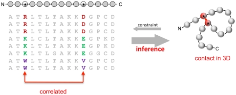
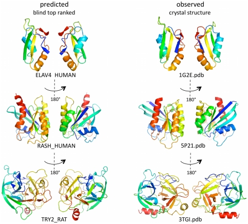
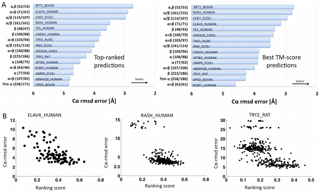
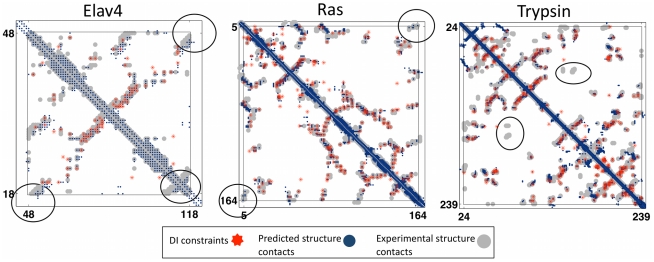
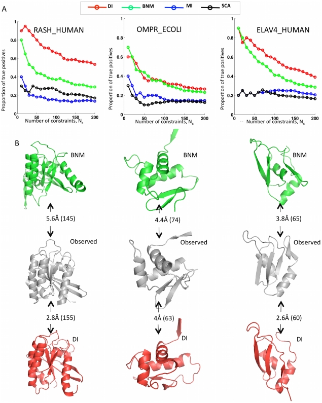
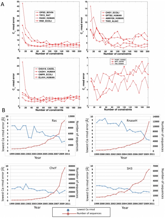
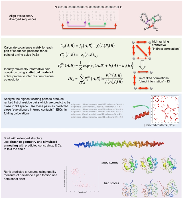
The evolutionary trajectory of a protein through sequence space is constrained by its function. Collections of sequence homologs record the outcomes of millions of evolutionary experiments in which the protein evolves according to these constraints. Deciphering the evolutionary record held in these sequences and exploiting it for predictive and engineering purposes presents a formidable challenge. The potential benefit of solving this challenge is amplified by the advent of inexpensive high-throughput genomic sequencing.In this paper we ask whether we can infer evolutionary constraints from a set of sequence homologs of a protein. The challenge is to distinguish true co-evolution couplings from the noisy set of observed correlations. We address this challenge using a maximum entropy model of the protein sequence, constrained by the statistics of the multiple sequence alignment, to infer residue pair couplings. Surprisingly, we find that the strength of these inferred couplings is an excellent predictor of residue-residue proximity in folded structures. Indeed, the top-scoring residue couplings are sufficiently accurate and well-distributed to define the 3D protein fold with remarkable accuracy.We quantify this observation by computing, from sequence alone, all-atom 3D structures of fifteen test proteins from different fold classes, ranging in size from 50 to 260 residues, including a G-protein coupled receptor. These blinded inferences are de novo, i.e., they do not use homology modeling or sequence-similar fragments from known structures. The co-evolution signals provide sufficient information to determine accurate 3D protein structure to 2.7-4.8 Å C(α)-RMSD error relative to the observed structure, over at least two-thirds of the protein (method called EVfold, details at http://EVfold.org). This discovery provides insight into essential interactions constraining protein evolution and will facilitate a comprehensive survey of the universe of protein structures, new strategies in protein and drug design, and the identification of functional genetic variants in normal and disease genomes.
Overview
The claims and statistics of this paper haven’t been checked yet.
Run the check — free, no account needed. It reads the open-access text and checks the sources it cites.
Integrity signals
Registry
Cites
Statistics
Figures
![Figure 3 Progress in contact prediction using the maximum entropy method. Extraction of evolutionary information about residue coupling and predicted contacts from multiple sequence alignments works much better using the global statistical model (right, Direct Information, DI, Equation 3) than the local statistical model (left, Mutual Information, MI, Equation 1). Predicted contacts for DI (red lines connecting the residues predicted to be coupled from sequence information) are better positioned in the experimentally observed structure (grey ribbon diagram), than those for MI (left, blue lines), shown here for the RAS protein (upper) and ELAV4 protein (lower). The DI residue pairs are also more evenly distributed along the chain and overlap more accurately with the contacts in the observed structure (red stars [predicted, grey circles [observed] in contact map; center, upper right triangle) than those using MI (blue [predicted], grey circles [observed]; center, lower left triangle). Details of contact maps for all proteins comparing predicted and observed contacts are in Figures S1 and S2, Text S1.](/paper-figures/ad0edff5338c41479018a65a69fdb4cd/protein-3d-structure-computed-from-evolutionary-sequence-variation-figure-3.jpg)
Verification
Claims the paper makes
The claims and statistics of this paper haven’t been checked yet.
Run the check — free, no account needed. It reads the open-access text and checks the sources it cites.
Claims other papers make about this one
Each is checked against this paper’s own text: does it say what the citing paper says it does?
In the citing paper
“The premise of methods to infer structure based on evolutionary couplings is that, because amino acids exist and evolve in the context of 3D structure, they are not free to evolve independently, but instead co-evolve in patterns reflective of the underlying structure.”
From Predicting multiple conformations via sequence clustering and AlphaFold2 (2024) · cites this paper as [9]
In the citing paper
“One of the major insights leveraged by AlphaFold 1,2 comes from the use of evolutionary covariations that can be extracted from large-scale multiple sequence alignments.[49][50][51][52][53][54]”
From AlphaFold and Implications for Intrinsically Disordered Proteins (2021) · cites this paper as [50]
Self-check
Percentages, sums and reported differences re-computed from the paper’s own numbers.
The self-check needs the paper’s full text.
Citations
Cites
The PyMOL Molecular Graphics System
DeLano, W. L. (2002) · Medical Entomology and Zoology · Cited by 18,147
Crystallography & NMR System: A New Software Suite for Macromolecular Structure Determination
Brünger, A. T., Adams, P. D., Clore, G. M., DeLano, W. L., Gros, P., Grosse‐Kunstleve, R. W., Jiang, J., Kuszewski, J. J., Nilges, M., Pannu, N., et al. (1998) · Acta Crystallographica Section D Biological Crystallography · doi:10.1107/s0907444998003254 · Cited by 15,723
The Pfam Protein Families Database
Bateman, A. G. (2002) · Nucleic Acids Research · doi:10.1093/nar/30.1.276 · Cited by 14,075
I-TASSER: a unified platform for automated protein structure and function prediction
Roy, A., Küçükural, A., & Zhang, Y. (2010) · Nature Protocols · doi:10.1038/nprot.2010.5 · Cited by 6,212
The Pfam protein families database
Finn, R. D., Mistry, J., Tate, J., Coggill, P., Heger, A., Pollington, J. E., Gavin, O. L., Gunasekaran, P., Ceric, G., Forslund, K., et al. (2010) · Nucleic Acids Research · doi:10.1093/nar/gkp985 · Cited by 2,694
Scoring function for automated assessment of protein structure template quality
Zhang, Y., & Skolnick, J. (2004) · Proteins: Structure, Function, and Bioinformatics · doi:10.1002/prot.20264 · Cited by 2,392
Atomic-level characterization of the structural dynamics of proteins
Shaw, D. E., Maragakis, P., Lindorff-Larsen, K., Piana, S., Dror, R. O., Eastwood, M. P., Bank, J. A., Jumper, J. M., Salmon, J. K., Shan, Y., et al. (2010) · Science · doi:10.1126/science.1187409 · Cited by 1,923
Weak pairwise correlations imply strongly correlated network states in a neural population
Schneidman, E., Berry, M. J., Segev, R., & Bialek, W. (2006) · Nature · doi:10.1038/nature04701 · Cited by 1,745
Holm, L., & Sander, C. (1996) · Science · doi:10.1126/science.273.5275.595 · Cited by 1,497
Self-citationMorcos, F., Pagnani, A., Lunt, B., Bertolino, A., Marks, D. S., Sander, C., Zecchina, R., Onuchic, J. N., Hwa, T., & Weigt, M. (2011) · Proceedings of the National Academy of Sciences · doi:10.1073/pnas.1111471108 · Cited by 1,434
Self-citation
Research works printed but not linked: 3
- KT Simons, 1999, Ab initio protein structure prediction of CASP III targets using ROSETTA., Proteins, Suppl 3, 171, 10.1002/(SICI)1097-0134(1999)37:3+<171::AID-PROT21>3.0.CO;2-Z · DOI
- MMT Mezard, 2009, Constraint satisfaction problems and neural networks: A statistical physics perspective., Journal of Physiology, 103, 107
- TR Lezon, 2006, Using the principle of entropy maximization to infer genetic interaction networks from gene expression patterns., Proceedings of the National Academy of Sciences of the United States of America, 103, 19033, 10.1073/pnas.0609152103 · DOI
Cited by
Sorted by citations
Items 401–500 of 1,005
Katsonis, P., & Lichtarge, O. (2017) · Human Mutation · doi:10.1002/humu.23266 · Cited by 29
Protein Structural Information and Evolutionary Landscape by In Vitro Evolution
Fantini, M., Lisi, S., Rios, P. D. L., Cattaneo, A., & Pastore, A. (2019) · Molecular Biology and Evolution · doi:10.1093/molbev/msz256 · Cited by 30
Accurate prediction of helix interactions and residue contacts in membrane proteins
Hönigschmid, P., & Frishman, D. (2016) · Journal of Structural Biology · doi:10.1016/j.jsb.2016.02.005 · Cited by 28
Comparing co-evolution methods and their application to template-free protein structure prediction
de Oliveira, S. H. P., Shi, J., & Deane, C. M. (2016) · Bioinformatics · doi:10.1093/bioinformatics/btw618 · Cited by 28
A large-scale comparative assessment of methods for residue–residue contact prediction
Wuyun, Q., Zheng, W. H., Peng, Z., & Yang, J. (2016) · Briefings in Bioinformatics · doi:10.1093/bib/bbw106 · Cited by 28
Morcos, F., & Onuchic, J. N. (2019) · Current Opinion in Structural Biology · doi:10.1016/j.sbi.2019.03.024 · Cited by 27
QuanTest2: benchmarking multiple sequence alignments using secondary structure prediction
Sievers, F., & Higgins, D. G. (2019) · Bioinformatics · doi:10.1093/bioinformatics/btz552 · Cited by 28
Peter, M. F., Gebhardt, C., Glaenzer, J., Schneberger, N., de Boer, M., Thomas, G. H., Cordes, T., & Hagelueken, G. (2020) · Journal of Molecular Biology · doi:10.1016/j.jmb.2020.166756 · Cited by 30
High throughput and quantitative enzymology in the genomic era
Mokhtari, D. A., Appel, M. J., Fordyce, P. M., & Herschlag, D. (2021) · Current Opinion in Structural Biology · doi:10.1016/j.sbi.2021.07.010 · Cited by 26
Zhou, Y., Jiao, L., Shen, J., Chi, H., Lu, Z., Liu, H., Lü, F., & Ping, Z. (2022) · International Journal of Molecular Sciences · doi:10.3390/ijms23179663 · Cited by 28
Coevolutionary Landscape of Kinase Family Proteins: Sequence Probabilities and Functional Motifs
Haldane, A., Flynn, W. F., He, P., & Levy, R. M. (2018) · Biophysical Journal · doi:10.1016/j.bpj.2017.10.028 · Cited by 28
Shah, N. H., & Kuriyan, J. (2018) · Nature Structural & Molecular Biology · doi:10.1038/s41594-018-0175-9 · Cited by 27
The emerging role of physical modeling in the future of structure determination
Gaalswyk, K., Muniyat, M. I., & MacCallum, J. L. (2018) · Current Opinion in Structural Biology · doi:10.1016/j.sbi.2018.03.005 · Cited by 28
Prediction Machines: Applied Machine Learning for Therapeutic Protein Design and Development
Kamerzell, T. J., & Middaugh, C. R. (2020) · Journal of Pharmaceutical Sciences · doi:10.1016/j.xphs.2020.11.034 · Cited by 28
Capturing coevolutionary signals inrepeat proteins
Espada, R., Parra, R. G., Mora, T., Walczak, A. M., & Ferreiro, D. U. (2015) · BMC Bioinformatics · doi:10.1186/s12859-015-0648-3 · Cited by 27
HCV E2 core structures and mAbs: something is still missing
Castelli, M., Clementi, N., Sautto, G. A., Pfaff, J. M., Kahle, K. M., Barnes, T., Doranz, B. J., Peraro, M. D., Clementi, M., Burioni, R., et al. (2014) · Drug Discovery Today · doi:10.1016/j.drudis.2014.08.011 · Cited by 27
Protein folding: a problem with multiple solutions
Bywater, R. P. (2012) · Journal of Biomolecular Structure and Dynamics · doi:10.1080/07391102.2012.703062 · Cited by 27
Co-evolution techniques are reshaping the way we do structural bioinformatics
de Oliveira, S. H. P., & Deane, C. M. (2017) · F1000Research · doi:10.12688/f1000research.11543.1 · Cited by 28
A protein coevolution method uncovers critical features of the Hepatitis C Virus fusion mechanism
Douam, F., Fusil, F., Enguehard, M., Dib, L., Nadalin, F., Schwaller, L., Hrebikova, G., Mancip, J., Mailly, L., Montserret, R., et al. (2018) · PLoS Pathogens · doi:10.1371/journal.ppat.1006908 · Cited by 27
DeepConPred2: An Improved Method for the Prediction of Protein Residue Contacts
Ding, W., Mao, W., Shao, D., Zhang, W., & Gong, H. (2018) · Computational and Structural Biotechnology Journal · doi:10.1016/j.csbj.2018.10.009 · Cited by 29
The intrinsic dimension of protein sequence evolution
Facco, E., Pagnani, A., Russo, E. T., & Laio, A. (2019) · PLoS Computational Biology · doi:10.1371/journal.pcbi.1006767 · Cited by 28
Self-citationA watershed moment for protein structure prediction
AlQuraishi, M. (2020) · Nature · doi:10.1038/d41586-019-03951-0 · Cited by 27
Inference of compressed Potts graphical models
Rizzato, F., Coucke, A., Leonardis, E. D., Barton, J. P., Tubiana, J., Monasson, R., & Cocco, S. (2020) · Physical review. E · doi:10.1103/physreve.101.012309 · Cited by 27
Deep Machine Learning for Computer-Aided Drug Design
Bajorath, J. (2022) · Frontiers in Drug Discovery · doi:10.3389/fddsv.2022.829043 · Cited by 28
Enhancing luciferase activity and stability through generative modeling of natural enzyme sequences
Xie, W.-J., Liu, D., Wang, X.-Y., Zhang, A., Wei, Q., Nandi, A., Dong, S.-W., & Warshel, A. (2023) · Proceedings of the National Academy of Sciences of the United States of America · doi:10.1073/pnas.2312848120 · Cited by 3
Global Quantitative Modeling of Chromatin Factor Interactions
Zhou, J., & Troyanskaya, O. G. (2014) · PLoS Computational Biology · doi:10.1371/journal.pcbi.1003525 · Cited by 27
Protein structure prediction: making AWSEM AWSEM‐ER by adding evolutionary restraints
Sirovetz, B. J., Schafer, N. P., & Wolynes, P. G. (2017) · Proteins Structure Function and Bioinformatics · doi:10.1002/prot.25367 · Cited by 27
Leone, V., & Faraldo‐Gómez, J. D. (2016) · The Journal of General Physiology · doi:10.1085/jgp.201611679 · Cited by 24
Evolutionary couplings and sequence variation effect predict protein binding sites
Littmann, M., Hopf, T. A., & Rost, B. (2018) · Proteins Structure Function and Bioinformatics · doi:10.1002/prot.25585 · Cited by 26
Self-citationAssessing the accuracy of direct-coupling analysis for RNA contact prediction
Cuturello, F., Tiana, G., & Bussi, G. (2020) · RNA · doi:10.1261/rna.074179.119 · Cited by 28
De novo protein structure prediction using ultra-fast molecular dynamics simulation
Cheung, N. J., & Yu, W. (2018) · PLoS ONE · doi:10.1371/journal.pone.0205819 · Cited by 26
Exploring the sequence fitness landscape of a bridge between protein folds
Tian, P., & Best, R. B. (2020) · PLoS Computational Biology · doi:10.1371/journal.pcbi.1008285 · Cited by 26
Shakeel, M., Du, J., Li, S., Zhou, Y.-J., Sarwar, N., & Bukhari, S. A. H. (2020) · Genes · doi:10.3390/genes11111258 · Cited by 26
Learning Graphs From Smooth and Graph-Stationary Signals With Hidden Variables
Buciulea, A., Rey, S., & Marques, A. G. (2022) · IEEE Transactions on Signal and Information Processing over Networks · doi:10.1109/tsipn.2022.3161079 · Cited by 27
Quantifying information of intracellular signaling: progress with machine learning
Tang, Y., & Hoffmann, A. (2022) · Reports on Progress in Physics · doi:10.1088/1361-6633/ac7a4a · Cited by 24
Achievements and challenges in structural bioinformatics and computational biophysics
Samish, I., Bourne, P. E., & Najmanovich, R. (2014) · Bioinformatics · doi:10.1093/bioinformatics/btu769 · Cited by 29
Parente, D. J., Ray, J. C. J., & Swint‐Kruse, L. (2015) · Proteins Structure Function and Bioinformatics · doi:10.1002/prot.24948 · Cited by 26
Improved Sampling Strategies for Protein Model Refinement Based on Molecular Dynamics Simulation
Heo, L., Arbour, C. F., Janson, G., & Feig, M. (2021) · Journal of Chemical Theory and Computation · doi:10.1021/acs.jctc.0c01238 · Cited by 26
Autofocused oracles for model-based designPreprint
Fannjiang, C., & Listgarten, J. (2020) · arXiv (Cornell University) · doi:10.48550/arxiv.2006.08052 · Cited by 37
Reliable and robust detection of coevolving protein residues†
Jeong, C.-S., & Kim, D. (2012) · Protein Engineering Design and Selection · doi:10.1093/protein/gzs081 · Cited by 25
Consensus designs and thermal stability determinants of a human glutamate transporter
Cirri, E., Brier, S., Assal, R., Canul-Tec, J. C., Chamot‐Rooke, J., & Reyes, N. (2018) · eLife · doi:10.7554/elife.40110 · Cited by 25
Deciphering the structure of the condensin protein complex
Krepel, D., Cheng, R. R., Pierro, M. D., & Onuchic, J. N. (2018) · Proceedings of the National Academy of Sciences · doi:10.1073/pnas.1812770115 · Cited by 26
Revealing evolutionary constraints on proteins through sequence analysis
Wang, S., Bitbol, A., & Wingreen, N. S. (2019) · PLoS Computational Biology · doi:10.1371/journal.pcbi.1007010 · Cited by 25
Zaucha, J. M., Heinzinger, M., Kulandaisamy, A., Kataka, E. S., Llorián-Salvador, Ó., Popov, P. A., Rost, B., Gromiha, M. M., Zhorov, B. S., & Frishman, D. (2020) · Briefings in Bioinformatics · doi:10.1093/bib/bbaa132 · Cited by 26
Mao, W., Kaya, C., Dutta, A., Horovitz, A., & Bahar, İ. (2015) · Bioinformatics · doi:10.1093/bioinformatics/btv103 · Cited by 25
Observation selection bias in contact prediction and its implications for structural bioinformatics
Orlando, G., Raimondi, D., & Vranken, W. (2016) · Scientific Reports · doi:10.1038/srep36679 · Cited by 25
Exploring protein sequence–function landscapes
Starr, T. N., & Thornton, J. W. (2017) · Nature Biotechnology · doi:10.1038/nbt.3786 · Cited by 25
Development of scoring functions for antibody sequence assessment and optimization
Seeliger, D. (2013) · PLoS ONE · doi:10.1371/journal.pone.0076909 · Cited by 24
Rossa, J., Protze, J., Kern, C., Piontek, A., Günzel, D., Krause, G., & Piontek, J. (2014) · Biochemical Journal · doi:10.1042/bj20140431 · Cited by 24
Simpkin, A. J., Thomas, J. M. H., Keegan, R. M., & Rigden, D. J. (2022) · Acta Crystallographica Section D Structural Biology · doi:10.1107/s2059798322003576 · Cited by 24
Natural Evolution Provides Strong Hints about Laboratory Evolution of Designer Enzymes
Xie, W. J., & Warshel, A. (2022) · Proceedings of the National Academy of Sciences · doi:10.1073/pnas.2207904119 · Cited by 23
Thirteen dubious ways to detect conserved structural RNAs
Gao, W. N. D., Yang, A., & Rivas, E. (2022) · IUBMB Life · doi:10.1002/iub.2694 · Cited by 21
Savojardo, C., Fariselli, P., Martelli, P. L., & Casadio, R. (2013) · Bioinformatics · doi:10.1093/bioinformatics/btt555 · Cited by 24
Emerging frontiers in protein structure prediction following the AlphaFold revolution
Rennie, M. L., & Oliver, M. R. (2025) · Journal of The Royal Society Interface · doi:10.1098/rsif.2024.0886 · Cited by 34
Population-specific design of de-immunized protein biotherapeutics
Schubert, B., Schärfe, C., Dönnes, P., Hopf, T. A., Marks, D. S., & Kohlbacher, O. (2018) · PLoS Computational Biology · doi:10.1371/journal.pcbi.1005983 · Cited by 24
Self-citationResidue co-evolution helps predict interaction sites in α-helical membrane proteins
Zeng, B., Hönigschmid, P., & Frishman, D. (2019) · Journal of Structural Biology · doi:10.1016/j.jsb.2019.02.009 · Cited by 23
Direct-acting antiviral resistance of Hepatitis C virus is promoted by epistasis
Zhang, H., Quadeer, A. A., & McKay, M. R. (2023) · Nature Communications · doi:10.1038/s41467-023-42550-6 · Cited by 23
Gupta, A. B., Wee, L. E., Zhou, Y., Hortsch, M., & Low, B. C. (2012) · PLoS ONE · doi:10.1371/journal.pone.0033863 · Cited by 23
ConEVA: a toolbox for comprehensive assessment of protein contacts
Adhikari, B., Nowotny, J., Bhattacharya, D., Hou, J., & Cheng, J. J. (2016) · BMC Bioinformatics · doi:10.1186/s12859-016-1404-z · Cited by 23
Proton-solute coupling mechanism of the maltose transporter from Saccharomyces cerevisiae
Henderson, R. K., & Poolman, B. (2017) · Scientific Reports · doi:10.1038/s41598-017-14438-1 · Cited by 25
Suplatov, D. A., Sharapova, Y., Timonina, D., Kopylov, K., & Švedas, V. K. (2017) · Journal of Bioinformatics and Computational Biology · doi:10.1142/s021972001840005x · Cited by 22
Using molecular simulation to explore the nanoscale dynamics of the plant kinome
Moffett, A. S., & Shukla, D. (2018) · Biochemical Journal · doi:10.1042/bcj20170299 · Cited by 23
DIRECT: RNA contact predictions by integrating structural patterns
Jian, Y., Wang, X., Qiu, J., Wang, H., Liu, Z., Zhao, Y., & Zeng, C. (2019) · BMC Bioinformatics · doi:10.1186/s12859-019-3099-4 · Cited by 24
Pucci, F., & Schug, A. H. (2019) · Methods · doi:10.1016/j.ymeth.2019.04.012 · Cited by 23
Deep generative models of genetic variation capture mutation effectsPreprint
Riesselman, A. J., Ingraham, J., & Marks, D. S. (2017) · bioRxiv (Cold Spring Harbor Laboratory) · doi:10.1101/235655 · Cited by 29
Self-citationCoevolving residues inform protein dynamics profiles and disease susceptibility of nSNVs
Butler, B. M., Kazan, I. C., Kumar, A., & Ozkan, S. B. (2018) · PLoS Computational Biology · doi:10.1371/journal.pcbi.1006626 · Cited by 19
Prediction and targeting of GPCR oligomer interfaces
Barreto, C. A. V., Baptista, S. J., Preto, A. J., Matos‐Filipe, P., Mourão, J., Melo, R., & Moreira, I. S. (2020) · Progress in molecular biology and translational science · doi:10.1016/bs.pmbts.2019.11.007 · Cited by 24
Cöster, M., Wittkopf, D., Kreuchwig, A., Kleinau, G., Thor, D., Krause, G., & Schöneberg, T. (2012) · The FASEB Journal · doi:10.1096/fj.12-203737 · Cited by 22
Predicting protein β-sheet contacts using a maximum entropy-based correlated mutation measure
Burkoff, N. S., Várnai, C., & Wild, D. L. (2013) · Bioinformatics · doi:10.1093/bioinformatics/btt005 · Cited by 22
Structural prediction of protein interactions and docking using conservation and coevolution
Andréani, J., Quignot, C., & Guérois, R. (2020) · Wiley Interdisciplinary Reviews Computational Molecular Science · doi:10.1002/wcms.1470 · Cited by 23
The role of local versus nonlocal physicochemical restraints in determining protein native structure
Skolnick, J., & Gao, M. (2021) · Current Opinion in Structural Biology · doi:10.1016/j.sbi.2020.10.008 · Cited by 21
Baltoumas, F. A., Karatzas, E., Páez-Espino, D., Venetsianou, N. K., Aplakidou, E., Oulas, A., Finn, R. D., Ovchinnikov, S., Pafilis, E., Kyrpides, N. C., et al. (2023) · Frontiers in Bioinformatics · doi:10.3389/fbinf.2023.1157956 · Cited by 21
Inferring Contacting Residues within and between Proteins: What Do the Probabilities Mean?
van Nimwegen, E. (2016) · PLoS Computational Biology · doi:10.1371/journal.pcbi.1004726 · Cited by 21
Kurt, F., Filiz, E., & Aydın, A. (2021) · Molecular Biology Reports · doi:10.1007/s11033-021-06620-6 · Cited by 21
Predicting Small Molecule Binding Nucleotides in RNA Structures Using RNA Surface Topography
Gao, J., Liu, H., Chen, Z., Zeng, C., & Zhao, Y. (2024) · Journal of Chemical Information and Modeling · doi:10.1021/acs.jcim.4c01264 · Cited by 21
Inverse Spin Glass and Related Maximum Entropy Problems
Castellana, M., & Bialek, W. (2014) · Physical Review Letters · doi:10.1103/physrevlett.113.117204 · Cited by 21
Emerging Diagnostic and Therapeutic Potentials of Human Hair Proteomics
Adeola, H. A., Wyk, J. C. V., Arowolo, A., Ngwanya, R. M., Mkentane, K., & Khumalo, N. P. (2017) · PROTEOMICS - CLINICAL APPLICATIONS · doi:10.1002/prca.201700048 · Cited by 21
Liu, H., & Liu, R. (2018) · Briefings in Bioinformatics · doi:10.1093/bib/bby123 · Cited by 22
Protein structure prediction assisted with sparse NMR data in CASP13
Sala, D., Huang, Y. J., Cole, C. A., Snyder, D. A., Liu, G., Ishida, Y., Swapna, G. V. T., Brock, K. P., Sander, C., Fidelis, K. A., et al. (2019) · Proteins Structure Function and Bioinformatics · doi:10.1002/prot.25837 · Cited by 23
Self-citationState-of-the-art web services for de novo protein structure prediction
Abriata, L. A., & Peraro, M. D. (2020) · Briefings in Bioinformatics · doi:10.1093/bib/bbaa139 · Cited by 21
Assessing the role of evolutionary information for enhancing protein language model embeddings
Erckert, K., & Rost, B. (2024) · Scientific Reports · doi:10.1038/s41598-024-71783-8 · Cited by 21
Combining Physicochemical and Evolutionary Information for Protein Contact Prediction
Schneider, M., & Brock, O. (2014) · PLoS ONE · doi:10.1371/journal.pone.0108438 · Cited by 20
Braun, T., Leman, J. K., & Lange, O. F. (2015) · PLoS Computational Biology · doi:10.1371/journal.pcbi.1004661 · Cited by 20
Structural insights into DNA sequence recognition by Type ISP restriction-modification enzymes
Kulkarni, M., Nirwan, N., van Aelst, K., Szczelkun, M. D., & Saikrishnan, K. (2016) · Nucleic Acids Research · doi:10.1093/nar/gkw154 · Cited by 20
Ramírez‐Guadiana, F. H., Rodrigues, C. D. A., Marquis, K. A., Campo, N., Barajas‐Ornelas, R. D. C., Brock, K. P., Marks, D. S., Kruse, A. C., & Rudner, D. Z. (2018) · PLoS Genetics · doi:10.1371/journal.pgen.1007753 · Cited by 21
Self-citationNon-Identifiability and the Blessings of Misspecification in Models of Molecular Fitness
Weinstein, E. N., Amin, A., Frazer, J., & Marks, D. S. (2022) · neural information processing systems · doi:10.52202/068431-0396 · Cited by 21
Self-citationSize and structure of the sequence space of repeat proteins
Marchi, J., Galpern, E. A., Espada, R., Ferreiro, D. U., Walczak, A. M., & Mora, T. (2019) · PLoS Computational Biology · doi:10.1371/journal.pcbi.1007282 · Cited by 20
Burkholz, S., Pokhrel, S., Kraemer, B. R., Mochly‐Rosen, D., Carback, R. T., Hodge, T., Harris, P. E., Ciotlos, S., Wang, L., Herst, C. V., et al. (2021) · Infection Genetics and Evolution · doi:10.1016/j.meegid.2021.104897 · Cited by 20
Bayesian approach to inverse statistical mechanics
Habeck, M. (2014) · Physical Review E · doi:10.1103/physreve.89.052113 · Cited by 20
Thermodynamics for a network of neurons: Signatures of criticalityPreprint
Tkačik, G., Mora, T., Marre, O., Amodei, D., Berry, M. J., & Bialek, W. (2014) · arXiv (Cornell University) · doi:10.48550/arxiv.1407.5946 · Cited by 20
Prediction of Contact Residue Pairs Based on Co-Substitution between Sites in Protein Structures
Miyazawa, S. (2013) · PLoS ONE · doi:10.1371/journal.pone.0054252 · Cited by 19
Hamborg, L., Granata, D., Olsen, J. G., Roche, J. V., Pedersen, L. E., Nielsen, A. T., Lindorff‐Larsen, K., & Teilum, K. (2021) · Communications Biology · doi:10.1038/s42003-021-02490-7 · Cited by 19
Extracting phylogenetic dimensions of coevolution reveals hidden functional signals
Colavin, A., Atolia, E., Bitbol, A., & Huang, K. C. (2022) · Scientific Reports · doi:10.1038/s41598-021-04260-1 · Cited by 21
Chinnam, N. B., Syed, A., Hura, G. L., Hammel, M., Tainer, J. A., & Tsutakawa, S. E. (2022) · Methods in enzymology on CD-ROM/Methods in enzymology · doi:10.1016/bs.mie.2022.09.023 · Cited by 19
Inferring protein fitness landscapes from laboratory evolution experiments
D’Costa, S., Hinds, E. C., Freschlin, C. R., Song, H., & Romero, P. A. (2023) · PLoS Computational Biology · doi:10.1371/journal.pcbi.1010956 · Cited by 19
Fluid protein fold space and its implications
Porter, L. L. (2023) · BioEssays · doi:10.1002/bies.202300057 · Cited by 19
Herrington, N. B., Li, Y. C., Li, Y. C., Pandey, G. S., & Schlessinger, A. (2024) · PLoS Computational Biology · doi:10.1371/journal.pcbi.1012302 · Cited by 27
The functional importance of co-evolving residues in proteins
Sandler, I., Zigdon, N., Levy, E., & Aharoni, A. (2013) · Cellular and Molecular Life Sciences · doi:10.1007/s00018-013-1458-2 · Cited by 19
Automated Procedure for Contact-Map-Based Protein Structure Reconstruction
Konopka, B. M., Ciombor, M., Kurczyńska, M., & Kotulska, M. (2014) · The Journal of Membrane Biology · doi:10.1007/s00232-014-9648-x · Cited by 19
A generative model for protein contact networks
Livi, L., Maiorino, E., Giuliani, A., Rizzi, A., & Sadeghian, A. (2015) · Journal of Biomolecular Structure and Dynamics · doi:10.1080/07391102.2015.1077736 · Cited by 19
Metadata
- Authors
- Marks, D. S., Colwell, L. J., Sheridan, R., Hopf, T. A., Pagnani, A., Zecchina, R., & Sander, C.
- Affiliations
- Harvard University (US)
- MRC Laboratory of Molecular Biology (GB)
- Memorial Sloan Kettering Cancer Center (US)
- Italian institute for Genomic Medicine (IT)
- Politecnico di Torino (IT)
- Corresponding author
- Marks, Debora S.
- Venue
- PLoS ONE
- Publisher
- Public Library of Science (PLoS)
- Published
- 2011-12-07
- Volume
- 6
- Issue
- 12
- Pages
- e28766
- ISSN
- 1932-6203 (ISSN-L)
- PMC
- PMC3233603
- OpenAlex
- W2061042699
- Cited by
- 1,144
- References
- 84
- Citations by year
- 2026: 61 · 2025: 60 · 2024: 47 · 2023: 72 · 2022: 96 · 2021: 104 · 2020: 90 · 2019: 110 · 2018: 91 · 2017: 107 · 2016: 71 · 2015: 74 · 2014: 72 · 2013: 56 · 2012: 29
- FWCI
- 20.79
- Type
- journal article
- Language
- en
- Open access
- gold
- doi.org
- Locations
- PLoS ONE (publishedVersion, cc-by)
- Europe PMC (PubMed Central) (publishedVersion, cc-by)
- PubMed Central (publishedVersion, cc-by)
- PubMed (publishedVersion)
- PORTO@Iris (submittedVersion, cc-by)
- DOAJ (DOAJ: Directory of Open Access Journals) (submittedVersion, cc-by-sa)
- Figshare (submittedVersion, cc-by)
- PORTO Publications Open Repository TOrino (Politecnico di Torino) (submittedVersion, cc-by)
- Funders
- Memorial Sloan-Kettering Cancer Center
- Studienstiftung des Deutschen Volkes
- National Institutes of Health (NIH U54, CA143798)
- Engineering and Physical Sciences Research Council (EP/H028064/1)
- National Cancer Institute (U54CA143798)
- National Institute of General Medical Sciences (R01GM106303)
- Topics
- Life Sciences › Biochemistry, Genetics and Molecular Biology › Molecular Biology › Protein Structure and Dynamics
- Life Sciences › Biochemistry, Genetics and Molecular Biology › Structural Biology › Enzyme Structure and Function
- Life Sciences › Biochemistry, Genetics and Molecular Biology › Molecular Biology › RNA and protein synthesis mechanisms
- Keywords
- protein structure prediction, coevolution, maximum entropy model, multiple sequence alignment, de novo structure prediction, protein evolution, G protein-coupled receptors, protein engineering
- MeSH terms
- Animals, Humans, Protein Conformation, Proteins, Rhodopsin, Trypsin, Genetic Variation, Drug Design, Reproducibility of Results, Models, Statistical, Genome, Protein Structure, Tertiary, Evolution, Molecular, Entropy, Computational Biology, Catalytic Domain, Imaging, Three-Dimensional
About these checks
Registry, retraction, Cites and statistics results are computed, not judged, with one exception: a printed reference that no index lists is read once by a model before it counts as a citation error. Claims are extracted by a model and kept only when their quote appears verbatim in the text; each cited source is then checked, with a model’s help, for whether it says what the claim relies on. Those verdicts can be wrong — read the claim’s quote and the cited source before relying on one.
Statistics and arithmetic are recomputed, not judged.
Confidence bands are shown only for fields calibrated on our annotated test set. None is calibrated yet, so no band is shown.
What counts as a claim ›How we measure confidence ›How we check this ›
Are you an author, or do you see a problem with this page? Request a review
