

The DOI matches its Crossref record; 83 of the 84 references it cites were found in a registry or an index.
Protein 3D Structure Computed from Evolutionary Sequence Variation
Open accessAbstract
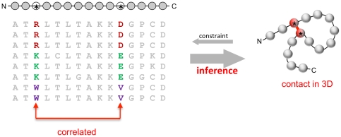
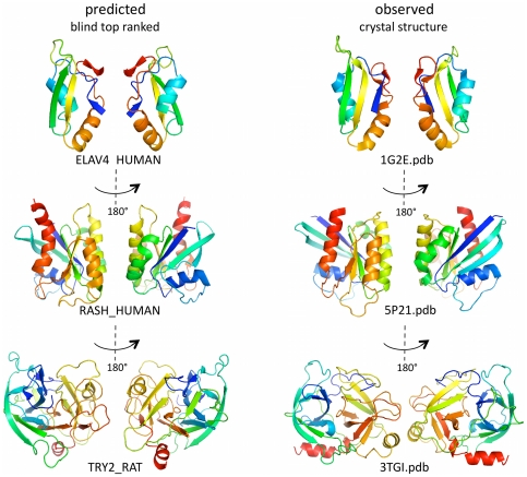
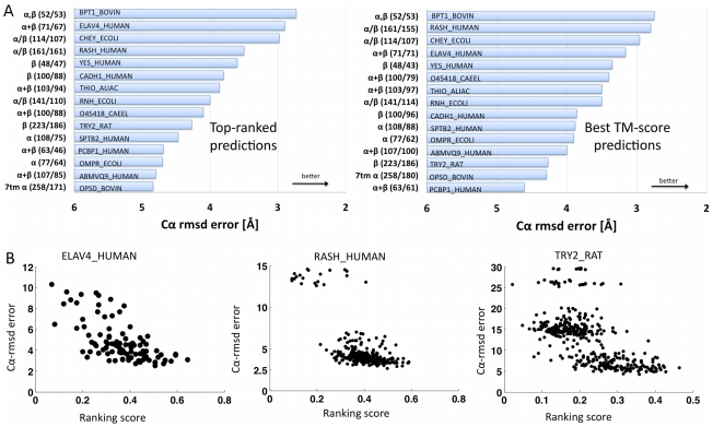
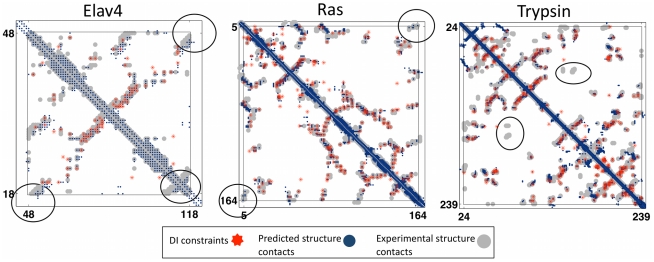
The evolutionary trajectory of a protein through sequence space is constrained by its function. Collections of sequence homologs record the outcomes of millions of evolutionary experiments in which the protein evolves according to these constraints. Deciphering the evolutionary record held in these sequences and exploiting it for predictive and engineering purposes presents a formidable challenge. The potential benefit of solving this challenge is amplified by the advent of inexpensive high-throughput genomic sequencing.In this paper we ask whether we can infer evolutionary constraints from a set of sequence homologs of a protein. The challenge is to distinguish true co-evolution couplings from the noisy set of observed correlations. We address this challenge using a maximum entropy model of the protein sequence, constrained by the statistics of the multiple sequence alignment, to infer residue pair couplings. Surprisingly, we find that the strength of these inferred couplings is an excellent predictor of residue-residue proximity in folded structures. Indeed, the top-scoring residue couplings are sufficiently accurate and well-distributed to define the 3D protein fold with remarkable accuracy.We quantify this observation by computing, from sequence alone, all-atom 3D structures of fifteen test proteins from different fold classes, ranging in size from 50 to 260 residues, including a G-protein coupled receptor. These blinded inferences are de novo, i.e., they do not use homology modeling or sequence-similar fragments from known structures. The co-evolution signals provide sufficient information to determine accurate 3D protein structure to 2.7-4.8 Å C(α)-RMSD error relative to the observed structure, over at least two-thirds of the protein (method called EVfold, details at http://EVfold.org). This discovery provides insight into essential interactions constraining protein evolution and will facilitate a comprehensive survey of the universe of protein structures, new strategies in protein and drug design, and the identification of functional genetic variants in normal and disease genomes.
Overview
The claims and statistics of this paper haven’t been checked yet.
Run the check — free, no account needed. It reads the open-access text and checks the sources it cites.
Integrity signals
Registry
Cites
Statistics
Figures
![Figure 3 Progress in contact prediction using the maximum entropy method. Extraction of evolutionary information about residue coupling and predicted contacts from multiple sequence alignments works much better using the global statistical model (right, Direct Information, DI, Equation 3) than the local statistical model (left, Mutual Information, MI, Equation 1). Predicted contacts for DI (red lines connecting the residues predicted to be coupled from sequence information) are better positioned in the experimentally observed structure (grey ribbon diagram), than those for MI (left, blue lines), shown here for the RAS protein (upper) and ELAV4 protein (lower). The DI residue pairs are also more evenly distributed along the chain and overlap more accurately with the contacts in the observed structure (red stars [predicted, grey circles [observed] in contact map; center, upper right triangle) than those using MI (blue [predicted], grey circles [observed]; center, lower left triangle). Details of contact maps for all proteins comparing predicted and observed contacts are in Figures S1 and S2, Text S1.](/paper-figures/ad0edff5338c41479018a65a69fdb4cd/protein-3d-structure-computed-from-evolutionary-sequence-variation-figure-3.jpg)
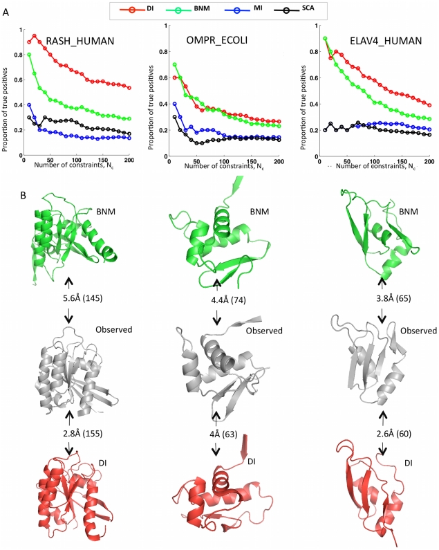
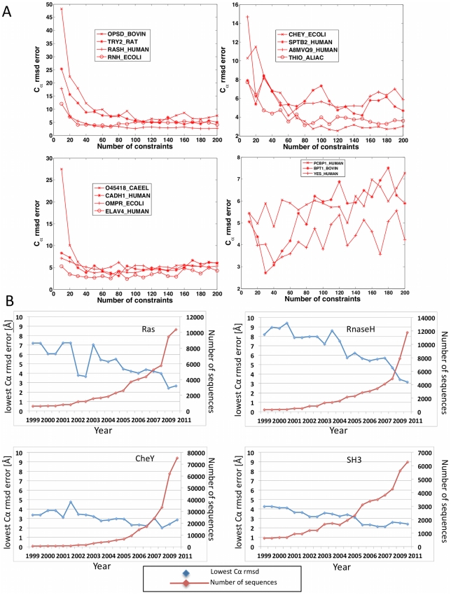
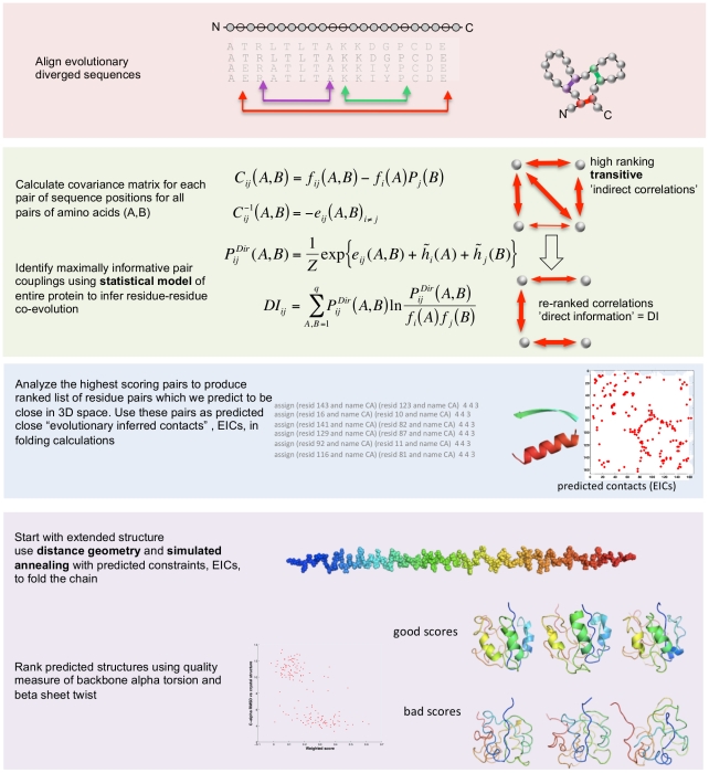
Verification
Claims the paper makes
The claims and statistics of this paper haven’t been checked yet.
Run the check — free, no account needed. It reads the open-access text and checks the sources it cites.
Claims other papers make about this one
Each is checked against this paper’s own text: does it say what the citing paper says it does?
In the citing paper
“The premise of methods to infer structure based on evolutionary couplings is that, because amino acids exist and evolve in the context of 3D structure, they are not free to evolve independently, but instead co-evolve in patterns reflective of the underlying structure.”
From Predicting multiple conformations via sequence clustering and AlphaFold2 (2024) · cites this paper as [9]
In the citing paper
“One of the major insights leveraged by AlphaFold 1,2 comes from the use of evolutionary covariations that can be extracted from large-scale multiple sequence alignments.[49][50][51][52][53][54]”
From AlphaFold and Implications for Intrinsically Disordered Proteins (2021) · cites this paper as [50]
Self-check
Percentages, sums and reported differences re-computed from the paper’s own numbers.
The self-check needs the paper’s full text.
Citations
Cites
The PyMOL Molecular Graphics System
DeLano, W. L. (2002) · Medical Entomology and Zoology · Cited by 18,147
Crystallography & NMR System: A New Software Suite for Macromolecular Structure Determination
Brünger, A. T., Adams, P. D., Clore, G. M., DeLano, W. L., Gros, P., Grosse‐Kunstleve, R. W., Jiang, J., Kuszewski, J. J., Nilges, M., Pannu, N., et al. (1998) · Acta Crystallographica Section D Biological Crystallography · doi:10.1107/s0907444998003254 · Cited by 15,723
The Pfam Protein Families Database
Bateman, A. G. (2002) · Nucleic Acids Research · doi:10.1093/nar/30.1.276 · Cited by 14,075
I-TASSER: a unified platform for automated protein structure and function prediction
Roy, A., Küçükural, A., & Zhang, Y. (2010) · Nature Protocols · doi:10.1038/nprot.2010.5 · Cited by 6,212
The Pfam protein families database
Finn, R. D., Mistry, J., Tate, J., Coggill, P., Heger, A., Pollington, J. E., Gavin, O. L., Gunasekaran, P., Ceric, G., Forslund, K., et al. (2010) · Nucleic Acids Research · doi:10.1093/nar/gkp985 · Cited by 2,694
Scoring function for automated assessment of protein structure template quality
Zhang, Y., & Skolnick, J. (2004) · Proteins: Structure, Function, and Bioinformatics · doi:10.1002/prot.20264 · Cited by 2,392
Atomic-level characterization of the structural dynamics of proteins
Shaw, D. E., Maragakis, P., Lindorff-Larsen, K., Piana, S., Dror, R. O., Eastwood, M. P., Bank, J. A., Jumper, J. M., Salmon, J. K., Shan, Y., et al. (2010) · Science · doi:10.1126/science.1187409 · Cited by 1,923
Weak pairwise correlations imply strongly correlated network states in a neural population
Schneidman, E., Berry, M. J., Segev, R., & Bialek, W. (2006) · Nature · doi:10.1038/nature04701 · Cited by 1,745
Holm, L., & Sander, C. (1996) · Science · doi:10.1126/science.273.5275.595 · Cited by 1,497
Self-citationMorcos, F., Pagnani, A., Lunt, B., Bertolino, A., Marks, D. S., Sander, C., Zecchina, R., Onuchic, J. N., Hwa, T., & Weigt, M. (2011) · Proceedings of the National Academy of Sciences · doi:10.1073/pnas.1111471108 · Cited by 1,434
Self-citation
Research works printed but not linked: 3
- KT Simons, 1999, Ab initio protein structure prediction of CASP III targets using ROSETTA., Proteins, Suppl 3, 171, 10.1002/(SICI)1097-0134(1999)37:3+<171::AID-PROT21>3.0.CO;2-Z · DOI
- MMT Mezard, 2009, Constraint satisfaction problems and neural networks: A statistical physics perspective., Journal of Physiology, 103, 107
- TR Lezon, 2006, Using the principle of entropy maximization to infer genetic interaction networks from gene expression patterns., Proceedings of the National Academy of Sciences of the United States of America, 103, 19033, 10.1073/pnas.0609152103 · DOI
Cited by
Sorted by citations
Items 701–800 of 1,005
Cheung, N. J., & Yu, W. (2019) · BMC Bioinformatics · doi:10.1186/s12859-019-2984-1 · Cited by 9
Klooster, J. S. V. ’., Bianchi, F., Doorn, R. B., Lorenzon, M., Lusseveld, J., Punter, C. M., & Poolman, B. (2020) · FEBS Journal · doi:10.1111/febs.15262 · Cited by 8
Gandarilla-Pérez, C. A., Mergny, P., Weigt, M., & Bitbol, A. (2020) · Physical review. E · doi:10.1103/physreve.101.032413 · Cited by 8
An engineered construct of cFLIP provides insight into DED1 structure and interactions
Panaitiu, A. E., Basiashvili, T., Mierke, D. F., & Pellegrini, M. (2021) · Structure · doi:10.1016/j.str.2021.10.011 · Cited by 8
Frederick, A. K., Thompson, S. L., Vakharia, Z. M., Cherney, M. M., Lei, H., Evenson, G. E., & Bowler, B. E. (2022) · Journal of Inorganic Biochemistry · doi:10.1016/j.jinorgbio.2022.111819 · Cited by 8
iBIS2Analyzer: a web server for a phylogeny-driven coevolution analysis of protein families
Oteri, F., Sarti, E., Nadalin, F., & Carbone, A. (2022) · Nucleic Acids Research · doi:10.1093/nar/gkac481 · Cited by 9
De Novo Membrane Protein Structure Prediction
Nugent, T. (2014) · Methods in molecular biology · doi:10.1007/978-1-4939-1465-4_15 · Cited by 9
Computational Identification of Novel Amino-Acid Interactions in HIV Gag via Correlated Evolution
Kalinina, O. V., Oberwinkler, H., Glass, B., Kräusslich, H., Russell, R. B., & Briggs, J. A. G. (2012) · PLoS ONE · doi:10.1371/journal.pone.0042468 · Cited by 8
Analysis of free modeling predictions by RBO aleph in CASP 11
Mabrouk, M. A., Werner, T., Schneider, M., Putz, I., & Brock, O. (2015) · Proteins Structure Function and Bioinformatics · doi:10.1002/prot.24950 · Cited by 8
Li, B., Mendenhall, J., Nguyen, E. D., Weiner, B. E., Fischer, A. W., & Meiler, J. (2017) · Proteins Structure Function and Bioinformatics · doi:10.1002/prot.25281 · Cited by 8
Hao, X., Zhang, G., & Zhou, X. (2018) · Computational Biology and Chemistry · doi:10.1016/j.compbiolchem.2018.02.003 · Cited by 8
Will Cryo-Electron Microscopy Shift the Current Paradigm in Protein Structure Prediction?
Abriata, L. A., & Peraro, M. D. (2020) · Journal of Chemical Information and Modeling · doi:10.1021/acs.jcim.0c00177 · Cited by 8
Roche, R., Bhattacharya, S., & Bhattacharya, D. (2021) · PLoS Computational Biology · doi:10.1371/journal.pcbi.1008753 · Cited by 10
Kinship and the Dynamics of the House: Rediscovering Dualism in the Pueblo Past
Heitman, C. C., & Plog, S. (2006) · Lincoln (University of Nebraska) · Cited by 8
Andreatta, M., Laplagne, S., Li, S. C., & Smale, S. T. (2013) · arXiv (Cornell University) · doi:10.48550/arxiv.1311.1301 · Cited by 8
Kilian, M., & Bischofs, I. B. (2023) · Molecular Microbiology · doi:10.1111/mmi.15169 · Cited by 8
RhoMax: Computational Prediction of Rhodopsin Absorption Maxima Using Geometric Deep Learning
Sela, M., Church, J. R., Schapiro, I., & Schneidman‐Duhovny, D. (2024) · Journal of Chemical Information and Modeling · doi:10.1021/acs.jcim.4c00467 · Cited by 7
Travers, T. S., Wang, K. J., López, C. A., & Gnanakaran, S. (2018) · Research in Microbiology · doi:10.1016/j.resmic.2018.01.002 · Cited by 7
A new approach for extracting information from protein dynamics
Liu, J., Amaral, L. A. N., & Keten, S. (2022) · Proteins Structure Function and Bioinformatics · doi:10.1002/prot.26421 · Cited by 7
Solution structure of a soluble fragment derived from a membrane protein by shotgun proteolysis
Allen, M. D., Christie, M., Jones, P. M., Porebski, B. T., Roome, B., Freund, S. M. V., Buckle, A. M., Bycroft, M., & Christ, D. (2015) · Protein Engineering Design and Selection · doi:10.1093/protein/gzv021 · Cited by 7
Polymorphic sites preferentially avoid co-evolving residues in MHC class I proteins
Dib, L., Salamin, N., & Gfeller, D. (2018) · PLoS Computational Biology · doi:10.1371/journal.pcbi.1006188 · Cited by 7
Huang, Y. J., Brock, K. P., Sander, C., Marks, D. S., & Montelione, G. T. (2018) · Advances in experimental medicine and biology · doi:10.1007/978-981-13-2200-6_10 · Cited by 7
Self-citationA Minimal yet Flexible Likelihood Framework to Assess Correlated Evolution
Behdenna, A., Godfroid, M., Petot, P., Pothier, J. F., Noûs, C., Lambert, A., & Achaz, G. (2021) · Systematic Biology · doi:10.1093/sysbio/syab092 · Cited by 7
Impact of population size on early adaptation in rugged fitness landscapes
Servajean, R., & Bitbol, A. (2023) · Philosophical Transactions of the Royal Society B Biological Sciences · doi:10.1098/rstb.2022.0045 · Cited by 6
Nemoto, W., Saito, A., & Oikawa, H. (2013) · Computational and Structural Biotechnology Journal · doi:10.5936/csbj.201308007 · Cited by 7
Kinjo, A. R. (2016) · Biophysics and Physicobiology · doi:10.2142/biophysico.13.0_45 · Cited by 7
Kamisetty, H., Ghosh, B., LANGMEAD, C. J., & Bailey‐Kellogg, C. (2015) · Journal of Computational Biology · doi:10.1089/cmb.2014.0289 · Cited by 7
Bioinformatics: A Way Forward to Explore “Plant Omics”
Rahman, M., Shaheen, T., Rahman, M.-U., Iqbal, M. A., & Zafar, Y. (2016) · InTech eBooks · doi:10.5772/64043 · Cited by 7
Pareto Optimization of Combinatorial Mutagenesis Libraries
Verma, D., Grigoryan, G., & Bailey‐Kellogg, C. (2018) · IEEE Transactions on Computational Biology and Bioinformatics · doi:10.1109/tcbb.2018.2858794 · Cited by 7
DCA-MOL: A PyMOL Plugin To Analyze Direct Evolutionary Couplings
Jarmolinska, A. I., Zhou, Q., Sulkowska, J. I., & Morcos, F. (2019) · Journal of Chemical Information and Modeling · doi:10.1021/acs.jcim.8b00690 · Cited by 7
Mukherjee, I., & Chakrabarti, S. (2021) · Computational and Structural Biotechnology Journal · doi:10.1016/j.csbj.2021.06.039 · Cited by 7
Protein alignment based on higher order conditional random fields for template-based modeling
Morales-Cordovilla, J. A., Sánchez, V., & Ratajczak, M. (2018) · PLoS ONE · doi:10.1371/journal.pone.0197912 · Cited by 7
Statistical investigations of protein residue direct couplings
Neuwald, A. F., & Altschul, S. F. (2018) · PLoS Computational Biology · doi:10.1371/journal.pcbi.1006237 · Cited by 7
Graphical Techniques to Visualize the Amphiphilic Structures of Antimicrobial Peptides
Phoenix, D. A., Dennison, S. R., & Harris, F. (2013) · Wiley eBooks · doi:10.1002/9783527652853.ch4 · Cited by 7
Bayesian Sparsity for Intractable DistributionsPreprint
Ingraham, J., & Marks, D. S. (2016) · arXiv (Cornell University) · Cited by 7
Self-citationPconsC4: fast, free, easy, and accurate contact predictionsPreprint
Michel, M., Hurtado, D. M., & Elofsson, A. (2018) · bioRxiv (Cold Spring Harbor Laboratory) · doi:10.1101/383133 · Cited by 7
Force-Field Benchmarking by Alternatives: A Systematic Study of Ten Small α- and β-ProteinsPreprint
Jana, K., & Kepp, K. P. (2020) · bioRxiv (Cold Spring Harbor Laboratory) · doi:10.1101/2020.03.03.974477 · Cited by 7
A structured observation distribution for generative biological sequence prediction and forecastingPreprint
Weinstein, E. N., & Marks, D. S. (2020) · bioRxiv (Cold Spring Harbor Laboratory) · doi:10.1101/2020.07.31.231381 · Cited by 8
Self-citationProtEx: A Retrieval-Augmented Approach for Protein Function PredictionPreprint
Shaw, P., Gurram, B., Belanger, D., Gane, A., Bileschi, M. L., Colwell, L. J., Toutanova, K., & Parikh, A. P. (2024) · bioRxiv (Cold Spring Harbor Laboratory) · doi:10.1101/2024.05.30.596539 · Cited by 8
Self-citationImproving prediction accuracy in chimeric proteins with windowed multiple sequence alignment
Vedula, S., Bronstein, A. M., & Marx, A. (2025) · Computational and Structural Biotechnology Journal · doi:10.1016/j.csbj.2025.07.039 · Cited by 7
Kaschner, M., Schillinger, O., Fettweiss, T., Nutschel, C., Krause, F., Fulton, A., Strodel, B., Stadler, A. M., Jaeger, K., & Krauß, U. (2017) · Scientific Reports · doi:10.1038/srep42592 · Cited by 6
Mulnaes, D., Schott‐Verdugo, S., Koenig, F., & Gohlke, H. (2021) · Journal of Chemical Theory and Computation · doi:10.1021/acs.jctc.1c00685 · Cited by 6
Gaber, A., Alsanie, W. F., Alhomrani, M., Alamri, A. S., Alyami, H., Shakya, S., Habeeballah, H. H., Alkhatabi, H. A., Felimban, R. I., Alamri, A., et al. (2022) · Molecules · doi:10.3390/molecules27185883 · Cited by 7
Machine Learning Applications in Drug Discovery
Ugurlu, S. Y., McDonald, D., & He, S. (2025) · French-Ukrainian Journal of Chemistry · doi:10.17721/fujcv13i1p1-66 · Cited by 6
Improved chemical shift prediction by Rosetta conformational sampling
Tian, Y., Opella, S. J., & Marassi, F. M. (2012) · Journal of Biomolecular NMR · doi:10.1007/s10858-012-9677-7 · Cited by 6
Modeling global changes induced by local perturbations to the HIV-1 capsid
Bergman, S., & Lezon, T. R. (2016) · Journal of Molecular Graphics and Modelling · doi:10.1016/j.jmgm.2016.12.003 · Cited by 6
Exploring RNA conformational space under sparse distance restraints
Taylor, W. R., & Hamilton, R. S. (2017) · Scientific Reports · doi:10.1038/srep44074 · Cited by 6
Protein Structures, Interactions and Function from Evolutionary Couplings
Hopf, T. A., & Marks, D. S. (2017) · Springer eBooks · doi:10.1007/978-94-024-1069-3_2 · Cited by 6
Self-citationMateos, G., Segarra, S., & Marques, A. G. (2018) · Elsevier eBooks · doi:10.1016/b978-0-12-813677-5.00013-4 · Cited by 6
Bittrich, S., Schroeder, M., & Labudde, D. (2018) · PLoS ONE · doi:10.1371/journal.pone.0206369 · Cited by 4
Entropy, Fluctuations, and Disordered Proteins
Faraggi, E., Dunker, A. K., Jernigan, R. L., & Kloczkowski, A. (2019) · Entropy · doi:10.3390/e21080764 · Cited by 6
Divergence in Dimerization and Activity of Primate APOBEC3C
Gaba, A., Hix, M. A., Suhail, S., Flath, B., Boysan, B., Williams, D. R., Pelletier, T., Emerman, M., Morcos, F., Cisneros, G. A., et al. (2021) · Journal of Molecular Biology · doi:10.1016/j.jmb.2021.167306 · Cited by 6
Liu, Z., Zhu, Y., Shen, L.-C., Xiao, X., Qiu, W., & Yu, D. (2023) · Computers in Biology and Medicine · doi:10.1016/j.compbiomed.2023.107529 · Cited by 6
Improved Multiple Sequence Alignments Using Coupled Pattern Mining
Hossain, K. S. M. T., Patnaik, D., Laxman, S., Jain, P., Bailey‐Kellogg, C., & Ramakrishnan, N. (2013) · IEEE Transactions on Computational Biology and Bioinformatics · doi:10.1109/tcbb.2013.36 · Cited by 6
Answering evolutionary questions: A guide for mechanistic biologists
Masel, J., & Promislow, D. (2016) · BioEssays · doi:10.1002/bies.201600029 · Cited by 7
Várnai, C., Burkoff, N. S., & Wild, D. L. (2017) · PLoS ONE · doi:10.1371/journal.pone.0169356 · Cited by 6
Protein Inter-Residue Contacts Prediction: Methods, Performances and Applications
Jing, X., Dong, Q., Lu, R., & Dong, Q. (2018) · Current Bioinformatics · doi:10.2174/1574893613666181109130430 · Cited by 6
Gopal, D., & Skariyachan, S. (2020) · Methods in pharmacology and toxicology · doi:10.1007/7653_2020_44 · Cited by 8
Krishnamohan, A., Hamilton, G. L., Goutam, R., Sanabria, H., & Morcos, F. (2023) · The Journal of Physical Chemistry B · doi:10.1021/acs.jpcb.2c06720 · Cited by 6
Two-Stage Distance Feature-based Optimization Algorithm for De novo Protein Structure Prediction
Zhang, G., Wang, X., Ma, L., Wang, L., Hu, J. S., & Zhou, X. (2019) · IEEE Transactions on Computational Biology and Bioinformatics · doi:10.1109/tcbb.2019.2917452 · Cited by 6
Wang, B., Yan, C., Lou, S., Emani, P. S., Li, B., Xu, M., Kong, X., Meyerson, W. U., Yang, Y., Lee, D., et al. (2019) · Structure · doi:10.1016/j.str.2019.06.001 · Cited by 6
Abriata, L. A. (2017) · bioRxiv (Cold Spring Harbor Laboratory) · doi:10.1101/123349 · Cited by 6
Green, A. G., Elhabashy, H., Brock, K. P., Maddamsetti, R., Kohlbacher, O., & Marks, D. S. (2019) · bioRxiv (Cold Spring Harbor Laboratory) · doi:10.1101/791293 · Cited by 6
Self-citationLiu, J., He, G.-X., Zhao, K., & Zhang, G. (2022) · bioRxiv (Cold Spring Harbor Laboratory) · doi:10.1101/2022.01.11.475831 · Cited by 7
Evolving Drug Design Methodology: from QSAR to AIDDPreprint
Xu, J. (2022) · ChemRxiv · doi:10.26434/chemrxiv-2022-9fwmg · Cited by 6
Blind Assessment of Monomeric AlphaFold2 Protein Structure Models with Experimental NMR DataPreprint
Li, E. H., Spaman, L. E., Tejero, R., Huang, Y. J., Ramelot, T. A., Fraga, K. J., Prestegard, J. H., Kennedy, M. A., & Montelione, G. T. (2023) · bioRxiv (Cold Spring Harbor Laboratory) · doi:10.1101/2023.01.22.525096 · Cited by 8
Birnbaum, F., & Keating, A. E. (2026) · Proceedings of the National Academy of Sciences · doi:10.1073/pnas.2535494123 · Cited by 7
Wang, J., Ye, H., Li, X., Lv, X., Lou, J., Chen, Y., Yu, S., & Zhang, L. (2023) · International Journal of Molecular Sciences · doi:10.3390/ijms25010406 · Cited by 5
Sacco, M., Testa, M. F., Ferretti, A., Basso, M., Lancellotti, S., Tardugno, M., Gennaro, L. D., Concolino, P., Minucci, A., Spoliti, C., et al. (2024) · Journal of Thrombosis and Haemostasis · doi:10.1016/j.jtha.2024.07.008 · Cited by 5
Kinshuk, S., Li, L., Meckes, B., & Chan, C. T. Y. (2024) · International Journal of Molecular Sciences · doi:10.3390/ijms25158320 · Cited by 5
Forecasting residue–residue contact prediction accuracy
Wozniak, P. P., Konopka, B. M., Xu, J., Vriend, G., & Kotulska, M. (2017) · Bioinformatics · doi:10.1093/bioinformatics/btx416 · Cited by 4
pycofitness—Evaluating the fitness landscape of RNA and protein sequences
Pucci, F., Zerihun, M. B., Rooman, M. J., & Schug, A. H. (2024) · Bioinformatics · doi:10.1093/bioinformatics/btae074 · Cited by 5
Direct coupling analysis and the attention mechanism
(2025) · doi:10.1186/s12859-025-06062-y · Cited by 5
Self-citationCocco, S., Monasson, R., & Weigt, M. (2013) · Journal of Physics Conference Series · doi:10.1088/1742-6596/473/1/012010 · Cited by 6
Two Rapidly Evolving Genes Contribute to Male Fitness in Drosophila
Reinhardt, J. A., & Jones, C. D. (2013) · Journal of Molecular Evolution · doi:10.1007/s00239-013-9594-8 · Cited by 5
Perlaza-Jiménez, L., & Walther, D. (2018) · Nucleic Acids Research · doi:10.1093/nar/gky576 · Cited by 5
Bittrich, S., Schroeder, M., & Labudde, D. (2019) · Scientific Reports · doi:10.1038/s41598-019-55047-4 · Cited by 7
Inferring couplings in networks across order-disorder phase transitions
Ngampruetikorn, V., Sachdeva, V., Torrence, J., Humplik, J., Schwab, D. J., & Palmer, S. E. (2022) · Physical Review Research · doi:10.1103/physrevresearch.4.023240 · Cited by 5
CAB-Align: A Flexible Protein Structure Alignment Method Based on the Residue-Residue Contact Area
Terashi, G., & Takeda‐Shitaka, M. (2015) · PLoS ONE · doi:10.1371/journal.pone.0141440 · Cited by 5
Chonofsky, M., de Oliveira, S. H. P., Krawczyk, K., & Deane, C. M. (2019) · Bioinformatics · doi:10.1093/bioinformatics/btz816 · Cited by 5
Inverse Ising techniques to infer underlying mechanisms from data*
Zeng, H.-L., & Aurell, E. (2020) · Chinese Physics B · doi:10.1088/1674-1056/ab8da6 · Cited by 8
Intragenic compensation through the lens of deep mutational scanning
Azbukina, N. V., Zharikova, A. A., & Ramensky, V. E. (2022) · Biophysical Reviews · doi:10.1007/s12551-022-01005-w · Cited by 6
Schlessinger, A. (2014) · Springer series in biophysics · doi:10.1007/978-3-642-53839-1_2 · Cited by 5
Evolution-based design of chorismate mutase enzymesPreprint
Russ, W. P., Figliuzzi, M., Stocker, C., Barrat-Charlaix, P., Socolich, M., Kast, P., Hilvert, D., Monasson, R., Cocco, S., Weigt, M., et al. (2020) · bioRxiv (Cold Spring Harbor Laboratory) · doi:10.1101/2020.04.01.020487 · Cited by 5
Tuned Fitness Landscapes for Benchmarking Model-Guided Protein DesignPreprint
Thomas, N., Agarwala, A., Belanger, D., Song, Y. S., & Colwell, L. J. (2022) · bioRxiv (Cold Spring Harbor Laboratory) · doi:10.1101/2022.10.28.514293 · Cited by 5
Self-citationMinimal epistatic networks from integrated sequence and mutational protein dataPreprint
Cocco, S., Posani, L., & Monasson, R. (2023) · bioRxiv (Cold Spring Harbor Laboratory) · doi:10.1101/2023.09.25.559251 · Cited by 4
McWhite, C. D., & Singh, M. (2022) · doi:10.1101/2022.10.21.513099 · Cited by 4
Shukla, D., Martin, J., Morcos, F., & Potoyan, D. A. (2025) · Journal of Chemical Theory and Computation · doi:10.1021/acs.jctc.4c01774 · Cited by 4
Minimax entropy: The statistical physics of optimal models
Carcamo, D. P., Weaver, N., Dixit, P. D., & Lynn, C. W. (2025) · Physical review. E · doi:10.1103/kr9x-q59y · Cited by 5
How Different are Self and Nonself?
Tiffeau-Mayer, A., Levine, J., Russo, C. J., Marcou, Q., Bialek, W., & Greenbaum, B. D. (2026) · PRX Life · doi:10.1103/fbct-vzwm · Cited by 10
Structural ontogeny of protein-protein interactions
Yang, A., Jiang, H., Jude, K. M., Akpinaroglu, D., Allenspach, S., Li, A. J., Bowden, J., Perez, C. P., Liu, L. D., Huang, P., et al. (2026) · Science · doi:10.1126/science.adx6931 · Cited by 5
Mechanistic basis of teichoic acid transport by a gatekeeper flippase
Cebrero, G., Chidananda, A. H., Cester, E., Dénéréaz, J., Demir, E. S., Mathew, A. T., Bhowmik, D. R., de Capitani, M., Reymond, J.-L., Kannan, N., et al. (2026) · Nature Communications · doi:10.1038/s41467-026-73616-w · Cited by 3
Bochtler, M. (2024) · BioEssays · doi:10.1002/bies.202400155 · Cited by 6
Inference for interacting linear waves in ordered and random media
Tyagi, P., Pagnani, A., Antenucci, F., Ibáñez-Berganza, M., & Leuzzi, L. (2015) · Journal of Statistical Mechanics Theory and Experiment · doi:10.1088/1742-5468/2015/05/p05031 · Cited by 4
Self-citationA Comparative Evaluation of the Structural and Dynamic Properties of Insect Odorant Binding Proteins
Tzotzos, G. T. (2022) · Biomolecules · doi:10.3390/biom12020282 · Cited by 5
Integrative structure determination of histones H3 and H4 using genetic interactions
Echeverria, I., Braberg, H., Krogan, N. J., & Šali, A. (2022) · FEBS Journal · doi:10.1111/febs.16435 · Cited by 4
Deep mutational scanning to probe specificity determinants in proteins
Bhowmick, J., Chandra, S., & Varadarajan, R. (2022) · Elsevier eBooks · doi:10.1016/b978-0-323-99533-7.00005-4 · Cited by 5
Extrinsic vs intrinsic criticality in systems with many components
Ngampruetikorn, V., Nemenman, I. M., & Schwab, D. J. (2025) · Physical Review Research · doi:10.1103/physrevresearch.7.013188 · Cited by 4
Xu, G., Guo, H. M., Yu, Z., Wang, S., Shen, D., Yang, L., Wu, J., Chen, B., & Yu, H. (2024) · Structure · doi:10.1016/j.str.2024.06.014 · Cited by 4
Symmetry, gauge freedoms, and the interpretability of sequence-function relationships
Posfai, A., McCandlish, D. M., & Kinney, J. B. (2025) · Physical Review Research · doi:10.1103/physrevresearch.7.023005 · Cited by 4
Metadata
- Authors
- Marks, D. S., Colwell, L. J., Sheridan, R., Hopf, T. A., Pagnani, A., Zecchina, R., & Sander, C.
- Affiliations
- Harvard University (US)
- MRC Laboratory of Molecular Biology (GB)
- Memorial Sloan Kettering Cancer Center (US)
- Italian institute for Genomic Medicine (IT)
- Politecnico di Torino (IT)
- Corresponding author
- Marks, Debora S.
- Venue
- PLoS ONE
- Publisher
- Public Library of Science (PLoS)
- Published
- 2011-12-07
- Volume
- 6
- Issue
- 12
- Pages
- e28766
- ISSN
- 1932-6203 (ISSN-L)
- PMC
- PMC3233603
- OpenAlex
- W2061042699
- Cited by
- 1,144
- References
- 84
- Citations by year
- 2026: 61 · 2025: 60 · 2024: 47 · 2023: 72 · 2022: 96 · 2021: 104 · 2020: 90 · 2019: 110 · 2018: 91 · 2017: 107 · 2016: 71 · 2015: 74 · 2014: 72 · 2013: 56 · 2012: 29
- FWCI
- 20.79
- Type
- journal article
- Language
- en
- Open access
- gold
- doi.org
- Locations
- PLoS ONE (publishedVersion, cc-by)
- Europe PMC (PubMed Central) (publishedVersion, cc-by)
- PubMed Central (publishedVersion, cc-by)
- PubMed (publishedVersion)
- PORTO@Iris (submittedVersion, cc-by)
- DOAJ (DOAJ: Directory of Open Access Journals) (submittedVersion, cc-by-sa)
- Figshare (submittedVersion, cc-by)
- PORTO Publications Open Repository TOrino (Politecnico di Torino) (submittedVersion, cc-by)
- Funders
- Memorial Sloan-Kettering Cancer Center
- Studienstiftung des Deutschen Volkes
- National Institutes of Health (NIH U54, CA143798)
- Engineering and Physical Sciences Research Council (EP/H028064/1)
- National Cancer Institute (U54CA143798)
- National Institute of General Medical Sciences (R01GM106303)
- Topics
- Life Sciences › Biochemistry, Genetics and Molecular Biology › Molecular Biology › Protein Structure and Dynamics
- Life Sciences › Biochemistry, Genetics and Molecular Biology › Structural Biology › Enzyme Structure and Function
- Life Sciences › Biochemistry, Genetics and Molecular Biology › Molecular Biology › RNA and protein synthesis mechanisms
- Keywords
- protein structure prediction, coevolution, maximum entropy model, multiple sequence alignment, de novo structure prediction, protein evolution, G protein-coupled receptors, protein engineering
- MeSH terms
- Animals, Humans, Protein Conformation, Proteins, Rhodopsin, Trypsin, Genetic Variation, Drug Design, Reproducibility of Results, Models, Statistical, Genome, Protein Structure, Tertiary, Evolution, Molecular, Entropy, Computational Biology, Catalytic Domain, Imaging, Three-Dimensional
About these checks
Registry, retraction, Cites and statistics results are computed, not judged, with one exception: a printed reference that no index lists is read once by a model before it counts as a citation error. Claims are extracted by a model and kept only when their quote appears verbatim in the text; each cited source is then checked, with a model’s help, for whether it says what the claim relies on. Those verdicts can be wrong — read the claim’s quote and the cited source before relying on one.
Statistics and arithmetic are recomputed, not judged.
Confidence bands are shown only for fields calibrated on our annotated test set. None is calibrated yet, so no band is shown.
What counts as a claim ›How we measure confidence ›How we check this ›
Are you an author, or do you see a problem with this page? Request a review
