

The DOI matches its Crossref record; 83 of the 84 references it cites were found in a registry or an index.
Protein 3D Structure Computed from Evolutionary Sequence Variation
Open accessAbstract
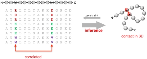
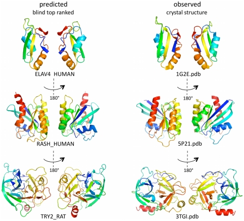
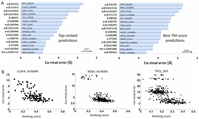
The evolutionary trajectory of a protein through sequence space is constrained by its function. Collections of sequence homologs record the outcomes of millions of evolutionary experiments in which the protein evolves according to these constraints. Deciphering the evolutionary record held in these sequences and exploiting it for predictive and engineering purposes presents a formidable challenge. The potential benefit of solving this challenge is amplified by the advent of inexpensive high-throughput genomic sequencing.In this paper we ask whether we can infer evolutionary constraints from a set of sequence homologs of a protein. The challenge is to distinguish true co-evolution couplings from the noisy set of observed correlations. We address this challenge using a maximum entropy model of the protein sequence, constrained by the statistics of the multiple sequence alignment, to infer residue pair couplings. Surprisingly, we find that the strength of these inferred couplings is an excellent predictor of residue-residue proximity in folded structures. Indeed, the top-scoring residue couplings are sufficiently accurate and well-distributed to define the 3D protein fold with remarkable accuracy.We quantify this observation by computing, from sequence alone, all-atom 3D structures of fifteen test proteins from different fold classes, ranging in size from 50 to 260 residues, including a G-protein coupled receptor. These blinded inferences are de novo, i.e., they do not use homology modeling or sequence-similar fragments from known structures. The co-evolution signals provide sufficient information to determine accurate 3D protein structure to 2.7-4.8 Å C(α)-RMSD error relative to the observed structure, over at least two-thirds of the protein (method called EVfold, details at http://EVfold.org). This discovery provides insight into essential interactions constraining protein evolution and will facilitate a comprehensive survey of the universe of protein structures, new strategies in protein and drug design, and the identification of functional genetic variants in normal and disease genomes.
Overview
The claims and statistics of this paper haven’t been checked yet.
Run the check — free, no account needed. It reads the open-access text and checks the sources it cites.
Integrity signals
Registry
Cites
Statistics
Figures
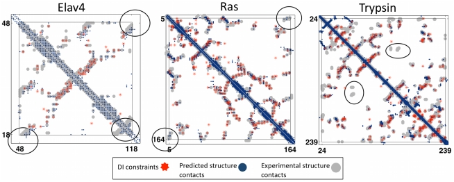
![Figure 3 Progress in contact prediction using the maximum entropy method. Extraction of evolutionary information about residue coupling and predicted contacts from multiple sequence alignments works much better using the global statistical model (right, Direct Information, DI, Equation 3) than the local statistical model (left, Mutual Information, MI, Equation 1). Predicted contacts for DI (red lines connecting the residues predicted to be coupled from sequence information) are better positioned in the experimentally observed structure (grey ribbon diagram), than those for MI (left, blue lines), shown here for the RAS protein (upper) and ELAV4 protein (lower). The DI residue pairs are also more evenly distributed along the chain and overlap more accurately with the contacts in the observed structure (red stars [predicted, grey circles [observed] in contact map; center, upper right triangle) than those using MI (blue [predicted], grey circles [observed]; center, lower left triangle). Details of contact maps for all proteins comparing predicted and observed contacts are in Figures S1 and S2, Text S1.](/paper-figures/ad0edff5338c41479018a65a69fdb4cd/protein-3d-structure-computed-from-evolutionary-sequence-variation-figure-3.jpg)
Verification
Claims the paper makes
The claims and statistics of this paper haven’t been checked yet.
Run the check — free, no account needed. It reads the open-access text and checks the sources it cites.
Claims other papers make about this one
Each is checked against this paper’s own text: does it say what the citing paper says it does?
In the citing paper
“The premise of methods to infer structure based on evolutionary couplings is that, because amino acids exist and evolve in the context of 3D structure, they are not free to evolve independently, but instead co-evolve in patterns reflective of the underlying structure.”
From Predicting multiple conformations via sequence clustering and AlphaFold2 (2024) · cites this paper as [9]
In the citing paper
“One of the major insights leveraged by AlphaFold 1,2 comes from the use of evolutionary covariations that can be extracted from large-scale multiple sequence alignments.[49][50][51][52][53][54]”
From AlphaFold and Implications for Intrinsically Disordered Proteins (2021) · cites this paper as [50]
Self-check
Percentages, sums and reported differences re-computed from the paper’s own numbers.
The self-check needs the paper’s full text.
Citations
Cites
The PyMOL Molecular Graphics System
DeLano, W. L. (2002) · Medical Entomology and Zoology · Cited by 18,147
Crystallography & NMR System: A New Software Suite for Macromolecular Structure Determination
Brünger, A. T., Adams, P. D., Clore, G. M., DeLano, W. L., Gros, P., Grosse‐Kunstleve, R. W., Jiang, J., Kuszewski, J. J., Nilges, M., Pannu, N., et al. (1998) · Acta Crystallographica Section D Biological Crystallography · doi:10.1107/s0907444998003254 · Cited by 15,723
The Pfam Protein Families Database
Bateman, A. G. (2002) · Nucleic Acids Research · doi:10.1093/nar/30.1.276 · Cited by 14,075
I-TASSER: a unified platform for automated protein structure and function prediction
Roy, A., Küçükural, A., & Zhang, Y. (2010) · Nature Protocols · doi:10.1038/nprot.2010.5 · Cited by 6,212
The Pfam protein families database
Finn, R. D., Mistry, J., Tate, J., Coggill, P., Heger, A., Pollington, J. E., Gavin, O. L., Gunasekaran, P., Ceric, G., Forslund, K., et al. (2010) · Nucleic Acids Research · doi:10.1093/nar/gkp985 · Cited by 2,694
Scoring function for automated assessment of protein structure template quality
Zhang, Y., & Skolnick, J. (2004) · Proteins: Structure, Function, and Bioinformatics · doi:10.1002/prot.20264 · Cited by 2,392
Atomic-level characterization of the structural dynamics of proteins
Shaw, D. E., Maragakis, P., Lindorff-Larsen, K., Piana, S., Dror, R. O., Eastwood, M. P., Bank, J. A., Jumper, J. M., Salmon, J. K., Shan, Y., et al. (2010) · Science · doi:10.1126/science.1187409 · Cited by 1,923
Weak pairwise correlations imply strongly correlated network states in a neural population
Schneidman, E., Berry, M. J., Segev, R., & Bialek, W. (2006) · Nature · doi:10.1038/nature04701 · Cited by 1,745
Holm, L., & Sander, C. (1996) · Science · doi:10.1126/science.273.5275.595 · Cited by 1,497
Self-citationMorcos, F., Pagnani, A., Lunt, B., Bertolino, A., Marks, D. S., Sander, C., Zecchina, R., Onuchic, J. N., Hwa, T., & Weigt, M. (2011) · Proceedings of the National Academy of Sciences · doi:10.1073/pnas.1111471108 · Cited by 1,434
Self-citation
Research works printed but not linked: 3
- KT Simons, 1999, Ab initio protein structure prediction of CASP III targets using ROSETTA., Proteins, Suppl 3, 171, 10.1002/(SICI)1097-0134(1999)37:3+<171::AID-PROT21>3.0.CO;2-Z · DOI
- MMT Mezard, 2009, Constraint satisfaction problems and neural networks: A statistical physics perspective., Journal of Physiology, 103, 107
- TR Lezon, 2006, Using the principle of entropy maximization to infer genetic interaction networks from gene expression patterns., Proceedings of the National Academy of Sciences of the United States of America, 103, 19033, 10.1073/pnas.0609152103 · DOI
Cited by
Sorted by citations
Items 101–200 of 1,005
Learning the pattern of epistasis linking genotype and phenotype in a protein
Poelwijk, F. J., Socolich, M., & Ranganathan, R. (2019) · Nature Communications · doi:10.1038/s41467-019-12130-8 · Cited by 174
Protein folding and de novo protein design for biotechnological applications
Khoury, G. A., Smadbeck, J. B., Kieslich, C. A., & Floudas, C. A. (2013) · Trends in biotechnology · doi:10.1016/j.tibtech.2013.10.008 · Cited by 162
MacCallum, J. L., Pérez, A., & Dill, K. A. (2015) · Proceedings of the National Academy of Sciences · doi:10.1073/pnas.1506788112 · Cited by 166
Hou, J., Wu, T., Cao, R., & Cheng, J. (2019) · Proteins Structure Function and Bioinformatics · doi:10.1002/prot.25697 · Cited by 162
Artificial Intelligence and Early Detection of Pancreatic Cancer: 2020 Summative Review
Kenner, B., Chari, S. T., Kelsen, D., Klimstra, D. S., Pandol, S. J., Rosenthal, M., Rustgi, A. K., Taylor, J. A., Yala, A., Abul-Husn, N., et al. (2021) · Pancreas · doi:10.1097/mpa.0000000000001762 · Cited by 158
Self-citation3D RNA and Functional Interactions from Evolutionary Couplings
Weinreb, C., Riesselman, A. J., Ingraham, J. B., Gross, T., Sander, C., & Marks, D. S. (2016) · Cell · doi:10.1016/j.cell.2016.03.030 · Cited by 159
Self-citationInferring protein 3D structure from deep mutation scans
Rollins, N. J., Brock, K. P., Poelwijk, F. J., Stiffler, M. A., Gauthier, N. P., Sander, C., & Marks, D. S. (2019) · Nature Genetics · doi:10.1038/s41588-019-0432-9 · Cited by 166
Self-citationImproved Contact Predictions Using the Recognition of Protein Like Contact Patterns
Skwark, M. J., Raimondi, D., Michel, M., & Elofsson, A. (2014) · PLoS Computational Biology · doi:10.1371/journal.pcbi.1003889 · Cited by 155
FreeContact: fast and free software for protein contact prediction from residue co-evolution
Kaján, L., Hopf, T. A., Kalaš, M., Marks, D. S., & Rost, B. (2014) · BMC Bioinformatics · doi:10.1186/1471-2105-15-85 · Cited by 152
Self-citationGenetic variation in human drug-related genes
Schärfe, C., Tremmel, R., Schwab, M. G., Kohlbacher, O., & Marks, D. S. (2017) · Genome Medicine · doi:10.1186/s13073-017-0502-5 · Cited by 152
Self-citationWen, B., Zeng, W., Liao, Y., Shi, Z., Savage, S. R., Jiang, W., & Zhang, B. (2020) · PROTEOMICS · doi:10.1002/pmic.201900335 · Cited by 160
CONFOLD: Residue-residue contact-guidedab initioprotein folding
Adhikari, B., Bhattacharya, D., Cao, R., & Cheng, J. J. (2015) · Proteins Structure Function and Bioinformatics · doi:10.1002/prot.24829 · Cited by 143
Baldassi, C., Zamparo, M., Feinauer, C., Procaccini, A., Zecchina, R., Weigt, M., & Pagnani, A. (2014) · PLoS ONE · doi:10.1371/journal.pone.0092721 · Cited by 150
Self-citationDetermining protein structures using deep mutagenesis
Schmiedel, J. M., & Lehner, B. (2019) · Nature Genetics · doi:10.1038/s41588-019-0431-x · Cited by 145
In vivo hypermutation and continuous evolution
Molina, R. S., Rix, G., Mengiste, A. A., Álvarez, B., Seo, D., Chen, H., Hurtado, J. E., Zhang, Q., García-García, J. D., Heins, Z., et al. (2022) · Nature Reviews Methods Primers · doi:10.1038/s43586-022-00119-5 · Cited by 144
Improved AlphaFold modeling with implicit experimental information
Terwilliger, T. C., Poon, B. K., Afonine, P. V., Schlicksup, C. J., Croll, T. I., Millán, C., Richardson, J. S., Read, R. J., & Adams, P. D. (2022) · Nature Methods · doi:10.1038/s41592-022-01645-6 · Cited by 140
Artificial intelligence in early drug discovery enabling precision medicine
Boniolo, F., Dorigatti, E., Ohnmacht, A. J., Saur, D., Schubert, B., & Menden, M. P. (2021) · Expert Opinion on Drug Discovery · doi:10.1080/17460441.2021.1918096 · Cited by 147
Protein language model embeddings for fast, accurate, alignment-free protein structure prediction
Weissenow, K., Heinzinger, M., & Rost, B. (2021) · doi:10.1101/2021.07.31.454572 · Cited by 139
ComplexContact: a web server for inter-protein contact prediction using deep learning
Zeng, H., Wang, S., Zhou, T., Zhao, F., Li, X., Wu, Q., & Xu, J. (2018) · Nucleic Acids Research · doi:10.1093/nar/gky420 · Cited by 137
Inferring interaction partners from protein sequences
Bitbol, A., Dwyer, R. S., Colwell, L. J., & Wingreen, N. S. (2016) · Proceedings of the National Academy of Sciences · doi:10.1073/pnas.1606762113 · Cited by 137
Self-citationPotts Hamiltonian models of protein co-variation, free energy landscapes, and evolutionary fitness
Levy, R. M., Haldane, A., & Flynn, W. F. (2016) · Current Opinion in Structural Biology · doi:10.1016/j.sbi.2016.11.004 · Cited by 134
Coevolution-based inference of amino acid interactions underlying protein function
Salinas, V. H., & Ranganathan, R. (2018) · eLife · doi:10.7554/elife.34300 · Cited by 132
From residue coevolution to protein conformational ensembles and functional dynamics
Sutto, L., Marsili, S., Valencia, A., & Gervasio, F. L. (2015) · Proceedings of the National Academy of Sciences · doi:10.1073/pnas.1508584112 · Cited by 130
Cocco, S., Monasson, R., & Weigt, M. (2013) · PLoS Computational Biology · doi:10.1371/journal.pcbi.1003176 · Cited by 134
Structure of the peptidoglycan polymerase RodA resolved by evolutionary coupling analysis
Sjodt, M., Brock, K. P., Dobihal, G. S., Rohs, P. D. A., Green, A. G., Hopf, T. A., Meeske, A. J., Srisuknimit, V., Kahne, D. E., Walker, S., et al. (2018) · Nature · doi:10.1038/nature25985 · Cited by 135
Self-citationAnalysis of distance‐based protein structure prediction by deep learning in CASP13
Xu, J., & Wang, S. (2019) · Proteins Structure Function and Bioinformatics · doi:10.1002/prot.25810 · Cited by 132
Role of distal sites in enzyme engineering
Gu, J., Xu, Y., & Nie, Y. (2023) · Biotechnology Advances · doi:10.1016/j.biotechadv.2023.108094 · Cited by 128
Structured States of Disordered Proteins from Genomic Sequences
Tóth-Petróczy, Á., Palmedo, P., Ingraham, J., Hopf, T. A., Berger, B., Sander, C., & Marks, D. S. (2016) · Cell · doi:10.1016/j.cell.2016.09.010 · Cited by 124
Self-citationA missing link between complex I and group 4 membrane-bound [NiFe] hydrogenases
Marreiros, B. C., Batista, A. P., Duarte, A. M. S., & Pereira, M. M. (2013) · Biochimica et Biophysica Acta (BBA) - Bioenergetics · doi:10.1016/j.bbabio.2012.09.012 · Cited by 120
PAIRpred: Partner‐specific prediction of interacting residues from sequence and structure
Minhas, F., Geiss, B. J., & Ben‐Hur, A. (2013) · Proteins Structure Function and Bioinformatics · doi:10.1002/prot.24479 · Cited by 127
Applications of Molecular Dynamics Simulation in Structure Prediction of Peptides and Proteins
Geng, H. R., Chen, F., Ye, J., & Jiang, F. (2019) · Computational and Structural Biotechnology Journal · doi:10.1016/j.csbj.2019.07.010 · Cited by 132
Mejhert, N., Kuruvilla, L., Gabriel, K. R., Elliott, S. D., Guie, M.-A., Wang, H., Lai, Z. W., Lane, E. A., Christiano, R., Danial, N. N., et al. (2020) · Molecular Cell · doi:10.1016/j.molcel.2020.01.014 · Cited by 121
Cheng, R. R., Morcos, F., Levine, H., & Onuchic, J. N. (2014) · Proceedings of the National Academy of Sciences · doi:10.1073/pnas.1323734111 · Cited by 121
Inferring Pairwise Interactions from Biological Data Using Maximum-Entropy Probability Models
Stein, R. R., Marks, D. S., & Sander, C. (2015) · PLoS Computational Biology · doi:10.1371/journal.pcbi.1004182 · Cited by 121
Self-citationHopf, T. A., Morinaga, S., Ihara, S., Touhara, K., Marks, D. S., & Benton, R. (2015) · Nature Communications · doi:10.1038/ncomms7077 · Cited by 118
Self-citationGauto, D. F., Estrozi, L. F., Schwieters, C. D., Effantin, G., Macek, P., Sounier, R., Sivertsen, A. C., Schmidt, E. Y., Kerfah, R., Mas, G., et al. (2019) · Nature Communications · doi:10.1038/s41467-019-10490-9 · Cited by 119
Pan-Cancer Analysis of Mutation Hotspots in Protein Domains
Miller, M. L., Reznik, E., Gauthier, N. P., Aksoy, B. A., Korkut, A., Gao, J., Ciriello, G., Schultz, N. D., & Sander, C. (2015) · Cell Systems · doi:10.1016/j.cels.2015.08.014 · Cited by 121
Self-citationGueudré, T., Baldassi, C., Zamparo, M., Weigt, M., & Pagnani, A. (2016) · Proceedings of the National Academy of Sciences · doi:10.1073/pnas.1607570113 · Cited by 116
Self-citationMembrane proteins structures: A review on computational modeling tools
de Almeida, J. G., Preto, A. J., Koukos, P. I., Bonvin, A. M. J. J., & Moreira, I. S. (2017) · Biochimica et Biophysica Acta (BBA) - Biomembranes · doi:10.1016/j.bbamem.2017.07.008 · Cited by 115
The simplest maximum entropy model for collective behavior in a neural network
Tkačik, G., Marre, O., Mora, T., Amodei, D., Berry, M. J., & Bialek, W. (2013) · Institute of Science and Technology Austria · Cited by 115
Enhancing Evolutionary Couplings with Deep Convolutional Neural Networks
Liu, Y., Palmedo, P., Ye, Q., Berger, B., & Peng, J. (2018) · Cell Systems · doi:10.1016/j.cels.2017.11.014 · Cited by 114
Green, A. G., Elhabashy, H., Brock, K. P., Maddamsetti, R., Kohlbacher, O., & Marks, D. S. (2021) · Nature Communications · doi:10.1038/s41467-021-21636-z · Cited by 114
Self-citationRix, G., Watkins‐Dulaney, E. J., Almhjell, P. J., Boville, C. E., Arnold, F. H., & Liu, C. C. (2020) · Nature Communications · doi:10.1038/s41467-020-19539-6 · Cited by 110
A new age in protein design empowered by deep learning
Khakzad, H., Igashov, I., Schneuing, A., Goverde, C. A., Bronstein, M. M., & Correia, B. E. (2023) · Cell Systems · doi:10.1016/j.cels.2023.10.006 · Cited by 121
Pokusaeva, V. O., Usmanova, D. R., Putintseva, E. V., Espinar, L., Sarkisyan, K. S., Mishin, A. S., Bogatyreva, N. S., Ivankov, D. N., Akopyan, A. V., Avvakumov, S. Y., et al. (2019) · PLoS Genetics · doi:10.1371/journal.pgen.1008079 · Cited by 125
Biomolecular modeling thrives in the age of technology
Tamar Schlick, A., & Portillo‐Ledesma, S. (2021) · Nature Computational Science · doi:10.1038/s43588-021-00060-9 · Cited by 114
The simplest maximum entropy model for collective behavior in a neural network
Tkačik, G., Marre, O., Mora, T., Amodei, D., Berry, M. J., II, & Bialek, W. (2013) · Journal of Statistical Mechanics Theory and Experiment · doi:10.1088/1742-5468/2013/03/p03011 · Cited by 128
De Novo Structure Prediction of Globular Proteins Aided by Sequence Variation-Derived Contacts
Kościółek, T., & Jones, D. T. (2014) · PLoS ONE · doi:10.1371/journal.pone.0092197 · Cited by 108
Uguzzoni, G., Lovis, S. J., Oteri, F., Schug, A. H., Szurmant, H., & Weigt, M. (2017) · Proceedings of the National Academy of Sciences · doi:10.1073/pnas.1615068114 · Cited by 108
Improved Protein Structure Prediction Using a New Multi‐Scale Network and Homologous Templates
Su, H. L., Wang, W., Du, Z., Peng, Z., Gao, S., Cheng, M., & Yang, J. (2021) · Advanced Science · doi:10.1002/advs.202102592 · Cited by 111
Deciphering protein evolution and fitness landscapes with latent space models
Ding, X., Zou, Z., & Brooks III, C. L. (2019) · Nature Communications · doi:10.1038/s41467-019-13633-0 · Cited by 106
Ma, J., Wang, S., Wang, Z., & Xu, J. (2015) · Bioinformatics · doi:10.1093/bioinformatics/btv472 · Cited by 107
Network of epistatic interactions within a yeast snoRNA
Puchta, O., Cseke, B., Czaja, H., Tollervey, D. W., Sanguinetti, G., & Kudla, G. (2016) · Science · doi:10.1126/science.aaf0965 · Cited by 112
Structure of a Functional Amyloid Protein Subunit Computed Using Sequence Variation
Tian, P., Boomsma, W., Wang, Y., Otzen, D. E., Jensen, M. H., & Lindorff‐Larsen, K. (2014) · Journal of the American Chemical Society · doi:10.1021/ja5093634 · Cited by 105
The Bacterial Cell Wall: From Lipid II Flipping to Polymerization
Kumar, S., Mollo, A., Kahne, D. E., & Ruiz, N. (2022) · Chemical Reviews · doi:10.1021/acs.chemrev.1c00773 · Cited by 105
Leonardis, E. D., Lutz, B., Ratz, S., Cocco, S., Monasson, R., Schug, A., & Weigt, M. (2015) · Nucleic Acids Research · doi:10.1093/nar/gkv932 · Cited by 103
Evaluation of residue–residue contact predictions in CASP9
Monastyrskyy, B., Fidelis, K. A., Tramontano, A. L., & Kryshtafovych, A. (2011) · Proteins Structure Function and Bioinformatics · doi:10.1002/prot.23160 · Cited by 102
PconsFold: improved contact predictions improve protein models
Michel, M., Hayat, S., Skwark, M. J., Sander, C., Marks, D. S., & Elofsson, A. (2014) · Bioinformatics · doi:10.1093/bioinformatics/btu458 · Cited by 101
Self-citationACE: adaptive cluster expansion for maximum entropy graphical model inference
Barton, J. P., Leonardis, E. D., Coucke, A., & Cocco, S. (2016) · Bioinformatics · doi:10.1093/bioinformatics/btw328 · Cited by 103
Structural basis for xenobiotic extrusion by eukaryotic MATE transporter
Miyauchi, H., Moriyama, S., Kusakizako, T., Kumazaki, K., Nakane, T., Yamashita, K., Hirata, K., Dohmae, N., Nishizawa, T., Ito, K., et al. (2017) · Nature Communications · doi:10.1038/s41467-017-01541-0 · Cited by 101
Biogenesis and structure of a type VI secretion baseplate
Cherrak, Y., Rapisarda, C., Pellarin, R., Bouvier, G., Bardiaux, B., Allain, F., Malosse, C., Rey, M., Chamot‐Rooke, J., Cascalès, E., et al. (2018) · Nature Microbiology · doi:10.1038/s41564-018-0260-1 · Cited by 107
Krause, G., Protze, J., & Piontek, J. (2015) · Seminars in Cell and Developmental Biology · doi:10.1016/j.semcdb.2015.04.010 · Cited by 99
Accelerating molecular simulations of proteins using Bayesian inference on weak information
Pérez, A., MacCallum, J. L., & Dill, K. A. (2015) · Proceedings of the National Academy of Sciences · doi:10.1073/pnas.1515561112 · Cited by 100
PconsC4: fast, accurate and hassle-free contact predictions
Michel, M., Hurtado, D. M., & Elofsson, A. (2018) · Bioinformatics · doi:10.1093/bioinformatics/bty1036 · Cited by 101
Protein Sectors: Statistical Coupling Analysis versus Conservation
Teşileanu, T., Colwell, L. J., & Leibler, S. (2015) · PLoS Computational Biology · doi:10.1371/journal.pcbi.1004091 · Cited by 96
Self-citationAnalysis of deep learning methods for blind protein contact prediction in CASP12
Wang, S., Sun, S., & Xu, J. (2017) · Proteins Structure Function and Bioinformatics · doi:10.1002/prot.25377 · Cited by 90
Protein–RNA interaction prediction with deep learning: structure matters
Wei, J., Chen, S., Zong, L., Gao, X., & Li, Y. (2021) · Briefings in Bioinformatics · doi:10.1093/bib/bbab540 · Cited by 97
Mortuza, S. M., Zheng, W. H., Zhang, C., Li, Y., Pearce, R., & Zhang, Y. (2021) · Nature Communications · doi:10.1038/s41467-021-25316-w · Cited by 93
Progress at protein structure prediction, as seen in CASP15
Elofsson, A. (2023) · Current Opinion in Structural Biology · doi:10.1016/j.sbi.2023.102594 · Cited by 97
Deep mutational scanning reveals the structural basis for α-synuclein activity
Newberry, R. W., Leong, J. T., Chow, E. D., Kampmann, M., & DeGrado, W. F. (2020) · Nature Chemical Biology · doi:10.1038/s41589-020-0480-6 · Cited by 95
Leonardis, E. D., Lutz, B., Ratz, S., Cocco, S., Monasson, R., & Martin, A. S. (2016) · Cited by 92
Chiasson, M. A., Rollins, N. J., Stephany, J. J., Sitko, K. A., Matreyek, K. A., Verby, M., Sun, S., Roth, F. P., DeSloover, D., Marks, D. S., et al. (2020) · eLife · doi:10.7554/elife.58026 · Cited by 93
Self-citationKim, D. E., DiMaio, F., Wang, R. Y., Song, Y., & Baker, D. A. (2013) · Proteins Structure Function and Bioinformatics · doi:10.1002/prot.24374 · Cited by 88
Abriata, L. A., Tamò, G. E., Monastyrskyy, B., Kryshtafovych, A., & Peraro, M. D. (2017) · Proteins Structure Function and Bioinformatics · doi:10.1002/prot.25423 · Cited by 88
Qiu, X., Li, H., Steeg, G. V., & Godzik, A. (2024) · Biomolecules · doi:10.3390/biom14030339 · Cited by 103
Bittrich, S., Bhikadiya, C., Bi, C., Chao, H., Duarte, J. M., Dutta, S., Fayazi, M., Henry, J., Khokhriakov, I., Lowe, R., et al. (2023) · Journal of Molecular Biology · doi:10.1016/j.jmb.2023.167994 · Cited by 85
Bacterial spore germination receptors are nutrient-gated ion channels
Gao, Y., Amon, J. D., Artzi, L., Ramírez‐Guadiana, F. H., Brock, K. P., Cofsky, J. C., Marks, D. S., Kruse, A. C., & Rudner, D. Z. (2023) · Science · doi:10.1126/science.adg9829 · Cited by 91
Self-citationImproving Contact Prediction along Three Dimensions
Feinauer, C., Skwark, M. J., Pagnani, A., & Aurell, E. M. (2014) · PLoS Computational Biology · doi:10.1371/journal.pcbi.1003847 · Cited by 88
Self-citationNews from the Protein Mutability Landscape
Hecht, M., Bromberg, Y., & Rost, B. (2013) · Journal of Molecular Biology · doi:10.1016/j.jmb.2013.07.028 · Cited by 88
Structural basis for amino acid export by DMT superfamily transporter YddG
Tsuchiya, H., Doki, S., Takemoto, M., Ikuta, T., Higuchi, T., Fukui, K., Usuda, Y., Tabuchi, E., Nagatoishi, S., Tsumoto, K., et al. (2016) · Nature · doi:10.1038/nature17991 · Cited by 92
Protein contact prediction using metagenome sequence data and residual neural networks
Wu, Q., Peng, Z., Anishchenko, I. V., Cong, Q., Baker, D. A., & Yang, J. (2019) · Bioinformatics · doi:10.1093/bioinformatics/btz477 · Cited by 87
A structural model of the active ribosome-bound membrane protein insertase YidC
Wickles, S., Singharoy, A., Andréani, J., Seemayer, S., Bischoff, L., Berninghausen, O., Söding, J., Schulten, K. J., van der Sluis, E. O., & Beckmann, R. (2014) · eLife · doi:10.7554/elife.03035 · Cited by 90
Lindorff-Larsen, K., & Kragelund, B. B. (2021) · Journal of Molecular Biology · doi:10.1016/j.jmb.2021.167196 · Cited by 85
Determinants of Endoplasmic Reticulum-to-Lipid Droplet Protein Targeting
Olarte, M.-J., Kim, S., Sharp, M. E., Swanson, J. M. J., Farese, R. V., & Walther, T. C. (2020) · Developmental Cell · doi:10.1016/j.devcel.2020.07.001 · Cited by 85
The emergence of protein complexes: quaternary structure, dynamics and allostery
Perica, T., Marsh, J. A., Sousa, F. L., Natan, E., Colwell, L. J., Ahnert, S. E., & Teichmann, S. A. (2012) · Biochemical Society Transactions · doi:10.1042/bst20120056 · Cited by 87
Self-citationJatana, N., Ascher, D. B., Pires, D. E. V., Gokhale, R. S., & Thukral, L. (2019) · Autophagy · doi:10.1080/15548627.2019.1606636 · Cited by 79
Ju, F., Zhu, J., Shao, B., Kong, L., Liu, T., Zheng, W., & Bu, D. (2021) · Nature Communications · doi:10.1038/s41467-021-22869-8 · Cited by 84
Ab Initio Protein Structure Prediction
Lee, J., Freddolino, P. L., & Zhang, Y. (2017) · Springer eBooks · doi:10.1007/978-94-024-1069-3_1 · Cited by 84
Folding Membrane Proteins by Deep Transfer Learning
Wang, S., Li, Z., Yu, Y., & Xu, J. (2017) · Cell Systems · doi:10.1016/j.cels.2017.09.001 · Cited by 82
Inter-residue, inter-protein and inter-family coevolution: bridging the scales
Szurmant, H., & Weigt, M. (2018) · Current Opinion in Structural Biology · doi:10.1016/j.sbi.2017.10.014 · Cited by 81
Kell, D. B., & Pretorius, E. (2014) · Integrative Biology · doi:10.1039/c4ib00173g · Cited by 81
New encouraging developments in contact prediction: assessment of the CASP11 results
Monastyrskyy, B., D'Andrea, D., Fidelis, K., Tramontano, A., & Kryshtafovych, A. (2015) · Proteins: Structure, Function, and Bioinformatics · doi:10.1002/prot.24943 · Cited by 95
Dimeric interactions and complex formation using direct coevolutionary couplings
dos Santos, R. N., Morcos, F., Jana, B., Andricopulo, A. D., & Onuchic, J. N. (2015) · Scientific Reports · doi:10.1038/srep13652 · Cited by 81
Recent advances in predicting and modeling protein–protein interactions
Durham, J., Zhang, J., Humphreys, I. R., Pei, J., & Cong, Q. (2023) · Trends in Biochemical Sciences · doi:10.1016/j.tibs.2023.03.003 · Cited by 83
AlphaFold Protein Structure Database 2025: a redesigned interface and updated structural coverage
Bertoni, D., Tsenkov, M. I., Magaña, P., Nair, S. S., Pidruchna, I., Afonso, M. Q. L., Midlik, A., Paramval, U., Lawal, D. K., Tanweer, A., et al. (2025) · Nucleic Acids Research · doi:10.1093/nar/gkaf1226 · Cited by 101
Evaluation of residue–residue contact prediction in CASP10
Monastyrskyy, B., D’Andrea, D., Fidelis, K. A., Tramontano, A. L., & Kryshtafovych, A. (2013) · Proteins Structure Function and Bioinformatics · doi:10.1002/prot.24340 · Cited by 79
Getting Momentum: From Biocatalysis to Advanced Synthetic Biology
Badenhorst, C. P. S., & Bornscheuer, U. T. (2018) · Trends in Biochemical Sciences · doi:10.1016/j.tibs.2018.01.003 · Cited by 80
Wilman, W., Wróbel, S., Bielska, W., Deszyński, P., Dudzic, P., Jaszczyszyn, I., Kaniewski, J., Młokosiewicz, J., Rouyan, A., Satława, T., et al. (2022) · Briefings in Bioinformatics · doi:10.1093/bib/bbac267 · Cited by 86
AlphaFold two years on: Validation and impact
Kovalevskiy, O. V., Mateos-Garcia, J. C., & Tunyasuvunakool, K. (2024) · Proceedings of the National Academy of Sciences · doi:10.1073/pnas.2315002121 · Cited by 92
Metadata
- Authors
- Marks, D. S., Colwell, L. J., Sheridan, R., Hopf, T. A., Pagnani, A., Zecchina, R., & Sander, C.
- Affiliations
- Harvard University (US)
- MRC Laboratory of Molecular Biology (GB)
- Memorial Sloan Kettering Cancer Center (US)
- Italian institute for Genomic Medicine (IT)
- Politecnico di Torino (IT)
- Corresponding author
- Marks, Debora S.
- Venue
- PLoS ONE
- Publisher
- Public Library of Science (PLoS)
- Published
- 2011-12-07
- Volume
- 6
- Issue
- 12
- Pages
- e28766
- ISSN
- 1932-6203 (ISSN-L)
- PMC
- PMC3233603
- OpenAlex
- W2061042699
- Cited by
- 1,144
- References
- 84
- Citations by year
- 2026: 61 · 2025: 60 · 2024: 47 · 2023: 72 · 2022: 96 · 2021: 104 · 2020: 90 · 2019: 110 · 2018: 91 · 2017: 107 · 2016: 71 · 2015: 74 · 2014: 72 · 2013: 56 · 2012: 29
- FWCI
- 20.79
- Type
- journal article
- Language
- en
- Open access
- gold
- doi.org
- Locations
- PLoS ONE (publishedVersion, cc-by)
- Europe PMC (PubMed Central) (publishedVersion, cc-by)
- PubMed Central (publishedVersion, cc-by)
- PubMed (publishedVersion)
- PORTO@Iris (submittedVersion, cc-by)
- DOAJ (DOAJ: Directory of Open Access Journals) (submittedVersion, cc-by-sa)
- Figshare (submittedVersion, cc-by)
- PORTO Publications Open Repository TOrino (Politecnico di Torino) (submittedVersion, cc-by)
- Funders
- Memorial Sloan-Kettering Cancer Center
- Studienstiftung des Deutschen Volkes
- National Institutes of Health (NIH U54, CA143798)
- Engineering and Physical Sciences Research Council (EP/H028064/1)
- National Cancer Institute (U54CA143798)
- National Institute of General Medical Sciences (R01GM106303)
- Topics
- Life Sciences › Biochemistry, Genetics and Molecular Biology › Molecular Biology › Protein Structure and Dynamics
- Life Sciences › Biochemistry, Genetics and Molecular Biology › Structural Biology › Enzyme Structure and Function
- Life Sciences › Biochemistry, Genetics and Molecular Biology › Molecular Biology › RNA and protein synthesis mechanisms
- Keywords
- protein structure prediction, coevolution, maximum entropy model, multiple sequence alignment, de novo structure prediction, protein evolution, G protein-coupled receptors, protein engineering
- MeSH terms
- Animals, Humans, Protein Conformation, Proteins, Rhodopsin, Trypsin, Genetic Variation, Drug Design, Reproducibility of Results, Models, Statistical, Genome, Protein Structure, Tertiary, Evolution, Molecular, Entropy, Computational Biology, Catalytic Domain, Imaging, Three-Dimensional
About these checks
Registry, retraction, Cites and statistics results are computed, not judged, with one exception: a printed reference that no index lists is read once by a model before it counts as a citation error. Claims are extracted by a model and kept only when their quote appears verbatim in the text; each cited source is then checked, with a model’s help, for whether it says what the claim relies on. Those verdicts can be wrong — read the claim’s quote and the cited source before relying on one.
Statistics and arithmetic are recomputed, not judged.
Confidence bands are shown only for fields calibrated on our annotated test set. None is calibrated yet, so no band is shown.
What counts as a claim ›How we measure confidence ›How we check this ›
Are you an author, or do you see a problem with this page? Request a review
