

The DOI matches its Crossref record; 83 of the 84 references it cites were found in a registry or an index.
Protein 3D Structure Computed from Evolutionary Sequence Variation
Open accessAbstract
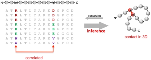
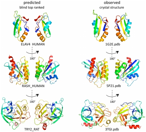
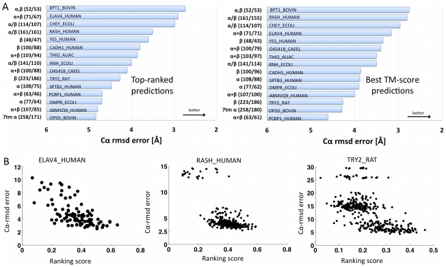
The evolutionary trajectory of a protein through sequence space is constrained by its function. Collections of sequence homologs record the outcomes of millions of evolutionary experiments in which the protein evolves according to these constraints. Deciphering the evolutionary record held in these sequences and exploiting it for predictive and engineering purposes presents a formidable challenge. The potential benefit of solving this challenge is amplified by the advent of inexpensive high-throughput genomic sequencing.In this paper we ask whether we can infer evolutionary constraints from a set of sequence homologs of a protein. The challenge is to distinguish true co-evolution couplings from the noisy set of observed correlations. We address this challenge using a maximum entropy model of the protein sequence, constrained by the statistics of the multiple sequence alignment, to infer residue pair couplings. Surprisingly, we find that the strength of these inferred couplings is an excellent predictor of residue-residue proximity in folded structures. Indeed, the top-scoring residue couplings are sufficiently accurate and well-distributed to define the 3D protein fold with remarkable accuracy.We quantify this observation by computing, from sequence alone, all-atom 3D structures of fifteen test proteins from different fold classes, ranging in size from 50 to 260 residues, including a G-protein coupled receptor. These blinded inferences are de novo, i.e., they do not use homology modeling or sequence-similar fragments from known structures. The co-evolution signals provide sufficient information to determine accurate 3D protein structure to 2.7-4.8 Å C(α)-RMSD error relative to the observed structure, over at least two-thirds of the protein (method called EVfold, details at http://EVfold.org). This discovery provides insight into essential interactions constraining protein evolution and will facilitate a comprehensive survey of the universe of protein structures, new strategies in protein and drug design, and the identification of functional genetic variants in normal and disease genomes.
Overview
The claims and statistics of this paper haven’t been checked yet.
Run the check — free, no account needed. It reads the open-access text and checks the sources it cites.
Integrity signals
Registry
Cites
Statistics
Figures
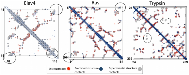
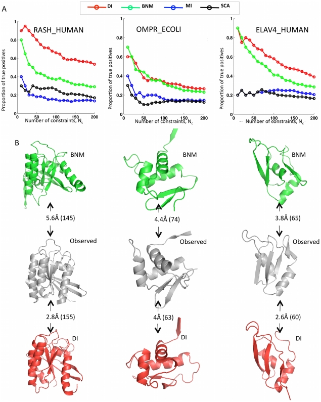
![Figure 3 Progress in contact prediction using the maximum entropy method. Extraction of evolutionary information about residue coupling and predicted contacts from multiple sequence alignments works much better using the global statistical model (right, Direct Information, DI, Equation 3) than the local statistical model (left, Mutual Information, MI, Equation 1). Predicted contacts for DI (red lines connecting the residues predicted to be coupled from sequence information) are better positioned in the experimentally observed structure (grey ribbon diagram), than those for MI (left, blue lines), shown here for the RAS protein (upper) and ELAV4 protein (lower). The DI residue pairs are also more evenly distributed along the chain and overlap more accurately with the contacts in the observed structure (red stars [predicted, grey circles [observed] in contact map; center, upper right triangle) than those using MI (blue [predicted], grey circles [observed]; center, lower left triangle). Details of contact maps for all proteins comparing predicted and observed contacts are in Figures S1 and S2, Text S1.](/paper-figures/ad0edff5338c41479018a65a69fdb4cd/protein-3d-structure-computed-from-evolutionary-sequence-variation-figure-3.jpg)
Verification
Claims the paper makes
The claims and statistics of this paper haven’t been checked yet.
Run the check — free, no account needed. It reads the open-access text and checks the sources it cites.
Claims other papers make about this one
Each is checked against this paper’s own text: does it say what the citing paper says it does?
In the citing paper
“The premise of methods to infer structure based on evolutionary couplings is that, because amino acids exist and evolve in the context of 3D structure, they are not free to evolve independently, but instead co-evolve in patterns reflective of the underlying structure.”
From Predicting multiple conformations via sequence clustering and AlphaFold2 (2024) · cites this paper as [9]
In the citing paper
“One of the major insights leveraged by AlphaFold 1,2 comes from the use of evolutionary covariations that can be extracted from large-scale multiple sequence alignments.[49][50][51][52][53][54]”
From AlphaFold and Implications for Intrinsically Disordered Proteins (2021) · cites this paper as [50]
Self-check
Percentages, sums and reported differences re-computed from the paper’s own numbers.
The self-check needs the paper’s full text.
Citations
Cites
The PyMOL Molecular Graphics System
DeLano, W. L. (2002) · Medical Entomology and Zoology · Cited by 18,147
Crystallography & NMR System: A New Software Suite for Macromolecular Structure Determination
Brünger, A. T., Adams, P. D., Clore, G. M., DeLano, W. L., Gros, P., Grosse‐Kunstleve, R. W., Jiang, J., Kuszewski, J. J., Nilges, M., Pannu, N., et al. (1998) · Acta Crystallographica Section D Biological Crystallography · doi:10.1107/s0907444998003254 · Cited by 15,723
The Pfam Protein Families Database
Bateman, A. G. (2002) · Nucleic Acids Research · doi:10.1093/nar/30.1.276 · Cited by 14,075
I-TASSER: a unified platform for automated protein structure and function prediction
Roy, A., Küçükural, A., & Zhang, Y. (2010) · Nature Protocols · doi:10.1038/nprot.2010.5 · Cited by 6,212
The Pfam protein families database
Finn, R. D., Mistry, J., Tate, J., Coggill, P., Heger, A., Pollington, J. E., Gavin, O. L., Gunasekaran, P., Ceric, G., Forslund, K., et al. (2010) · Nucleic Acids Research · doi:10.1093/nar/gkp985 · Cited by 2,694
Scoring function for automated assessment of protein structure template quality
Zhang, Y., & Skolnick, J. (2004) · Proteins: Structure, Function, and Bioinformatics · doi:10.1002/prot.20264 · Cited by 2,392
Atomic-level characterization of the structural dynamics of proteins
Shaw, D. E., Maragakis, P., Lindorff-Larsen, K., Piana, S., Dror, R. O., Eastwood, M. P., Bank, J. A., Jumper, J. M., Salmon, J. K., Shan, Y., et al. (2010) · Science · doi:10.1126/science.1187409 · Cited by 1,923
Weak pairwise correlations imply strongly correlated network states in a neural population
Schneidman, E., Berry, M. J., Segev, R., & Bialek, W. (2006) · Nature · doi:10.1038/nature04701 · Cited by 1,745
Holm, L., & Sander, C. (1996) · Science · doi:10.1126/science.273.5275.595 · Cited by 1,497
Self-citationMorcos, F., Pagnani, A., Lunt, B., Bertolino, A., Marks, D. S., Sander, C., Zecchina, R., Onuchic, J. N., Hwa, T., & Weigt, M. (2011) · Proceedings of the National Academy of Sciences · doi:10.1073/pnas.1111471108 · Cited by 1,434
Self-citation
Research works printed but not linked: 3
- KT Simons, 1999, Ab initio protein structure prediction of CASP III targets using ROSETTA., Proteins, Suppl 3, 171, 10.1002/(SICI)1097-0134(1999)37:3+<171::AID-PROT21>3.0.CO;2-Z · DOI
- MMT Mezard, 2009, Constraint satisfaction problems and neural networks: A statistical physics perspective., Journal of Physiology, 103, 107
- TR Lezon, 2006, Using the principle of entropy maximization to infer genetic interaction networks from gene expression patterns., Proceedings of the National Academy of Sciences of the United States of America, 103, 19033, 10.1073/pnas.0609152103 · DOI
Cited by
Sorted by citations
Items 501–600 of 1,005
Avila-Herrera, A., & Pollard, K. S. (2015) · BMC Bioinformatics · doi:10.1186/s12859-015-0677-y · Cited by 20
Lee, G. R., Heo, L., & Seok, C. (2017) · Proteins Structure Function and Bioinformatics · doi:10.1002/prot.25404 · Cited by 19
Machine learning applications in macromolecular X-ray crystallography
Vollmar, M., & Evans, G. (2021) · Crystallography Reviews · doi:10.1080/0889311x.2021.1982914 · Cited by 20
Vu, P. J. D., Yao, X., Momin, M., & Hamelberg, D. (2018) · ACS Catalysis · doi:10.1021/acscatal.7b04263 · Cited by 19
Oops I Took A Gradient: Scalable Sampling for Discrete DistributionsPreprint
Grathwohl, W., Swersky, K., Hashemi, M., Duvenaud, D., & Maddison, C. J. (2021) · arXiv (Cornell University) · doi:10.48550/arxiv.2102.04509 · Cited by 19
Increasing the Efficacy of Seproxetine as an Antidepressant Using Charge–Transfer Complexes
Alsanie, W. F., Alamri, A. S., Alyami, H., Alhomrani, M., Shakya, S., Habeeballah, H. H., Alkhatabi, H. A., Felimban, R. I., Alzahrani, A. S., Alhabeeb, A. A., et al. (2022) · Molecules · doi:10.3390/molecules27103290 · Cited by 18
Sun, J., Kulandaisamy, A., Liu, J., Hu, K., Gromiha, M. M., & Zhang, Y. (2023) · Computational and Structural Biotechnology Journal · doi:10.1016/j.csbj.2023.01.036 · Cited by 21
Williams, J. A., Biancucci, M., Lessen, L. N., Tian, S., Balsaraf, A., Chen, L., Chesterman, C. C., Maruggi, G., Vandepaer, S., Huang, Y., et al. (2023) · Science Advances · doi:10.1126/sciadv.adg0330 · Cited by 18
Gültaş, M., Düzgün, G., Herzog, S., Jäger, S., Meckbach, C., Wingender, E., & Waack, S. (2014) · BMC Bioinformatics · doi:10.1186/1471-2105-15-96 · Cited by 18
Fox, G., Sievers, F., & Higgins, D. G. (2015) · Bioinformatics · doi:10.1093/bioinformatics/btv592 · Cited by 18
Abriata, L. A., Bovigny, C., & Peraro, M. D. (2016) · BMC Bioinformatics · doi:10.1186/s12859-016-1124-4 · Cited by 18
Evolutionary couplings detect side-chain interactions
Hockenberry, A. J., & Wilke, C. O. (2019) · PeerJ · doi:10.7717/peerj.7280 · Cited by 16
do Amaral, M. J., de Araújo, T. S., Cirauqui, N., Accornero, F., Polycarpo, C. R., Cordeiro, Y., Cabral, K. M. D. S., & Almeida, M. S. (2020) · Journal of Molecular Biology · doi:10.1016/j.jmb.2020.02.030 · Cited by 17
MIIC online: a web server to reconstruct causal or non-causal networks from non-perturbative data
Sella, N., Verny, L., Uguzzoni, G., Affeldt, S., & Isambert, H. (2017) · Bioinformatics · doi:10.1093/bioinformatics/btx844 · Cited by 18
MacGowan, S. A., Madeira, F., Britto‐Borges, T., Schmittner, M. S., Cole, C., & Barton, G. J. (2017) · bioRxiv (Cold Spring Harbor Laboratory) · doi:10.1101/127050 · Cited by 18
Optimal Design of Stochastic DNA Synthesis Protocols based on Generative Sequence ModelsPreprint
Weinstein, E. N., Amin, A. N., Grathwohl, W., Kassler, D., Disset, J., & Marks, D. S. (2021) · bioRxiv (Cold Spring Harbor Laboratory) · doi:10.1101/2021.10.28.466307 · Cited by 19
Self-citationRecent progress and future challenges in structure-based protein-protein interaction prediction
Yuan, R., Zhang, J., Zhou, J., & Cong, Q. (2025) · Molecular Therapy · doi:10.1016/j.ymthe.2025.04.003 · Cited by 25
RBO Aleph: leveraging novel information sources for protein structure prediction
Mabrouk, M. A., Putz, I., Werner, T., Schneider, M., Neeb, M., Bartels, P., & Brock, O. (2015) · Nucleic Acids Research · doi:10.1093/nar/gkv357 · Cited by 17
Recent Advances in Protein Homology Detection Propelled by Inter-Residue Interaction Map Threading
Bhattacharya, S., Roche, R., Shuvo, M. H., & Bhattacharya, D. (2021) · Frontiers in Molecular Biosciences · doi:10.3389/fmolb.2021.643752 · Cited by 18
Wangchuk, J., Chatterjee, A., Patil, S. D., Madugula, S. K., & Kondabagil, K. R. (2021) · Virology · doi:10.1016/j.virol.2021.09.004 · Cited by 17
Artificial intelligence for template-free protein structure prediction: a comprehensive review
Mufassirin, M. M. M., Newton, M. A. H., & Sattar, A. (2022) · Artificial Intelligence Review · doi:10.1007/s10462-022-10350-x · Cited by 19
General features of transmembrane beta barrels from a large database
Montezano, D., Bernstein, R., Copeland, M. M., & Slusky, J. S. G. (2023) · Proceedings of the National Academy of Sciences · doi:10.1073/pnas.2220762120 · Cited by 17
An outlook on structural biology after A lpha F old: tools, limits and perspectives
Rosignoli, S., Pacelli, M., Manganiello, F., & Paiardini, A. (2024) · FEBS Open Bio · doi:10.1002/2211-5463.13902 · Cited by 21
Savojardo, C., Fariselli, P., Martelli, P. L., & Casadio, R. (2013) · BMC Bioinformatics · doi:10.1186/1471-2105-14-s1-s10 · Cited by 17
Prediction of contact matrix for protein–protein interaction
González, A. J., Liao, L., & Wu, C. H. (2013) · Bioinformatics · doi:10.1093/bioinformatics/btt076 · Cited by 17
Kassem, M. M., Wang, Y., Boomsma, W., & Lindorff‐Larsen, K. (2016) · Biophysical Journal · doi:10.1016/j.bpj.2016.04.039 · Cited by 17
Accelerating physical simulations of proteins by leveraging external knowledge
Pérez, A., Morrone, J. A., & Dill, K. A. (2017) · Wiley Interdisciplinary Reviews Computational Molecular Science · doi:10.1002/wcms.1309 · Cited by 17
A view of estimation of distribution algorithms through the lens of expectation-maximization
Brookes, D. H., Busia, A., Fannjiang, C., Murphy, K., & Listgarten, J. (2020) · Genetic and Evolutionary Computation Conference Companion (GECCO Companion) · doi:10.1145/3377929.3389938 · Cited by 17
Structural change in GadD2 of Listeria monocytogenes field isolates supports nisin resistance
Szendy, M., Kalkhof, S., Bittrich, S., Kaiser, F., Leberecht, C., Labudde, D., & Noll, M. (2019) · International Journal of Food Microbiology · doi:10.1016/j.ijfoodmicro.2019.108240 · Cited by 17
Phylogenetic Weighting Does Little to Improve the Accuracy of Evolutionary Coupling Analyses
Hockenberry, A. J., & Wilke, C. O. (2019) · Entropy · doi:10.3390/e21101000 · Cited by 18
Gupta, A., Dsouza, N. R., Zárate, Y. A., Lombardo, R. C., Hopkin, R. J., Linehan, A. R., Simpson, J., McCarrier, J., Agre, K. E., Gavrilova, R. H., et al. (2019) · European Journal of Medical Genetics · doi:10.1016/j.ejmg.2019.103817 · Cited by 17
Aligning biological sequences by exploiting residue conservation and coevolution
Muntoni, A. P., Pagnani, A., Weigt, M., & Zamponi, F. (2020) · Physical review. E · doi:10.1103/physreve.102.062409 · Cited by 20
Self-citationŠimkovic, F., Thomas, J. M. H., Keegan, R. M., Winn, M., Mayans, O. M., & Rigden, D. J. (2016) · IUCrJ · doi:10.1107/s2052252516008113 · Cited by 17
Coevolutionary Analysis of Protein Subfamilies by Sequence Reweighting
Malinverni, D., & Barducci, A. (2019) · Entropy · doi:10.3390/e21111127 · Cited by 17
Reverse-engineering biological networks from large data setsPreprint
Natale, J. L., Hofmann, D., Hernández, D. G., & Nemenman, I. M. (2017) · bioRxiv (Cold Spring Harbor Laboratory) · doi:10.1101/142034 · Cited by 20
Protein Structural Alignments From SequencePreprint
Morton, J. T., Strauss, C. E. M., Blackwell, R., Berenberg, D., Gligorijević, V., & Bonneau, R. (2020) · bioRxiv (Cold Spring Harbor Laboratory) · doi:10.1101/2020.11.03.365932 · Cited by 17
Learning protein constitutive motifs from sequence data
Tubiana, J., Cocco, S., & Monasson, R. (2019) · eLife · doi:10.7554/elife.39397 · Cited by 129
Multiple conformations facilitate PilT function in the type IV pilus
McCallum, M., Benlekbir, S., Nguyen, S., Tammam, S. D., Rubinstein, J. L., Burrows, L. L., & Howell, P. L. (2019) · Nature Communications · doi:10.1038/s41467-019-13070-z · Cited by 28
ProtMamba: a homology-aware but alignment-free protein state space model
Sgarbossa, D., Malbranke, C., & Bitbol, A.-F. (2025) · Bioinformatics · doi:10.1093/bioinformatics/btaf348 · Cited by 27
The Biodiversity Cell Atlas: mapping the tree of life at cellular resolution
Sebé-Pedrós, A., Tanay, A., Lawniczak, M., Arendt, D., Aerts, S., Archibald, J. M., Arnone, M. I., Blaxter, M., Cleves, P. A., Coelho, S. M., et al. (2025) · Nature · doi:10.1038/s41586-025-09312-4 · Cited by 22
Arsiwala, A., Bhatt, R., van Niekerk, L., Quintero-Cadena, P., Ao, X., Rosenbaum, A., Bhatt, A., Smith, A., Yang, Y., Anderson, K., et al. (2025) · mAbs · doi:10.1080/19420862.2025.2593055 · Cited by 16
From propensities to patterns to principles in protein folding
Rose, G. D. (2023) · Proteins Structure Function and Bioinformatics · doi:10.1002/prot.26540 · Cited by 20
Kewalramani, N., Emili, A., & Crovella, M. E. (2023) · PROTEOMICS · doi:10.1002/pmic.202200292 · Cited by 17
Learning millisecond protein dynamics from what is missing in NMR spectra
Wayment-Steele, H. K., Nesr, G. E., Hettiarachchi, R., Ojoawo, A. M., Kariyawasam, H., Ovchinnikov, S., & Kern, D. (2026) · Nature · doi:10.1038/s41586-026-10989-4 · Cited by 16
Hinsen, K., Vaïtinadapoulé, A., Ostuni, M. A., Etchebest, C., & Lacapère, J.-J. (2014) · Biochimica et Biophysica Acta (BBA) - Biomembranes · doi:10.1016/j.bbamem.2014.10.028 · Cited by 16
Kim, D., Han, S. K., Lee, K., Kim, I., Kong, J., & Kim, S. (2019) · Nucleic Acids Research · doi:10.1093/nar/gkz536 · Cited by 15
Discovery of a New Neisseria gonorrhoeae Type IV Pilus Assembly Factor, TfpC
Hu, L. I., Yin, S., Ozer, E. A., Sewell, L., Rehman, S., Garnett, J. A., & Seifert, H. S. (2020) · mBio · doi:10.1128/mbio.02528-20 · Cited by 16
Leri: A web-server for identifying protein functional networks from evolutionary couplings
Cheung, N. J., Peter, A. T. J., & Kornmann, B. (2021) · Computational and Structural Biotechnology Journal · doi:10.1016/j.csbj.2021.06.002 · Cited by 16
Woo, J., Robertson, D. L., & Lovell, S. C. (2014) · Virology · doi:10.1016/j.virol.2014.01.029 · Cited by 16
Correlated positions in protein evolution and engineering
Franceus, J., Verhaeghe, T., & Desmet, T. (2016) · Journal of Industrial Microbiology & Biotechnology · doi:10.1007/s10295-016-1811-1 · Cited by 16
Miyazawa, S. (2017) · Journal of Theoretical Biology · doi:10.1016/j.jtbi.2017.08.018 · Cited by 18
Structural Studies of TSPO, a Mitochondrial Membrane Protein
Lacapère, J.-J., Iatmanen-Harbi, S., Sénicourt, L., Lequin, O., Tékély, P., Purusottam, R. N., Hellwig, P., Kriegel, S., Ravaud, S., Juillan‐Binard, C., et al. (2014) · Springer eBooks · doi:10.1007/978-1-4939-0662-8_14 · Cited by 18
Artificial intelligence-based methods for protein structure prediction: a survey
Zhan, Z., Hong, J., Li, J.-Y., Wang, C., He, L., Xu, Z., & Zhang, J. (2025) · Artificial Intelligence Review · doi:10.1007/s10462-025-11325-4 · Cited by 17
Gene regulatory networks: from correlative models to causal explanations
Maizels, R. J., & Briscoe, J. (2026) · Nature Reviews Genetics · doi:10.1038/s41576-026-00939-1 · Cited by 21
Lal, M., Bhardwaj, E., Chahar, N., Yadav, S., & Das, S. (2022) · Functional & Integrative Genomics · doi:10.1007/s10142-022-00836-w · Cited by 15
Complex fitness landscape shapes variation in a hyperpolymorphic species
Stolyarova, A. V., Neretina, T. V., ZVYAGINA, E. A., Fedotova, A., Kondrashov, A. S., & Bazykin, G. A. (2022) · eLife · doi:10.7554/elife.76073 · Cited by 14
Niitsu, A., & Sugita, Y. (2023) · Physical Chemistry Chemical Physics · doi:10.1039/d2cp03972a · Cited by 15
Liu, J., Yu, C., Hu, X., Kim, J., Bierma, J. C., Jun, H.-I., Rychnovsky, S. D., Huang, L., & Qiao, F. (2015) · Cell Reports · doi:10.1016/j.celrep.2015.08.043 · Cited by 17
Using Analyses of Amino Acid Coevolution to Understand Protein Structure and Function
Ashenberg, O., & Laub, M. T. (2013) · Methods in enzymology on CD-ROM/Methods in enzymology · doi:10.1016/b978-0-12-394292-0.00009-6 · Cited by 15
Novel therapies for diabetes mellitus in pregnancy
Feghali, M. N., & Scifres, C. (2018) · BMJ · doi:10.1136/bmj.k2034 · Cited by 16
Molecular replacement using structure predictions from databases
Simpkin, A. J., Thomas, J. M. H., Šimkovic, F., Keegan, R. M., & Rigden, D. J. (2019) · Acta Crystallographica Section D Structural Biology · doi:10.1107/s2059798319013962 · Cited by 14
Pravin, N., Kumar, R., Tripathi, S., Kumar, P., Mohite, G. M., Navalkar, A., Panigrahi, R., Singh, N., Gadhe, L. G., Manchanda, S., et al. (2020) · Journal of Neurochemistry · doi:10.1111/jnc.15138 · Cited by 15
Co-evolutionary distance predictions contain flexibility information
Schwarz, D., Georges, G. J., Kelm, S., Shi, J., Vangone, A., & Deane, C. M. (2021) · Bioinformatics · doi:10.1093/bioinformatics/btab562 · Cited by 17
Quantum Convolutional Neural Network on Protein Distance Prediction
Hong, Z., Wang, J., Qu, X., Zhu, X., Liu, J., & Xiao, J. (2021) · International Joint Conference on Neural Networks (IJCNN) · doi:10.1109/ijcnn52387.2021.9533405 · Cited by 16
Self-organized computation in the far-from-equilibrium cell
England, J. L. (2022) · Biophysics Reviews · doi:10.1063/5.0103151 · Cited by 15
Adaptive Thouless-Anderson-Palmer approach to inverse Ising problems with quenched random fields
Huang, H., & Kabashima, Y. (2013) · Physical Review E · doi:10.1103/physreve.87.062129 · Cited by 15
Tetchner, S., Kościółek, T., & Jones, D. T. (2014) · Bio-Algorithms and Med-Systems · doi:10.1515/bams-2014-0013 · Cited by 14
Zhang, M., Case, D. A., & Peng, J. W. (2018) · Structure · doi:10.1016/j.str.2018.07.014 · Cited by 15
A primer on model-guided exploration of fitness landscapes for biological sequence designPreprint
Sinai, S., & Kelsic, E. D. (2020) · arXiv (Cornell University) · doi:10.48550/arxiv.2010.10614 · Cited by 29
Elofsson, A. (2026) · Proteins: Structure, Function, and Bioinformatics · doi:10.1002/prot.70044 · Cited by 24
Evolutionary Context-Integrated Deep Sequence Modeling for Protein Engineering
Luo, Y., Vo, L. T., Ding, H., Su, Y., Liu, Y., Qian, W. W., Zhao, H., & Peng, J. (2020) · Lecture notes in computer science · doi:10.1007/978-3-030-45257-5_30 · Cited by 15
Sun, J., & Frishman, D. (2020) · Journal of Structural Biology · doi:10.1016/j.jsb.2020.107574 · Cited by 15
Using metagenomic data to boost protein structure prediction and discovery
Hou, Q., Pucci, F., Pan, F., Xue, F., Rooman, M. J., & Feng, Q. (2022) · Computational and Structural Biotechnology Journal · doi:10.1016/j.csbj.2021.12.030 · Cited by 18
Wu, M., Lv, K., Li, J., Wu, B., & He, B. (2022) · Biotechnology and Bioengineering · doi:10.1002/bit.28113 · Cited by 14
Kalogeropoulos, K., Bohn, M., Jenkins, D. E., Ledergerber, J., Sørensen, C. V., Hofmann, N., Wade, J., Fryer, T., Nguyen, G. T. T., Keller, U. A. D., et al. (2023) · Toxicon · doi:10.1016/j.toxicon.2023.107559 · Cited by 18
Knowledge-augmented Graph Machine Learning for Drug Discovery: A Survey
Zhong, Z., Barkova, A., & Mottin, D. (2025) · ACM Computing Surveys · doi:10.1145/3744237 · Cited by 20
Clark, G. W., Ackerman, S. H., Tillier, E. R. M., & Gatti, D. L. (2014) · BMC Bioinformatics · doi:10.1186/1471-2105-15-157 · Cited by 14
Accurate Simulation and Detection of Coevolution Signals in Multiple Sequence Alignments
Ackerman, S. H., Tillier, E. R. M., & Gatti, D. L. (2012) · PLoS ONE · doi:10.1371/journal.pone.0047108 · Cited by 14
Validating a Coarse-Grained Potential Energy Function through Protein Loop Modelling
MacDonald, J. T., Kelley, L. A., & Freemont, P. S. (2013) · PLoS ONE · doi:10.1371/journal.pone.0065770 · Cited by 14
Rao, S. J. A., & Shetty, N. P. (2021) · Life Sciences · doi:10.1016/j.lfs.2021.119774 · Cited by 14
Page, B. M., Martin, T. A., Wright, C. L., Fenton, L. A., Villar, M. T., Tang, Q., Artigues, A., Lamb, A. L., Fenton, A. W., & Swint‐Kruse, L. (2022) · Protein Science · doi:10.1002/pro.4336 · Cited by 13
Prediction of protein domain boundaries from inverse covariances
Sadowski, M. I. (2012) · Proteins Structure Function and Bioinformatics · doi:10.1002/prot.24181 · Cited by 14
Inferring joint sequence-structural determinants of protein functional specificity
Neuwald, A. F., Aravind, L., & Altschul, S. F. (2018) · eLife · doi:10.7554/elife.29880 · Cited by 13
Tafazoli, A., Wawrusiewicz‐Kurylonek, N., Posmyk, R., & Miltyk, W. (2020) · Journal of Clinical Medicine · doi:10.3390/jcm10010034 · Cited by 13
Computational strategies and tools for protein tertiary structure prediction
Kumar, R., & Sharma, A. K. (2023) · Elsevier eBooks · doi:10.1016/b978-0-12-816109-8.00015-5 · Cited by 14
Brüderlin, M., Böhm, R., Fadel, F., Hiller, S., Schirmer, T., & Dubey, B. N. (2023) · Nature Communications · doi:10.1038/s41467-023-36597-8 · Cited by 13
Gandarilla-Pérez, C. A., Pinilla, S., Bitbol, A.-F., & Weigt, M. (2023) · PLOS Computational Biology · doi:10.1371/journal.pcbi.1011010 · Cited by 13
Identification of hidden N4-like viruses and their interactions with hosts
Zheng, K., Liang, Y., Páez-Espino, D., 黄, 思., Zou, X., Gao, C., Jiang, Y., He, H., Guo, C., 邵, 红., et al. (2023) · mSystems · doi:10.1128/msystems.00197-23 · Cited by 15
Machine learning in biological physics: From biomolecular prediction to design
Martin, J., Mateos, M., Onuchic, J. N., Coluzza, I., & Morcos, F. (2024) · Proceedings of the National Academy of Sciences · doi:10.1073/pnas.2311807121 · Cited by 14
Luo, J., Song, C., Cui, W., Wang, Q., Zhou, Z., & Han, L. (2024) · Chemical Science · doi:10.1039/d4sc02058h · Cited by 13
Statistical mechanics for networks of real neurons
Meshulam, L., & Bialek, W. (2025) · Reviews of Modern Physics · doi:10.1103/jcrn-3nrc · Cited by 19
Characterizing Conformational Dynamics of Proteins Using Evolutionary Couplings
Feng, J., & Shukla, D. (2018) · The Journal of Physical Chemistry B · doi:10.1021/acs.jpcb.7b07529 · Cited by 14
Subramaniya, S. R. M. V., Terashi, G., Jain, A., Kagaya, Y., & Kihara, D. (2021) · Bioinformatics · doi:10.1093/bioinformatics/btab220 · Cited by 23
Design of a Protein with Improved Thermal Stability by an Evolution‐Based Generative Model
Tian, P., Lemaire, A., Sénéchal, F., Habrylo, O., Antonietti, V., Sonnet, P., Lefebvre, V., Marin, F. I., Best, R. B., Pelloux, J., et al. (2022) · Angewandte Chemie International Edition · doi:10.1002/anie.202202711 · Cited by 13
Did α-Synuclein and Glucocerebrosidase Coevolve? Implications for Parkinson’s Disease
Gruschus, J. M. (2015) · PLoS ONE · doi:10.1371/journal.pone.0133863 · Cited by 13
Integrated Analysis of Residue Coevolution and Protein Structure in ABC Transporters
Gulyás-Kovács, A. (2012) · PLoS ONE · doi:10.1371/journal.pone.0036546 · Cited by 13
Co-evolution analysis to predict protein–protein interactions within influenza virus envelope
Mintaev, R. R., Alexeevski, A. V., & Kordyukova, L. V. (2014) · Journal of Bioinformatics and Computational Biology · doi:10.1142/s021972001441008x · Cited by 13
Circuit topology of linear polymers: a statistical mechanical treatment
Mashaghi, A., & Ramezanpour, A. (2015) · RSC Advances · doi:10.1039/c5ra08106h · Cited by 13
Noel, J. K., Morcos, F., & Onuchic, J. N. (2016) · F1000Research · doi:10.12688/f1000research.7186.1 · Cited by 13
ConKit: a python interface to contact predictions
Šimkovic, F., Thomas, J. M. H., & Rigden, D. J. (2017) · Bioinformatics · doi:10.1093/bioinformatics/btx148 · Cited by 14
Metadata
- Authors
- Marks, D. S., Colwell, L. J., Sheridan, R., Hopf, T. A., Pagnani, A., Zecchina, R., & Sander, C.
- Affiliations
- Harvard University (US)
- MRC Laboratory of Molecular Biology (GB)
- Memorial Sloan Kettering Cancer Center (US)
- Italian institute for Genomic Medicine (IT)
- Politecnico di Torino (IT)
- Corresponding author
- Marks, Debora S.
- Venue
- PLoS ONE
- Publisher
- Public Library of Science (PLoS)
- Published
- 2011-12-07
- Volume
- 6
- Issue
- 12
- Pages
- e28766
- ISSN
- 1932-6203 (ISSN-L)
- PMC
- PMC3233603
- OpenAlex
- W2061042699
- Cited by
- 1,144
- References
- 84
- Citations by year
- 2026: 61 · 2025: 60 · 2024: 47 · 2023: 72 · 2022: 96 · 2021: 104 · 2020: 90 · 2019: 110 · 2018: 91 · 2017: 107 · 2016: 71 · 2015: 74 · 2014: 72 · 2013: 56 · 2012: 29
- FWCI
- 20.79
- Type
- journal article
- Language
- en
- Open access
- gold
- doi.org
- Locations
- PLoS ONE (publishedVersion, cc-by)
- Europe PMC (PubMed Central) (publishedVersion, cc-by)
- PubMed Central (publishedVersion, cc-by)
- PubMed (publishedVersion)
- PORTO@Iris (submittedVersion, cc-by)
- DOAJ (DOAJ: Directory of Open Access Journals) (submittedVersion, cc-by-sa)
- Figshare (submittedVersion, cc-by)
- PORTO Publications Open Repository TOrino (Politecnico di Torino) (submittedVersion, cc-by)
- Funders
- Memorial Sloan-Kettering Cancer Center
- Studienstiftung des Deutschen Volkes
- National Institutes of Health (NIH U54, CA143798)
- Engineering and Physical Sciences Research Council (EP/H028064/1)
- National Cancer Institute (U54CA143798)
- National Institute of General Medical Sciences (R01GM106303)
- Topics
- Life Sciences › Biochemistry, Genetics and Molecular Biology › Molecular Biology › Protein Structure and Dynamics
- Life Sciences › Biochemistry, Genetics and Molecular Biology › Structural Biology › Enzyme Structure and Function
- Life Sciences › Biochemistry, Genetics and Molecular Biology › Molecular Biology › RNA and protein synthesis mechanisms
- Keywords
- protein structure prediction, coevolution, maximum entropy model, multiple sequence alignment, de novo structure prediction, protein evolution, G protein-coupled receptors, protein engineering
- MeSH terms
- Animals, Humans, Protein Conformation, Proteins, Rhodopsin, Trypsin, Genetic Variation, Drug Design, Reproducibility of Results, Models, Statistical, Genome, Protein Structure, Tertiary, Evolution, Molecular, Entropy, Computational Biology, Catalytic Domain, Imaging, Three-Dimensional
About these checks
Registry, retraction, Cites and statistics results are computed, not judged, with one exception: a printed reference that no index lists is read once by a model before it counts as a citation error. Claims are extracted by a model and kept only when their quote appears verbatim in the text; each cited source is then checked, with a model’s help, for whether it says what the claim relies on. Those verdicts can be wrong — read the claim’s quote and the cited source before relying on one.
Statistics and arithmetic are recomputed, not judged.
Confidence bands are shown only for fields calibrated on our annotated test set. None is calibrated yet, so no band is shown.
What counts as a claim ›How we measure confidence ›How we check this ›
Are you an author, or do you see a problem with this page? Request a review
