

The DOI matches its Crossref record; 83 of the 84 references it cites were found in a registry or an index.
Protein 3D Structure Computed from Evolutionary Sequence Variation
Open accessAbstract
The evolutionary trajectory of a protein through sequence space is constrained by its function. Collections of sequence homologs record the outcomes of millions of evolutionary experiments in which the protein evolves according to these constraints. Deciphering the evolutionary record held in these sequences and exploiting it for predictive and engineering purposes presents a formidable challenge. The potential benefit of solving this challenge is amplified by the advent of inexpensive high-throughput genomic sequencing.In this paper we ask whether we can infer evolutionary constraints from a set of sequence homologs of a protein. The challenge is to distinguish true co-evolution couplings from the noisy set of observed correlations. We address this challenge using a maximum entropy model of the protein sequence, constrained by the statistics of the multiple sequence alignment, to infer residue pair couplings. Surprisingly, we find that the strength of these inferred couplings is an excellent predictor of residue-residue proximity in folded structures. Indeed, the top-scoring residue couplings are sufficiently accurate and well-distributed to define the 3D protein fold with remarkable accuracy.We quantify this observation by computing, from sequence alone, all-atom 3D structures of fifteen test proteins from different fold classes, ranging in size from 50 to 260 residues, including a G-protein coupled receptor. These blinded inferences are de novo, i.e., they do not use homology modeling or sequence-similar fragments from known structures. The co-evolution signals provide sufficient information to determine accurate 3D protein structure to 2.7-4.8 Å C(α)-RMSD error relative to the observed structure, over at least two-thirds of the protein (method called EVfold, details at http://EVfold.org). This discovery provides insight into essential interactions constraining protein evolution and will facilitate a comprehensive survey of the universe of protein structures, new strategies in protein and drug design, and the identification of functional genetic variants in normal and disease genomes.
Overview
The claims and statistics of this paper haven’t been checked yet.
Run the check — free, no account needed. It reads the open-access text and checks the sources it cites.
Integrity signals
Registry
Cites
Statistics
Figures
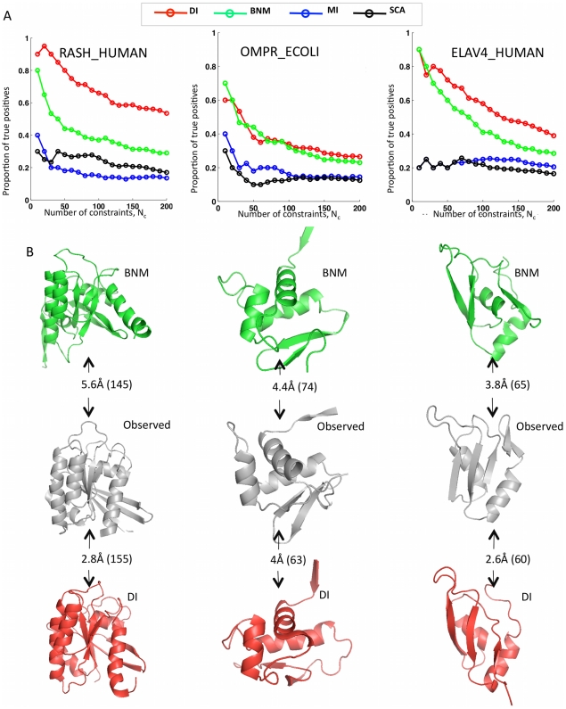
![Figure 3 Progress in contact prediction using the maximum entropy method. Extraction of evolutionary information about residue coupling and predicted contacts from multiple sequence alignments works much better using the global statistical model (right, Direct Information, DI, Equation 3) than the local statistical model (left, Mutual Information, MI, Equation 1). Predicted contacts for DI (red lines connecting the residues predicted to be coupled from sequence information) are better positioned in the experimentally observed structure (grey ribbon diagram), than those for MI (left, blue lines), shown here for the RAS protein (upper) and ELAV4 protein (lower). The DI residue pairs are also more evenly distributed along the chain and overlap more accurately with the contacts in the observed structure (red stars [predicted, grey circles [observed] in contact map; center, upper right triangle) than those using MI (blue [predicted], grey circles [observed]; center, lower left triangle). Details of contact maps for all proteins comparing predicted and observed contacts are in Figures S1 and S2, Text S1.](/paper-figures/ad0edff5338c41479018a65a69fdb4cd/protein-3d-structure-computed-from-evolutionary-sequence-variation-figure-3.jpg)
Verification
Claims the paper makes
The claims and statistics of this paper haven’t been checked yet.
Run the check — free, no account needed. It reads the open-access text and checks the sources it cites.
Claims other papers make about this one
Each is checked against this paper’s own text: does it say what the citing paper says it does?
In the citing paper
“The premise of methods to infer structure based on evolutionary couplings is that, because amino acids exist and evolve in the context of 3D structure, they are not free to evolve independently, but instead co-evolve in patterns reflective of the underlying structure.”
From Predicting multiple conformations via sequence clustering and AlphaFold2 (2024) · cites this paper as [9]
In the citing paper
“One of the major insights leveraged by AlphaFold 1,2 comes from the use of evolutionary covariations that can be extracted from large-scale multiple sequence alignments.[49][50][51][52][53][54]”
From AlphaFold and Implications for Intrinsically Disordered Proteins (2021) · cites this paper as [50]
Self-check
Percentages, sums and reported differences re-computed from the paper’s own numbers.
The self-check needs the paper’s full text.
Citations
Cites
The PyMOL Molecular Graphics System
DeLano, W. L. (2002) · Medical Entomology and Zoology · Cited by 18,147
Crystallography & NMR System: A New Software Suite for Macromolecular Structure Determination
Brünger, A. T., Adams, P. D., Clore, G. M., DeLano, W. L., Gros, P., Grosse‐Kunstleve, R. W., Jiang, J., Kuszewski, J. J., Nilges, M., Pannu, N., et al. (1998) · Acta Crystallographica Section D Biological Crystallography · doi:10.1107/s0907444998003254 · Cited by 15,723
The Pfam Protein Families Database
Bateman, A. G. (2002) · Nucleic Acids Research · doi:10.1093/nar/30.1.276 · Cited by 14,075
I-TASSER: a unified platform for automated protein structure and function prediction
Roy, A., Küçükural, A., & Zhang, Y. (2010) · Nature Protocols · doi:10.1038/nprot.2010.5 · Cited by 6,212
The Pfam protein families database
Finn, R. D., Mistry, J., Tate, J., Coggill, P., Heger, A., Pollington, J. E., Gavin, O. L., Gunasekaran, P., Ceric, G., Forslund, K., et al. (2010) · Nucleic Acids Research · doi:10.1093/nar/gkp985 · Cited by 2,694
Scoring function for automated assessment of protein structure template quality
Zhang, Y., & Skolnick, J. (2004) · Proteins: Structure, Function, and Bioinformatics · doi:10.1002/prot.20264 · Cited by 2,392
Atomic-level characterization of the structural dynamics of proteins
Shaw, D. E., Maragakis, P., Lindorff-Larsen, K., Piana, S., Dror, R. O., Eastwood, M. P., Bank, J. A., Jumper, J. M., Salmon, J. K., Shan, Y., et al. (2010) · Science · doi:10.1126/science.1187409 · Cited by 1,923
Weak pairwise correlations imply strongly correlated network states in a neural population
Schneidman, E., Berry, M. J., Segev, R., & Bialek, W. (2006) · Nature · doi:10.1038/nature04701 · Cited by 1,745
Holm, L., & Sander, C. (1996) · Science · doi:10.1126/science.273.5275.595 · Cited by 1,497
Self-citationMorcos, F., Pagnani, A., Lunt, B., Bertolino, A., Marks, D. S., Sander, C., Zecchina, R., Onuchic, J. N., Hwa, T., & Weigt, M. (2011) · Proceedings of the National Academy of Sciences · doi:10.1073/pnas.1111471108 · Cited by 1,434
Self-citation
Research works printed but not linked: 3
- KT Simons, 1999, Ab initio protein structure prediction of CASP III targets using ROSETTA., Proteins, Suppl 3, 171, 10.1002/(SICI)1097-0134(1999)37:3+<171::AID-PROT21>3.0.CO;2-Z · DOI
- MMT Mezard, 2009, Constraint satisfaction problems and neural networks: A statistical physics perspective., Journal of Physiology, 103, 107
- TR Lezon, 2006, Using the principle of entropy maximization to infer genetic interaction networks from gene expression patterns., Proceedings of the National Academy of Sciences of the United States of America, 103, 19033, 10.1073/pnas.0609152103 · DOI
Cited by
Sorted by citations
Items 601–700 of 1,005
Sequential search leads to faster, more efficient fragment-basedde novoprotein structure prediction
de Oliveira, S. H. P., Law, E. C., Shi, J., & Deane, C. M. (2017) · Bioinformatics · doi:10.1093/bioinformatics/btx722 · Cited by 13
Wang, Y., Marrero, M. C., Medema, M. H., & van Dijk, A. D. J. (2020) · Bioinformatics · doi:10.1093/bioinformatics/btaa595 · Cited by 14
Deep representation learning improves prediction of LacI-mediated transcriptional repression
Garruss, A. S., Collins, K. M., & Church, G. M. (2021) · Proceedings of the National Academy of Sciences · doi:10.1073/pnas.2022838118 · Cited by 14
Ali, N. F., Khan, S., & Zahid, S. (2025) · Computational Biology and Chemistry · doi:10.1016/j.compbiolchem.2025.108430 · Cited by 13
CorNet: Assigning function to networks of co-evolving residues by automated literature mining
van den Bergh, T., Tamò, G. E., Nobili, A., Tao, Y., Tan, T., Bornscheuer, U. T., Kuipers, R. K. P., Vroling, B., de Jong, R. M., Subramanian, K., et al. (2017) · PLoS ONE · doi:10.1371/journal.pone.0176427 · Cited by 12
Simpkin, A. J., Caballero, I., McNicholas, S. J., Stevenson, K., Jiménez, E., Rodríguez, F. S., Fando, M. S., Uski, V., Ballard, C. C., Chojnowski, G., et al. (2023) · Acta Crystallographica Section D Structural Biology · doi:10.1107/s2059798323006289 · Cited by 13
Hybrid approaches to molecular simulation
Ho, B. K., Pérahia, D., & Buckle, A. M. (2012) · Current Opinion in Structural Biology · doi:10.1016/j.sbi.2012.05.005 · Cited by 12
Molecular Models for the Core Components of the Flagellar Type-III Secretion Complex
Taylor, W. R., Matthews-Palmer, T. R. S., & Beeby, M. (2016) · PLoS ONE · doi:10.1371/journal.pone.0164047 · Cited by 12
Tondnevis, F., Dudenhausen, E. E., Miller, A. M., McKENNA, R. V., Altschul, S. F., Bloom, L. B., & Neuwald, A. F. (2020) · Scientific Reports · doi:10.1038/s41598-019-55118-6 · Cited by 12
De novo Protein Structure Prediction by Coupling Contact With Distance Profile
Peng, C., Zhou, X., & Zhang, G. (2020) · IEEE Transactions on Computational Biology and Bioinformatics · doi:10.1109/tcbb.2020.3000758 · Cited by 13
Chasing coevolutionary signals in intrinsically disordered proteins complexes
Iserte, J., Lázár, T., Tosatto, S. C. E., Tompa, P., & Marino‐Buslje, C. (2020) · Scientific Reports · doi:10.1038/s41598-020-74791-6 · Cited by 13
Luppino, F., Adzhubei, I. A., Cassa, C. A., & Toth-Petroczy, A. (2023) · Nature Communications · doi:10.1038/s41467-023-37661-z · Cited by 12
Evolution of the Immunoglobulin Isotypes—Variations of Biophysical Properties among Animal Classes
Pomarici, N. D., Cacciato, R., Kokot, J., Fernández‐Quintero, M. L., & Liedl, K. R. (2023) · Biomolecules · doi:10.3390/biom13050801 · Cited by 11
Szurmant, H., & Hoch, J. A. (2012) · Molecular Microbiology · doi:10.1111/mmi.12128 · Cited by 12
Raimondi, D., Orlando, G., & Vranken, W. (2014) · Bioinformatics · doi:10.1093/bioinformatics/btu794 · Cited by 12
Pelé, J., Moreau, M., Abdi, H., Rodien, P., Castel, H., & Chabbert, M. (2014) · Proteins Structure Function and Bioinformatics · doi:10.1002/prot.24570 · Cited by 12
Integrated Structural Biology for α-Helical Membrane Protein Structure Determination
Xia, Y., Fischer, A. W., Teixeira, P. L., Weiner, B., & Meiler, J. (2018) · Structure · doi:10.1016/j.str.2018.02.006 · Cited by 13
Quantifying steric hindrance and topological obstruction to protein structure superposition
Røgen, P. (2021) · Algorithms for Molecular Biology · doi:10.1186/s13015-020-00180-3 · Cited by 13
Liu, J., Zhao, K., He, G.-X., Wang, L., Zhou, X., & Zhang, G. (2021) · Bioinformatics · doi:10.1093/bioinformatics/btab620 · Cited by 12
Repertoire-based diagnostics using statistical biophysics
Arora, R., Kaplinsky, J., Li, A., & Arnaout, R. (2019) · bioRxiv (Cold Spring Harbor Laboratory) · doi:10.1101/519108 · Cited by 10
Carosella, N., Brock, K. P., Zambelli, B., Musiani, F., Sander, C., & Ciurli, S. (2023) · Frontiers in Chemical Biology · doi:10.3389/fchbi.2023.1243564 · Cited by 12
Self-citationTowards parsimonious generative modeling of RNA families
Calvanese, F., Lambert, C. N., Nghe, P., Zamponi, F., & Weigt, M. (2024) · Nucleic Acids Research · doi:10.1093/nar/gkae289 · Cited by 11
ZEPPI: Proteome-scale sequence-based evaluation of protein–protein interaction models
Zhao, H., Petrey, D. S., Murray, D., & Honig, B. H. (2024) · Proceedings of the National Academy of Sciences · doi:10.1073/pnas.2400260121 · Cited by 13
Protein binding and folding through an evolutionary lens
Jemth, P. (2025) · Current Opinion in Structural Biology · doi:10.1016/j.sbi.2024.102980 · Cited by 11
Gauge fixing for sequence-function relationships
(2025) · doi:10.1371/journal.pcbi.1012818 · Cited by 11
Valentin, J. B., Andreetta, C., Boomsma, W., Bottaro, S., Ferkinghoff‐Borg, J., Frellsen, J., Mardia, K. V., Tian, P., & Hamelryck, T. (2013) · Proteins Structure Function and Bioinformatics · doi:10.1002/prot.24386 · Cited by 11
Basu, S., & Bahadur, R. P. (2021) · Proteins Structure Function and Bioinformatics · doi:10.1002/prot.26261 · Cited by 11
Undersampling and the inference of coevolution in proteins
Kleeorin, Y., Russ, W. P., Rivoire, O., & Ranganathan, R. (2023) · Cell Systems · doi:10.1016/j.cels.2022.12.013 · Cited by 15
Protein dynamics provide mechanistic insights about epistasis among common missense polymorphisms
Ose, N. J., Campitelli, P., Patel, R., Kumar, S., & Ozkan, S. B. (2023) · Biophysical Journal · doi:10.1016/j.bpj.2023.01.037 · Cited by 11
Deng, X., & Cheng, J. (2014) · BMC Bioinformatics · doi:10.1186/1471-2105-15-252 · Cited by 11
Structure, Dynamics and Implied Gating Mechanism of a Human Cyclic Nucleotide-Gated Channel
Gofman, Y., Schärfe, C., Marks, D. S., Haliloǧlu, T., & Ben‐Tal, N. (2014) · PLoS Computational Biology · doi:10.1371/journal.pcbi.1003976 · Cited by 11
Self-citationHu, J. P., Liu, H., Sun, J., Wang, J., & Liu, R. (2018) · Protein Science · doi:10.1002/pro.3448 · Cited by 11
Coevolutionary Signals and Structure-Based Models for the Prediction of Protein Native Conformations
dos Santos, R. N., Jiang, X., Martı́nez, L., & Morcos, F. (2018) · Methods in molecular biology · doi:10.1007/978-1-4939-8736-8_5 · Cited by 11
Fongang, B., Cunningham, K. A., Rowicka, M., & Kudlicki, A. S. (2019) · Neuroscience · doi:10.1016/j.neuroscience.2019.05.013 · Cited by 14
Coevolutionary Analysis of Protein Sequences for Molecular Modeling
Malinverni, D., & Barducci, A. (2019) · Methods in molecular biology · doi:10.1007/978-1-4939-9608-7_16 · Cited by 10
AlignmentViewer: Sequence Analysis of Large Protein Families
Reguant, R., Antipin, Y., Sheridan, R., Dallago, C., Diamantoukos, D., Luna, A., Sander, C., & Gauthier, N. P. (2020) · F1000Research · doi:10.12688/f1000research.22242.1 · Cited by 11
Self-citationA Model for Allosteric Communication in Drug Transport by the AcrAB-TolC Tripartite Efflux Pump
Webber, A., Ratnaweera, M., Harris, A., Luisi, B. F., & Enguéné, V. Y. N. (2022) · Antibiotics · doi:10.3390/antibiotics11010052 · Cited by 11
Entropic effects in a nonequilibrium system: Flocks of birds
Castellana, M., Bialek, W., Cavagna, A., & Giardina, I. (2016) · Physical review. E · doi:10.1103/physreve.93.052416 · Cited by 11
Evans, D. L., & Joshi, S. V. (2016) · Journal of Theoretical Biology · doi:10.1016/j.jtbi.2016.07.025 · Cited by 11
Combining Evolutionary Covariance and NMR Data for Protein Structure Determination
Huang, Y. J., Brock, K. P., Ishida, Y., Swapna, G. V. T., Inouye, M., Marks, D. S., Sander, C., & Montelione, G. T. (2018) · Methods in enzymology on CD-ROM/Methods in enzymology · doi:10.1016/bs.mie.2018.11.004 · Cited by 10
Self-citationComputing the Pathogenicity of Wilson’s Disease ATP7B Mutations: Implications for Disease Prevalence
Tang, N., Sandahl, T. D., Ott, P., & Kepp, K. P. (2019) · Journal of Chemical Information and Modeling · doi:10.1021/acs.jcim.9b00852 · Cited by 12
Co‐Evolutionary Fitness Landscapes for Sequence Design
Tian, P., Louis, J. M., Baber, J. L., Aniana, A., & Best, R. B. (2018) · Angewandte Chemie · doi:10.1002/ange.201713220 · Cited by 11
Exploring the Druggable Conformational Space of Protein Kinases Using AI-Generated StructuresPreprint
Herrington, N. B., Stein, D. F., Li, Y. C., Pandey, G. S., & Schlessinger, A. (2023) · bioRxiv (Cold Spring Harbor Laboratory) · doi:10.1101/2023.08.31.555779 · Cited by 11
Gauge fixing for sequence-function relationships
Posfai, A., Zhou, J., McCandlish, D. M., & Kinney, J. B. (2024) · doi:10.1101/2024.05.12.593772 · Cited by 11
A Newfound Cancer-Activating Mutation Reshapes the Energy Landscape of Estrogen-Binding Domain
Huang, W., Ravikumar, K. M., & Yang, S. (2014) · Journal of Chemical Theory and Computation · doi:10.1021/ct500313e · Cited by 10
Hao, X., Zhang, G., & Zhou, X. (2017) · IEEE Transactions on NanoBioscience · doi:10.1109/tnb.2017.2749243 · Cited by 10
Delarue, M. H., & Koehl, P. (2018) · F1000Research · doi:10.12688/f1000research.14870.1 · Cited by 10
Thompson, M. J., Domville, J. A., & Baenziger, J. E. (2020) · Journal of Biological Chemistry · doi:10.1074/jbc.ra120.013751 · Cited by 11
Gomes, G., do Amaral, M. J., Bagri, K. M., Vasconcellos, L. M., Almeida, M. S., Alvares, L. E., & Mermelstein, C. (2021) · International Journal of Molecular Sciences · doi:10.3390/ijms222312885 · Cited by 10
The role of self-organization in developmental evolution
Bozorgmehr, J. H. (2014) · Theory in Biosciences · doi:10.1007/s12064-014-0200-4 · Cited by 10
Motoyama, T., Nakano, S., Yamamoto, Y., Tokiwa, H., Asano, Y., & Ito, S. (2017) · Biochemistry · doi:10.1021/acs.biochem.7b00832 · Cited by 10
From Levinthal’s Paradox to the Effects of Cell Environmental Perturbation on Protein Folding
Zeng, J., & Huang, Z. (2018) · Current Medicinal Chemistry · doi:10.2174/0929867325666181017160857 · Cited by 10
Repository of Enriched Structures of Proteins Involved in the Red Blood Cell Environment (RESPIRE)
Télétchéa, S., Santuz, H., Léonard, S., & Etchebest, C. (2019) · PLoS ONE · doi:10.1371/journal.pone.0211043 · Cited by 10
FingerprintContacts: Predicting Alternative Conformations of Proteins from Coevolution
Feng, J., & Shukla, D. (2020) · The Journal of Physical Chemistry B · doi:10.1021/acs.jpcb.9b11869 · Cited by 9
Neuwald, A. F., Lanczycki, C. J., Hodges, T. K., & Marchler‐Bauer, A. (2020) · Database · doi:10.1093/database/baaa042 · Cited by 10
GISA: using Gauss Integrals to identify rare conformations in protein structures
Grønbæk, C., Hamelryck, T., & Røgen, P. (2020) · PeerJ · doi:10.7717/peerj.9159 · Cited by 10
Experimental determination and data-driven prediction of homotypic transmembrane domain interfaces
Xiao, Y., Zeng, B., Berner, N., Frishman, D., Langosch, D., & Teese, M. G. (2020) · Computational and Structural Biotechnology Journal · doi:10.1016/j.csbj.2020.09.035 · Cited by 10
Fitness landscape of a dynamic RNA structure
Soo, V. W. C., Swadling, J. B., Faure, A. J., & Warnecke, T. (2021) · PLoS Genetics · doi:10.1371/journal.pgen.1009353 · Cited by 10
Enhancing and inhibitory motifs regulate CD4 activity
Lee, M. S., Tuohy, P. J., Kim, C. Y., Lichauco, K., Parrish, H. L., Doorslaer, K. V., & Kuhns, M. S. (2022) · eLife · doi:10.7554/elife.79508 · Cited by 10
Exploring the miRNA Regulatory Network Using Evolutionary Correlations
Obermayer, B., & Levine, E. (2014) · PLoS Computational Biology · doi:10.1371/journal.pcbi.1003860 · Cited by 11
Identifying functionally informative evolutionary sequence profiles
Gil, N., & Fiser, A. (2017) · Bioinformatics · doi:10.1093/bioinformatics/btx779 · Cited by 10
Critiquing Protein Family Classification Models Using Sufficient Input Subsets
Carter, B., Bileschi, M. L., Smith, J., Sanderson, T., Bryant, D., Belanger, D., & Colwell, L. J. (2019) · Journal of Computational Biology · doi:10.1089/cmb.2019.0339 · Cited by 10
Self-citationInverse Ising inference with correlated samples
Obermayer, B., & Levine, E. (2014) · MDC Repository (Max-Delbrueck-Center for Molecular Medicine) · Cited by 10
Comparative Protein Structure Modelling
Fiser, A. (2017) · Springer eBooks · doi:10.1007/978-94-024-1069-3_4 · Cited by 11
Deciphering membrane protein structures from protein sequences
Flock, T., Venkatakrishnan, A., Vinothkumar, K., & Babu, M. M. (2012) · Genome biology · doi:10.1186/gb-2012-13-6-160 · Cited by 10
3D protein structure from genetic epistasis experimentsPreprint
Rollins, N. J., Brock, K. P., Poelwijk, F. J., Stiffler, M. A., Gauthier, N. P., Sander, C., & Marks, D. S. (2018) · bioRxiv (Cold Spring Harbor Laboratory) · doi:10.1101/320721 · Cited by 10
Self-citationIllig, A.-M., Siedhoff, N. E., Schwaneberg, U., & Davari, M. D. (2022) · bioRxiv (Cold Spring Harbor Laboratory) · doi:10.1101/2022.06.07.495081 · Cited by 10
Feng, T., Liu, J., Zhang, X., Fan, D., & Bai, Y. (2023) · Nanoscale Advances · doi:10.1039/d3na00515a · Cited by 9
Exact minimax entropy models of large-scale neuronal activity
Lynn, C. W., Yu, Q., Pang, R., Palmer, S. E., & Bialek, W. (2025) · Physical review. E · doi:10.1103/physreve.111.054411 · Cited by 12
Allostery is a widespread cause of loss-of-function variant pathogenicity
Liao, X., & Lehner, B. (2026) · Nature Communications · doi:10.1038/s41467-026-74517-8 · Cited by 13
Chakravarty, D., & Porter, L. L. (2025) · Annual Review of Biophysics · doi:10.1146/annurev-biophys-022924-012038 · Cited by 11
Probabilistic grammatical model for helix‐helix contact site classification
Dyrka, W., Nebel, J., & Kotulska, M. (2013) · Algorithms for Molecular Biology · doi:10.1186/1748-7188-8-31 · Cited by 9
OMPcontact: An Outer Membrane Protein Inter-Barrel Residue Contact Prediction Method
Zhang, L., Wang, H., Yan, L., Su, L., & Xu, D. H. (2016) · Journal of Computational Biology · doi:10.1089/cmb.2015.0236 · Cited by 9
Hernández, D. G., Sober, S. J., & Nemenman, I. M. (2022) · eLife · doi:10.7554/elife.68192 · Cited by 9
Nadaradjane, A. A., Quignot, C., Traoré, S., Andréani, J., & Guérois, R. (2019) · Proteins Structure Function and Bioinformatics · doi:10.1002/prot.25857 · Cited by 9
Inter-protein residue covariation information unravels physically interacting protein dimers
Salmanian, S., Pezeshk, H., & Sadeghi, M. (2020) · BMC Bioinformatics · doi:10.1186/s12859-020-03930-7 · Cited by 5
Computational compensatory mutation discovery approach: Predicting a PARP1 variant rescue mutation
Ravishankar, K., Jiang, X., Leddin, E. M., Morcos, F., & Cisneros, G. A. (2022) · Biophysical Journal · doi:10.1016/j.bpj.2022.05.036 · Cited by 10
Deep Antimicrobial Activity and Stability Analysis Inform Lysin Sequence–Function Mapping
Tresnak, D. T., & Hackel, B. J. (2023) · ACS Synthetic Biology · doi:10.1021/acssynbio.2c00509 · Cited by 10
Inverse Ising inference with correlated samples
Obermayer, B., & Levine, E. (2014) · New Journal of Physics · doi:10.1088/1367-2630/16/12/123017 · Cited by 9
Janda, J.-O., Popal, A., Bauer, J., Busch, M. R., Klocke, M., Spitzer, W., Keller, J., & Merkl, R. (2014) · BMC Bioinformatics · doi:10.1186/1471-2105-15-118 · Cited by 9
Computational studies of G protein-coupled receptor complexes: Structure and dynamics
Şensoy, Ö., de Almeida, J. G., Shabbir, J., Moreira, I. S., & Morra, G. (2017) · Methods in cell biology · doi:10.1016/bs.mcb.2017.07.011 · Cited by 9
Extending the Horizon of Homology Detection with Coevolution-based Structure Prediction
Sánchez‐Pulido, L., & Ponting, C. P. (2021) · Journal of Molecular Biology · doi:10.1016/j.jmb.2021.167106 · Cited by 10
Increasing the accuracy of protein loop structure prediction with evolutionary constraints
Marks, C., & Deane, C. M. (2018) · Bioinformatics · doi:10.1093/bioinformatics/bty996 · Cited by 8
Abriata, L. A. (2015) · bioRxiv (Cold Spring Harbor Laboratory) · doi:10.1101/013581 · Cited by 9
Protein Three-Dimensional Structure Prediction
Abeln, S., Feenstra, K. A., & Heringa, J. (2018) · Elsevier eBooks · doi:10.1016/b978-0-12-809633-8.20505-0 · Cited by 9
Protein Contact Map Denoising Using Generative Adversarial NetworksPreprint
Subramaniya, S. R. M. V., Terashi, G., Jain, A., Kagaya, Y., & Kihara, D. (2020) · bioRxiv (Cold Spring Harbor Laboratory) · doi:10.1101/2020.06.26.174300 · Cited by 9
Inverse Potts model improves accuracy of phylogenetic profiling
Fukunaga, T., & Iwasaki, W. (2022) · Bioinformatics · doi:10.1093/bioinformatics/btac034 · Cited by 8
Impact of phylogeny on structural contact inference from protein sequence data
Dietler, N., Lupo, U., & Bitbol, A.-F. (2023) · Journal of The Royal Society Interface · doi:10.1098/rsif.2022.0707 · Cited by 6
Insights into DNA cleavage by MutL homologs from analysis of conserved motifs in eukaryotic Mlh1
Putnam, C. D., & Kolodner, R. D. (2023) · BioEssays · doi:10.1002/bies.202300031 · Cited by 10
Gu, J., Sim, B. R., Li, J., Yu, Y., Qin, L., Wu, L., Liu, H., Xu, Y., Zhao, Y., & Nie, Y. (2025) · International Journal of Biological Macromolecules · doi:10.1016/j.ijbiomac.2025.141233 · Cited by 8
Ahn, J. J., Yu, T. C., Dadonaite, B., Radford, C. E., & Bloom, J. D. (2026) · Virus Evolution · doi:10.1093/ve/veag018 · Cited by 11
Identifying the structural and kinetic elements in protein large-amplitude conformational motions
Chu, J., & Yang, H. (2017) · International Reviews in Physical Chemistry · doi:10.1080/0144235x.2017.1283885 · Cited by 9
Machine-Learned Molecular Surface and Its Application to Implicit Solvent Simulations
Wei, H., Zhao, Z., & Luo, R. (2021) · Journal of Chemical Theory and Computation · doi:10.1021/acs.jctc.1c00492 · Cited by 8
Kennedy, E. N., Foster, C. A., Barr, S. A., & Bourret, R. B. (2022) · Biochemical Society Transactions · doi:10.1042/bst20220849 · Cited by 8
Exactly solvable statistical physics models for large neuronal populations
Lynn, C. W., Yu, Q., Pang, R., Bialek, W., & Palmer, S. E. (2025) · Physical Review Research · doi:10.1103/physrevresearch.7.l022039 · Cited by 8
Evolutionary decision rules for predicting protein contact maps
Marquez-Chamorro, A. E., Cortés, G. A., Divina, F., & Aguilar–Ruiz, J. S. (2012) · Pattern Analysis and Applications · doi:10.1007/s10044-012-0297-3 · Cited by 8
The evolution of logic circuits for the purpose of protein contact map prediction
Chapman, S., Adami, C., Wilke, C. O., & KC, D. B. (2017) · PeerJ · doi:10.7717/peerj.3139 · Cited by 8
Zhao, Z. J., Zhang, H., Shu, D., Montemagno, C. D., Ding, B., Li, J., & Guo, P. (2016) · Small · doi:10.1002/smll.201601600 · Cited by 8
Zerihun, M. B., & Schug, A. H. (2017) · Biochemical Society Transactions · doi:10.1042/bst20170063 · Cited by 10
Metadata
- Authors
- Marks, D. S., Colwell, L. J., Sheridan, R., Hopf, T. A., Pagnani, A., Zecchina, R., & Sander, C.
- Affiliations
- Harvard University (US)
- MRC Laboratory of Molecular Biology (GB)
- Memorial Sloan Kettering Cancer Center (US)
- Italian institute for Genomic Medicine (IT)
- Politecnico di Torino (IT)
- Corresponding author
- Marks, Debora S.
- Venue
- PLoS ONE
- Publisher
- Public Library of Science (PLoS)
- Published
- 2011-12-07
- Volume
- 6
- Issue
- 12
- Pages
- e28766
- ISSN
- 1932-6203 (ISSN-L)
- PMC
- PMC3233603
- OpenAlex
- W2061042699
- Cited by
- 1,144
- References
- 84
- Citations by year
- 2026: 61 · 2025: 60 · 2024: 47 · 2023: 72 · 2022: 96 · 2021: 104 · 2020: 90 · 2019: 110 · 2018: 91 · 2017: 107 · 2016: 71 · 2015: 74 · 2014: 72 · 2013: 56 · 2012: 29
- FWCI
- 20.79
- Type
- journal article
- Language
- en
- Open access
- gold
- doi.org
- Locations
- PLoS ONE (publishedVersion, cc-by)
- Europe PMC (PubMed Central) (publishedVersion, cc-by)
- PubMed Central (publishedVersion, cc-by)
- PubMed (publishedVersion)
- PORTO@Iris (submittedVersion, cc-by)
- DOAJ (DOAJ: Directory of Open Access Journals) (submittedVersion, cc-by-sa)
- Figshare (submittedVersion, cc-by)
- PORTO Publications Open Repository TOrino (Politecnico di Torino) (submittedVersion, cc-by)
- Funders
- Memorial Sloan-Kettering Cancer Center
- Studienstiftung des Deutschen Volkes
- National Institutes of Health (NIH U54, CA143798)
- Engineering and Physical Sciences Research Council (EP/H028064/1)
- National Cancer Institute (U54CA143798)
- National Institute of General Medical Sciences (R01GM106303)
- Topics
- Life Sciences › Biochemistry, Genetics and Molecular Biology › Molecular Biology › Protein Structure and Dynamics
- Life Sciences › Biochemistry, Genetics and Molecular Biology › Structural Biology › Enzyme Structure and Function
- Life Sciences › Biochemistry, Genetics and Molecular Biology › Molecular Biology › RNA and protein synthesis mechanisms
- Keywords
- protein structure prediction, coevolution, maximum entropy model, multiple sequence alignment, de novo structure prediction, protein evolution, G protein-coupled receptors, protein engineering
- MeSH terms
- Animals, Humans, Protein Conformation, Proteins, Rhodopsin, Trypsin, Genetic Variation, Drug Design, Reproducibility of Results, Models, Statistical, Genome, Protein Structure, Tertiary, Evolution, Molecular, Entropy, Computational Biology, Catalytic Domain, Imaging, Three-Dimensional
About these checks
Registry, retraction, Cites and statistics results are computed, not judged, with one exception: a printed reference that no index lists is read once by a model before it counts as a citation error. Claims are extracted by a model and kept only when their quote appears verbatim in the text; each cited source is then checked, with a model’s help, for whether it says what the claim relies on. Those verdicts can be wrong — read the claim’s quote and the cited source before relying on one.
Statistics and arithmetic are recomputed, not judged.
Confidence bands are shown only for fields calibrated on our annotated test set. None is calibrated yet, so no band is shown.
What counts as a claim ›How we measure confidence ›How we check this ›
Are you an author, or do you see a problem with this page? Request a review
