

The DOI matches its Crossref record; 83 of the 84 references it cites were found in a registry or an index.
Protein 3D Structure Computed from Evolutionary Sequence Variation
Open accessAbstract
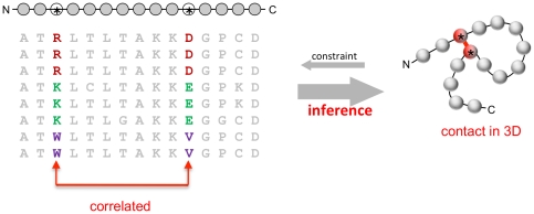
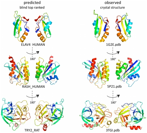
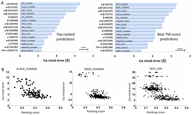
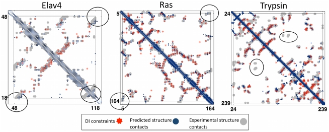
The evolutionary trajectory of a protein through sequence space is constrained by its function. Collections of sequence homologs record the outcomes of millions of evolutionary experiments in which the protein evolves according to these constraints. Deciphering the evolutionary record held in these sequences and exploiting it for predictive and engineering purposes presents a formidable challenge. The potential benefit of solving this challenge is amplified by the advent of inexpensive high-throughput genomic sequencing.In this paper we ask whether we can infer evolutionary constraints from a set of sequence homologs of a protein. The challenge is to distinguish true co-evolution couplings from the noisy set of observed correlations. We address this challenge using a maximum entropy model of the protein sequence, constrained by the statistics of the multiple sequence alignment, to infer residue pair couplings. Surprisingly, we find that the strength of these inferred couplings is an excellent predictor of residue-residue proximity in folded structures. Indeed, the top-scoring residue couplings are sufficiently accurate and well-distributed to define the 3D protein fold with remarkable accuracy.We quantify this observation by computing, from sequence alone, all-atom 3D structures of fifteen test proteins from different fold classes, ranging in size from 50 to 260 residues, including a G-protein coupled receptor. These blinded inferences are de novo, i.e., they do not use homology modeling or sequence-similar fragments from known structures. The co-evolution signals provide sufficient information to determine accurate 3D protein structure to 2.7-4.8 Å C(α)-RMSD error relative to the observed structure, over at least two-thirds of the protein (method called EVfold, details at http://EVfold.org). This discovery provides insight into essential interactions constraining protein evolution and will facilitate a comprehensive survey of the universe of protein structures, new strategies in protein and drug design, and the identification of functional genetic variants in normal and disease genomes.
Overview
The claims and statistics of this paper haven’t been checked yet.
Run the check — free, no account needed. It reads the open-access text and checks the sources it cites.
Integrity signals
Registry
Cites
Statistics
Figures
![Figure 3 Progress in contact prediction using the maximum entropy method. Extraction of evolutionary information about residue coupling and predicted contacts from multiple sequence alignments works much better using the global statistical model (right, Direct Information, DI, Equation 3) than the local statistical model (left, Mutual Information, MI, Equation 1). Predicted contacts for DI (red lines connecting the residues predicted to be coupled from sequence information) are better positioned in the experimentally observed structure (grey ribbon diagram), than those for MI (left, blue lines), shown here for the RAS protein (upper) and ELAV4 protein (lower). The DI residue pairs are also more evenly distributed along the chain and overlap more accurately with the contacts in the observed structure (red stars [predicted, grey circles [observed] in contact map; center, upper right triangle) than those using MI (blue [predicted], grey circles [observed]; center, lower left triangle). Details of contact maps for all proteins comparing predicted and observed contacts are in Figures S1 and S2, Text S1.](/paper-figures/ad0edff5338c41479018a65a69fdb4cd/protein-3d-structure-computed-from-evolutionary-sequence-variation-figure-3.jpg)
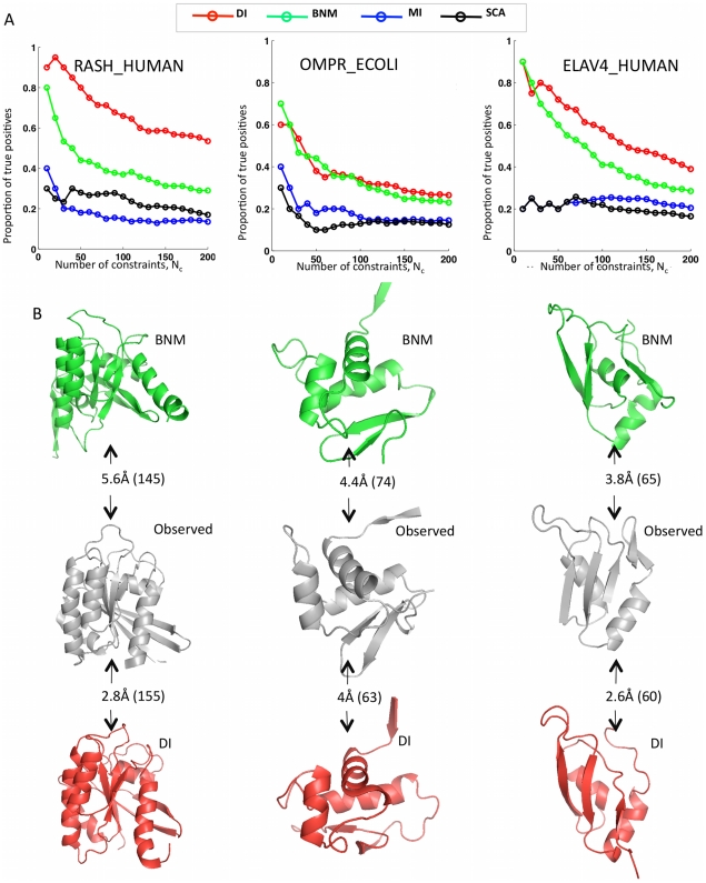
Verification
Claims the paper makes
The claims and statistics of this paper haven’t been checked yet.
Run the check — free, no account needed. It reads the open-access text and checks the sources it cites.
Claims other papers make about this one
Each is checked against this paper’s own text: does it say what the citing paper says it does?
In the citing paper
“The premise of methods to infer structure based on evolutionary couplings is that, because amino acids exist and evolve in the context of 3D structure, they are not free to evolve independently, but instead co-evolve in patterns reflective of the underlying structure.”
From Predicting multiple conformations via sequence clustering and AlphaFold2 (2024) · cites this paper as [9]
In the citing paper
“One of the major insights leveraged by AlphaFold 1,2 comes from the use of evolutionary covariations that can be extracted from large-scale multiple sequence alignments.[49][50][51][52][53][54]”
From AlphaFold and Implications for Intrinsically Disordered Proteins (2021) · cites this paper as [50]
Self-check
Percentages, sums and reported differences re-computed from the paper’s own numbers.
The self-check needs the paper’s full text.
Citations
Cites
The PyMOL Molecular Graphics System
DeLano, W. L. (2002) · Medical Entomology and Zoology · Cited by 18,147
Crystallography & NMR System: A New Software Suite for Macromolecular Structure Determination
Brünger, A. T., Adams, P. D., Clore, G. M., DeLano, W. L., Gros, P., Grosse‐Kunstleve, R. W., Jiang, J., Kuszewski, J. J., Nilges, M., Pannu, N., et al. (1998) · Acta Crystallographica Section D Biological Crystallography · doi:10.1107/s0907444998003254 · Cited by 15,723
The Pfam Protein Families Database
Bateman, A. G. (2002) · Nucleic Acids Research · doi:10.1093/nar/30.1.276 · Cited by 14,075
I-TASSER: a unified platform for automated protein structure and function prediction
Roy, A., Küçükural, A., & Zhang, Y. (2010) · Nature Protocols · doi:10.1038/nprot.2010.5 · Cited by 6,212
The Pfam protein families database
Finn, R. D., Mistry, J., Tate, J., Coggill, P., Heger, A., Pollington, J. E., Gavin, O. L., Gunasekaran, P., Ceric, G., Forslund, K., et al. (2010) · Nucleic Acids Research · doi:10.1093/nar/gkp985 · Cited by 2,694
Scoring function for automated assessment of protein structure template quality
Zhang, Y., & Skolnick, J. (2004) · Proteins: Structure, Function, and Bioinformatics · doi:10.1002/prot.20264 · Cited by 2,392
Atomic-level characterization of the structural dynamics of proteins
Shaw, D. E., Maragakis, P., Lindorff-Larsen, K., Piana, S., Dror, R. O., Eastwood, M. P., Bank, J. A., Jumper, J. M., Salmon, J. K., Shan, Y., et al. (2010) · Science · doi:10.1126/science.1187409 · Cited by 1,923
Weak pairwise correlations imply strongly correlated network states in a neural population
Schneidman, E., Berry, M. J., Segev, R., & Bialek, W. (2006) · Nature · doi:10.1038/nature04701 · Cited by 1,745
Holm, L., & Sander, C. (1996) · Science · doi:10.1126/science.273.5275.595 · Cited by 1,497
Self-citationMorcos, F., Pagnani, A., Lunt, B., Bertolino, A., Marks, D. S., Sander, C., Zecchina, R., Onuchic, J. N., Hwa, T., & Weigt, M. (2011) · Proceedings of the National Academy of Sciences · doi:10.1073/pnas.1111471108 · Cited by 1,434
Self-citation
Research works printed but not linked: 3
- KT Simons, 1999, Ab initio protein structure prediction of CASP III targets using ROSETTA., Proteins, Suppl 3, 171, 10.1002/(SICI)1097-0134(1999)37:3+<171::AID-PROT21>3.0.CO;2-Z · DOI
- MMT Mezard, 2009, Constraint satisfaction problems and neural networks: A statistical physics perspective., Journal of Physiology, 103, 107
- TR Lezon, 2006, Using the principle of entropy maximization to infer genetic interaction networks from gene expression patterns., Proceedings of the National Academy of Sciences of the United States of America, 103, 19033, 10.1073/pnas.0609152103 · DOI
Cited by
Sorted by citations
Items 301–400 of 1,005
Topology of membrane proteins — predictions, limitations and variations
Tsirigos, K. D., Sudha, G., Bassot, C., Västermark, Å., Lamb, J., Shu, N., & Elofsson, A. (2017) · Current Opinion in Structural Biology · doi:10.1016/j.sbi.2017.10.003 · Cited by 49
Entropy and Information within Intrinsically Disordered Protein Regions
Pritišanac, I., Vernon, R. M., Moses, A. M., & Forman-Kay, J. D. (2019) · Entropy · doi:10.3390/e21070662 · Cited by 46
pydca v1.0: a comprehensive software for direct coupling analysis of RNA and protein sequences
Zerihun, M. B., Pucci, F., Peter, E. K., & Schug, A. H. (2019) · Bioinformatics · doi:10.1093/bioinformatics/btz892 · Cited by 44
Fukuda, H., & Tomii, K. (2020) · BMC Bioinformatics · doi:10.1186/s12859-019-3190-x · Cited by 44
Modeling protein–ligand interactions for drug discovery in the era of deep learning
Wang, Y. X., Li, Y., Chen, J., & Lai, L. (2025) · Chemical Society Reviews · doi:10.1039/d5cs00415b · Cited by 53
Applications of contact predictions to structural biology
Šimkovic, F., Ovchinnikov, S., Baker, D. A., & Rigden, D. J. (2017) · IUCrJ · doi:10.1107/s2052252517005115 · Cited by 42
MitImpact 3: modeling the residue interaction network of the Respiratory Chain subunits
Castellana, S., Biagini, T., Petrizzelli, F., Parca, L., Panzironi, N., Caputo, V., Vescovi, A. L., Carella, M., & Mazza, T. (2020) · Nucleic Acids Research · doi:10.1093/nar/gkaa1032 · Cited by 47
Data-driven computational protein design
Frappier, V., & Keating, A. E. (2021) · Current Opinion in Structural Biology · doi:10.1016/j.sbi.2021.03.009 · Cited by 44
Latent generative landscapes as maps of functional diversity in protein sequence space
Ziegler, C., Martin, J., Sinner, C., & Morcos, F. (2023) · Nature Communications · doi:10.1038/s41467-023-37958-z · Cited by 43
Global Shifts in Genome and Proteome Composition Are Very Tightly Coupled
Brbić, M., Warnecke, T., Kriško, A., & Supek, F. (2015) · Genome Biology and Evolution · doi:10.1093/gbe/evv088 · Cited by 36
Large-scale structure prediction by improved contact predictions and model quality assessment
Michel, M., Hurtado, D. M., Uziela, K., & Elofsson, A. (2017) · Bioinformatics · doi:10.1093/bioinformatics/btx239 · Cited by 38
Engineering repressors with coevolutionary cues facilitates toggle switches with a master reset
Dimas, R. P., Jiang, X., de la Paz, J. A., Morcos, F., & Chan, C. T. Y. (2019) · Nucleic Acids Research · doi:10.1093/nar/gkz280 · Cited by 41
Artificial intelligence powers protein-folding predictions
Eisenstein, M. S. (2021) · Nature · doi:10.1038/d41586-021-03499-y · Cited by 42
Protein Residue Contacts and Prediction Methods
Adhikari, B., & Cheng, J. J. (2016) · Methods in molecular biology · doi:10.1007/978-1-4939-3572-7_24 · Cited by 40
Lessons from (co‐)evolution in the docking of proteins and peptides for CAPRI Rounds 28–35
Yu, J., Andréani, J., Ochsenbein, F., & Guérois, R. (2016) · Proteins Structure Function and Bioinformatics · doi:10.1002/prot.25180 · Cited by 40
Xiong, D., Zeng, J., & Gong, H. (2017) · Bioinformatics · doi:10.1093/bioinformatics/btx296 · Cited by 40
Predicting accurate contacts in thousands of Pfam domain families using PconsC3
Michel, M., Skwark, M. J., Hurtado, D. M., Ekeberg, M., & Elofsson, A. (2017) · Bioinformatics · doi:10.1093/bioinformatics/btx332 · Cited by 41
Analysis of several key factors influencing deep learning-based inter-residue contact prediction
Wu, T., Hou, J., Adhikari, B., & Cheng, J. (2019) · Bioinformatics · doi:10.1093/bioinformatics/btz679 · Cited by 38
Coevolution-based prediction of key allosteric residues for protein function regulation
Xie, J., Zhang, W., Zhu, X., Deng, M., & Lai, L. (2023) · eLife · doi:10.7554/elife.81850 · Cited by 40
Gao, Y., Barajas‐Ornelas, R. D. C., Amon, J. D., Ramírez‐Guadiana, F. H., Alon, A., Brock, K. P., Marks, D. S., Kruse, A. C., & Rudner, D. Z. (2022) · Genes & Development · doi:10.1101/gad.349488.122 · Cited by 41
Self-citationBhattacharya, D., Cao, R., & Cheng, J. (2016) · Bioinformatics · doi:10.1093/bioinformatics/btw316 · Cited by 38
A Comprehensive Analysis of Anion–Quadrupole Interactions in Protein Structures
Chakravarty, S., Ung, A. R., Moore, B. G., Shore, J. S., & Alshamrani, M. S. (2018) · Biochemistry · doi:10.1021/acs.biochem.7b01006 · Cited by 40
Molecular replacement: tricks and treats
Abergel, C. (2013) · Acta Crystallographica Section D Biological Crystallography · doi:10.1107/s0907444913015291 · Cited by 36
Engineering highly active nuclease enzymes with machine learning and high-throughput screening
Thomas, N., Belanger, D., Xu, C. A., Lee, H., Hirano, K., Iwai, K., Polic, V., Nyberg, K. D., Hoff, K. G., Frenz, L., et al. (2025) · Cell Systems · doi:10.1016/j.cels.2025.101236 · Cited by 41
Self-citationProtein Design and Variant Prediction Using Autoregressive Generative ModelsPreprint
Shin, J.-E., Riesselman, A. J., Kollasch, A. W., McMahon, C., Simon, E. P., Sander, C., Manglik, A., Kruse, A. C., & Marks, D. S. (2019) · bioRxiv (Cold Spring Harbor Laboratory) · doi:10.1101/757252 · Cited by 40
Self-citationGenetic interaction mapping informs integrative structure determination of protein complexes
Braberg, H., Echeverria, I., Bohn, S., Cimermancic, P., Shiver, A., Alexander, R., Xu, J., Shales, M., Dronamraju, R., Jiang, S., et al. (2020) · Science · doi:10.1126/science.aaz4910 · Cited by 37
Condon, S. G. F., Mahbuba, D., Armstrong, C. R., Vázquez, G. D., Craven, S. J., LaPointe, L. M., Khadria, A. S., Chadda, R., Crooks, J. A., Rangarajan, N., et al. (2017) · Journal of Biological Chemistry · doi:10.1074/jbc.ra117.000426 · Cited by 35
Selection of sequence motifs and generative Hopfield-Potts models for protein families
Shimagaki, K. S., & Weigt, M. (2019) · Physical review. E · doi:10.1103/physreve.100.032128 · Cited by 41
Oehme, D. P., Shafee, T. M. A., Downton, M. T., Bacic, A., & Doblin, M. S. (2019) · PLoS ONE · doi:10.1371/journal.pone.0224442 · Cited by 36
Soft computing methods for the prediction of protein tertiary structures: A survey
Marquez-Chamorro, A. E., Cortés, G. A., Santiesteban-Toca, C. E., & Aguilar–Ruiz, J. S. (2015) · Applied Soft Computing · doi:10.1016/j.asoc.2015.06.024 · Cited by 38
Computational Protein Design Quantifies Structural Constraints on Amino Acid Covariation
Ollikainen, N., & Kortemme, T. (2013) · PLoS Computational Biology · doi:10.1371/journal.pcbi.1003313 · Cited by 36
bbcontacts: prediction of β -strand pairing from direct coupling patterns
Andréani, J., & Söding, J. (2015) · Bioinformatics · doi:10.1093/bioinformatics/btv041 · Cited by 36
Lukose, V., Luo, L., Kozakov, D., Vajda, S., Allen, K. N., & Imperiali, B. (2015) · Biochemistry · doi:10.1021/acs.biochem.5b01086 · Cited by 36
Sahoo, A., Khare, S., Devanarayanan, S., Jain, P. C., & Varadarajan, R. (2015) · eLife · doi:10.7554/elife.09532 · Cited by 36
Wang, Y., & Barth, P. (2015) · Nature Communications · doi:10.1038/ncomms8196 · Cited by 36
Sung, Y.-M., Wilkins, A. D., Rodriguez, G. J., Wensel, T. G., & Lichtarge, O. (2016) · Proceedings of the National Academy of Sciences · doi:10.1073/pnas.1516579113 · Cited by 36
Identification of Protein–Protein Interactions by Detecting Correlated Mutation at the Interface
Guo, F., Ding, Y., Li, Z., & Tang, J. (2015) · Journal of Chemical Information and Modeling · doi:10.1021/acs.jcim.5b00320 · Cited by 36
Agarwala, A., & Fisher, D. S. (2018) · Theoretical Population Biology · doi:10.1016/j.tpb.2019.09.011 · Cited by 41
Wang, Y., Shi, Q., Yang, P., Zhang, C., Mortuza, S. M., Xue, Z., Ning, K., & Zhang, Y. (2019) · Genome biology · doi:10.1186/s13059-019-1823-z · Cited by 35
Protein folding ‐ seeing is deceiving
Rose, G. D. (2021) · Protein Science · doi:10.1002/pro.4096 · Cited by 36
Alhomrani, M., Alsanie, W. F., Alamri, A. S., Alyami, H., Habeeballah, H. H., Alkhatabi, H. A., Felimban, R. I., Haynes, J. M., Shakya, S., Raafat, B. M., et al. (2022) · Pharmaceuticals · doi:10.3390/ph15030285 · Cited by 36
Green function of correlated genes in a minimal mechanical model of protein evolution
Dutta, S., Eckmann, J., Libchaber, A. J., & Tlusty, T. (2018) · Proceedings of the National Academy of Sciences · doi:10.1073/pnas.1716215115 · Cited by 35
McCafferty, C. L., Pennington, E. L., Papoulas, O., Taylor, D. W., & Marcotte, E. M. (2023) · Communications Biology · doi:10.1038/s42003-023-04773-7 · Cited by 35
Pei, J., Zhang, J., & Cong, Q. (2022) · Bioinformatics · doi:10.1093/bioinformatics/btac527 · Cited by 35
Quantitative approaches for decoding the specificity of the human T cell repertoire
Ghoreyshi, Z. S., & George, J. T. (2023) · Frontiers in Immunology · doi:10.3389/fimmu.2023.1228873 · Cited by 41
Finding the needle in the haystack: towards solving the protein-folding problem computationally
Li, B., Fooksa, M., Heinze, S., & Meiler, J. (2017) · Critical Reviews in Biochemistry and Molecular Biology · doi:10.1080/10409238.2017.1380596 · Cited by 39
Experimentally-driven protein structure modeling
Dokholyan, N. V. (2020) · Journal of Proteomics · doi:10.1016/j.jprot.2020.103777 · Cited by 37
Wang, X., Jing, X., Deng, Y., Nie, Y., Xu, F., Xu, Y., Zhao, Y., Hunt, J. F., Montelione, G. T., & Szyperski, T. (2019) · FEBS Letters · doi:10.1002/1873-3468.13652 · Cited by 35
Big-data approaches to protein structure prediction
Söding, J. (2017) · Science · doi:10.1126/science.aal4512 · Cited by 36
Fundamentals to function: Quantitative and scalable approaches for measuring protein stability
Atsavapranee, B., Stark, C., Sunden, F., Thompson, S., & Fordyce, P. M. (2021) · Cell Systems · doi:10.1016/j.cels.2021.05.009 · Cited by 38
The relaxin receptor RXFP1 signals through a mechanism of autoinhibition
Erlandson, S. C., Rawson, S., Osei‐Owusu, J., Brock, K. P., Liu, X., Paulo, J. A., Mintseris, J., Gygi, S. P., Marks, D. S., Cong, X., et al. (2023) · Nature Chemical Biology · doi:10.1038/s41589-023-01321-6 · Cited by 34
Self-citationPrediction of contacts from correlated sequence substitutions
Taylor, W. R., Hamilton, R. S., & Sadowski, M. I. (2013) · Current Opinion in Structural Biology · doi:10.1016/j.sbi.2013.04.001 · Cited by 34
Phylogenetic correlations can suffice to infer protein partners from sequences
Marmier, G., Weigt, M., & Bitbol, A. (2019) · PLoS Computational Biology · doi:10.1371/journal.pcbi.1007179 · Cited by 34
Recent Advances and Challenges in Protein Structure Prediction
Peng, C., Liang, F., Xia, Y., Zhao, K., Hou, M., & Zhang, G. (2023) · Journal of Chemical Information and Modeling · doi:10.1021/acs.jcim.3c01324 · Cited by 42
Raval, A., Piana, S., Eastwood, M. P., & Shaw, D. E. (2015) · Protein Science · doi:10.1002/pro.2770 · Cited by 33
The network of stabilizing contacts in proteins studied by coevolutionary data
Lui, S., & Tiana, G. (2013) · The Journal of Chemical Physics · doi:10.1063/1.4826096 · Cited by 33
GtrA Protein Rv3789 Is Required for Arabinosylation of Arabinogalactan in Mycobacterium tuberculosis
Kolly, G. S., Mukherjee, R., Kilacsková, E., Abriata, L. A., Raccaud, M., Blaško, J., Sala, C., Peraro, M. D., Mikušová, K., & Cole, S. T. (2015) · Journal of Bacteriology · doi:10.1128/jb.00628-15 · Cited by 33
Colloquium : Proteins: The physics of amorphous evolving matter
Eckmann, J., Rougemont, J., & Tlusty, T. (2019) · Reviews of Modern Physics · doi:10.1103/revmodphys.91.031001 · Cited by 34
Messelink, J. J. B., van Teeseling, M. C. F., Janssen, J., Thanbichler, M. R., & Broedersz, C. P. (2021) · Nature Communications · doi:10.1038/s41467-021-22189-x · Cited by 31
StarMap: a user-friendly workflow for Rosetta-driven molecular structure refinement
Lugmayr, W., Kotov, V., Goessweiner‐Mohr, N., Wald, J., DiMaio, F., & Marlovits, T. C. (2022) · Nature Protocols · doi:10.1038/s41596-022-00757-9 · Cited by 33
Blind assessment of monomeric AlphaFold2 protein structure models with experimental NMR data
Li, E. H., Spaman, L. E., Tejero, R., Huang, Y. J., Ramelot, T. A., Fraga, K. J., Prestegard, J. H., Kennedy, M. A., & Montelione, G. T. (2023) · Journal of Magnetic Resonance · doi:10.1016/j.jmr.2023.107481 · Cited by 34
Generative AAV capsid diversification by latent interpolationPreprint
Sinai, S., Jain, N. K., Church, G. M., & Kelsic, E. D. (2021) · bioRxiv (Cold Spring Harbor Laboratory) · doi:10.1101/2021.04.16.440236 · Cited by 39
Dwyer, R. S., Ricci, D. P., Colwell, L. J., Silhavy, T. J., & Wingreen, N. S. (2013) · Genetics · doi:10.1534/genetics.113.155861 · Cited by 32
Self-citationChampeimont, R., Laine, É., Hu, S., Pénin, F., & Carbone, A. (2016) · Scientific Reports · doi:10.1038/srep26401 · Cited by 30
ISPRED4: interaction sites PREDiction in protein structures with a refining grammar model
Savojardo, C., Fariselli, P., Martelli, P. L., & Casadio, R. (2017) · Bioinformatics · doi:10.1093/bioinformatics/btx044 · Cited by 34
Overcoming Immunological Challenges Limiting Capsid-Mediated Gene Therapy With Machine Learning
Wec, A. Z., Lin, K. S., Kwasnieski, J. C., Sinai, S., Gerold, J., & Kelsic, E. D. (2021) · Frontiers in Immunology · doi:10.3389/fimmu.2021.674021 · Cited by 35
Simultaneous enhancement of multiple functional properties using evolution-informed protein design
Fram, B., Su, Y., Truebridge, I., Riesselman, A. J., Ingraham, J. B., Passera, A., Napier, E., Thadani, N. N., Lim, S., Roberts, K., et al. (2024) · Nature Communications · doi:10.1038/s41467-024-49119-x · Cited by 34
Self-citationPairing interacting protein sequences using masked language modeling
Lupo, U., Sgarbossa, D., & Bitbol, A.-F. (2024) · Proceedings of the National Academy of Sciences · doi:10.1073/pnas.2311887121 · Cited by 31
Repeat proteins challenge the concept of structural domains
Espada, R., Parra, R. G., Sippl, M. J., Mora, T., Walczak, A. M., & Ferreiro, D. U. (2015) · Biochemical Society Transactions · doi:10.1042/bst20150083 · Cited by 31
Recent advances in sequence-based protein structure prediction: Table 1
KC, D. B. (2016) · Briefings in Bioinformatics · doi:10.1093/bib/bbw070 · Cited by 31
Applications of sequence coevolution in membrane protein biochemistry
Nicoludis, J. M., & Gaudet, R. (2017) · Biochimica et Biophysica Acta (BBA) - Biomembranes · doi:10.1016/j.bbamem.2017.10.004 · Cited by 30
Nicoludis, J. M., Green, A. G., Walujkar, S., May, E., Sotomayor, M. M., Marks, D. S., & Gaudet, R. (2019) · Proceedings of the National Academy of Sciences · doi:10.1073/pnas.1821063116 · Cited by 31
Self-citationNew amino acid substitution matrix brings sequence alignments into agreement with structure matches
Jia, K., & Jernigan, R. L. (2021) · Proteins Structure Function and Bioinformatics · doi:10.1002/prot.26050 · Cited by 32
Physics‐based protein structure refinement in the era of artificial intelligence
Heo, L., Janson, G., & Feig, M. (2021) · Proteins Structure Function and Bioinformatics · doi:10.1002/prot.26161 · Cited by 33
Barton, J. P., Cocco, S., Leonardis, E. D., & Monasson, R. (2014) · Physical Review E · doi:10.1103/physreve.90.012132 · Cited by 31
Nerli, S., & Sgourakis, N. G. (2018) · Methods in enzymology on CD-ROM/Methods in enzymology · doi:10.1016/bs.mie.2018.07.005 · Cited by 32
EVfold.org: Evolutionary Couplings and Protein 3D Structure PredictionPreprint
Sheridan, R., Fieldhouse, R. J., Hayat, S., Sun, Y., Antipin, Y., Yang, L., Hopf, T. A., Marks, D. S., & Sander, C. (2015) · bioRxiv (Cold Spring Harbor Laboratory) · doi:10.1101/021022 · Cited by 34
Self-citationScalable matrix inversion using MapReduce
Xiang, J., Meng, H., & Aboulnaga, A. (2014) · ACM International Symposium on High-Performance Parallel and Distributed Computing (HPDC) · doi:10.1145/2600212.2600220 · Cited by 30
Maximum-Entropy Models of Sequenced Immune Repertoires Predict Antigen-Antibody Affinity
Asti, L., Uguzzoni, G., Marcatili, P., & Pagnani, A. (2016) · PLoS Computational Biology · doi:10.1371/journal.pcbi.1004870 · Cited by 30
Self-citationSynthetic protein alignments by CCMgen quantify noise in residue-residue contact prediction
Vorberg, S., Seemayer, S., & Söding, J. (2018) · PLoS Computational Biology · doi:10.1371/journal.pcbi.1006526 · Cited by 36
Liu, J., Zhou, X., Zhang, Y., & Zhang, G. (2019) · Bioinformatics · doi:10.1093/bioinformatics/btz943 · Cited by 29
Machine Learning Approaches for Quality Assessment of Protein Structures
Chen, J., & Siu, S. W. I. (2020) · Biomolecules · doi:10.3390/biom10040626 · Cited by 37
CryoEM map of Pseudomonas aeruginosa PilQ enables structural characterization of TsaP
McCallum, M., Tammam, S. D., Rubinstein, J. L., Burrows, L. L., & Howell, P. L. (2020) · Structure · doi:10.1016/j.str.2020.11.019 · Cited by 30
Extending the New Generation of Structure Predictors to Account for Dynamics and Allostery
Fleishman, S. J., & Horovitz, A. (2021) · Journal of Molecular Biology · doi:10.1016/j.jmb.2021.167007 · Cited by 28
(2022) · doi:10.1126/sciadv.abo1126 · Cited by 30
Self-citationResearch trends of artificial intelligence in pancreatic cancer: a bibliometric analysis
Yin, H., Zhang, F., Yang, X., Meng, X., Yu, M., Hussain, M. S. N., Yang, L., & Li, Z. (2022) · Frontiers in Oncology · doi:10.3389/fonc.2022.973999 · Cited by 32
Eickholt, J., & Cheng, J. J. (2013) · BMC Bioinformatics · doi:10.1186/1471-2105-14-s14-s12 · Cited by 30
Teppa, E., Zea, D. J., & Marino‐Buslje, C. (2017) · Protein Science · doi:10.1002/pro.3318 · Cited by 30
MISTIC2: comprehensive server to study coevolution in protein families
Colell, E. A., Iserte, J., Simonetti, F. L., & Marino‐Buslje, C. (2018) · Nucleic Acids Research · doi:10.1093/nar/gky419 · Cited by 26
Cohan, M. C., Ruff, K. M., & Pappu, R. V. (2019) · Protein Engineering Design and Selection · doi:10.1093/protein/gzz014 · Cited by 28
Strong evidence for protein epistasis, weak evidence against it
Pollock, D. D., & Goldstein, R. A. (2014) · Proceedings of the National Academy of Sciences · doi:10.1073/pnas.1401112111 · Cited by 30
Contact Potential for Structure Prediction of Proteins and Protein Complexes from Potts Model
Anishchenko, I., Kundrotas, P. J., & Vakser, I. A. (2018) · Biophysical Journal · doi:10.1016/j.bpj.2018.07.035 · Cited by 29
Structural Constraints Identified with Covariation Analysis in Ribosomal RNA
Shang, L., Xu, W., Ozer, S., & Gutell, R. R. (2012) · PLoS ONE · doi:10.1371/journal.pone.0039383 · Cited by 29
Zhang, H., Huang, Q., Bei, Z., Wei, Y., & Floudas, C. A. (2016) · Proteins Structure Function and Bioinformatics · doi:10.1002/prot.24979 · Cited by 29
Recruiting machine learning methods for molecular simulations of proteins
Mittal, S., & Shukla, D. (2018) · Molecular Simulation · doi:10.1080/08927022.2018.1448976 · Cited by 28
Jain, A., Terashi, G., Kagaya, Y., Subramaniya, S. R. M. V., Christoffer, C. W., & Kihara, D. (2021) · Scientific Reports · doi:10.1038/s41598-021-87204-z · Cited by 30
From systems to structure — using genetic data to model protein structures
Braberg, H., Echeverria, I., Kaake, R. M., Šali, A., & Krogan, N. J. (2022) · Nature Reviews Genetics · doi:10.1038/s41576-021-00441-w · Cited by 30
I-COMS: Interprotein-COrrelated Mutations Server
Iserte, J., Simonetti, F. L., Zea, D. J., Teppa, E., & Marino‐Buslje, C. (2015) · Nucleic Acids Research · doi:10.1093/nar/gkv572 · Cited by 29
Metadata
- Authors
- Marks, D. S., Colwell, L. J., Sheridan, R., Hopf, T. A., Pagnani, A., Zecchina, R., & Sander, C.
- Affiliations
- Harvard University (US)
- MRC Laboratory of Molecular Biology (GB)
- Memorial Sloan Kettering Cancer Center (US)
- Italian institute for Genomic Medicine (IT)
- Politecnico di Torino (IT)
- Corresponding author
- Marks, Debora S.
- Venue
- PLoS ONE
- Publisher
- Public Library of Science (PLoS)
- Published
- 2011-12-07
- Volume
- 6
- Issue
- 12
- Pages
- e28766
- ISSN
- 1932-6203 (ISSN-L)
- PMC
- PMC3233603
- OpenAlex
- W2061042699
- Cited by
- 1,144
- References
- 84
- Citations by year
- 2026: 61 · 2025: 60 · 2024: 47 · 2023: 72 · 2022: 96 · 2021: 104 · 2020: 90 · 2019: 110 · 2018: 91 · 2017: 107 · 2016: 71 · 2015: 74 · 2014: 72 · 2013: 56 · 2012: 29
- FWCI
- 20.79
- Type
- journal article
- Language
- en
- Open access
- gold
- doi.org
- Locations
- PLoS ONE (publishedVersion, cc-by)
- Europe PMC (PubMed Central) (publishedVersion, cc-by)
- PubMed Central (publishedVersion, cc-by)
- PubMed (publishedVersion)
- PORTO@Iris (submittedVersion, cc-by)
- DOAJ (DOAJ: Directory of Open Access Journals) (submittedVersion, cc-by-sa)
- Figshare (submittedVersion, cc-by)
- PORTO Publications Open Repository TOrino (Politecnico di Torino) (submittedVersion, cc-by)
- Funders
- Memorial Sloan-Kettering Cancer Center
- Studienstiftung des Deutschen Volkes
- National Institutes of Health (NIH U54, CA143798)
- Engineering and Physical Sciences Research Council (EP/H028064/1)
- National Cancer Institute (U54CA143798)
- National Institute of General Medical Sciences (R01GM106303)
- Topics
- Life Sciences › Biochemistry, Genetics and Molecular Biology › Molecular Biology › Protein Structure and Dynamics
- Life Sciences › Biochemistry, Genetics and Molecular Biology › Structural Biology › Enzyme Structure and Function
- Life Sciences › Biochemistry, Genetics and Molecular Biology › Molecular Biology › RNA and protein synthesis mechanisms
- Keywords
- protein structure prediction, coevolution, maximum entropy model, multiple sequence alignment, de novo structure prediction, protein evolution, G protein-coupled receptors, protein engineering
- MeSH terms
- Animals, Humans, Protein Conformation, Proteins, Rhodopsin, Trypsin, Genetic Variation, Drug Design, Reproducibility of Results, Models, Statistical, Genome, Protein Structure, Tertiary, Evolution, Molecular, Entropy, Computational Biology, Catalytic Domain, Imaging, Three-Dimensional
About these checks
Registry, retraction, Cites and statistics results are computed, not judged, with one exception: a printed reference that no index lists is read once by a model before it counts as a citation error. Claims are extracted by a model and kept only when their quote appears verbatim in the text; each cited source is then checked, with a model’s help, for whether it says what the claim relies on. Those verdicts can be wrong — read the claim’s quote and the cited source before relying on one.
Statistics and arithmetic are recomputed, not judged.
Confidence bands are shown only for fields calibrated on our annotated test set. None is calibrated yet, so no band is shown.
What counts as a claim ›How we measure confidence ›How we check this ›
Are you an author, or do you see a problem with this page? Request a review
