

The DOI matches its Crossref record; 83 of the 84 references it cites were found in a registry or an index.
Protein 3D Structure Computed from Evolutionary Sequence Variation
Open accessAbstract
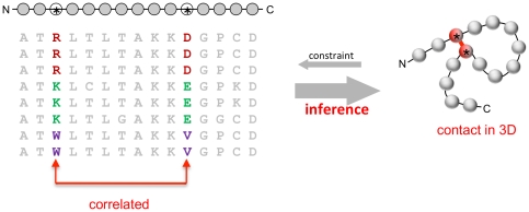
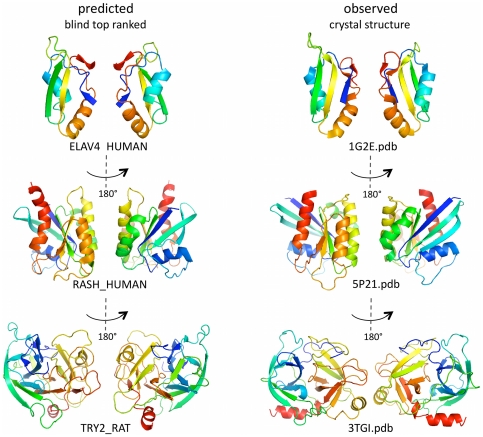
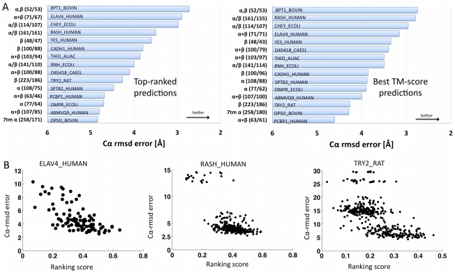
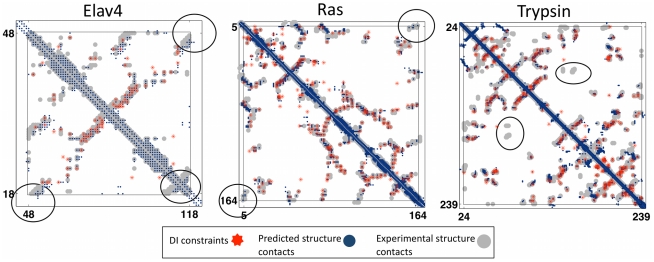
The evolutionary trajectory of a protein through sequence space is constrained by its function. Collections of sequence homologs record the outcomes of millions of evolutionary experiments in which the protein evolves according to these constraints. Deciphering the evolutionary record held in these sequences and exploiting it for predictive and engineering purposes presents a formidable challenge. The potential benefit of solving this challenge is amplified by the advent of inexpensive high-throughput genomic sequencing.In this paper we ask whether we can infer evolutionary constraints from a set of sequence homologs of a protein. The challenge is to distinguish true co-evolution couplings from the noisy set of observed correlations. We address this challenge using a maximum entropy model of the protein sequence, constrained by the statistics of the multiple sequence alignment, to infer residue pair couplings. Surprisingly, we find that the strength of these inferred couplings is an excellent predictor of residue-residue proximity in folded structures. Indeed, the top-scoring residue couplings are sufficiently accurate and well-distributed to define the 3D protein fold with remarkable accuracy.We quantify this observation by computing, from sequence alone, all-atom 3D structures of fifteen test proteins from different fold classes, ranging in size from 50 to 260 residues, including a G-protein coupled receptor. These blinded inferences are de novo, i.e., they do not use homology modeling or sequence-similar fragments from known structures. The co-evolution signals provide sufficient information to determine accurate 3D protein structure to 2.7-4.8 Å C(α)-RMSD error relative to the observed structure, over at least two-thirds of the protein (method called EVfold, details at http://EVfold.org). This discovery provides insight into essential interactions constraining protein evolution and will facilitate a comprehensive survey of the universe of protein structures, new strategies in protein and drug design, and the identification of functional genetic variants in normal and disease genomes.
Overview
The claims and statistics of this paper haven’t been checked yet.
Run the check — free, no account needed. It reads the open-access text and checks the sources it cites.
Integrity signals
Registry
Cites
Statistics
Figures
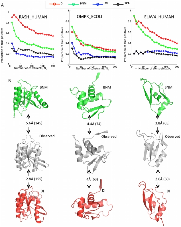
![Figure 3 Progress in contact prediction using the maximum entropy method. Extraction of evolutionary information about residue coupling and predicted contacts from multiple sequence alignments works much better using the global statistical model (right, Direct Information, DI, Equation 3) than the local statistical model (left, Mutual Information, MI, Equation 1). Predicted contacts for DI (red lines connecting the residues predicted to be coupled from sequence information) are better positioned in the experimentally observed structure (grey ribbon diagram), than those for MI (left, blue lines), shown here for the RAS protein (upper) and ELAV4 protein (lower). The DI residue pairs are also more evenly distributed along the chain and overlap more accurately with the contacts in the observed structure (red stars [predicted, grey circles [observed] in contact map; center, upper right triangle) than those using MI (blue [predicted], grey circles [observed]; center, lower left triangle). Details of contact maps for all proteins comparing predicted and observed contacts are in Figures S1 and S2, Text S1.](/paper-figures/ad0edff5338c41479018a65a69fdb4cd/protein-3d-structure-computed-from-evolutionary-sequence-variation-figure-3.jpg)
Verification
Claims the paper makes
The claims and statistics of this paper haven’t been checked yet.
Run the check — free, no account needed. It reads the open-access text and checks the sources it cites.
Claims other papers make about this one
Each is checked against this paper’s own text: does it say what the citing paper says it does?
In the citing paper
“The premise of methods to infer structure based on evolutionary couplings is that, because amino acids exist and evolve in the context of 3D structure, they are not free to evolve independently, but instead co-evolve in patterns reflective of the underlying structure.”
From Predicting multiple conformations via sequence clustering and AlphaFold2 (2024) · cites this paper as [9]
In the citing paper
“One of the major insights leveraged by AlphaFold 1,2 comes from the use of evolutionary covariations that can be extracted from large-scale multiple sequence alignments.[49][50][51][52][53][54]”
From AlphaFold and Implications for Intrinsically Disordered Proteins (2021) · cites this paper as [50]
Self-check
Percentages, sums and reported differences re-computed from the paper’s own numbers.
The self-check needs the paper’s full text.
Citations
Cites
The PyMOL Molecular Graphics System
DeLano, W. L. (2002) · Medical Entomology and Zoology · Cited by 18,147
Crystallography & NMR System: A New Software Suite for Macromolecular Structure Determination
Brünger, A. T., Adams, P. D., Clore, G. M., DeLano, W. L., Gros, P., Grosse‐Kunstleve, R. W., Jiang, J., Kuszewski, J. J., Nilges, M., Pannu, N., et al. (1998) · Acta Crystallographica Section D Biological Crystallography · doi:10.1107/s0907444998003254 · Cited by 15,723
The Pfam Protein Families Database
Bateman, A. G. (2002) · Nucleic Acids Research · doi:10.1093/nar/30.1.276 · Cited by 14,075
I-TASSER: a unified platform for automated protein structure and function prediction
Roy, A., Küçükural, A., & Zhang, Y. (2010) · Nature Protocols · doi:10.1038/nprot.2010.5 · Cited by 6,212
The Pfam protein families database
Finn, R. D., Mistry, J., Tate, J., Coggill, P., Heger, A., Pollington, J. E., Gavin, O. L., Gunasekaran, P., Ceric, G., Forslund, K., et al. (2010) · Nucleic Acids Research · doi:10.1093/nar/gkp985 · Cited by 2,694
Scoring function for automated assessment of protein structure template quality
Zhang, Y., & Skolnick, J. (2004) · Proteins: Structure, Function, and Bioinformatics · doi:10.1002/prot.20264 · Cited by 2,392
Atomic-level characterization of the structural dynamics of proteins
Shaw, D. E., Maragakis, P., Lindorff-Larsen, K., Piana, S., Dror, R. O., Eastwood, M. P., Bank, J. A., Jumper, J. M., Salmon, J. K., Shan, Y., et al. (2010) · Science · doi:10.1126/science.1187409 · Cited by 1,923
Weak pairwise correlations imply strongly correlated network states in a neural population
Schneidman, E., Berry, M. J., Segev, R., & Bialek, W. (2006) · Nature · doi:10.1038/nature04701 · Cited by 1,745
Holm, L., & Sander, C. (1996) · Science · doi:10.1126/science.273.5275.595 · Cited by 1,497
Self-citationMorcos, F., Pagnani, A., Lunt, B., Bertolino, A., Marks, D. S., Sander, C., Zecchina, R., Onuchic, J. N., Hwa, T., & Weigt, M. (2011) · Proceedings of the National Academy of Sciences · doi:10.1073/pnas.1111471108 · Cited by 1,434
Self-citation
Research works printed but not linked: 3
- KT Simons, 1999, Ab initio protein structure prediction of CASP III targets using ROSETTA., Proteins, Suppl 3, 171, 10.1002/(SICI)1097-0134(1999)37:3+<171::AID-PROT21>3.0.CO;2-Z · DOI
- MMT Mezard, 2009, Constraint satisfaction problems and neural networks: A statistical physics perspective., Journal of Physiology, 103, 107
- TR Lezon, 2006, Using the principle of entropy maximization to infer genetic interaction networks from gene expression patterns., Proceedings of the National Academy of Sciences of the United States of America, 103, 19033, 10.1073/pnas.0609152103 · DOI
Cited by
Sorted by citations
Items 201–300 of 1,005
InterPLM: discovering interpretable features in protein language models via sparse autoencoders
Simon, E. P., & Zou, J. (2025) · Nature Methods · doi:10.1038/s41592-025-02836-7 · Cited by 89
Molecular hyperdiversity and evolution in very large populations
Cutter, A. D., Jovelin, R., & Dey, A. (2013) · Molecular Ecology · doi:10.1111/mec.12281 · Cited by 76
Co‐Evolutionary Fitness Landscapes for Sequence Design
Tian, P., Louis, J. M., Baber, J. L., Aniana, A., & Best, R. B. (2018) · Angewandte Chemie International Edition · doi:10.1002/anie.201713220 · Cited by 76
Towards Structural Systems Pharmacology to Study Complex Diseases and Personalized Medicine
Xie, L., Ge, X., Tan, H., Xie, L., Zhang, Y., Hart, T. D., Yang, X., & Bourne, P. E. (2014) · PLoS Computational Biology · doi:10.1371/journal.pcbi.1003554 · Cited by 75
PconsC: combination of direct information methods and alignments improves contact prediction
Skwark, M. J., Abdel‐Rehim, A., & Elofsson, A. (2013) · Bioinformatics · doi:10.1093/bioinformatics/btt259 · Cited by 76
Robust enzyme design: Bioinformatic tools for improved protein stability
Suplatov, D. A., Воеводин, В. В., & Švedas, V. K. (2014) · Biotechnology Journal · doi:10.1002/biot.201400150 · Cited by 77
Protein structure determination by combining sparse NMR data with evolutionary couplings
Tang, Y., Huang, Y. J., Hopf, T. A., Sander, C., Marks, D. S., & Montelione, G. T. (2015) · Nature Methods · doi:10.1038/nmeth.3455 · Cited by 75
Self-citationEcology and Biogenesis of Functional Amyloids in Pseudomonas
Rouse, S., Matthews, S., & Dueholm, M. S. (2018) · Journal of Molecular Biology · doi:10.1016/j.jmb.2018.05.004 · Cited by 65
Structural and Functional Characterization of the Bacterial Type III Secretion Export Apparatus
Dietsche, T., Mebrhatu, M. T., Brunner, M. J., Abrusci, P., Yan, J., Franz‐Wachtel, M., Schärfe, C., Zilkenat, S., Grin, I., Galán, J. E., et al. (2016) · PLoS Pathogens · doi:10.1371/journal.ppat.1006071 · Cited by 74
Integration of QUARK and I‐TASSER for Ab Initio Protein Structure Prediction in CASP11
Zhang, W., Yang, J., He, B., Walker, S. E., Zhang, H., Govindarajoo, B., Virtanen, J., Xue, Z., Shen, H., & Zhang, Y. (2015) · Proteins Structure Function and Bioinformatics · doi:10.1002/prot.24930 · Cited by 72
Zhao, J. W., Krystofiak, E., Ballesteros, Á., Cui, R., Itallie, C. M. V., Anderson, J. M., Fenollar‐Ferrer, C., & Kachar, B. (2018) · Communications Biology · doi:10.1038/s42003-018-0051-5 · Cited by 73
Structure and mutagenic analysis of the lipid II flippase MurJ from Escherichia coli
Zheng, S., Sham, L., Rubino, F. A., Brock, K. P., Robins, W. P., Mekalanos, J. J., Marks, D. S., Bernhardt, T. G., & Kruse, A. C. (2018) · Proceedings of the National Academy of Sciences · doi:10.1073/pnas.1802192115 · Cited by 76
Self-citationNovel machine learning approaches revolutionize protein knowledge
Bordin, N., Dallago, C., Heinzinger, M., Kim, S., Littmann, M., Rauer, C., Steinegger, M., Rost, B., & Orengo, C. (2023) · Trends in Biochemical Sciences · doi:10.1016/j.tibs.2022.11.001 · Cited by 72
He, B., Mortuza, S. M., Wang, Y., Shen, H., & Zhang, Y. (2017) · Bioinformatics · doi:10.1093/bioinformatics/btx164 · Cited by 72
The genetic architecture of protein stability
Faure, A. J., Martí-Aranda, A., Hidalgo-Carcedo, C., Beltran, T., Schmiedel, J. M., & Lehner, B. (2024) · Nature · doi:10.1038/s41586-024-07966-0 · Cited by 73
Marsh, J. A., & Teichmann, S. A. (2013) · BioEssays · doi:10.1002/bies.201300134 · Cited by 72
Evolutionary conservation of RNA sequence and structure
Rivas, E. (2021) · Wiley Interdisciplinary Reviews - RNA · doi:10.1002/wrna.1649 · Cited by 68
Chemical shift-based methods in NMR structure determination
Nerli, S., McShan, A. C., & Sgourakis, N. G. (2018) · Progress in Nuclear Magnetic Resonance Spectroscopy · doi:10.1016/j.pnmrs.2018.03.002 · Cited by 74
Continuous evolution of user-defined genes at 1 million times the genomic mutation rate
Rix, G., Williams, R. L., Hu, V. J., Spinner, A., Pisera, A. (., Marks, D. S., & Liu, C. C. (2024) · Science · doi:10.1126/science.adm9073 · Cited by 71
Self-citationShammas, M. K., Huang, X., Wu, B. P., Fessler, E., Song, I. Y., Randolph, N., Li, Y., Bleck, C. K. E., Springer, D., Fratter, C., et al. (2022) · Journal of Clinical Investigation · doi:10.1172/jci157504 · Cited by 68
Harnessing generative AI to decode enzyme catalysis and evolution for enhanced engineering
Xie, W. J., & Warshel, A. (2023) · National Science Review · doi:10.1093/nsr/nwad331 · Cited by 69
Prediction of multiple conformational states by combining sequence clustering with AlphaFold2Preprint
Wayment-Steele, H. K., Ovchinnikov, S., Colwell, L. J., & Kern, D. (2022) · bioRxiv (Cold Spring Harbor Laboratory) · doi:10.1101/2022.10.17.512570 · Cited by 66
Self-citationHybrid methods for combined experimental and computational determination of protein structure
Seffernick, J. T., & Lindert, S. (2020) · The Journal of Chemical Physics · doi:10.1063/5.0026025 · Cited by 70
Hasanzadeh, A., Hamblin, M. R., Kiani, J., Noori, H., Hardie, J. M., Karimi, M., & Shafiee, H. (2022) · Nano Today · doi:10.1016/j.nantod.2022.101665 · Cited by 73
Protein language models trained on multiple sequence alignments learn phylogenetic relationships
Lupo, U., Sgarbossa, D., & Bitbol, A.-F. (2022) · Nature Communications · doi:10.1038/s41467-022-34032-y · Cited by 67
Blind protein structure prediction using accelerated free-energy simulations
Pérez, A., Morrone, J. A., Brini, E., MacCallum, J. L., & Dill, K. A. (2016) · Science Advances · doi:10.1126/sciadv.1601274 · Cited by 67
De novo design of symmetric ferredoxins that shuttle electrons in vivo
Mutter, A. C., Tyryshkin, A. M., Campbell, I. J., Poudel, S., Bennett, G. N., Silberg, J. J., Nanda, V., & Falkowski, P. G. (2019) · Proceedings of the National Academy of Sciences · doi:10.1073/pnas.1905643116 · Cited by 64
AlphaSpace: Fragment-Centric Topographical Mapping To Target Protein–Protein Interaction Interfaces
Rooklin, D. W., Wang, C., Katigbak, J., Arora, P. S., & Zhang, Y. (2015) · Journal of Chemical Information and Modeling · doi:10.1021/acs.jcim.5b00103 · Cited by 68
The breakthrough in protein structure prediction
Lupas, A. N., Pereira, J., Alva, V., Merino, F., Coles, M. P., & Hartmann, M. D. (2021) · Biochemical Journal · doi:10.1042/bcj20200963 · Cited by 64
Evolution of protein interactions: From interactomes to interfaces
Andréani, J., & Guérois, R. (2014) · Archives of Biochemistry and Biophysics · doi:10.1016/j.abb.2014.05.010 · Cited by 70
Direct Coupling Analysis for Protein Contact Prediction
Morcos, F., Terence Hwa, B., Onuchic, J. N., & Weigt, M. (2014) · Methods in molecular biology · doi:10.1007/978-1-4939-0366-5_5 · Cited by 63
Power law tails in phylogenetic systems
Qin, C., & Colwell, L. J. (2018) · Proceedings of the National Academy of Sciences · doi:10.1073/pnas.1711913115 · Cited by 63
Self-citationPredicting protein conformational motions using energetic frustration analysis and AlphaFold2
Guan, X., Tang, Q.-Y., Ren, W., Chen, M., Wang, W., Wolynes, P. G., & Li, W. (2024) · Proceedings of the National Academy of Sciences · doi:10.1073/pnas.2410662121 · Cited by 62
Malinverni, D., Marsili, S., Barducci, A., & Rios, P. D. L. (2015) · PLoS Computational Biology · doi:10.1371/journal.pcbi.1004262 · Cited by 62
Structural propensities of kinase family proteins from a Potts model of residue co‐variation
Haldane, A., Flynn, W. F., He, P., Vijayan, R., & Levy, R. M. (2016) · Protein Science · doi:10.1002/pro.2954 · Cited by 62
DESTINI: A deep-learning approach to contact-driven protein structure prediction
Gao, M., Zhou, H., & Skolnick, J. (2019) · Scientific Reports · doi:10.1038/s41598-019-40314-1 · Cited by 61
Nicoludis, J. M., Lau, S.-Y., Schärfe, C., Marks, D. S., Weihofen, W. A., & Gaudet, R. (2015) · Structure · doi:10.1016/j.str.2015.09.005 · Cited by 59
Self-citationProtein Structural Bioinformatics: An Overview
Gromiha, M. M., Raju, N., & Selvaraj, S. (2018) · Elsevier eBooks · doi:10.1016/b978-0-12-809633-8.20278-1 · Cited by 61
Feinauer, C., Szurmant, H., Weigt, M., & Pagnani, A. (2016) · PLoS ONE · doi:10.1371/journal.pone.0149166 · Cited by 60
Self-citationRecent developments in deep learning applied to protein structure prediction
Kandathil, S. M., Greener, J. G., & Jones, D. T. (2019) · Proteins Structure Function and Bioinformatics · doi:10.1002/prot.25824 · Cited by 63
Structural coverage of the proteome for pharmaceutical applications
Somody, J. C., MacKinnon, S. S., & Windemuth, A. (2017) · Drug Discovery Today · doi:10.1016/j.drudis.2017.08.004 · Cited by 62
Applied Directional Statistics
Garc\'ia-Portugu\'es, E., Golden, M., S{\o}rensen, M., Mardia, K. V., Hamelryck, T., & Hein, J. (2018) · Chapman & Hall/CRC eBooks · doi:10.1201/9781315228570 · Cited by 59
Wang, X., Nie, Y., & Xu, Y. (2019) · Bioresource Technology · doi:10.1016/j.biortech.2019.01.098 · Cited by 60
Deep embedding and alignment of protein sequences
Llinares-López, F., Berthet, Q., Blondel, M., Teboul, O., & Vert, J.-P. (2023) · Nature Methods · doi:10.1038/s41592-022-01700-2 · Cited by 58
Lam, S. D., Das, S., Sillitoe, I., & Orengo, C. A. (2017) · Acta Crystallographica Section D Structural Biology · doi:10.1107/s2059798317008920 · Cited by 59
Selective Nutrient Transport in Bacteria: Multicomponent Transporter Systems Reign Supreme
Davies, J. S., Currie, M. J., Wright, J. D., Newton-Vesty, M. C., North, R. A., Mace, P. D., Allison, J. R., & Dobson, R. C. J. (2021) · Frontiers in Molecular Biosciences · doi:10.3389/fmolb.2021.699222 · Cited by 60
CoinFold: a web server for protein contact prediction and contact-assisted protein folding
Wang, S., Li, W., Zhang, R., Liu, S., & Xu, J. (2016) · Nucleic Acids Research · doi:10.1093/nar/gkw307 · Cited by 58
Global and local selection acting on the pathogen Stenotrophomonas maltophilia in the human lung
Chung, H., Lieberman, T. D., Vargas, S. O., Flett, K. B., McAdam, A. J., Priebe, G. P., & Kishony, R. (2017) · Nature Communications · doi:10.1038/ncomms14078 · Cited by 58
Metropolized Knockoff Sampling
Bates, S., Candès, E. J., Janson, L., & Wang, W. (2020) · Journal of the American Statistical Association · doi:10.1080/01621459.2020.1729163 · Cited by 57
Accurate contact predictions using covariation techniques and machine learning
Kościółek, T., & Jones, D. T. (2015) · Proteins Structure Function and Bioinformatics · doi:10.1002/prot.24863 · Cited by 56
Revolutionizing protein–protein interaction prediction with deep learning
Zhang, J., Durham, J., & Cong, Q. (2024) · Current Opinion in Structural Biology · doi:10.1016/j.sbi.2024.102775 · Cited by 58
Sfriso, P., Duran‐Frigola, M., Mosca, R., Emperador, A., Aloy, P., & Orozco, M. (2015) · Structure · doi:10.1016/j.str.2015.10.025 · Cited by 55
Automated structure modeling of large protein assemblies using crosslinks as distance restraints
Ferber, M., Kosiński, J., Ori, A., Rashid, U. J., Moreno‐Morcillo, M., Simon, B., Bouvier, G., Batista, P. R., Müller, C. W., Beck, M., et al. (2016) · Nature Methods · doi:10.1038/nmeth.3838 · Cited by 56
EigenTHREADER: analogous protein fold recognition by efficient contact map threading
Buchan, D. W. A., & Jones, D. T. (2017) · Bioinformatics · doi:10.1093/bioinformatics/btx217 · Cited by 57
Integrative Structural Biology in the Era of Accurate Structure Prediction
Masrati, G., Landau, M., Ben-Tal, N., Lupas, A., Kosloff, M., & Kosinski, J. (2021) · Journal of Molecular Biology · doi:10.1016/j.jmb.2021.167127 · Cited by 53
On the dimensionality of behavior
Bialek, W. (2022) · Proceedings of the National Academy of Sciences · doi:10.1073/pnas.2021860119 · Cited by 54
Jana, B., Morcos, F., & Onuchic, J. N. (2014) · Physical Chemistry Chemical Physics · doi:10.1039/c3cp55275f · Cited by 55
Making automated multiple alignments of very large numbers of protein sequences
Sievers, F., Dineen, D. G., Wilm, A., & Higgins, D. G. (2013) · Bioinformatics · doi:10.1093/bioinformatics/btt093 · Cited by 55
AlphaFold Models of Small Proteins Rival the Accuracy of Solution NMR Structures
Tejero, R., Huang, Y. J., Ramelot, T. A., & Montelione, G. T. (2022) · Frontiers in Molecular Biosciences · doi:10.3389/fmolb.2022.877000 · Cited by 56
Inferring interaction partners from protein sequences using mutual information
Bitbol, A. (2018) · PLoS Computational Biology · doi:10.1371/journal.pcbi.1006401 · Cited by 53
Huang, Y. J., Zhang, N., Bersch, B., Fidelis, K., Inouye, M., Ishida, Y., Kryshtafovych, A., Kobayashi, N., Kuroda, Y., Liu, G., et al. (2021) · Proteins: Structure, Function, and Bioinformatics · doi:10.1002/prot.26246 · Cited by 49
Improved protein contact predictions with the MetaPSICOV2 server in CASP12
Buchan, D. W. A., & Jones, D. T. (2017) · Proteins Structure Function and Bioinformatics · doi:10.1002/prot.25379 · Cited by 53
Cooper, C. J., Zheng, K., Rush, K. W., Johs, A., Sanders, B., Pavlopoulos, G. A., Kyrpides, N. C., Podar, M., Ovchinnikov, S., Ragsdale, S. W., et al. (2020) · Communications Biology · doi:10.1038/s42003-020-1047-5 · Cited by 59
In vivo hypermutation and continuous evolution
Molina, R. S., Rix, G., Mengiste, A. A., Álvarez, B., Seo, D., Chen, H., Hurtado, J., Zhang, Q., García-García, J. D., Heins, Z., et al. (2022) · Nature Reviews Methods Primers · doi:10.1038/s43586-022-00130-w · Cited by 54
Covariation Is a Poor Measure of Molecular Coevolution
Talavera, D., Lovell, S. C., & Whelan, S. (2015) · Molecular Biology and Evolution · doi:10.1093/molbev/msv109 · Cited by 54
Domain-swap polymerization drives the self-assembly of the bacterial flagellar motor
Baker, M. A. B., Hynson, R. M. G., Ganuelas, L. A., Mohammadi, N. S., Liew, C. W., Rey, A. A., Duff, A. P., Whitten, A. E., Jeffries, C. M., Delalez, N. J., et al. (2016) · Nature Structural & Molecular Biology · doi:10.1038/nsmb.3172 · Cited by 53
Enhanced unbiased sampling of protein dynamics using evolutionary coupling information
Shamsi, Z., Moffett, A. S., & Shukla, D. (2017) · Scientific Reports · doi:10.1038/s41598-017-12874-7 · Cited by 52
How Many Protein Sequences Fold to a Given Structure? A Coevolutionary Analysis
Tian, P., & Best, R. B. (2017) · Biophysical Journal · doi:10.1016/j.bpj.2017.08.039 · Cited by 52
Integrative Structure Modeling: Overview and Assessment
Braitbard, M., Schneidman‐Duhovny, D., & Kalisman, N. (2019) · Annual Review of Biochemistry · doi:10.1146/annurev-biochem-013118-111429 · Cited by 52
Model-based reinforcement learning for biological sequence design
Angermueller, C., Dohan, D., Belanger, D., Deshpande, R., Murphy, K. J., & Colwell, L. J. (2020) · International Conference on Learning Representations · Cited by 51
Self-citationAll-atom 3D structure prediction of transmembrane β-barrel proteins from sequences
Hayat, S., Sander, C., Marks, D. S., & Elofsson, A. (2015) · Proceedings of the National Academy of Sciences · doi:10.1073/pnas.1419956112 · Cited by 51
Self-citationSchäfer, N., Maierhofer, T., Herrmann, J., Jørgensen, M. E., Lind, C., von Meyer, K., Lautner, S., Fromm, J. H., Felder, M., Hetherington, A. M., et al. (2018) · Current Biology · doi:10.1016/j.cub.2018.03.027 · Cited by 51
Schubert, B., Maddamsetti, R., Nyman, J., Farhat, M. R., & Marks, D. S. (2018) · Nature Microbiology · doi:10.1038/s41564-018-0309-1 · Cited by 51
Self-citationLarge-scale state-dependent membrane remodeling by a transporter protein
Zhou, W., Fiorin, G., Anselmi, C., Karimi‐Varzaneh, H. A., Poblete, H., Forrest, L. R., & Faraldo‐Gómez, J. D. (2019) · eLife · doi:10.7554/elife.50576 · Cited by 51
Dormant spores sense amino acids through the B subunits of their germination receptors
Artzi, L., Alon, A., Brock, K. P., Green, A. G., Tam, A., Ramírez‐Guadiana, F. H., Marks, D. S., Kruse, A. C., & Rudner, D. Z. (2021) · Nature Communications · doi:10.1038/s41467-021-27235-2 · Cited by 51
Self-citationAddressing epistasis in the design of protein function
Lipsh‐Sokolik, R., & Fleishman, S. J. (2024) · Proceedings of the National Academy of Sciences · doi:10.1073/pnas.2314999121 · Cited by 55
What evolution tells us about protein physics, and protein physics tells us about evolution
Bastolla, U., Dehouck, Y., & Echave, J. (2016) · Current Opinion in Structural Biology · doi:10.1016/j.sbi.2016.10.020 · Cited by 52
de la Paz, J. A., Nartey, C. M., Yuvaraj, M., & Morcos, F. (2020) · Proceedings of the National Academy of Sciences · doi:10.1073/pnas.1913071117 · Cited by 50
DEEPCON: protein contact prediction using dilated convolutional neural networks with dropout
Adhikari, B. (2019) · Bioinformatics · doi:10.1093/bioinformatics/btz593 · Cited by 49
Feher, V. A., Randall, A. Z., Baldi, P., Bush, R. M., de la Maza, L. M., & Amaro, R. E. (2013) · PLoS ONE · doi:10.1371/journal.pone.0068934 · Cited by 49
Kukić, P., Mirabello, C., Tradigo, G., Walsh, I., Veltri, P., & Pollastri, G. (2014) · BMC Bioinformatics · doi:10.1186/1471-2105-15-6 · Cited by 49
AIDA: ab initio domain assembly server
Xu, D. H., Jaroszewski, L., Li, Z., & Godzik, A. (2014) · Nucleic Acids Research · doi:10.1093/nar/gku369 · Cited by 49
Perspectives on theory at the interface of physics and biology
Bialek, W. (2017) · Reports on Progress in Physics · doi:10.1088/1361-6633/aa995b · Cited by 60
Subnanometer resolution cryo-EM structure of Arabidopsis thaliana ATG9
Lai, L. T. F., Yu, C., Wong, J. S. K., Lo, H. S., Benlekbir, S., Jiang, L., & Lau, W. C. Y. (2019) · Autophagy · doi:10.1080/15548627.2019.1639300 · Cited by 53
Critical Review of Synthesis, Toxicology and Detection of Acyclovir
Wei, Y., Yao, L., Wu, Y., Liu, X., Peng, L., Tian, Y., Ding, J., Li, K.-H., & He, Q. (2021) · Molecules · doi:10.3390/molecules26216566 · Cited by 49
Are protein language models the new universal key?
Weißenow, K., & Rost, B. (2025) · Current Opinion in Structural Biology · doi:10.1016/j.sbi.2025.102997 · Cited by 54
Secondary Structure and Contact Guided Differential Evolution for Protein Structure Prediction
Zhang, G., Ma, L., Wang, X., & Zhou, X. (2018) · IEEE Transactions on Computational Biology and Bioinformatics · doi:10.1109/tcbb.2018.2873691 · Cited by 48
Shahmoradi, A., Sydykova, D. K., Spielman, S. J., Jackson, E. L., Dawson, E. T., Meyer, A. G., & Wilke, C. O. (2014) · Journal of Molecular Evolution · doi:10.1007/s00239-014-9644-x · Cited by 50
Schep, D. G., Zhao, J., & Rubinstein, J. L. (2016) · Proceedings of the National Academy of Sciences · doi:10.1073/pnas.1521990113 · Cited by 47
Enhancing computational enzyme design by a maximum entropy strategy
Xie, W. J., Asadi, M., & Warshel, A. (2022) · Proceedings of the National Academy of Sciences · doi:10.1073/pnas.2122355119 · Cited by 50
Wang, Z., Cao, R., Taylor, K. H., Briley, A., Caldwell, C. W., & Cheng, J. J. (2013) · PLoS ONE · doi:10.1371/journal.pone.0058793 · Cited by 46
Nicoludis, J. M., Vogt, B. E., Green, A. G., Schärfe, C. P., Marks, D. S., & Gaudet, R. (2016) · eLife · doi:10.7554/elife.18449 · Cited by 46
Self-citationZhu, J., Zhang, H., Li, S. C., Wang, C., Kong, L., Sun, S., Zheng, W., & Bu, D. (2017) · Bioinformatics · doi:10.1093/bioinformatics/btx514 · Cited by 46
CryoEM Reveals the Complexity and Diversity of ATP Synthases
Courbon, G. M., & Rubinstein, J. L. (2022) · Frontiers in Microbiology · doi:10.3389/fmicb.2022.864006 · Cited by 45
Yang, J., He, B.-J., Jang, R., Zhang, Y., & Shen, H. (2015) · Bioinformatics · doi:10.1093/bioinformatics/btv459 · Cited by 47
BIS2Analyzer: a server for co-evolution analysis of conserved protein families
Oteri, F., Nadalin, F., Champeimont, R., & Carbone, A. (2017) · Nucleic Acids Research · doi:10.1093/nar/gkx336 · Cited by 44
Protein Structure from Experimental Evolution
Stiffler, M. A., Poelwijk, F. J., Brock, K. P., Stein, R. R., Riesselman, A. J., Teyra, J., Sidhu, S. S., Marks, D. S., Gauthier, N. P., & Sander, C. (2019) · Cell Systems · doi:10.1016/j.cels.2019.11.008 · Cited by 45
Self-citationVariational auto-encoding of protein sequencesPreprint
Sinai, S., Kelsic, E. D., Church, G. M., & Nowak, M. A. (2017) · arXiv (Cornell University) · doi:10.48550/arxiv.1712.03346 · Cited by 77
Zero-shot antibody design in a 24-well platePreprint
Chai Discovery Team, Boitreaud, J., Dent, J., Geisz, D., McPartlon, M., Meier, J., Qiao, Z., Rogozhnikov, A., Rollins, N., Wollenhaupt, P., et al. (2025) · bioRxiv (Cold Spring Harbor Laboratory) · doi:10.1101/2025.07.05.663018 · Cited by 56
Protein sequence‐to‐structure learning: Is this the end(‐to‐end revolution)?
Laine, E., Eismann, S., Elofsson, A., & Grudinin, S. (2021) · Proteins: Structure, Function, and Bioinformatics · doi:10.1002/prot.26235 · Cited by 44
“They enabled the modelling of 3D structures for large protein families [9, 10, 11, 12, 13, 14].”
BackgroundMethod
Metadata
- Authors
- Marks, D. S., Colwell, L. J., Sheridan, R., Hopf, T. A., Pagnani, A., Zecchina, R., & Sander, C.
- Affiliations
- Harvard University (US)
- MRC Laboratory of Molecular Biology (GB)
- Memorial Sloan Kettering Cancer Center (US)
- Italian institute for Genomic Medicine (IT)
- Politecnico di Torino (IT)
- Corresponding author
- Marks, Debora S.
- Venue
- PLoS ONE
- Publisher
- Public Library of Science (PLoS)
- Published
- 2011-12-07
- Volume
- 6
- Issue
- 12
- Pages
- e28766
- ISSN
- 1932-6203 (ISSN-L)
- PMC
- PMC3233603
- OpenAlex
- W2061042699
- Cited by
- 1,144
- References
- 84
- Citations by year
- 2026: 61 · 2025: 60 · 2024: 47 · 2023: 72 · 2022: 96 · 2021: 104 · 2020: 90 · 2019: 110 · 2018: 91 · 2017: 107 · 2016: 71 · 2015: 74 · 2014: 72 · 2013: 56 · 2012: 29
- FWCI
- 20.79
- Type
- journal article
- Language
- en
- Open access
- gold
- doi.org
- Locations
- PLoS ONE (publishedVersion, cc-by)
- Europe PMC (PubMed Central) (publishedVersion, cc-by)
- PubMed Central (publishedVersion, cc-by)
- PubMed (publishedVersion)
- PORTO@Iris (submittedVersion, cc-by)
- DOAJ (DOAJ: Directory of Open Access Journals) (submittedVersion, cc-by-sa)
- Figshare (submittedVersion, cc-by)
- PORTO Publications Open Repository TOrino (Politecnico di Torino) (submittedVersion, cc-by)
- Funders
- Memorial Sloan-Kettering Cancer Center
- Studienstiftung des Deutschen Volkes
- National Institutes of Health (NIH U54, CA143798)
- Engineering and Physical Sciences Research Council (EP/H028064/1)
- National Cancer Institute (U54CA143798)
- National Institute of General Medical Sciences (R01GM106303)
- Topics
- Life Sciences › Biochemistry, Genetics and Molecular Biology › Molecular Biology › Protein Structure and Dynamics
- Life Sciences › Biochemistry, Genetics and Molecular Biology › Structural Biology › Enzyme Structure and Function
- Life Sciences › Biochemistry, Genetics and Molecular Biology › Molecular Biology › RNA and protein synthesis mechanisms
- Keywords
- protein structure prediction, coevolution, maximum entropy model, multiple sequence alignment, de novo structure prediction, protein evolution, G protein-coupled receptors, protein engineering
- MeSH terms
- Animals, Humans, Protein Conformation, Proteins, Rhodopsin, Trypsin, Genetic Variation, Drug Design, Reproducibility of Results, Models, Statistical, Genome, Protein Structure, Tertiary, Evolution, Molecular, Entropy, Computational Biology, Catalytic Domain, Imaging, Three-Dimensional
About these checks
Registry, retraction, Cites and statistics results are computed, not judged, with one exception: a printed reference that no index lists is read once by a model before it counts as a citation error. Claims are extracted by a model and kept only when their quote appears verbatim in the text; each cited source is then checked, with a model’s help, for whether it says what the claim relies on. Those verdicts can be wrong — read the claim’s quote and the cited source before relying on one.
Statistics and arithmetic are recomputed, not judged.
Confidence bands are shown only for fields calibrated on our annotated test set. None is calibrated yet, so no band is shown.
What counts as a claim ›How we measure confidence ›How we check this ›
Are you an author, or do you see a problem with this page? Request a review
