

Single-nucleus and single-cell transcriptomes compared in matched cortical cell types
Open accessPublished version of a preprint
The DOI matches its Crossref record; 58 of the 58 references it cites were found in a registry or an index.
Abstract
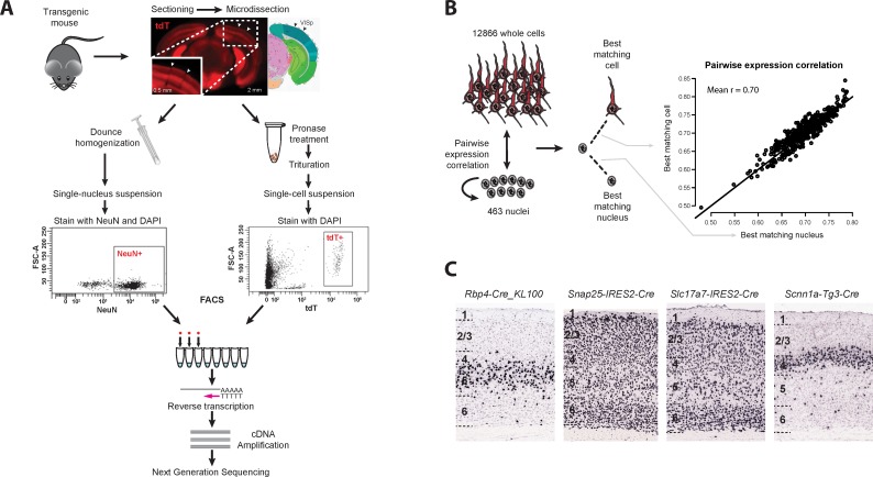
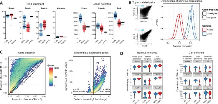
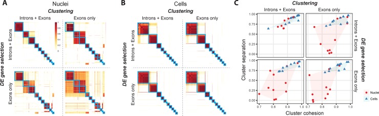
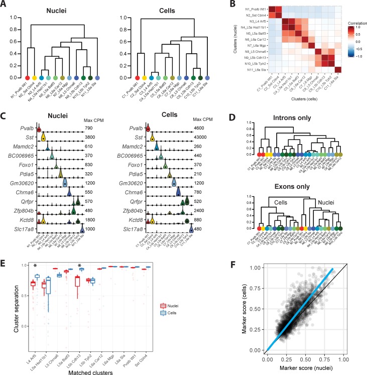
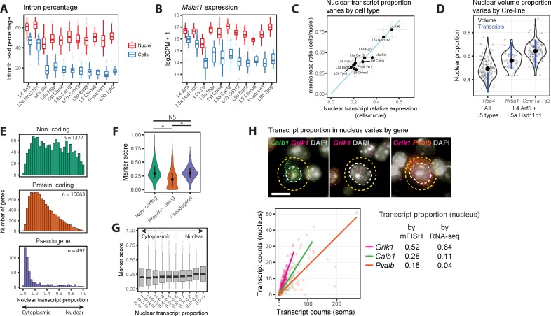
Transcriptomic profiling of complex tissues by single-nucleus RNA-sequencing (snRNA-seq) affords some advantages over single-cell RNA-sequencing (scRNA-seq). snRNA-seq provides less biased cellular coverage, does not appear to suffer cell isolation-based transcriptional artifacts, and can be applied to archived frozen specimens. We used well-matched snRNA-seq and scRNA-seq datasets from mouse visual cortex to compare cell type detection. Although more transcripts are detected in individual whole cells (~11,000 genes) than nuclei (~7,000 genes), we demonstrate that closely related neuronal cell types can be similarly discriminated with both methods if intronic sequences are included in snRNA-seq analysis. We estimate that the nuclear proportion of total cellular mRNA varies from 20% to over 50% for large and small pyramidal neurons, respectively. Together, these results illustrate the high information content of nuclear RNA for characterization of cellular diversity in brain tissues.
Overview
The claims and statistics of this paper haven’t been checked yet.
Free: it reads the open-access text and checks the first 5 claims that cite a source we can check. Create a free account to check every claim.
Integrity signals
Registry
Cites
Statistics
Figures
Verification
Claims the paper makes
The claims and statistics of this paper haven’t been checked yet.
Free: it reads the open-access text and checks the first 5 claims that cite a source we can check. Create a free account to check every claim.
Claims other papers make about this one
Each is checked against this paper’s own text: does it say what the citing paper says it does?
The source backs part of the claim, not all of it — or backs it for a different population, condition or comparison.
In the citing paper
“While scRNA-seq data are analyzed by mapping sequenced fragments to exons, snRNA-seq data analysis should also consider the abundance of intronic reads from pre-mRNA, which may account for >75% of fragments (Bakken et al., 2018; Gaidatzis et al., 2015).”
From Single-Cell Sequencing of Brain Cell Transcriptomes and Epigenomes (2021) · cites this paper as Bakken et al., 2018
Experimental — accuracy not yet measuredThe source backs part of the claim, not all of it — or backs it for a different population, condition or comparison.
In the citing paper
“Nuclear transcripts comprise ~20%−50% of all the RNA in the cell (Bakken et al., 2018), including immature and unspliced RNA molecules containing introns (Fig. 2a).”
From Single-Cell Sequencing of Brain Cell Transcriptomes and Epigenomes (2021) · cites this paper as Bakken et al., 2018
Experimental — accuracy not yet measuredThe source backs part of the claim, not all of it — or backs it for a different population, condition or comparison.
In the citing paper
“Fresh tissue samples are deemed appropriate for single-cell sequencing, whereas freeze-thaw samples are generally recommended for snRNA-seq [242, 243].”
From Single-cell sequencing to multi-omics: technologies and applications (2024) · cites this paper as [243]
Experimental — accuracy not yet measuredThe source backs part of the claim, not all of it — or backs it for a different population, condition or comparison.
In the citing paper
“Differential expression analysis comparing individual cell types profiled by snRNA-seq and scRNA-seq suggested higher expression of long noncoding RNAs in snRNA-seq libraries and higher expression of genes related to mitochondrial and ribosomal functions in scRNA-seq, in agreement with previous reports [24, 26]”
From Systematic assessment of tissue dissociation and storage biases in single-cell and single-nucleus RNA-seq workflows (2020) · cites this paper as [24]
Experimental — accuracy not yet measuredThe source backs part of the claim, not all of it — or backs it for a different population, condition or comparison.
In the citing paper
“whereas longer CDSs were enriched (NES = 1.91, padj = 3.6e-05), as already observed in earlier snRNA-seq studies (Bakken et al., 2018).”
From Single-Nucleus RNA-Seq Is Not Suitable for Detection of Microglial Activation Genes in Humans (2020) · cites this paper as Bakken et al., 2018
Experimental — accuracy not yet measured
6 more claims about this paper
Create a free account to see every claim other papers make about this one.
Self-check
Percentages, sums and reported differences re-computed from the paper’s own numbers.
The self-check needs the paper’s full text.
Citations
Cites
STAR: ultrafast universal RNA-seq aligner
Dobin, A., Davis, C. A., Schlesinger, F., Drenkow, J., Zaleski, C., Jha, S., Batut, P., Chaisson, M., & Gingeras, T. R. (2013) · Bioinformatics · doi:10.1093/bioinformatics/bts635 · Cited by 51,709
limma powers differential expression analyses for RNA-sequencing and microarray studies
Ritchie, M. E., Phipson, B., Wu, D., Hu, Y., Law, C. W., Shi, W., & Smyth, G. K. (2015) · Nucleic Acids Research · doi:10.1093/nar/gkv007 · Cited by 40,458
ggplot2: Elegant Graphics for Data Analysis
Wickham, H. (2009) · doi:10.1007/978-0-387-98141-3 · Cited by 14,447
Highly parallel genome-wide expression profiling of individual cells using nanoliter droplets.
Macosko, E. Z., Basu, A., Satija, R., Nemesh, J., Shekhar, K., Goldman, M., Tirosh, I., Bialas, A. R., Kamitaki, N., Martersteck, E. M., et al. (2015) · Cell · doi:10.1016/j.cell.2015.05.002 · Cited by 7,110
A robust and high-throughput Cre reporting and characterization system for the whole mouse brain
Madisen, L., Zwingman, T. A., Sunkin, S. M., Oh, S. W., Zariwala, H. A., Gu, H., Ng, L. L., Palmiter, R. D., Hawrylycz, M. J., Jones, A. R., et al. (2010) · Nature Neuroscience · doi:10.1038/nn.2467 · Cited by 6,907
Self-citationREVIGO summarizes and visualizes long lists of gene ontology terms
Supek, F., Bošnjak, M., Škunca, N., & Šmuc, T. (2011) · PLoS ONE · doi:10.1371/journal.pone.0021800 · Cited by 6,264
Genome-wide atlas of gene expression in the adult mouse brain
Lein, E. S., Hawrylycz, M. J., Ao, N., Ayres, M., Bensinger, A., Bernard, A., Boe, A. F., Boguski, M. S., Brockway, K. S., Byrnes, E. J., et al. (2007) · Nature · doi:10.1038/nature05453 · Cited by 5,572
Self-citationLandscape of transcription in human cells
Djebali, S., Davis, C. A., Merkel, A., Dobin, A., Lassmann, T., Mortazavi, A., Tanzer, A., Lagarde, J., Lin, W., Schlesinger, F., et al. (2012) · Nature · doi:10.1038/nature11233 · Cited by 5,187
Software for Computing and Annotating Genomic Ranges
Lawrence, M., Huber, W., Pagès, H., Aboyoun, P., Carlson, M., Gentleman, R., Morgan, M. T., & Carey, V. J. (2013) · PLoS Computational Biology · doi:10.1371/journal.pcbi.1003118 · Cited by 4,398
Cell types in the mouse cortex and hippocampus revealed by single-cell RNA-seq
Zeisel, A., Muñoz-Manchado, A. B., Codeluppi, S., Lönnerberg, P., La Manno, G., Juréus, A., Marques, S., Munguba, H., He, L., Betsholtz, C., et al. (2015) · Science · doi:10.1126/science.aaa1934 · Cited by 3,124
Research works printed but not linked: 1
- Aronesty E. “ ea-utils: Command-line tools for processing biological sequencing data.” Expr Anal. 2011; http://code.google.com/p/ea-utils
Cited by
Items 101–200 of 629
The Impact of Single-Cell Genomics on Adipose Tissue Research
Deutsch, A., Feng, D., Pessin, J. E., & Shinoda, K. (2020) · International Journal of Molecular Sciences · doi:10.3390/ijms21134773 · Cited by 66
“Recent literature comparing well-matched single-nuclei RNA-sequencing (snRNA-seq) and scRNA-seq datasets has revealed that snRNA-seq is comparable to scRNA-seq in cell-type detection and superior in the cellular coverage for complex tissues, such as the brain, despite the known loss of mRNA from the cytoplasm and othe…”
BackgroundCell type discovery using single-cell transcriptomics: implications for ontological representation
Aevermann, B. D., Novotny, M., Bakken, T., Miller, J. A., Diehl, A. D., Osumi-Sutherland, D., Lasken, R. S., Lein, E. S., & Scheuermann, R. H. (2018) · Human Molecular Genetics · doi:10.1093/hmg/ddy100 · Cited by 63
Self-citationSingle‐nucleus RNA sequencing of plant tissues using a nanowell‐based system
Sunaga, D. Y., Muiño, J. M., Braeuning, C., Xu, X., Zong, M., Smaczniak, C., Yan, W., Fischer, C., Vidal, R. O., Kliem, M., et al. (2021) · The Plant Journal · doi:10.1111/tpj.15458 · Cited by 70
Yang, S., Liu, C., Jiang, M., Liu, X., Geng, L., Zhang, Y., Sun, S., Wang, K., Yin, J., Ma, S., et al. (2024) · Protein & Cell · doi:10.1093/procel/pwad039 · Cited by 62
“Using single-nucleus RNA-seq, which recapitulates cell proportions better than scRNA-seq, and is superior for an unbiased dissection of all cell types present in the liver (Bakken et al., 2018; He et al., 2020), we interro-gated the contribution of different cell types to primate liver aging.”
An integrated transcriptomic and epigenomic atlas of mouse primary motor cortex cell types
Yao, Z., Liu, H., Xie, F., Fischer, S., Booeshaghi, A. S., Adkins, R. S., Aldridge, A. I., Ament, S. A., Pinto-Duarte, A., Bartlett, A., et al. (2020) · doi:10.1101/2020.02.29.970558 · Cited by 397
Self-citationSingle-cell genomics in plants: current state, future directions, and hurdles to overcome
Cuperus, J. T. (2021) · PLANT PHYSIOLOGY · doi:10.1093/plphys/kiab478 · Cited by 62
“Although nuclei-based methods capture fewer molecules, these may be more biologically informative (Bakken et al., 2018; Farmer et al., 2021).”
BackgroundTuddenham, J. F., Taga, M. F., Haage, V. C., Marshe, V. S., Roostaei, T., White, C. C., Lee, A., Fujita, M., Khairallah, A., Zhang, Y., et al. (2024) · Nature Neuroscience · doi:10.1038/s41593-024-01764-7 · Cited by 85
“…Nature Portfolio reporting summaries, source data, extended data, supplementary information, acknowledgements, peer review information; details of author contributions and competing interests; and statements of data and code availability are available at https://doi.org/10.1038/s41593-024-01764-7.”
InfluentialIsolation of Plant Root Nuclei for Single Cell RNA Sequencing
Thibivilliers, S., Anderson, D. K., & Libault, M. (2020) · Current Protocols in Plant Biology · doi:10.1002/cppb.20120 · Cited by 62
Single-cell transcriptomic landscape of the developing human spinal cord
Andersen, J., Thom, N., Shadrach, J. L., Chen, X., Onesto, M. M., Amin, N. D., Yoon, S.-J., Li, L., Greenleaf, W. J., Müller, F., et al. (2023) · Nature Neuroscience · doi:10.1038/s41593-023-01311-w · Cited by 61
“Single-cell and single-nucleus samples displayed some differences, as reported 13 .”
Isolation of Nuclei from Mammalian Cells and Tissues for Single‐Nucleus Molecular Profiling
Nadelmann, E. R., Gorham, J. M., Reichart, D., Delaughter, D. M., Wakimoto, H., Lindberg, E. L., Litviňukova, M., Maatz, H., Curran, J. J., Ischiu Gutierrez, D., et al. (2021) · Current Protocols · doi:10.1002/cpz1.132 · Cited by 61
“BASIC PROTOCOL 2 NUCLEI ISOLATION The use of isolated nuclei, rather than whole cells, allows for the transcriptomic profiling of difficult-to-isolate cells in both fresh and frozen tissues (Del-Aguila et al., 2019).”
BackgroundCell type-specific changes identified by single-cell transcriptomics in Alzheimer’s disease
Luquez, T., Gaur, P., Kosater, I. M., Lam, M., Lee, D. I., Mares, J., Paryani, F., Yadav, A., & Menon, V. (2022) · Genome Medicine · doi:10.1186/s13073-022-01136-5 · Cited by 61
“However, side-by-side studies of the two approaches [26, 27] have shown that the overall distribution of transcripts in the nucleus is sufficient to recapitulate cell type signatures found by scRNA-seq, even with the reduced transcript detection in cell classes such as microglia [28].”
BackgroundAn atlas of late prenatal human neurodevelopment resolved by single-nucleus transcriptomics
Ramos, S. I., Mussa, Z. M., Falk, E. N., Pai, B., Giotti, B., Allette, K., Cai, P., Dekio, F., Sebra, R., Beaumont, K. G., et al. (2022) · Nature Communications · doi:10.1038/s41467-022-34975-2 · Cited by 60
“0.1) by mapping to the GRCh38 human genome pre-mRNA transcriptome sequences; such mapping enables comparable cluster resolution to single-cell RNA sequencing (scRNA-seq) 66 .”
InfluentialKang, R. B., Li, Y., Rosselot, C., Zhang, T., Siddiq, M., Rajbhandari, P., Stewart, A. F., Scott, D. K., Garcia-Ocana, A., & Lu, G. (2023) · Genome Medicine · doi:10.1186/s13073-023-01179-2 · Cited by 62
“scRNA-seq analyzes both nuclear and cytoplasmic transcripts with a majority being cytoplasmic, whereas snRNA-seq profiles mostly nuclear transcripts with minimal transcripts derived from cytoplasm or rough ER during nuclei isolation [47, 48].”
BackgroundSingle-cell spatial transcriptomic atlas of the whole mouse brain
Han, L., Liu, Z., Jing, Z., Liu, Y., Chang, H., Lei, J., Peng, Y., Wang, K., Xu, Y., Liu, W., et al. (2025) · Neuron · doi:10.1016/j.neuron.2025.02.015 · Cited by 84
Transcriptomic Insight Into the Polygenic Mechanisms Underlying Psychiatric Disorders
Hernandez, L. M., Kim, M., Hoftman, G. D., Haney, J. R., de la Torre-Ubieta, L., Paşaniuc, B., & Gandal, M. J. (2020) · Biological Psychiatry · doi:10.1016/j.biopsych.2020.06.005 · Cited by 70
“snRNA-seq is generally comparable to single-cell RNA-seq, although it detects more intronic reads and fewer total genes (114,115).”
BackgroundIsolation of Adult Spinal Cord Nuclei for Massively Parallel Single-nucleus RNA Sequencing
Matson, K. J. E., Sathyamurthy, A., Johnson, K. R., Kelly, M. C., Kelley, M. W., & Levine, A. J. (2018) · Journal of Visualized Experiments · doi:10.3791/58413 · Cited by 60
The Effects of Opioids on HIV Neuropathogenesis
Murphy, A., Barbaro, J. M., Martínez-Aguado, P., Chilunda, V., Jaureguiberry-Bravo, M., & Berman, J. W. (2019) · Frontiers in Immunology · doi:10.3389/fimmu.2019.02445 · Cited by 62
Wang, X., Sun, Q., Liu, T., Lu, H., Lin, X., Wang, W., Liu, Y., Huang, Y., Huang, G., Sun, H., et al. (2024) · Science Advances · doi:10.1126/sciadv.adn4306 · Cited by 71
Hierarchical progressive learning of cell identities in single-cell data
Michielsen, L., Reinders, M. J. T., & Mahfouz, A. E. (2021) · Nature Communications · doi:10.1038/s41467-021-23196-8 · Cited by 61
Mapping visual functions onto molecular cell types in the mouse superior colliculus
Liu, Y., Savier, E. L., DePiero, V. J., Chen, C., Schwalbe, D. C., Abraham‐Fan, R., Chen, H., Campbell, J. N., & Cang, J. (2023) · Neuron · doi:10.1016/j.neuron.2023.03.036 · Cited by 65
Molecular Connectomics Reveals a Glucagon-Like Peptide 1 Sensitive Neural Circuit for Satiety
Webster, A. N., Becker, J. J., Li, C., Schwalbe, D. C., Kerspern, D., Karolczak, E. O., Bundon, C., Onoharigho, R. A., Crook, M., Jalil, M., et al. (2023) · doi:10.1101/2023.10.31.564990 · Cited by 56
“We chose to profile nuclei instead of whole cells because enzymatic dissociation appears to deplete rabies-infected neurons, potentially due to poor cell viability 23 , and because a cell’s nuclear RNA profile is sufficient to identify its molecular type 24-26 .”
BackgroundOpportunities and challenges in the application of single-cell and spatial transcriptomics in plants
Chen, C., Ge, Y., & Lu, L. (2023) · Frontiers in Plant Science · doi:10.3389/fpls.2023.1185377 · Cited by 71
Molecular connectomics reveals a glucagon-like peptide 1-sensitive neural circuit for satiety
Webster, A. N., Becker, J. J., Li, C., Schwalbe, D. C., Kerspern, D., Karolczak, E. O., Bundon, C. B., Onoharigho, R. A., Crook, M., Jalil, M., et al. (2024) · Nature Metabolism · doi:10.1038/s42255-024-01168-8 · Cited by 55
“We chose to profile nuclei instead of whole cells because enzymatic dissociation appears to deplete rabies-infected neurons, potentially due to poor cell viability 24 , and because a cell’s nuclear RNA profile is sufficient to identify its molecular type 25–27 .”
Hegenbarth, J.-C., Lezzoche, G., Windt, L. J. D., & Stoll, M. (2022) · Frontiers in Molecular Medicine · doi:10.3389/fmmed.2022.839338 · Cited by 67
Brain atrophy in prodromal synucleinopathy is shaped by structural connectivity and gene expression
Rahayel, S., Tremblay, C., Vo, A., Zheng, Y. Q., Lehéricy, S., Arnulf, I., Vidailhet, M., Corvol, J., ICEBERG Study Group, Vidailhet, M., et al. (2022) · Brain · doi:10.1093/brain/awac187 · Cited by 57
Single‐cell RNA sequencing in Drosophila: Technologies and applications
Li, H. (2021) · WIREs Developmental Biology · doi:10.1002/wdev.396 · Cited by 60
Scaled, high fidelity electrophysiological, morphological, and transcriptomic cell characterization
Lee, B. R., Budzillo, A., Hadley, K., Miller, J. A., Jarsky, T., Baker, K., Hill, D., Kim, L., Mann, R., Ng, L., et al. (2021) · eLife · doi:10.7554/elife.65482 · Cited by 54
“A powerful aspect of the FACS-based transcriptomic studies is the ability to use similar methods to profile and compare neurons from different brain regions or species (Bakken et al. 2018; Hodge et al. 2019; Kalmbach et al. 2020).”
BackgroundMethodSelf-citationWhytock, K. L., Sun, Y., Divoux, A., Yu, G., Smith, S. R., Walsh, M. J., & Sparks, L. M. (2022) · iScience · doi:10.1016/j.isci.2022.104772 · Cited by 58
Methods and Insights from Single-Cell Expression Quantitative Trait Loci
Kang, J. B., Raveane, A., Nathan, A., Soranzo, N., & Raychaudhuri, S. (2023) · Annual Review of Genomics and Human Genetics · doi:10.1146/annurev-genom-101422-100437 · Cited by 66
“An alternative to scRNA-seq is snRNA-seq, which sequences isolated nuclei instead of cells (40). snRNA-seq can be especially useful for tissues that are difficult to dissociate due to cell size or fragility, such as brain tissue or frozen tumor samples (11, 18, 131).”
BackgroundMaden, S. K., Kwon, S. H., Huuki-Myers, L. A., Collado‐Torres, L., Hicks, S. C., & Maynard, K. R. (2023) · Genome biology · doi:10.1186/s13059-023-03123-4 · Cited by 66
Single-Cell Quantification of mRNA Expression in The Human Brain
Jolly, S., Lang, V., Koelzer, V. H., Frigerio, C. S., Magno, L., Salinas, P. C., Whiting, P. J., & Palomer, E. (2019) · Scientific Reports · doi:10.1038/s41598-019-48787-w · Cited by 59
The Acute Stress Response in the Multiomic Era
Floriou-Servou, A., von Ziegler, L., Waag, R., Schläppi, C., Germain, P.-L., & Bohacek, J. (2021) · Biological Psychiatry · doi:10.1016/j.biopsych.2020.12.031 · Cited by 51
Plasticity and heterogeneity of thermogenic adipose tissue
Sun, W., Modica, S., Dong, H., & Wolfrum, C. (2021) · Nature Metabolism · doi:10.1038/s42255-021-00417-4 · Cited by 54
Fröhlich, A. S., Gerstner, N., Gagliardi, M., Ködel, M., Yusupov, N., Matosin, N., Czamara, D., Sauer, S., Roeh, S., Murek, V., et al. (2024) · Nature Neuroscience · doi:10.1038/s41593-024-01742-z · Cited by 66
“Although nuclei have been demonstrated to be comparable to whole-cell transcriptomes 69,70 , certain aspects such as mitochondrial transcription, an important pathway affected in aging and neurodegeneration, cannot be profiled.”
BackgroundSingle-Nucleus Sequencing of an Entire Mammalian Heart: Cell Type Composition and Velocity
Wolfien, M., Galow, A.-M., Müller, P., Bartsch, M., Brunner, R. M., Goldammer, T., Wolkenhauer, O., Hoeflich, A., & David, R. (2020) · Cells · doi:10.3390/cells9020318 · Cited by 56
Employing core regulatory circuits to define cell identity
Almeida, N., Chung, M. W. H., Drudi, E. M., Engquist, E. N., Hamrud, E., Isaacson, A., Tsang, V. S. K., Watt, F. M., & Spagnoli, F. M. (2021) · The EMBO Journal · doi:10.15252/embj.2020106785 · Cited by 49
“Experimental conditions need therefore to be carefully designed to examine such biologically relevant variability without compromising cell identity regulation.”
CSMD1 regulates brain complement activity and circuit development
Baum, M. L., Wilton, D. K., Fox, R. G., Carey, A. E., Hsu, Y.-H. H., Hu, R., Jäntti, H., Fahey, J. B., Muthukumar, A. K., Salla, N., et al. (2024) · Brain Behavior and Immunity · doi:10.1016/j.bbi.2024.03.041 · Cited by 52
“Such variation could in principle alter global, regional, cell-type, or developmental regulation of CSMD1 expression, nuclear retention of transcript (Bakken et al., 2018; Tuffery-Giraud et al., 2017), or susceptibility to double-stranded DNA breaks (Wei et al., 2018, 2016).”
BackgroundSaraswathy, V. M., Zhou, L., & Mokalled, M. H. (2024) · Nature Communications · doi:10.1038/s41467-024-50628-y · Cited by 58
Gupta, A., Shamsi, F., Altemose, N., Dorlhiac, G., Cypess, A. M., White, A. P., Yosef, N., Patti, M., Tseng, Y.-H., & Streets, A. (2022) · Genome Research · doi:10.1101/gr.275509.121 · Cited by 56
Emerging technologies to study glial cells
Hirbec, H., Déglon, N., Foo, L. C., Goshen, I., Grutzendler, J., Hangen, E., Kreisel, T., Linck, N., Muffat, J., Regio, S., et al. (2020) · Glia · doi:10.1002/glia.23780 · Cited by 47
“Still, high-depth snRNA-seq has proved sufficient to discriminate between closely related neuronal cell types (Bakken et al., 2018). snRNA-seq was recently used to analyze no less than 80,000 nuclei isolated from the prefrontal cortex of 48 patients at different stages of Alzheimer pathology…”
MethodDeng, G., Morselli, L. L., Wagner, V. A., Balapattabi, K., Sapouckey, S. A., Knudtson, K. L., Rahmouni, K., Cui, H., Sigmund, C. D., Kwitek, A. E., et al. (2020) · Hypertension · doi:10.1161/hypertensionaha.120.15137 · Cited by 49
A Systems Approach to Brain Tumor Treatment
Park, J. H., de Lomana, A. L. G., Marzese, D. M., Juarez, T., Feroze, A. H., Hothi, P., Cobbs, C., Patel, A. P., Kesari, S., Huang, S., et al. (2021) · Cancers · doi:10.3390/cancers13133152 · Cited by 54
Goldfarb, C. N., Karri, K., Pyatkov, M., & Waxman, D. J. (2022) · Endocrinology · doi:10.1210/endocr/bqac059 · Cited by 46
Single-cell sequencing technology in skin wound healing
Xu, C., Wang, Z., Rui, W., Feng, Z., Shuai, H., & Wang, Z. (2024) · Burns & Trauma · doi:10.1093/burnst/tkae043 · Cited by 47
“Currently, snRNA-seq is mostly used for SCS of hard-to-isolate tissues such as the brain and kidneys [82, 83].”
MethodHaglund, A., Zuber, V., Abouzeid, M., Yang, Y., Ko, J. H., Wiemann, L., Otero‐Jimenez, M., Muhammed, L., Feleke, R., Nott, A., et al. (2025) · Nature Genetics · doi:10.1038/s41588-024-02050-9 · Cited by 57
“As shown in the earlier studies, this results in a greater number of genes detected per nucleus, as well as better cell-type classification 36,37 .”
Taylor, R. S., Daniels, R. R., Dobie, R., Naseer, S., Clark, T. C., Henderson, N. C., Boudinot, P., Martín, S., & Macqueen, D. J. (2022) · Frontiers in Immunology · doi:10.3389/fimmu.2022.984799 · Cited by 48
Berrío, R. T., & Dubois, M. (2024) · Journal of Experimental Botany · doi:10.1093/jxb/erae107 · Cited by 44
Inherent constraints on a polyfunctional tissue lead to a reproduction-immunity tradeoff
Gupta, V., Frank, A. M., Matolka, N., & Lazzaro, B. P. (2022) · BMC Biology · doi:10.1186/s12915-022-01328-w · Cited by 55
“For snRNASeq reads, we counted reads mapping to introns as well as exons, as this results in a greater number of genes detected per nucleus, more nuclei passing quality control and better cell type identification, as previously described [41, 42].”
MethodHuang, Y., Wang, M., Ni, H., Zhang, J., Li, A., Hu, B., Junqueira Alves, C., Wahane, S., Rios de Anda, M., Ho, L., et al. (2024) · Nature Neuroscience · doi:10.1038/s41593-024-01664-w · Cited by 42
“Of note, due to technical factors related to tissue dissociation 41 , neurons were underrepresented in this analysis.”
Williams, K., Yokomori, K., & Mortazavi, A. (2022) · Frontiers in Genetics · doi:10.3389/fgene.2022.835099 · Cited by 53
“Single-nucleus RNA-seq has been widely used for neurons since their long and intricate morphologies can cause them to clump or be too large for scRNA-seq microfluidics systems (Krishnaswami et al., 2016; Habib et al., 2017; Lake et al., 2017; Bakken et al., 2018).”
MethodAn integrated organoid omics map extends modeling potential of kidney disease
Lassé, M., Saghir, J. E., Berthier, C. C., Eddy, S. F., Fischer, M., Laufer, S. D., Kylies, D., Hutzfeldt, A. D., Bonin, L. L., Dumoulin, B., et al. (2023) · Nature Communications · doi:10.1038/s41467-023-39740-7 · Cited by 50
“Heatmaps and clustering were carried out in Per-seus using mean-subtracted data and maximum distance clustering or using the R package complexHeatmap 32 .”
Methods for Single-Cell Isolation and Preparation
Pensold, D., & Zimmer‐Bensch, G. (2020) · Advances in experimental medicine and biology · doi:10.1007/978-981-15-4494-1_2 · Cited by 49
Forrest, S. L., Lee, S., Nassir, N., Martínez-Valbuena, I., Sackmann, V., Li, J., Ahmed, A., Tartaglia, M. C., Ittner, L. M., Lang, A. E., et al. (2023) · Acta Neuropathologica · doi:10.1007/s00401-023-02604-x · Cited by 49
“This has been demonstrated for other genes in the cortex [3] and other tissues [70, 72].”
BackgroundAn optimized FACS-free single-nucleus RNA sequencing (snRNA-seq) method for plant science research
Wang, K., Zhao, C., Xiang, S., Duan, K., Chen, X., Guo, X. L., & Sahu, S. K. (2022) · Plant Science · doi:10.1016/j.plantsci.2022.111535 · Cited by 48
Abramson, B. W., Novotny, M. A., Hartwick, N. T., Colt, K., Aevermann, B. D., Scheuermann, R. H., & Michael, T. P. (2021) · PLANT PHYSIOLOGY · doi:10.1093/plphys/kiab564 · Cited by 43
Self-citationOpportunities and Challenges in Advancing Plant Research with Single-cell Omics
Rhaman, M. S., Ali, M., Ye, W., & Li, B. (2024) · Genomics Proteomics & Bioinformatics · doi:10.1093/gpbjnl/qzae026 · Cited by 53
“Despite being more generally applicable and less likely to be impacted by the isolation process, snRNA-seq captures fewer transcripts than scRNA-seq and results in more immature messenger RNAs (mRNAs) [38,39].”
BackgroundConsistent cross-modal identification of cortical neurons with coupled autoencoders
Gala, R., Budzillo, A., Baftizadeh, F., Miller, J., Gouwens, N., Arkhipov, A., Murphy, G., Tasic, B., Zeng, H., Hawrylycz, M., et al. (2021) · Nature Computational Science · doi:10.1038/s43588-021-00030-1 · Cited by 39
Self-citationSimultaneous profiling of chromatin architecture and transcription in single cells
Qu, J., Sun, J., Zhao, C., Liu, X., Zhang, X., Jiang, S., Wei, C., Yu, H., Zeng, X., Fan, L., et al. (2023) · Nature Structural & Molecular Biology · doi:10.1038/s41594-023-01066-9 · Cited by 41
Cell Atlas technologies and insights into tissue architecture
Wilbrey-Clark, A., Roberts, K., & Teichmann, S. A. (2020) · Biochemical Journal · doi:10.1042/bcj20190341 · Cited by 42
Single-Cell Mapping of GLP-1 and GIP Receptor Expression in the Dorsal Vagal Complex
Ludwig, M. Q., Todorov, P. V., Egerod, K. L., Olson, D. P., & Pers, T. H. (2021) · Diabetes · doi:10.2337/dbi21-0003 · Cited by 38
“Although nucleusbased approaches do not assay cytosolic RNAs, RNAs in whole cells and nuclei are similarly distributed so that nuclei-based protocols are expected to provide an equally good estimate of the cells’ transcriptional state (23).”
BackgroundSingle cell genomics as a transformative approach for aquaculture research and innovation
Daniels, R. R., Taylor, R. S., Robledo, D., & Macqueen, D. J. (2023) · Reviews in Aquaculture · doi:10.1111/raq.12806 · Cited by 39
“A large number of studies have considered the relative merits of scRNA-seq and snRNA-seq across diverse biological contexts and tissues, including mammalian brain, 123 – 125 kidney, 126 liver, 127 cardiomyocytes, 128 adipose tissue, 129 peripheral blood mononuclear cells, and cell lines.”
BackgroundCelf4 controls mRNA translation underlying synaptic development in the prenatal mammalian neocortex
Salamon, I., Park, Y., Miškić, T., Kopić, J., Matteson, P. G., Page, N. F., Roque, A., McAuliffe, G., Favate, J. S., García-Forn, M., et al. (2023) · Nature Communications · doi:10.1038/s41467-023-41730-8 · Cited by 41
“…the assumption that single nucleus transcriptome pro fi les recapitulate faithfully many of the transcriptomic changes found in intact single cells 60,61 , as carefully demonstrated by other side-by-side studies showing high correlation between single nucleus and whole cell transcriptomes 62 – 64 .”
Svoboda, M., Frost, H. R., & Bosco, G. (2022) · NAR Genomics and Bioinformatics · doi:10.1093/nargab/lqac035 · Cited by 50
Qiu, M., Zong, J., He, Q., Liu, Y., Wan, Y., Li, M., Zhou, Y., Wu, J., & Hu, B. (2022) · Aging and Disease · doi:10.14336/ad.2022.0212 · Cited by 41
“Nuclear transcripts account for 20%–50% RNA in the cells, including unspliced and immature RNA molecules [192].”
BackgroundHaltom, A. R., Hassen, W. E., Hensel, J., Kim, J., Sugimoto, H., Li, B., McAndrews, K. M., Conner, M. R., Kirtley, M. L., Luo, X., et al. (2022) · Extracellular Vesicle · doi:10.1016/j.vesic.2022.100014 · Cited by 36
Neuronal and glial DNA methylation and gene expression changes in early epileptogenesis
Berger, T. C., Vigeland, M. D., Hjorthaug, H. S., Etholm, L., Nome, C. G., Taubøll, E., Heuser, K., & Selmer, K. K. (2019) · PLoS ONE · doi:10.1371/journal.pone.0226575 · Cited by 39
Basile, G., Kahraman, S., Dirice, E., Pan, H., Dreyfuss, J. M., & Kulkarni, R. N. (2021) · Genome Medicine · doi:10.1186/s13073-021-00941-8 · Cited by 37
“sequencing efficiency was similar between scRNA-seq and snRNA-seq procedures, consistent with reports on other metabolic tissues [35, 67, 71].”
BackgroundResultAdvances in long-read single-cell transcriptomics
Kumari, P., Kaur, M., Dindhoria, K., Ashford, B., Amarasinghe, S. L., & Thind, A. S. (2024) · Human Genetics · doi:10.1007/s00439-024-02678-x · Cited by 42
“…from the cytoplasm and organelles), RNA degradation & more difficult tissue preparation (isolation of whole cells is more difficult than nuclei and may cause more mechanical damage), and challenges studying specific cell types or subpopulations of cells (Nguyen et al. 2018) (Bakken et al. 2018).”
BackgroundPuvogel, S., Alsema, A. M., Kracht, L., Webster, M. J., Weickert, C. S., Sommer, I. E., & Eggen, B. J. L. (2022) · Molecular Psychiatry · doi:10.1038/s41380-022-01796-0 · Cited by 39
Berg, J., Sorensen, S. A., Ting, J. T., Miller, J. A., Chartrand, T., Buchin, A., Bakken, T. E., Budzillo, A., Dee, N., Ding, S.-L., et al. (2020) · bioRxiv (Cold Spring Harbor Laboratory) · doi:10.1101/2020.03.31.018820 · Cited by 33
Self-citationStöber, M. C., González, R. C., Brückner, L., Conrad, T., Wittstruck, N., Szymansky, A., Eggert, A., Schulte, J. H., Koche, R. P., Henssen, A. G., et al. (2024) · Cell Reports · doi:10.1016/j.celrep.2024.114711 · Cited by 37
“While both approaches were found to be comparable with similar sensitivity, 56–58 single-nuclei sequencing can be prone to a higher gene-dropout rate, which might affect the size of the discovered gene sets.”
Zerti, D., Collin, J., Queen, R. A., Cockell, S., & Lako, M. (2020) · Current Eye Research · doi:10.1080/02713683.2019.1697453 · Cited by 34
Approaches for single-cell RNA sequencing across tissues and cell types
Sant, P., Rippe, K., & Mallm, J. (2023) · Transcription · doi:10.1080/21541264.2023.2200721 · Cited by 39
“Several comparative studies between single-nucleus and scRNA-seq reported that similar number of genes were detected between nuclei and whole cells, allowing identification of diverse cell types present in the tissue [46,54,55], even though nuclei contain lower amounts of mRNA and are enriched in lncRNAs and unspliced…”
BackgroundFiorini, M. R., Dilliott, A. A., Thomas, R. A., & Farhan, S. M. K. (2024) · Molecular Neurobiology · doi:10.1007/s12035-024-04124-5 · Cited by 41
“One limitation of scRNA-seq is that it may provide a biased representation of captured cell types, as certain cellular species are more vulnerable to the dissociation process [21].”
BackgroundA comprehensive analysis framework for evaluating commercial single-cell RNA sequencing technologies
Simone, M. C. D., Hoover, J., Lau, J., Bennett, H. M., Wu, B., Chen, C., Menon, H., Au‐Yeung, A., Lear, S. P., Vaidya, S., et al. (2024) · Nucleic Acids Research · doi:10.1093/nar/gkae1186 · Cited by 67
“Several studies have suggested that a distinguishing feature of nuclear transcripts is a relatively high degree of intron retention due to incomplete splicing ( 12 , 39 , 40 ).”
Single cell RNA sequencing approaches to cardiac development and congenital heart disease
Samad, T., & Wu, S. M. (2021) · Seminars in Cell and Developmental Biology · doi:10.1016/j.semcdb.2021.04.023 · Cited by 34
Li, J., Pinto‐Duarte, A., Zander, M., Cuoco, M. S., Lai, C.-Y., Osteen, J. K., Fang, L., Luo, C., Lucero, J. D., Castanon, R., et al. (2022) · eLife · doi:10.7554/elife.66909 · Cited by 41
A community-based transcriptomics classification and nomenclature of neocortical cell types
Yuste, R., Hawrylycz, M., Aalling, N., Aguilar-Valles, A., Arendt, D., Armañanzas, R., Ascoli, G. A., Bielza, C., Bokharaie, V., Bergmann, T. B., et al. (2020) · Nature Neuroscience · doi:10.1038/s41593-020-0685-8 · Cited by 32
Self-citationBrain aging and rejuvenation at single-cell resolution
Sun, E., Nagvekar, R., Pogson, A. N., & Brunet, A. (2025) · Neuron · doi:10.1016/j.neuron.2024.12.007 · Cited by 38
Wang, W., Zhang, X., Zhang, Y., Zhang, Z., Yang, C., Cao, W., Liang, Y., Zhou, Q., Hu, Q., Zhang, Y., et al. (2025) · Advanced Science · doi:10.1002/advs.202416846 · Cited by 44
Machine learning methods to model multicellular complexity and tissue specificity
Sealfon, R. S. G., Wong, A. K., & Troyanskaya, O. G. (2021) · Nature Reviews Materials · doi:10.1038/s41578-021-00339-3 · Cited by 32
Machine learning for cell type classification from single nucleus RNA sequencing data
Le, H. Q., Peng, B., Uy, J., Carrillo, D., Zhang, Y., Aevermann, B. D., & Scheuermann, R. H. (2022) · PLoS ONE · doi:10.1371/journal.pone.0275070 · Cited by 36
“Each nucleus was classified into one of 75 transcriptionally distinct cell types, including 45 inhibitory neuron types, 24 excitatory neuron types, and 6 non-neuronal types, using unsupervised iterative Louvain clustering in principal component analysis (PCA) space as described [20].”
MethodSelf-citationSantiago, C. P., Gimmen, M. Y., Lu, Y., McNally, M. M., Duncan, L. H., Creamer, T. J., Orzolek, L. D., Blackshaw, S., & Singh, M. S. (2023) · Ophthalmology Science · doi:10.1016/j.xops.2023.100335 · Cited by 29
Molecular pathways and diagnosis in spatially resolved Alzheimer’s hippocampal atlas
Wang, P., Han, L., Wang, L., Tao, Q., Guo, Z., Luo, T., He, Y., Xu, Z. P., Yu, J., Liu, Y., et al. (2025) · Neuron · doi:10.1016/j.neuron.2025.03.002 · Cited by 42
The promise of single-cell genomics in plants
McFaline‐Figueroa, J. L., Trapnell, C., & Cuperus, J. T. (2020) · Current Opinion in Plant Biology · doi:10.1016/j.pbi.2020.04.002 · Cited by 31
Elucidating the cellular dynamics of the brain with single-cell RNA sequencing
Cardona‐Alberich, A., Tourbez, M., Pearce, S. F., & Sibley, C. R. (2021) · RNA Biology · doi:10.1080/15476286.2020.1870362 · Cited by 34
Piantadosi, S. C., McClain, L. L., Klei, L., Wang, J., Chamberlain, B. L., Springer, S. A., Lewis, D. A., Devlin, B., & Ahmari, S. E. (2021) · Translational Psychiatry · doi:10.1038/s41398-021-01290-1 · Cited by 34
Andresen, A. M. S., Taylor, R. S., Grimholt, U., Daniels, R. R., Sun, J., Dobie, R., Henderson, N. C., Martín, S., Macqueen, D. J., & Fosse, J. H. (2024) · Fish & Shellfish Immunology · doi:10.1016/j.fsi.2024.109357 · Cited by 34
“In line with previous reports of differences in cell type recovery between the two methods [7,9,111,112], four additional cell populations were recovered by snRNA-seq only.”
MethodResultIntegrative Cluster Analysis of Whole Hearts Reveals Proliferative Cardiomyocytes in Adult Mice
Galow, A.-M., Wolfien, M., Müller, P., Bartsch, M., Brunner, R. M., Hoeflich, A., Wolkenhauer, O., David, R., & Goldammer, T. (2020) · Cells · doi:10.3390/cells9051144 · Cited by 34
“This was a first essential processing step because the datasets were assayed with two different, yet comparable approaches in which actual biological differences might be interspersed with technical differences [23,40].”
Heavner, W. E., Ji, S., Notwell, J. H., Dyer, E., Tseng, A. M., Birgmeier, J. A., Yoo, B., Bejerano, G., & McConnell, S. K. (2020) · Proceedings of the National Academy of Sciences · doi:10.1073/pnas.2008013117 · Cited by 34
Chung, H., Melnikov, A., Aubin, C., Drokhlyansky, E., Wittenberghe, N. V., Magee, E. M., Waldman, J., Spira, A. E., Chen, F., Mazzilli, S. A., et al. (2022) · bioRxiv (Cold Spring Harbor Laboratory) · doi:10.1101/2022.08.25.505257 · Cited by 29
STAMP: Single-cell transcriptomics analysis and multimodal profiling through imaging
Pitino, E., Pascual-Reguant, A., Segato-Dezem, F., Wise, K., Salvador-Martinez, I., Crowell, H. L., Marção, M., Ruiz, M., Courtois, E., Flynn, W. F., et al. (2025) · Cell · doi:10.1016/j.cell.2025.05.027 · Cited by 28
“45,46 Using the mouse reference cell atlas for high-level annotation, 47 we identified 44 different cell types across all organs (Figure 7G).”
Methods and applications of in vivo CRISPR screening
Santinha, A. J., Strano, A., & Platt, R. J. (2025) · Nature Reviews Genetics · doi:10.1038/s41576-025-00873-8 · Cited by 34
“for cell or nuclei isolation from complex tissues 109,110 .”
Astrocyte diversity and subtypes: aligning transcriptomics with multimodal perspectives
Hennes, M., Richter, M. L., Fischer-Sternjak, J., & Götz, M. (2025) · EMBO Reports · doi:10.1038/s44319-025-00529-y · Cited by 37
“This led to a lower number of transcripts detected in snRNA (~7000/nuclei) compared to scRNA analysis (~11,000/cell) (Bakken et al, 2018).”
Single nucleus RNA-sequencing: how it's done, applications and limitations
Fischer, J., & Ayers, T. (2021) · Emerging Topics in Life Sciences · doi:10.1042/etls20210074 · Cited by 31
Márquez-Galera, Á., de la Prida, L. M., & López‐Atalaya, J. P. (2022) · STAR Protocols · doi:10.1016/j.xpro.2022.101121 · Cited by 35
Marmolejo-Garza, A., Medeiros-Furquim, T., Rao, R. G., Eggen, B. J. L., Boddeke, E., & Dolga, A. M. (2022) · Biochimica et Biophysica Acta (BBA) - Molecular Cell Research · doi:10.1016/j.bbamcr.2022.119326 · Cited by 31
“In snRNA-seq, nuclei are isolated from frozen samples and serve as a proxy to the whole-cell transcriptome of fresh samples of neurons [182] and microglia [183,184].”
Diverse cell-specific patterns of alternative polyadenylation in Drosophila
Lee, S., Chen, Y., Gillen, A. E., Taliaferro, J. M., Deplancke, B., Li, H., & Lai, E. C. (2022) · Nature Communications · doi:10.1038/s41467-022-32305-0 · Cited by 31
Slota, J. A., Sajesh, B. V., Frost, K. F., Medina, S. J., & Booth, S. A. (2022) · Acta Neuropathologica Communications · doi:10.1186/s40478-022-01450-4 · Cited by 30
Single-cell atlas of a non-human primate reveals new pathogenic mechanisms of COVID-19Preprint
Han, L., Wei, X., Liu, C., Volpe, G., Wang, Z., Pan, T., Yuan, Y., Lei, Y., Lai, Y., Ward, C., et al. (2020) · bioRxiv (Cold Spring Harbor Laboratory) · doi:10.1101/2020.04.10.022103 · Cited by 34
Metadata
- Authors
- Bakken, T. E., Hodge, R. D., Miller, J. A., Yao, Z., Nguyen, T. N., Aevermann, B., Barkan, E., Bertagnolli, D., Casper, T., Dee, N., Garren, E., Goldy, J., Graybuck, L. T., Kroll, M., Lasken, R. S., Lathia, K., Parry, S., Rimorin, C., Scheuermann, R. H., Schork, N. J., Shehata, S. I., Tieu, M., Phillips, J. W., Bernard, A., Smith, K. A., Zeng, H., Lein, E. S., & Tasic, B.
- Affiliations
- Allen Institute for Brain Science (US)
- J. Craig Venter Institute (US)
- Corresponding author
- Bakken, Trygve E.
- Hodge, Rebecca D.
- Miller, Jeremy A.
- Yao, Zizhen
- Nguyen, Thuc Nghi
- Aevermann, Brian
- Barkan, Eliza
- Bertagnolli, Darren
- Casper, Tamara
- Dee, Nick
- Garren, Emma
- Goldy, Jeff
- Graybuck, Lucas T.
- Kroll, Matthew
- Lasken, Roger S.
- Lathia, Kanan
- Parry, Sheana
- Rimorin, Christine
- Scheuermann, Richard H.
- Schork, Nicholas J.
- Shehata, Soraya I.
- Tieu, Michael
- Phillips, John W.
- Bernard, Amy
- Smith, Kimberly A.
- Zeng, Hongkui
- Lein, Ed S.
- Tasic, Bosiljka
- ORCID
- Bakken, Trygve E.
- Hodge, Rebecca D.
- Miller, Jeremy A.
- Yao, Zizhen
- Nguyen, Thuc Nghi
- Aevermann, Brian
- Barkan, Eliza
- Bertagnolli, Darren
- Casper, Tamara
- Dee, Nick
- Goldy, Jeff
- Graybuck, Lucas T.
- Kroll, Matthew
- Lathia, Kanan
- Rimorin, Christine
- Scheuermann, Richard H.
- Schork, Nicholas J.
- Shehata, Soraya I.
- Tieu, Michael
- Phillips, John W.
- Bernard, Amy
- Smith, Kimberly A.
- Zeng, Hongkui
- Lein, Ed S.
- Tasic, Bosiljka
- Venue
- PLoS ONE
- Publisher
- Public Library of Science (PLoS)
- Published
- 2018-12-26
- Volume
- 13
- Issue
- 12
- Pages
- e0209648
- ISSN
- 1932-6203 (ISSN-L)
- PMC
- PMC6306246
- OpenAlex
- W2906213364
- Cited by
- 608
- References
- 57
- Citations by year
- 2026: 75 · 2025: 78 · 2024: 86 · 2023: 86 · 2022: 89 · 2021: 84 · 2020: 80 · 2019: 24 · 2018: 5
- FWCI
- 17.25
- Type
- journal article
- Language
- en
- Open access
- gold
- doi.org
- Licence
- Locations
- PLoS ONE (publishedVersion, cc-by)
- bioRxiv (Cold Spring Harbor Laboratory) (acceptedVersion)
- Europe PMC (PubMed Central) (publishedVersion, cc-by)
- PubMed Central (publishedVersion, cc-by)
- PubMed (publishedVersion)
- DOAJ (DOAJ: Directory of Open Access Journals) (submittedVersion, cc-by-sa)
- Figshare (submittedVersion, cc-by)
- Funders
- National Institute of Mental Health (U01MH105982)
- National Eye Institute (R01EY023173)
- Topics
- Life Sciences › Biochemistry, Genetics and Molecular Biology › Molecular Biology › Single-cell and spatial transcriptomics
- Life Sciences › Biochemistry, Genetics and Molecular Biology › Molecular Biology › Genomics and Phylogenetic Studies
- Life Sciences › Biochemistry, Genetics and Molecular Biology › Molecular Biology › Genomics and Chromatin Dynamics
- Keywords
- single-nucleus RNA sequencing, single-cell RNA sequencing, cell type annotation, mouse visual cortex, nuclear RNA, introns, pyramidal neurons, neuronal cell types, transcriptome comparison
- MeSH terms
- Animals, Cell Nucleus, Neurons, Visual Cortex, Sequence Analysis, RNA, Cell Lineage, Gene Expression Profiling, Mice, Single-Cell Analysis, Transcriptome
About these checks
Registry, retraction, Cites and statistics results are computed, not judged, with one exception: a printed reference that no index lists is read once by a model before it counts as a citation error. Claims are extracted by a model and kept only when their quote appears verbatim in the text; each cited source is then checked, with a model’s help, for whether it says what the claim relies on. Those verdicts can be wrong — read the claim’s quote and the cited source before relying on one.
Statistics and arithmetic are recomputed, not judged.
Confidence bands are shown only for fields calibrated on our annotated test set. None is calibrated yet, so no band is shown.
What counts as a claim ›How we measure confidence ›How we check this ›
Are you an author, or do you see a problem with this page? Request a review
Data sources
Bibliographic data: Semantic Scholar, OpenAlex, Crossref.
Contains information from the Semantic Scholar Academic Graph Datasets, made available under the ODC Attribution License (ODC-BY 1.0).
Semantic Scholar’s data is described in: Kinney, R., et al. (2023). The Semantic Scholar Open Data Platform. arXiv:2301.10140.
