

The DOI matches its Crossref record; 53 of the 55 references it cites were found in a registry or an index.
Chronic Interleukin-1β Expression in Mouse Brain Leads to Leukocyte Infiltration and Neutrophil-Independent Blood–Brain Barrier Permeability without Overt Neurodegeneration
Open accessAbstract
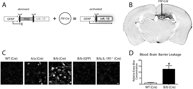
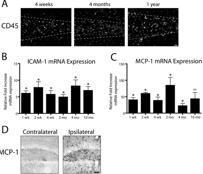
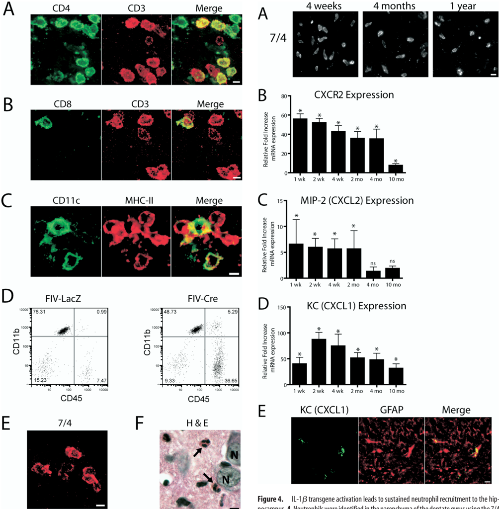
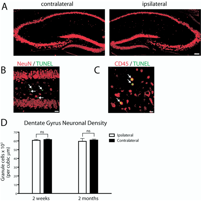
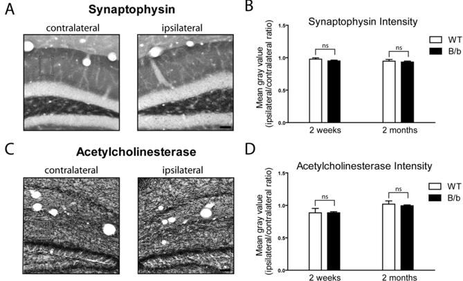
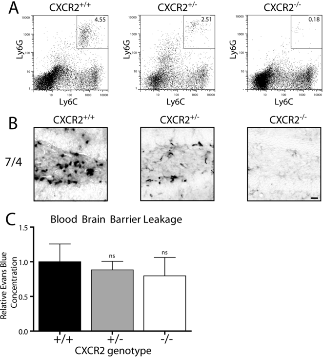
The proinflammatory cytokine interleukin-1beta (IL-1beta) plays a significant role in leukocyte recruitment to the CNS. Although acute effects of IL-1beta signaling in the mouse brain have been well described, studies elucidating the downstream effects of sustained upregulation have been lacking. Using the recently described IL-1beta(XAT) transgenic mouse model, we triggered sustained unilateral hippocampal overexpression of IL-1beta. Transgene induction led to blood-brain barrier leakage, induction of MCP-1 (monocyte chemoattractant protein 1) (CCL2), ICAM-1 (intercellular adhesion molecule 1), and dramatic infiltration of CD45-positive leukocytes comprised of neutrophils, T-cells, macrophages, and dendritic cells. Despite prolonged cellular infiltration of the hippocampus, there was no evidence of neuronal degeneration. Surprisingly, neutrophils were observed in the hippocampal parenchyma as late as 1 year after transgene induction. Their presence was coincident with upregulation of the potent neutrophil chemotactic chemokines KC (keratinocyte-derived chemokine) (CXCL1) and MIP-2 (macrophage inflammatory protein 2) (CXCL2). Knock-out of their sole receptor CXCR2 abrogated neutrophil infiltration but failed to reduce leakage of the blood-brain barrier.
Overview
The claims and statistics of this paper haven’t been checked yet.
Run the check — free, no account needed. It reads the open-access text and checks the sources it cites.
Integrity signals
Registry
Cites
Statistics
Figures
Verification
Claims the paper makes
The claims and statistics of this paper haven’t been checked yet.
Run the check — free, no account needed. It reads the open-access text and checks the sources it cites.
Claims other papers make about this one
Each is checked against this paper’s own text: does it say what the citing paper says it does?
The cited source says what the claim attributes to it.
In the citing paper
“Previous work from our lab has shown that IL-1β induces significant recruitment of neutrophils [30]”
From IL-1β-driven amyloid plaque clearance is associated with an expansion of transcriptionally reprogrammed microglia (2019) · cites this paper as [30]
The cited source says what the claim attributes to it.
In the citing paper
“we have previously shown that IL-1β induces recruitment of CD4+ and CD8+ T cells [30].”
From IL-1β-driven amyloid plaque clearance is associated with an expansion of transcriptionally reprogrammed microglia (2019) · cites this paper as [30]
The check wasn’t confident either way, so it gives no verdict — never disputed.
In the citing paper
“In agreement with our previous observations [28, 30] there was minimal recruitment of CD45hi peripheral immune cells (CD11b+ and CD11b−) in APP/PS1-Phe mice”
From IL-1β-driven amyloid plaque clearance is associated with an expansion of transcriptionally reprogrammed microglia (2019) · cites this paper as [30]
Experimental — accuracy not yet measured
Self-check
Percentages, sums and reported differences re-computed from the paper’s own numbers.
The self-check needs the paper’s full text.
Citations
Cites
Neutrophils and immunity: challenges and opportunities
Nathan, C. (2006) · Nature reviews. Immunology · doi:10.1038/nri1785 · Cited by 2,971
Mouse and human dendritic cell subtypes
Shortman, K., & Liu, Y.-J. (2002) · Nature reviews. Immunology · doi:10.1038/nri746 · Cited by 2,289
Monocyte chemoattractant protein 1 acts as a T-lymphocyte chemoattractant.
Carr, M. W., Roth, S. J., Luther, E., Rose, S. S., & Springer, T. A. (1994) · Proceedings of the National Academy of Sciences · doi:10.1073/pnas.91.9.3652 · Cited by 1,198
Three or more routes for leukocyte migration into the central nervous system
Ransohoff, R. M., Kivisäkk, P., & Kidd, G. (2003) · Nature Reviews Immunology · doi:10.1038/nri1130 · Cited by 1,052
Interleukin-1 and neuronal injury
Allan, S. M., Tyrrell, P. J., & Rothwell, N. J. (2005) · Nature Reviews Immunology · doi:10.1038/nri1664 · Cited by 992
Inflammatory response in acute traumatic brain injury: a double-edged sword
Morganti-Kossmann, M., Rancan, M., Stahel, P., & Kossmann, T. (2002) · Current Opinion in Critical Care · doi:10.1097/00075198-200204000-00002 · Cited by 702
Neutrophil and B Cell Expansion in Mice That Lack the Murine IL-8 Receptor Homolog
Cacalano, G., Lee, J., Kikly, K. K., Ryan, A. M., Pitts-Meek, S., Hultgren, B., Wood, W. I., & Moore, M. W. (1994) · Science · doi:10.1126/science.8036519 · Cited by 663
Ford, A. L., Goodsall, A. L., Hickey, W. F., & Sedgwick, J. D. (1995) · The Journal of Immunology · doi:10.4049/jimmunol.154.9.4309 · Cited by 631
Izikson, L., Klein, R. S., Charo, I. F., Weiner, H. L., & Luster, A. D. (2000) · The Journal of Experimental Medicine · doi:10.1084/jem.192.7.1075 · Cited by 584
Huang, D., Wang, J., Kivisäkk, P., Rollins, B. J., & Ransohoff, R. M. (2001) · The Journal of Experimental Medicine · doi:10.1084/jem.193.6.713 · Cited by 576
Cited by
Sorted by citations
Neuroinflammation in Alzheimer's disease
Heneka, M. T., Carson, M. J., Khoury, J. E., Landreth, G. E., Brosseron, F., Feinstein, D. L., Jacobs, A. H., Wyss-Coray, T., Vitorica, J., Ransohoff, R. M., et al. (2015) · The Lancet Neurology · doi:10.1016/s1474-4422(15)70016-5 · Cited by 6,226
NLRP3 is activated in Alzheimer’s disease and contributes to pathology in APP/PS1 mice
Heneka, M. T., Kummer, M. P., Stutz, A., Delekate, A., Schwartz, S., Vieira-Saecker, A., Griep, A., Axt, D., Remus, A., Tzeng, T.-C., et al. (2013) · Nature · doi:10.1038/nature11729 · Cited by 2,898
Inflammatory cytokines in depression: neurobiological mechanisms and therapeutic implications
Felger, J. C., & Lotrich, F. E. (2013) · Neuroscience · doi:10.1016/j.neuroscience.2013.04.060 · Cited by 1,180
Neuroinflammatory mechanisms of blood-brain barrier damage in ischemic stroke
Yang, C., Hawkins, K. E., Doré, S., & Candelario-Jalil, E. (2019) · American Journal of Physiology-Cell Physiology · doi:10.1152/ajpcell.00136.2018 · Cited by 871
Cytokine networks in neuroinflammation
Becher, B., Spath, S., & Goverman, J. M. (2016) · Nature reviews. Immunology · doi:10.1038/nri.2016.123 · Cited by 707
Blood–Brain Barrier Pathophysiology in Traumatic Brain Injury
Chodobski, A., Zink, B. J., & Szmydynger‐Chodobska, J. (2011) · Translational Stroke Research · doi:10.1007/s12975-011-0125-x · Cited by 672
Brain Tissue Responses to Neural Implants Impact Signal Sensitivity and Intervention Strategies
Kozai, T. D. Y., Jaquins-Gerstl, A. S., Vazquez, A. L., Michael, A. C., & Cui, X. T. (2015) · ACS Chemical Neuroscience · doi:10.1021/cn500256e · Cited by 622
Dysregulation of brain and choroid plexus cell types in severe COVID-19
Yang, A. C., Kern, F., Losada, P. M., Agam, M. R., Maat, C. A., Schmartz, G. P., Fehlmann, T., Stein, J. A., Schaum, N., Lee, D. P., et al. (2021) · Nature · doi:10.1038/s41586-021-03710-0 · Cited by 620
The role of interleukin-1 in neuroinflammation and Alzheimer disease: an evolving perspective
Shaftel, S. S., Griffin, W. S. T., & O'Banion, M. K. (2008) · Journal of Neuroinflammation · doi:10.1186/1742-2094-5-7 · Cited by 565
Self-citationInflammation: the link between comorbidities, genetics, and Alzheimer’s disease
Newcombe, E. A., Camats-Perna, J., Silva, M. L., Valmas, N., Huat, T. J., & Medeiros, R. (2018) · Journal of Neuroinflammation · doi:10.1186/s12974-018-1313-3 · Cited by 532
Felger, J. C., & Miller, A. H. (2012) · Frontiers in Neuroendocrinology · doi:10.1016/j.yfrne.2012.09.003 · Cited by 413
Ghosh, S., Wu, M. D., Shaftel, S. S., Kyrkanides, S., LaFerla, F. M., Olschowka, J. A., & O'Banion, M. K. (2013) · The Journal of Neuroscience · doi:10.1523/jneurosci.4361-12.2013 · Cited by 390
Self-citationNLRP3 inflammasome-driven pathways in depression: clinical and preclinical findings
Kaufmann, F. N., Costa, A. P., Ghisleni, G., Diaz, A. P., Rodrigues, A. L. S., Peluffo, H., & Kaster, M. P. (2017) · Brain, Behavior, and Immunity · doi:10.1016/j.bbi.2017.03.002 · Cited by 370
Pathogenesis of acute stroke and the role of inflammasomes
Fann, D. Y., Lee, S.-Y., Manzanero, S., Chunduri, P., Sobey, C. G., & Arumugam, T. V. (2013) · Ageing Research Reviews · doi:10.1016/j.arr.2013.09.004 · Cited by 334
The multifaceted profile of activated microglia
Lynch, M. A. (2009) · Molecular Neurobiology · doi:10.1007/s12035-009-8077-9 · Cited by 268
Redox Control of Microglial Function: Molecular Mechanisms and Functional Significance
Rojo, A. I., McBean, G. J., Cindrić, M., Egea, J., López, M. G., Rada, P., Žarković, N., & Cuadrado, A. (2014) · Antioxidants and Redox Signaling · doi:10.1089/ars.2013.5745 · Cited by 323
The pathogenic role of the inflammasome in neurodegenerative diseases
Freeman, L. C., & Ting, J. P. ‐. (2016) · Journal of Neurochemistry · doi:10.1111/jnc.13217 · Cited by 276
Jha, S., Srivastava, S. Y., Brickey, W. J., Iocca, H., Toews, A., Morrison, J. P., Chen, V. S., Gris, D., Matsushima, G. K., & Ting, J. P.-Y. (2010) · The Journal of Neuroscience · doi:10.1523/jneurosci.4088-10.2010 · Cited by 310
The IL-1β phenomena in neuroinflammatory diseases
Mendiola, A. S., & Cardona, A. E. (2017) · Journal of Neural Transmission · doi:10.1007/s00702-017-1732-9 · Cited by 328
Sun, Y., Koyama, Y., & Shimada, S. (2022) · Frontiers in Aging Neuroscience · doi:10.3389/fnagi.2022.903455 · Cited by 304
Antagonism of peripheral inflammation reduces the severity of status epilepticus
Marchi, N., Fan, Q., Ghosh, C., Fazio, V. J., Bertolini, F., Betto, G., Batra, A., Carlton, E. F., Najm, I., Granata, T., et al. (2008) · Neurobiology of Disease · doi:10.1016/j.nbd.2008.10.002 · Cited by 284
Inflammation in epileptogenesis after traumatic brain injury
Webster, K. M., Sun, M., Crack, P. J., O'Brien, T. J., Shultz, S. R., & Semple, B. D. (2017) · Journal of Neuroinflammation · doi:10.1186/s12974-016-0786-1 · Cited by 281
Innate Immune Cell Death in Neuroinflammation and Alzheimer’s Disease
Rajesh, Y., & Kanneganti, T. (2022) · Cells · doi:10.3390/cells11121885 · Cited by 269
Propson, N. E., Roy, E. R., Litvinchuk, A., Köhl, J., & Zheng, H. (2021) · Journal of Clinical Investigation · doi:10.1172/jci140966 · Cited by 258
The blood-brain barrier, chemokines and multiple sclerosis
Holman, D. W., Klein, R. S., & Ransohoff, R. M. (2011) · Biochimica et Biophysica Acta (BBA) - Molecular Basis of Disease · doi:10.1016/j.bbadis.2010.07.019 · Cited by 247
Potter, K. A., Buck, A. C., Self, W. K., & Capadona, J. R. (2012) · Journal of Neural Engineering · doi:10.1088/1741-2560/9/4/046020 · Cited by 242
A review of experimental evidence linking neurotoxic organophosphorus compounds and inflammation
Banks, C. N., & Lein, P. J. (2012) · NeuroToxicology · doi:10.1016/j.neuro.2012.02.002 · Cited by 240
Rosenzweig, N., Dvir-Szternfeld, R., Tsitsou-Kampeli, A., Keren‐Shaul, H., Ben-Yehuda, H., Weill-Raynal, P., Cahalon, L., Kertser, A., Baruch, K., Amit, I., et al. (2019) · Nature Communications · doi:10.1038/s41467-019-08352-5 · Cited by 230
Cai, H. Q., Catts, V. S., Webster, M. J., Galletly, C. A., Liu, D., O׳Donnell, M., Weickert, T. W., & Weickert, C. S. (2018) · Molecular Psychiatry · doi:10.1038/s41380-018-0235-x · Cited by 229
Shechter, R., & Schwartz, M. (2012) · The Journal of Pathology · doi:10.1002/path.4106 · Cited by 237
Sustained hippocampal IL-1β overexpression impairs contextual and spatial memory in transgenic mice
Hein, A. M., Stasko, M. R., Matousek, S. B., Scott-McKean, J. J., Maier, S. F., Olschowka, J. A., Costa, A. C. S., & O’Banion, M. K. (2009) · Brain Behavior and Immunity · doi:10.1016/j.bbi.2009.10.002 · Cited by 235
Self-citationParkinson's Disease and Systemic Inflammation
Ferrari, C. C., & Tarelli, R. (2011) · Parkinson s Disease · doi:10.4061/2011/436813 · Cited by 223
Cazareth, J., Guyon, A., Heurteaux, C., Chabry, J., & Petit-Paitel, A. (2014) · Journal of Neuroinflammation · doi:10.1186/1742-2094-11-132 · Cited by 206
Saresella, M., Calabrese, E., Marventano, I., Piancone, F., Gatti, A. V., Alberoni, M., Nemni, R., & Clerici, M. (2010) · Brain Behavior and Immunity · doi:10.1016/j.bbi.2010.12.004 · Cited by 194
Neutrophils in traumatic brain injury (TBI): friend or foe?
Liu, Y., Li, S., & Dai, S.-S. (2018) · Journal of Neuroinflammation · doi:10.1186/s12974-018-1173-x · Cited by 197
The role of neutrophils in neuro-immune modulation
Kanashiro, A., Hiroki, C. H., da Fonseca, D. M., Birbrair, A., Ferreira, R. G., Bassi, G. S., Fonseca, M. D., Kusuda, R., Cebinelli, G. C. M., da Silva, K. P., et al. (2019) · Pharmacological Research · doi:10.1016/j.phrs.2019.104580 · Cited by 186
Neuroinflammation and Memory: The Role of Prostaglandins
Hein, A. M., & O’Banion, M. K. (2009) · Molecular Neurobiology · doi:10.1007/s12035-009-8066-z · Cited by 176
Self-citationInterleukin-2 improves amyloid pathology, synaptic failure and memory in Alzheimer’s disease mice
Alves, S., Churlaud, G., Audrain, M., Michaelsen‐Preusse, K., Fol, R. R., Souchet, B., Braudeau, J., Körte, M., Klatzmann, D., & Cartier, N. (2016) · Brain · doi:10.1093/brain/aww330 · Cited by 178
Cell‐specific blood–brain barrier regulation in health and disease: a focus on hypoxia
Engelhardt, S., Patkar, S., & Ogunshola, O. O. (2013) · British Journal of Pharmacology · doi:10.1111/bph.12489 · Cited by 168
Inflammasomes link vascular disease with neuroinflammation and brain disorders
Lénárt, N., Brough, D., & Dénes, Á. (2016) · Journal of Cerebral Blood Flow & Metabolism · doi:10.1177/0271678x16662043 · Cited by 170
Brown, C. M., Mulcahey, T. A., Filipek, N. C., & Wise, P. M. (2010) · Endocrinology · doi:10.1210/en.2010-0371 · Cited by 166
Major Depression: One Brain, One Disease, One Set of Intertwined Processes
Filatova, E. V., Shadrina, M. I., & Slominsky, P. A. (2021) · Cells · doi:10.3390/cells10061283 · Cited by 186
Sustained expression of interleukin-1β in mouse hippocampus impairs spatial memory
Moore, A. H., Wu, M., Shaftel, S. S., Graham, K. A., & O’Banion, M. K. (2009) · Neuroscience · doi:10.1016/j.neuroscience.2009.08.073 · Cited by 159
Self-citationInitiation and Progression of Axonopathy in Experimental Autoimmune Encephalomyelitis
Soulika, A. M., Lee, E., McCauley, E., Miers, L. A., Bannerman, P. G. C., & Pleasure, D. (2009) · Journal of Neuroscience · doi:10.1523/jneurosci.3794-09.2009 · Cited by 161
Israelsson, C., Bengtsson, H., Kylberg, A., Kullander, K., Lewén, A., Hillered, L., & Ebendal, T. (2008) · Journal of Neurotrauma · doi:10.1089/neu.2008.0562 · Cited by 161
Advances in the Potential Biomarkers of Epilepsy
Kobylarek, D., Iwanowski, P. S., Lewandowska, Z., Limphaibool, N., Szafranek, S., Labrzycka, A., & Kozubski, W. P. (2019) · Frontiers in Neurology · doi:10.3389/fneur.2019.00685 · Cited by 157
Microglial inflammation and phagocytosis in Alzheimer's disease: Potential therapeutic targets
Nizami, S., Hall‐Roberts, H., Warrier, S., Cowley, S. A., & Di Daniel, E. (2019) · British Journal of Pharmacology · doi:10.1111/bph.14618 · Cited by 147
Interleukin‐1β: a bridge between inflammation and excitotoxicity?
Fogal, B., & Hewett, S. J. (2008) · Journal of Neurochemistry · doi:10.1111/j.1471-4159.2008.05315.x · Cited by 151
Saresella, M., Calabrese, E., Marventano, I., Piancone, F., Gatti, A. V., Calvo, M. G., Nemni, R., & Clerici, M. (2010) · Journal of Alzheimer s Disease · doi:10.3233/jad-2010-091696 · Cited by 146
Alterations in Immune Cells and Mediators in the Brain: It's Not Always Neuroinflammation!
Estes, M. L., & McAllister, A. K. (2014) · Brain Pathology · doi:10.1111/bpa.12198 · Cited by 150
Kozai, T. D. Y., Li, X., Bodily, L. M., Caparosa, E. M., Zenonos, G. A., Carlisle, D. L., Friedlander, R. M., & Cui, X. T. (2014) · Biomaterials · doi:10.1016/j.biomaterials.2014.08.006 · Cited by 136
Inflammatory monocytes and the pathogenesis of viral encephalitis
Terry, R. L., Getts, D. R., Deffrasnes, C., van Vreden, C., Campbell, I. L., & King, N. J. C. (2012) · Journal of Neuroinflammation · doi:10.1186/1742-2094-9-270 · Cited by 138
Wu, M. D., Hein, A. M., Moravan, M. J., Shaftel, S. S., Olschowka, J. A., & O’Banion, M. K. (2011) · Brain Behavior and Immunity · doi:10.1016/j.bbi.2011.09.012 · Cited by 134
Self-citationStress responses: the contribution of prostaglandin E2 and its receptors
Furuyashiki, T., & Narumiya, S. (2010) · Nature Reviews Endocrinology · doi:10.1038/nrendo.2010.194 · Cited by 132
Zhang, N., Zhang, X., Liu, X., Wang, H., Xue, J., Yu, J., Kang, N., & Wang, X. (2014) · Mediators of Inflammation · doi:10.1155/2014/370530 · Cited by 126
Targeting innate immunity for neurodegenerative disorders of the central nervous system
Andreasson, K. I., Bachstetter, A. D., Colonna, M., Ginhoux, F., Holmes, C., Lamb, B., Landreth, G., Lee, D. C., Low, D., Lynch, M. A., et al. (2016) · Journal of Neurochemistry · doi:10.1111/jnc.13667 · Cited by 126
Self-citationNeuroinflammation as an etiological trigger for depression comorbid with inflammatory bowel disease
Craig, C. F., Filippone, R. T., Stavely, R., Bornstein, J. C., Apostolopoulos, V., & Nurgali, K. (2022) · Journal of Neuroinflammation · doi:10.1186/s12974-021-02354-1 · Cited by 124
Cardiovascular comorbidities, inflammation, and cerebral small vessel disease
Evans, L., Taylor, J. L., Smith, C. J., Pritchard, H. A. T., Greenstein, A. S., & Allan, S. M. (2021) · Cardiovascular Research · doi:10.1093/cvr/cvab284 · Cited by 127
Yang, G., Meng, Y. J., Li, W., Yong, Y., Fan, Z., Ding, H., Wei, Y., Luo, J., & Ke, Z. (2010) · Brain Pathology · doi:10.1111/j.1750-3639.2010.00445.x · Cited by 124
Alfonso‐Loeches, S., Ureña‐Peralta, J. R., Morillo-Bargues, M. J., Gómez‐Pinedo, U., & Guerri, C. (2015) · Neurochemical Research · doi:10.1007/s11064-015-1760-5 · Cited by 126
Xu, J., & Ikezu, T. (2008) · Journal of Neuroimmune Pharmacology · doi:10.1007/s11481-008-9136-0 · Cited by 120
Shigemoto‐Mogami, Y., Hoshikawa, K., & Sato, K. (2018) · Frontiers in Cellular Neuroscience · doi:10.3389/fncel.2018.00494 · Cited by 118
Principles of inflammasome priming and inhibition: implications for psychiatric disorders
Herman, F. J., & Pasinetti, G. M. (2018) · Brain, Behavior, and Immunity · doi:10.1016/j.bbi.2018.06.010 · Cited by 115
Osteoarthritis accelerates and exacerbates Alzheimer's disease pathology in mice
Kyrkanides, S., Tallents, R. H., Miller, J. H., Olschowka, M. E., Johnson, R. E., Yang, M., Olschowka, J. A., Brouxhon, S. M., & O’Banion, M. K. (2011) · Journal of Neuroinflammation · doi:10.1186/1742-2094-8-112 · Cited by 116
Self-citationJohnson, E. A., Dao, T. L., Guignet, M. A., Geddes, C. E., Koemeter-Cox, A., & Kan, R. K. (2011) · Journal of Neuroinflammation · doi:10.1186/1742-2094-8-41 · Cited by 119
Semple, B. D., Bye, N., Ziebell, J. M., & Morganti-Kossmann, M. C. (2010) · Neurobiology of Disease · doi:10.1016/j.nbd.2010.06.015 · Cited by 116
Graham, L. C., Harder, J. M., Soto, I., de Vries, W. N., John, S. W. M., & Howell, G. R. (2016) · Scientific Reports · doi:10.1038/srep21568 · Cited by 113
Jacobs, J. P., Ortíz‐López, A., Campbell, J. J., Gerard, C. J., Mathis, D. J., & Benoist, C. O. (2010) · Arthritis & Rheumatism · doi:10.1002/art.27470 · Cited by 110
Riis, J. L., Out, D., Dorn, L. D., Beal, S. J., Denson, L. A., Pabst, S., Jaedicke, K., & Granger, D. A. (2014) · Developmental Psychobiology · doi:10.1002/dev.21149 · Cited by 108
Macrophages in Alzheimer’s disease: the blood-borne identity
Gate, D., Rezai-Zadeh, K., Jodry, D., Rentsendorj, A., & Town, T. (2010) · Journal of Neural Transmission · doi:10.1007/s00702-010-0422-7 · Cited by 107
Inflammation in Ischemic Stroke: Mechanisms, Consequences and Possible Drug Targets
Ahmad, M., Dar, N. J., Bhat, Z. S., Hussain, A., Shah, A., Liu, H., & Graham, S. H. (2014) · CNS & Neurological Disorders - Drug Targets · doi:10.2174/1871527313666141023094720 · Cited by 102
Carl, S. M., Lindley, D. J., Couraud, P. O., Weksler, B. B., Romero, I., Mowery, S. A., & Knipp, G. T. (2010) · Molecular Pharmaceutics · doi:10.1021/mp900178j · Cited by 88
DiSabato, D. J., Nemeth, D. P., Liu, X., Witcher, K. G., O’Neil, S. M., Oliver, B., Bray, C. E., Sheridan, J. F., Godbout, J. P., & Quan, N. (2020) · Molecular Psychiatry · doi:10.1038/s41380-020-0788-3 · Cited by 93
McCandless, E. E., Budde, M. D., Lees, J. R., Dorsey, D. A., Lyng, E., & Klein, R. S. (2009) · The Journal of Immunology · doi:10.4049/jimmunol.0802258 · Cited by 90
Echeverría, V., Iarkov, A., & Aliev, G. (2016) · Progress in Neurobiology · doi:10.1016/j.pneurobio.2016.01.002 · Cited by 95
Ataka, K., Asakawa, A., Nagaishi, K., Kaimoto, K., Sawada, A., Hayakawa, Y., Tatezawa, R., Inui, A., & Fujimiya, M. (2013) · PLoS ONE · doi:10.1371/journal.pone.0081744 · Cited by 89
Leptin regulates leukocyte recruitment into the brain following systemic LPS-induced inflammation
Rummel, C., Inoue, W., Poole, S., & Luheshi, G. N. (2010) · Molecular Psychiatry · doi:10.1038/mp.2009.98 · Cited by 88
Brain injury-induced dysfunction of the blood brain barrier as a risk for dementia
Abrahamson, E. E., & Ikonomović, M. D. (2020) · Experimental Neurology · doi:10.1016/j.expneurol.2020.113257 · Cited by 91
Matousek, S. B., Ghosh, S., Shaftel, S. S., Kyrkanides, S., Olschowka, J. A., & O’Banion, M. K. (2012) · Journal of Neuroimmune Pharmacology · doi:10.1007/s11481-011-9331-2 · Cited by 86
“In an earlier study, we found that sustained elevation of IL- 1β leads to profound reduction in plaque pathology in APPswe/PS1dE9 mice on a mixed B6C3F1/J background (Shaftel et al. 2007a).”
InfluentialSelf-citationFisher, Y., Nemirovsky, A., Baron, R., & Monsonego, A. (2010) · PLoS ONE · doi:10.1371/journal.pone.0010830 · Cited by 83
Elshatory, Y. M., & Gan, L. (2008) · Journal of Neuroscience · doi:10.1523/jneurosci.5730-07.2008 · Cited by 80
Understanding the Origin and Diversity of Macrophages to Tailor Their Targeting in Solid Cancers
Kielbassa-Elkadi, K., Vegna, S., Ramirez, C. F. A., & Akkari, L. (2019) · Frontiers in Immunology · doi:10.3389/fimmu.2019.02215 · Cited by 82
Zhang, H., Park, J. H., Maharjan, S., Park, J. A., Choi, K.-S., Park, H., Jeong, Y., Ahn, J. H., Kim, I. H., Lee, J. C., et al. (2017) · Journal of Neuroinflammation · doi:10.1186/s12974-017-0897-3 · Cited by 79
Repeated Iron–Soot Exposure and Nose-to-brain Transport of Inhaled Ultrafine Particles
Hopkins, L. E., Laing, E. A., Peake, J. L., Uyeminami, D. L., Mack, S. M., Li, X., Smiley‐Jewell, S. M., & Pinkerton, K. E. (2017) · Toxicologic Pathology · doi:10.1177/0192623317729222 · Cited by 79
Durrant, D. M., Robinette, M. L., & Klein, R. S. (2013) · The Journal of Experimental Medicine · doi:10.1084/jem.20121897 · Cited by 81
Wu, M. D., Montgomery, S. L., Rivera‐Escalera, F., Olschowka, J. A., & O’Banion, M. K. (2013) · Brain Behavior and Immunity · doi:10.1016/j.bbi.2013.03.003 · Cited by 80
Self-citationPivotal Roles of Monocytes/Macrophages in Stroke
Chiba, T., & Umegaki, K. (2013) · Mediators of Inflammation · doi:10.1155/2013/759103 · Cited by 81
Rivera-Escalera, F., Pinney, J. J., Owlett, L., Ahmed, H., Thakar, J., Olschowka, J. A., Elliott, M. R., & O’Banion, M. K. (2019) · Journal of Neuroinflammation · doi:10.1186/s12974-019-1645-7 · Cited by 75
“2b, Additional file 3) and we have previously shown that IL-1β induces recruitment of CD4 and CD8 T cells [30].”
InfluentialSelf-citationLocal microvascular leakage promotes trafficking of activated neutrophils to remote organs
Owen-Woods, C., Joulia, R., Barkaway, A., Rolas, L., Ma, B., Nottebaum, A. F., Arkill, K. P., Stein, M., Girbl, T., Golding, M., et al. (2020) · Journal of Clinical Investigation · doi:10.1172/jci133661 · Cited by 78
The Role of Cytokines in the Regulation of Neurotransmission
Camacho‐Arroyo, I., López-Griego, L., & Morales‐Montor, J. (2008) · NeuroImmunoModulation · doi:10.1159/000179661 · Cited by 78
Rodríguez‐Arias, M., Montagud‐Romero, S., Rubio‐Araiz, A., Aguilar, M. A., Martín‐García, E., Cabrera, R., Maldonado, R. J., Porcu, F., Colado, M. I., & Miñarro, J. (2015) · Addiction Biology · doi:10.1111/adb.12301 · Cited by 76
Galvão, R. P., & Zong, H. (2013) · Current Pathobiology Reports · doi:10.1007/s40139-012-0006-3 · Cited by 75
Mediators of oligodendrocyte differentiation during remyelination
Patel, J. R., & Klein, R. S. (2011) · FEBS Letters · doi:10.1016/j.febslet.2011.04.037 · Cited by 76
CD 4 T cells in immunity and immunotherapy of A lzheimer's disease
Monsonego, A., Nemirovsky, A., & Harpaz, I. (2013) · Immunology · doi:10.1111/imm.12103 · Cited by 72
Jeon, S., Jha, M. K., Ock, J., Seo, J.-W., Jin, M., Cho, H., Lee, W., & Suk, K. (2013) · Journal of Biological Chemistry · doi:10.1074/jbc.m113.454140 · Cited by 69
Kong, Q., Peterson, T. S., Baker, O. J., Stanley, E., Camden, J. M., Seye, C. I., Erb, L., Simonyi, Á., Wood, W. G., Sun, G. Y., et al. (2009) · Journal of Neurochemistry · doi:10.1111/j.1471-4159.2009.06048.x · Cited by 69
Ben‐Baruch, A. (2011) · Cancer Microenvironment · doi:10.1007/s12307-011-0094-3 · Cited by 69
Role of the IL-1 Pathway in Dopaminergic Neurodegeneration and Decreased Voluntary Movement
Stojakovic, A., Paz-Filho, G., Arcos‐Burgos, M., Licinio, J., Wong, M., & Mastronardi, C. A. (2016) · Molecular Neurobiology · doi:10.1007/s12035-016-9988-x · Cited by 66
Kim, J.-E., Ryu, H. J., Yeo, S., & Kang, T. (2010) · Journal of Neuroinflammation · doi:10.1186/1742-2094-7-65 · Cited by 64
Garnett, C. N., Watson, J. B., Higgins, E. K., Quan, N., & Bachstetter, A. D. (2021) · Frontiers in Immunology · doi:10.3389/fimmu.2021.688254 · Cited by 62
Metadata
- Authors
- Shaftel, S. S., Carlson, T. J., Olschowka, J. A., Kyrkanides, S., Matousek, S. B., & O'Banion, M. K.
- Affiliations
- University of Rochester Medicine (US)
- Venue
- The Journal of Neuroscience
- Publisher
- Society for Neuroscience
- Published
- 2007-08-29
- Volume
- 27
- Issue
- 35
- Pages
- 9301-9309
- ISSN
- 0270-6474 (ISSN-L)
- 1529-2401
- PMC
- PMC6673122
- OpenAlex
- W2094676206
- Cited by
- 243
- References
- 55
- Citations by year
- 2026: 6 · 2025: 7 · 2024: 5 · 2023: 9 · 2022: 14 · 2021: 13 · 2020: 10 · 2019: 18 · 2018: 22 · 2017: 9 · 2016: 15 · 2015: 13 · 2014: 19 · 2013: 19 · 2012: 10
- FWCI
- 8.68
- Type
- journal article
- Language
- en
- Open access
- hybrid
- doi.org
- Licence
- Locations
- Journal of Neuroscience (publishedVersion, cc-by-nc-sa)
- PubMed Central (publishedVersion)
- PubMed (publishedVersion)
- Funders
- University of Rochester
- National Institutes of Health
- National Institute of General Medical Sciences (GM 07356, T32GM007356)
- National Institute of Neurological Disorders and Stroke (NS048522, R29NS033553, NS 33553, R21NS048522, R01NS033553)
- Topics
- Life Sciences › Neuroscience › Neurology › Neuroinflammation and Neurodegeneration Mechanisms
- Life Sciences › Neuroscience › Neurology › Barrier Structure and Function Studies
- Life Sciences › Neuroscience › Biological Psychiatry › Tryptophan and brain disorders
- Keywords
- IL-1β, chronic neuroinflammation, blood-brain barrier permeability, leukocyte infiltration, neutrophils, CXCR2, CXCL1, CXCL2, MCP-1, ICAM-1, hippocampus, transgenic mouse model, neurodegeneration, chemokines
- MeSH terms
- Animals, Blood-Brain Barrier, Hippocampus, Leukocytes, Mice, Inbred C57BL, Mice, Transgenic, Neutrophils, Monokines, Up-Regulation, Leukocyte Common Antigens, Intercellular Adhesion Molecule-1, Chemokine CCL2, Mice, Interleukin-1beta, Chemokine CXCL2
About these checks
Registry, retraction, Cites and statistics results are computed, not judged, with one exception: a printed reference that no index lists is read once by a model before it counts as a citation error. Claims are extracted by a model and kept only when their quote appears verbatim in the text; each cited source is then checked, with a model’s help, for whether it says what the claim relies on. Those verdicts can be wrong — read the claim’s quote and the cited source before relying on one.
Statistics and arithmetic are recomputed, not judged.
Confidence bands are shown only for fields calibrated on our annotated test set. None is calibrated yet, so no band is shown.
What counts as a claim ›How we measure confidence ›How we check this ›
Are you an author, or do you see a problem with this page? Request a review
