

The DOI matches its Crossref record; 408 of the 412 references it cites were found in a registry or an index.
Neurosteroid Actions in Memory and Neurologic/Neuropsychiatric Disorders
Open accessAbstract
Memory dysfunction is a symptomatic feature of many neurologic and neuropsychiatric disorders; however, the basic underlying mechanisms of memory and altered states of circuitry function associated with disorders of memory remain a vast unexplored territory. The initial discovery of endogenous neurosteroids triggered a quest to elucidate their role as neuromodulators in normal and diseased brain function. In this review, based on the perspective of our own research, the advances leading to the discovery of positive and negative neurosteroid allosteric modulators of GABA type-A (GABAA), NMDA, and non-NMDA type glutamate receptors are brought together in a historical and conceptual framework. We extend the analysis toward a state-of-the art view of how neurosteroid modulation of neural circuitry function may affect memory and memory deficits. By aggregating the results from multiple laboratories using both animal models for disease and human clinical research on neuropsychiatric and age-related neurodegenerative disorders, elements of a circuitry level view begins to emerge. Lastly, the effects of both endogenously active and exogenously administered neurosteroids on neural networks across the life span of women and men point to a possible underlying pharmacological connectome by which these neuromodulators might act to modulate memory across diverse altered states of mind.
Overview
The claims and statistics of this paper haven’t been checked yet.
Run the check — free, no account needed. It reads the open-access text and checks the sources it cites.
Integrity signals
Registry
Cites
Statistics
Figures
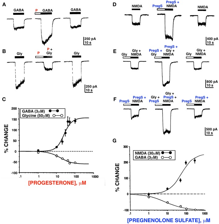
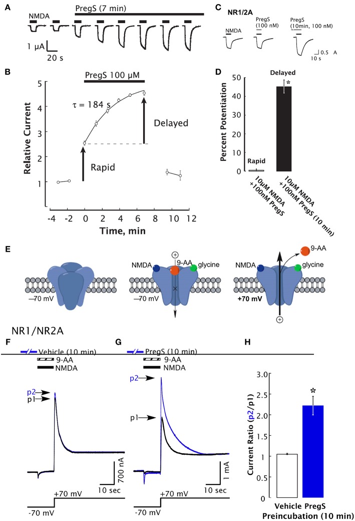
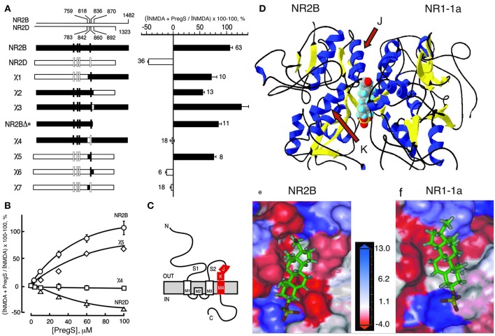
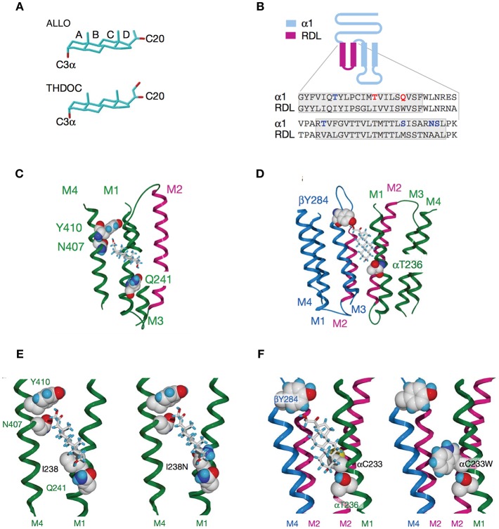
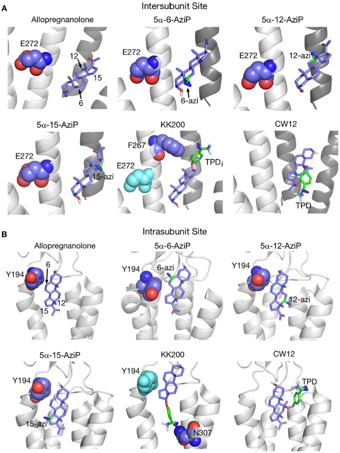
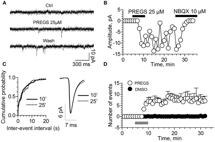
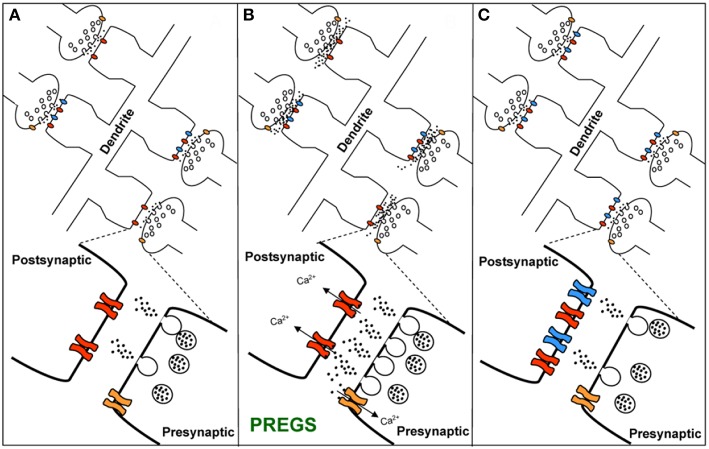
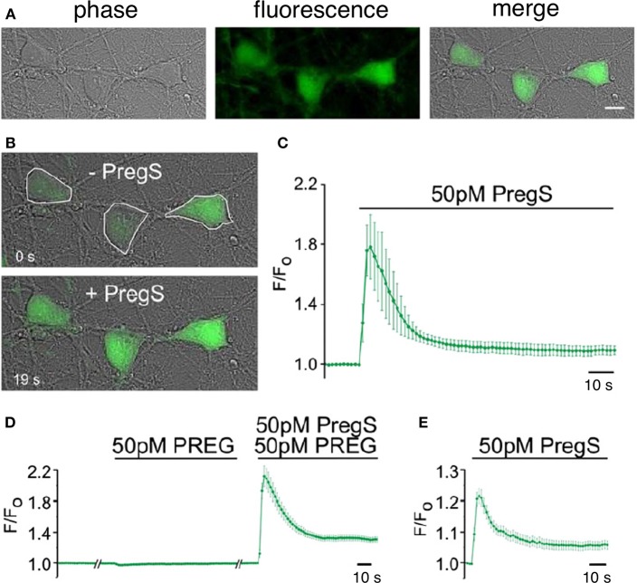
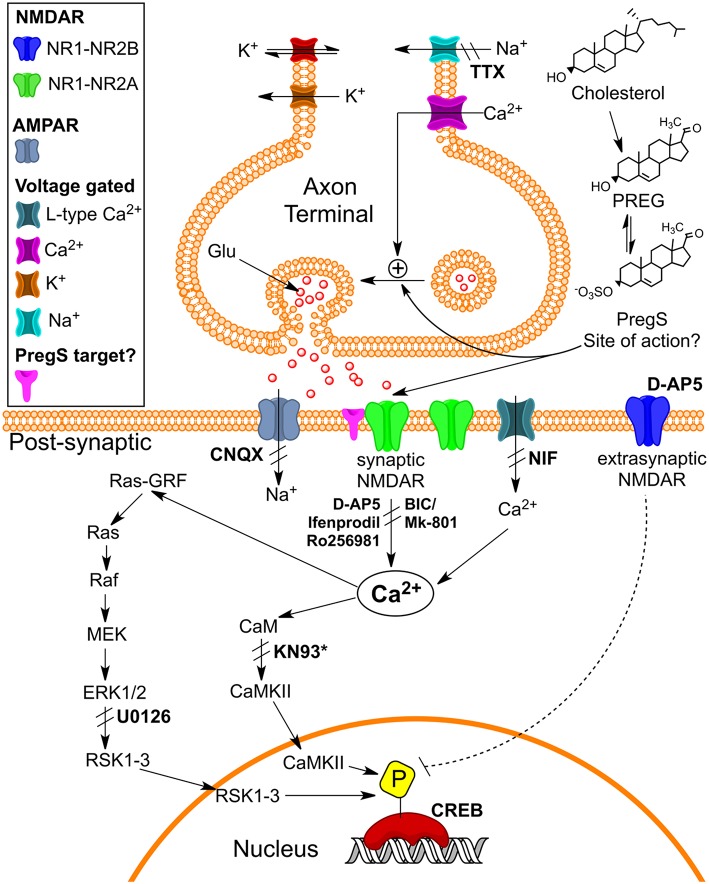
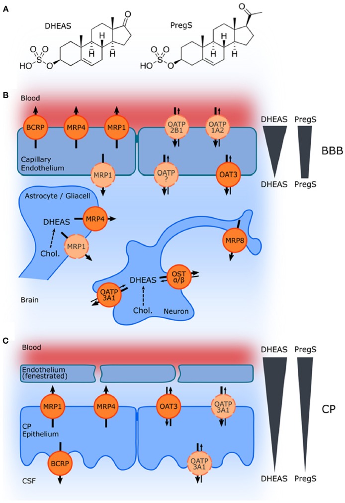
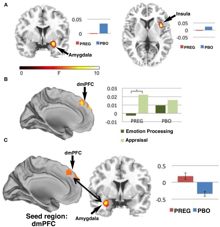
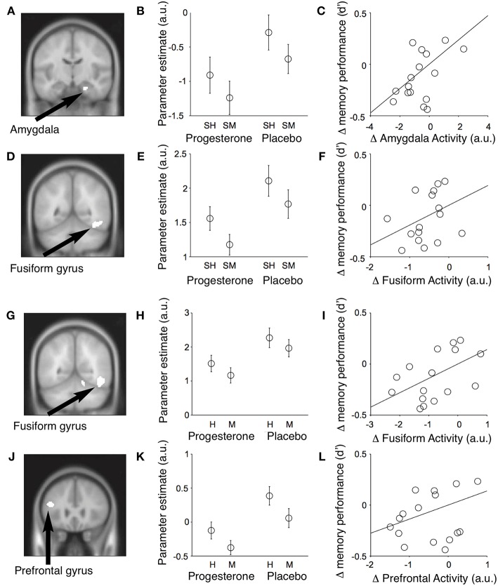
Verification
Claims the paper makes
The claims and statistics of this paper haven’t been checked yet.
Run the check — free, no account needed. It reads the open-access text and checks the sources it cites.
Claims other papers make about this one
Each is checked against this paper’s own text: does it say what the citing paper says it does?
The source backs part of the claim, not all of it — or backs it for a different population, condition or comparison.
In the citing paper
“Such a prediction is consistent with reported correlations between neurosteroids and a series of neurological disorders, including migraine, postpartum-depression, epilepsy, anxiety, cognitive and psychiatric disorders [56,144,145].”
From Fibromyalgia pathogenesis explained by a neuroendocrine multistable model (2024) · cites this paper as [144]
Experimental — accuracy not yet measured
Self-check
Percentages, sums and reported differences re-computed from the paper’s own numbers.
The self-check needs the paper’s full text.
Citations
Cites
Kessler, R. C., Berglund, P., Demler, O., Jin, R., Merikangas, K. R., & Walters, E. E. (2005) · Archives of General Psychiatry · doi:10.1001/archpsyc.62.6.593 · Cited by 20,391
Molecular Mechanisms of Depression: Perspectives on New Treatment Strategies
Lang, U. E., & Borgwardt, S. (2013) · Cellular Physiology and Biochemistry · doi:10.1159/000350094 · Cited by 8,526
Merikangas, K. R., He, J.-P., Burstein, M., Swanson, S. A., Avenevoli, S., Cui, L., Benjet, C., Georgiades, K., & Swendsen, J. (2010) · Journal of the American Academy of Child & Adolescent Psychiatry · doi:10.1016/j.jaac.2010.05.017 · Cited by 6,278
Large-scale brain networks and psychopathology: a unifying triple network model
Menon, V. (2011) · Trends in Cognitive Sciences · doi:10.1016/j.tics.2011.08.003 · Cited by 4,168
Morris, R. G. M., Anderson, E., Lynch, G. S., & Baudry, M. (1986) · Nature · doi:10.1038/319774a0 · Cited by 3,640
Glutamate Receptor Ion Channels: Structure, Regulation, and Function
Traynelis, S. F., Wollmuth, L. P., McBain, C. J., Menniti, F. S., Vance, K. M., Ogden, K. K., Hansen, K. B., Yuan, H., Myers, S. J., & Dingledine, R. (2010) · Pharmacological Reviews · doi:10.1124/pr.109.002451 · Cited by 3,568
Parvalbumin neurons and gamma rhythms enhance cortical circuit performance
Sohal, V. S., Zhang, F., Yizhar, O., & Deisseroth, K. (2009) · Nature · doi:10.1038/nature07991 · Cited by 2,606
Hyperlocomotion and indifference to cocaine and amphetamine in mice lacking the dopamine transporter
Giros, B., Jaber, M., Jones, S. R., Wightman, R. M., & Caron, M. G. (1996) · Nature · doi:10.1038/379606a0 · Cited by 2,476
Collingridge, G. L., Kehl, S. J., & McLennan, H. (1983) · The Journal of Physiology · doi:10.1113/jphysiol.1983.sp014478 · Cited by 2,413
Steroid hormone metabolites are barbiturate-like modulators of the GABA receptor
Majewska, M. D., Harrison, N. L., Schwartz, R. D., Barker, J. L., & Paul, S. M. (1986) · Science · doi:10.1126/science.2422758 · Cited by 2,225
Wisden, W., Laurie, D. J., Monyer, H., & Seeburg, P. (1992) · Journal of Neuroscience · doi:10.1523/jneurosci.12-03-01040.1992 · Cited by 1,543
Epidemiology of mental disorders in children and adolescents
Merikangas, K. R., Nakamura, E. F., & Kessler, R. C. (2009) · Dialogues in Clinical Neuroscience · doi:10.31887/dcns.2009.11.1/krmerikangas · Cited by 1,246
Verret, L., Mann, E. O., Hang, G. B., Barth, A. M. I., Cobos, I., Ho, K., Devidze, N., Masliah, E., Kreitzer, A. C., Mody, I., et al. (2012) · Cell · doi:10.1016/j.cell.2012.02.046 · Cited by 1,216
Sierra-Mercado, D., Padilla-Coreano, N., & Quirk, G. J. (2011) · Neuropsychopharmacology · doi:10.1038/npp.2010.184 · Cited by 1,190
Prevalence and Treatment of Mental Disorders Among US Children in the 2001–2004 NHANES
Merikangas, K. R., He, J.-P., Brody, D., Fisher, P. W., Bourdon, K., & Koretz, D. S. (2010) · Pediatrics · doi:10.1542/peds.2008-2598 · Cited by 1,172
Neurotransmitter Transporters: Recent Progress
Amara, S., & Kuhar, M. J. (1993) · Annual Review of Neuroscience · doi:10.1146/annurev.ne.16.030193.000445 · Cited by 1,088
Network abnormalities and interneuron dysfunction in Alzheimer disease
Palop, J. J., & Mucke, L. (2016) · Nature reviews. Neuroscience · doi:10.1038/nrn.2016.141 · Cited by 1,022
Lisman, J. E., Coyle, J. T., Green, R. W., Javitt, D. C., Benes, F. M., Heckers, S., & Grace, A. A. (2008) · Trends in Neurosciences · doi:10.1016/j.tins.2008.02.005 · Cited by 1,010
Purdy, R. H., Morrow, A. L., Moore, P. H., & Paul, S. M. (1991) · Proceedings of the National Academy of Sciences · doi:10.1073/pnas.88.10.4553 · Cited by 951
Reduction of Hippocampal Hyperactivity Improves Cognition in Amnestic Mild Cognitive Impairment
Bakker, A., Krauss, G. L., Albert, M., Speck, C. L., Jones, L. R., Stark, C. E. L., Yassa, M. A., Bassett, S. S., Shelton, A. L., & Gallagher, M. (2012) · Neuron · doi:10.1016/j.neuron.2012.03.023 · Cited by 940
Setiawan, E., Wilson, A. A., Mizrahi, R., Rusjan, P. M., Miler, L., Rajkowska, G., Suridjan, I., Kennedy, J. L., Rekkas, P. V., Houle, S., et al. (2015) · JAMA Psychiatry · doi:10.1001/jamapsychiatry.2014.2427 · Cited by 939
Neuronal activity enhances tau propagation and tau pathology in vivo
Wu, J. W., Hussaini, S. A., Bastille, I. M., Rodriguez, G. A., Mrejeru, A., Rilett, K., Sanders, D. W., Cook, C., Fu, H., Boonen, R. A. C. M., et al. (2016) · Nature Neuroscience · doi:10.1038/nn.4328 · Cited by 820
Stell, B. M., Brickley, S. G., Tang, C., Farrant, M., & Módy, I. (2003) · Proceedings of the National Academy of Sciences · doi:10.1073/pnas.2435457100 · Cited by 799
Maninger, N., Wolkowitz, O. M., Reus, V. I., Epel, E. S., & Mellon, S. H. (2009) · Frontiers in Neuroendocrinology · doi:10.1016/j.yfrne.2008.11.002 · Cited by 769
McIntyre, R. S., Cha, D. S., Soczynska, J. K., Woldeyohannes, H. O., Gallaugher, L. A., Kudlow, P., Alsuwaidan, M., & Baskaran, A. (2013) · Depression and Anxiety · doi:10.1002/da.22063 · Cited by 765
Endogenous neurosteroids regulate GABAA receptors through two discrete transmembrane sites
Hosie, A. M., Wilkins, M. E., da Silva, H. M. A., & Smart, T. G. (2006) · Nature · doi:10.1038/nature05324 · Cited by 752
Chang, M. M., & Leeman, S. E. (1970) · Journal of Biological Chemistry · doi:10.1016/s0021-9258(18)62860-1 · Cited by 699
Dopamine transporter density in patients with attention deficit hyperactivity disorder
Dougherty, D. D., Bonab, A. A., Spencer, T. J., Rauch, S. L., Madras, B. K., & Fischman, A. J. (1999) · The Lancet · doi:10.1016/s0140-6736(99)04030-1 · Cited by 684
Uzunova, V., Sheline, Y. I., Davis, J. M., Rasmusson, A. M., Uzunov, D. P., Costa, E., & Guidotti, A. (1998) · Proceedings of the National Academy of Sciences · doi:10.1073/pnas.95.6.3239 · Cited by 672
Carlén, M., Meletis, K., Siegle, J. H., Cardin, J. A., Futai, K., Vierling-Claassen, D., Rühlmann, C., Jones, S. R., Deisseroth, K., Sheng, M., et al. (2012) · Molecular Psychiatry · doi:10.1038/mp.2011.31 · Cited by 669
Characterization and measurement of dehydroepiandrosterone sulfate in rat brain
Corpéchot, C., Robel, P., Axelson, M., Sjövall, J., & Baulieu, E. E. (1981) · Proceedings of the National Academy of Sciences · doi:10.1073/pnas.78.8.4704 · Cited by 656
Assessing rodent hippocampal involvement in the novel object recognition task. A review
Cohen, S. J., & Stackman, R. W. (2014) · Behavioural Brain Research · doi:10.1016/j.bbr.2014.08.002 · Cited by 592
Canteras, N. S., & Swanson, L. W. (1992) · The Journal of Comparative Neurology · doi:10.1002/cne.903240204 · Cited by 558
Pregnenolone sulfate: a positive allosteric modulator at the N-methyl-D-aspartate receptor.
Wu, F., Gibbs, T. T., & Farb, D. H. (1991) · Molecular Pharmacology · doi:10.1016/s0026-895x(25)12923-4 · Cited by 534
Self-citationAlger, B. E., & Nicoll, R. A. (1982) · The Journal of Physiology · doi:10.1113/jphysiol.1982.sp014256 · Cited by 520
Mihalek, R., Banerjee, P., Korpi, E. R., Quinlan, J. J., Firestone, L. L., Mi, Z.-P., Lagenaur, C. F., Tretter, V. E., Sieghart, W., Anagnostaras, S., et al. (1999) · Proceedings of the National Academy of Sciences · doi:10.1073/pnas.96.22.12905 · Cited by 520
The paraventricular thalamus controls a central amygdala fear circuit
Penzo, M. A., Robert, V., Tucciarone, J., De Bundel, D., Wang, M., Van Aelst, L., Darvas, M., Parada, L. F., Palmiter, R. D., He, M., et al. (2015) · Nature · doi:10.1038/nature13978 · Cited by 506
Modulation of the GABA receptor complex by a steroid anaesthetic
Harrison, N. L., & Simmonds, M. A. (1984) · Brain Research · doi:10.1016/0006-8993(84)90299-3 · Cited by 501
Control of cortical GABA circuitry development by Nrg1 and ErbB4 signalling
Fazzari, P., Paternain, A. V., Valiente, M., Pla, R., Luján, R., Lloyd, K., Lerma, J., Marín, O., & Rico, B. (2010) · Nature · doi:10.1038/nature08928 · Cited by 501
TRPA1 channels regulate astrocyte resting calcium and inhibitory synapse efficacy through GAT-3
Shigetomi, E., Tong, X., Kwan, K. Y., Corey, D. P., & Khakh, B. S. (2011) · Nature Neuroscience · doi:10.1038/nn.3000 · Cited by 491
Brexanolone (SAGE-547 injection) in post-partum depression: a randomised controlled trial.
Kanes, S., Colquhoun, H., Gunduz-Bruce, H., Raines, S., Arnold, R., Schacterle, A., Doherty, J., Epperson, C. N., Deligiannidis, K. M., Riesenberg, R., et al. (2017) · The Lancet · doi:10.1016/s0140-6736(17)31264-3 · Cited by 479
Gonzalez-Burgos, G., & Lewis, D. A. (2012) · Schizophrenia Bulletin · doi:10.1093/schbul/sbs010 · Cited by 478
Age-related memory deficits linked to circuit-specific disruptions in the hippocampus
Yassa, M. A., Mattfeld, A. T., Stark, S. M., & Stark, C. E. L. (2011) · Proceedings of the National Academy of Sciences · doi:10.1073/pnas.1101567108 · Cited by 476
Stress and depression: possible links to neuron death in the hippocampus
Lee, A. L., Ogle, W. O., & Sapolsky, R. M. (2002) · Bipolar Disorders · doi:10.1034/j.1399-5618.2002.01144.x · Cited by 470
Transport of Steroid Hormones through the Rat Blood-Brain Barrier
Pardridge, W. M., & Mietus, L. J. (1979) · Journal of Clinical Investigation · doi:10.1172/jci109433 · Cited by 445
Converging Evidence of NMDA Receptor Hypofunction in the Pathophysiology of Schizophrenia
Coyle, J. T., Tsai, G. E., & GOFF, D. C. (2003) · Annals of the New York Academy of Sciences · doi:10.1196/annals.1300.020 · Cited by 443
Kimonides, V. G., Khatibi, N. H., Svendsen, C. N., Sofroniew, M. V., & Herbert, J. (1998) · Proceedings of the National Academy of Sciences · doi:10.1073/pnas.95.4.1852 · Cited by 438
Memory-enhancing effects in male mice of pregnenolone and steroids metabolically derived from it
Flood, J. F., Morley, J. E., & Roberts, E. (1992) · Proceedings of the National Academy of Sciences · doi:10.1073/pnas.89.5.1567 · Cited by 436
Plasma Cortisol and Progression of Dementia in Subjects With Alzheimer-Type Dementia
Csernansky, J. G., Dong, H., Fagan, A. M., Wang, L., Xiong, C., Holtzman, D. M., & Morris, J. C. (2006) · American Journal of Psychiatry · doi:10.1176/ajp.2006.163.12.2164 · Cited by 433
The neurosteroid dehydroepiandrosterone sulfate is an allosteric antagonist of the GABAA receptor
Majewska, M. D., Demirgo¨ren, S., Spivak, C. E., & London, E. D. (1990) · Brain Research · doi:10.1016/0006-8993(90)90261-9 · Cited by 433
Plasma Cortisol and Progression of Dementia in Subjects With Alzheimer-Type Dementia
Csernansky, J. G., Dong, H., Fagan, A. M., Wang, L., Xiong, C., Holtzman, D. M., & Morris, J. C. (2006) · American Journal of Psychiatry · doi:10.1176/appi.ajp.163.12.2164 · Cited by 426
The Regulation of Steroid Action by Sulfation and Desulfation
Mueller, J. W., Gilligan, L. C., Idkowiak, J., Arlt, W., & Foster, P. A. (2015) · Endocrine Reviews · doi:10.1210/er.2015-1036 · Cited by 419
Sripada, R. K., King, A. P., Welsh, R. C., Garfinkel, S. N., Wang, X., Sripada, C. S., & Liberzon, I. (2012) · Psychosomatic Medicine · doi:10.1097/psy.0b013e318273bf33 · Cited by 412
Monoamine Transporters: From Genes to Behavior
Gainetdinov, R. R., & Caron, M. G. (2003) · The Annual Review of Pharmacology and Toxicology · doi:10.1146/annurev.pharmtox.43.050802.112309 · Cited by 398
Excitatory Actions of GABA in the Cortex
Gulledge, A. T., & Stuart, G. J. (2003) · Neuron · doi:10.1016/s0896-6273(02)01146-7 · Cited by 393
Circulating Levels of Allopregnanolone in Humans: Gender, Age, and Endocrine Influences
Genazzani, A. R., Petraglia, F., Bernardi, F., Casarosa, E., Salvestroni, C., Tonetti, A., Nappi, R. E., Luisi, S. V., Palumbo, M. A., Purdy, R. H., et al. (1998) · The Journal of Clinical Endocrinology & Metabolism · doi:10.1210/jcem.83.6.4905 · Cited by 392
Transient receptor potential M3 channels are ionotropic steroid receptors in pancreatic β cells
Wagner, T. F. J., Loch, S., Lambert, S., Straub, I., Mannebach, S., Mathar, I., Düfer, M., Lis, A., Flockerzi, V., Philipp, S. E., et al. (2008) · Nature Cell Biology · doi:10.1038/ncb1801 · Cited by 392
Selective serotonin reuptake inhibitors directly alter activity of neurosteroidogenic enzymes
Griffin, L. D., & Mellon, S. H. (1999) · Proceedings of the National Academy of Sciences · doi:10.1073/pnas.96.23.13512 · Cited by 387
Staley, K. J., & Módy, I. (1992) · Journal of Neurophysiology · doi:10.1152/jn.1992.68.1.197 · Cited by 386
Pregnenolone and its sulfate ester in the rat brain
Corpéchot, C., Synguelakis, M., Talha, S., Axelson, M., Sjövall, J., Vihko, R., Baulieu, E.-E., & Robel, P. (1983) · Brain Research · doi:10.1016/0006-8993(83)90797-7 · Cited by 385
Characterization of brain neurons that express enzymes mediating neurosteroid biosynthesis
Agís‐Balboa, R. C., Pinna, G., Zhubi, A., Maloku, E., Veldić, M., Costa, E., & Guidotti, A. (2006) · Proceedings of the National Academy of Sciences · doi:10.1073/pnas.0606544103 · Cited by 384
Anesthetic Effect of Steroid Hormones.
Selye, H. (1941) · Experimental Biology and Medicine · doi:10.3181/00379727-46-11907 · Cited by 375
Long-term potentiation of NMDA receptor-mediated synaptic transmission in the hippocampus
Bashir, Z. I., Alford, S. T., Davies, S. N., Randall, A. D., & Collingridge, G. L. (1991) · Nature · doi:10.1038/349156a0 · Cited by 370
Neurosteroids: Beginning of the story
Baulieu, E. E., Röbel, P., & Schumacher, M. (2001) · International review of neurobiology · doi:10.1016/s0074-7742(01)46057-0 · Cited by 364
Park-Chung, M., Malayev, A., Purdy, R. H., Gibbs, T. T., & Farb, D. H. (1999) · Brain Research · doi:10.1016/s0006-8993(99)01381-5 · Cited by 356
Self-citationChung, B. C., Matteson, K. J., Voutilainen, R., Mohandas, T. K., & Miller, W. L. (1986) · Proceedings of the National Academy of Sciences · doi:10.1073/pnas.83.23.8962 · Cited by 354
Wang, M., Seippel, L., Purdy, R. H., & Bäckström, T. C. (1996) · The Journal of Clinical Endocrinology & Metabolism · doi:10.1210/jcem.81.3.8772579 · Cited by 353
Altered resting-state amygdala functional connectivity in men with posttraumatic stress disorder
Sripada, R. K., King, A. P., Garfinkel, S. N., Wang, X., Sripada, C. S., Welsh, R. C., & Liberzon, I. (2012) · Journal of Psychiatry and Neuroscience · doi:10.1503/jpn.110069 · Cited by 352
In vivo radioligand binding to translocator protein correlates with severity of Alzheimer’s disease
Kreisl, W. C., Lyoo, C. H., McGwier, M., Snow, J., Jenko, K. J., Kimura, N., Corona, W., Morse, C. L., Zoghbi, S. S., Pike, V. W., et al. (2013) · Brain · doi:10.1093/brain/awt145 · Cited by 350
ABCG2 Transports Sulfated Conjugates of Steroids and Xenobiotics
Suzuki, M., Suzuki, H., Sugimoto, Y., & Sugiyama, Y. (2003) · Journal of Biological Chemistry · doi:10.1074/jbc.m212399200 · Cited by 342
The influence of subunit composition on the interaction of neurosteroids with GABAA receptors
Belelli, D., Casula, A. M., Ling, A., & Lambert, J. J. (2002) · Neuropharmacology · doi:10.1016/s0028-3908(02)00172-7 · Cited by 335
Weill-Engerer, S., David, J.-P., Sazdovitch, V., Liere, P., Eychenne, B., Pianos, A., Schumacher, M., Delacourte, A., Baulieu, E.-E., & Akwa, Y. (2002) · The Journal of Clinical Endocrinology & Metabolism · doi:10.1210/jc.2002-020878 · Cited by 333
Pregnenolone-sulfate: an endogenous antagonist of the γ-aminobutyric acid receptor complex in brain?
Majewska, M. D., & Schwartz, R. D. (1987) · Brain Research · doi:10.1016/0006-8993(87)91394-1 · Cited by 327
Pregnenolone Can Protect the Brain from Cannabis Intoxication
Vallée, M., Vitiello, S., Bellocchio, L., Hébert-Chatelain, É., Monlezun, S., Martín‐García, E., Kasanetz, F., Baillie, G. L., Panin, F., Cathala, A., et al. (2014) · Science · doi:10.1126/science.1243985 · Cited by 321
VanDoren, M. J., Matthews, D. B., Janis, G. C., Grobin, A. C., Devaud, L. L., & Morrow, A. L. (2000) · Journal of Neuroscience · doi:10.1523/jneurosci.20-05-01982.2000 · Cited by 314
Synaptic inhibition of Purkinje cells mediates consolidation of vestibulo-cerebellar motor learning
Wulff, P., Schonewille, M., Renzi, M., Viltono, L., Sassoé‐Pognetto, M., Badura, A., Gao, Z., Hoebeek, F. E., van Dorp, S., Wisden, W., et al. (2009) · Nature Neuroscience · doi:10.1038/nn.2348 · Cited by 303
Vallée, M., Mayo, W., Darnaudéry, M., Corpechot, C., Young, J., Koehl, M., Moal, M. L., Baulieu, É.-É., Röbel, P., & Simon, H. (1997) · Proceedings of the National Academy of Sciences · doi:10.1073/pnas.94.26.14865 · Cited by 294
Modulation of the GABAA receptor by depressant barbiturates and pregnane steroids
Peters, J. A., Kirkness, E. F., Callachan, H., Lambert, J. J., & Turner, A. J. (1988) · British Journal of Pharmacology · doi:10.1111/j.1476-5381.1988.tb11646.x · Cited by 294
Brain levels of sex steroid hormones in men and women during normal aging and in Alzheimer's disease
Rosario, E. R., Chang, L., Head, E. H., Stanczyk, F. Z., & Pike, C. J. (2011) · Neurobiology of Aging · doi:10.1016/j.neurobiolaging.2009.04.008 · Cited by 289
Decreased Cerebrospinal Fluid Allopregnanolone Levels in Women with Posttraumatic Stress Disorder
Rasmusson, A. M., Pinna, G., Paliwal, P., Weisman, D., Gottschalk, C. H., Charney, D. S., Krystal, J. H., & Guidotti, A. (2006) · Biological Psychiatry · doi:10.1016/j.biopsych.2006.03.026 · Cited by 287
Bixo, M., Andersson, A., Winblad, B. G., Purdy, R. H., & Bäckström, T. C. (1997) · Brain Research · doi:10.1016/s0006-8993(97)00455-1 · Cited by 280
Chlordiazepoxide selectively augments GABA action in spinal cord cell cultures
CHOI, D. W., Farb, D. H., & Fischbach, G. D. (1977) · Nature · doi:10.1038/269342a0 · Cited by 278
Self-citationFirst in vivo evidence of an NMDA receptor deficit in medication-free schizophrenic patients
Pilowsky, L. S., Bressan, R. A., Stone, J. M., Erlandsson, K., Mulligan, R. S., Krystal, J. H., & Ell, P. J. (2006) · Molecular Psychiatry · doi:10.1038/sj.mp.4001751 · Cited by 278
Paul, S. M., Doherty, J., Robichaud, A. J., Belfort, G. M., Chow, B. Y., Hammond, R., Crawford, D. C., Linsenbardt, A. J., Shu, H., Izumi, Y., et al. (2013) · Journal of Neuroscience · doi:10.1523/jneurosci.2619-13.2013 · Cited by 276
The developmental course of anxiety symptoms during adolescence: the TRAILS study
van Oort, F. V. A., Greaves‐Lord, K., Verhulst, F. C., Ormel, J., & Huizink, A. C. (2009) · Journal of Child Psychology and Psychiatry · doi:10.1111/j.1469-7610.2009.02092.x · Cited by 274
Puberty and the emergence of gender differences in psychopathology
Hayward, C., & Sanborn, K. (2002) · Journal of Adolescent Health · doi:10.1016/s1054-139x(02)00336-1 · Cited by 270
Associations of Salivary Cortisol With Cognitive Function in the Baltimore Memory Study
Lee, B. K., Glass, T. A., McAtee, M. J., Wand, G. S., Bandeen‐Roche, K., Bolla, K. I., & Schwartz, B. S. (2007) · Archives of General Psychiatry · doi:10.1001/archpsyc.64.7.810 · Cited by 270
Allopregnanolone reverses neurogenic and cognitive deficits in mouse model of Alzheimer's disease
Wang, J. M., Singh, C., Liu, L., Irwin, R. W., Chen, S., Chung, E. J., Thompson, R. F., & Brinton, R. D. (2010) · Proceedings of the National Academy of Sciences · doi:10.1073/pnas.1001422107 · Cited by 269
Allopregnanolone levels and reactivity to mental stress in premenstrual dysphoric disorder.
Girdler, S., Straneva, P. A., Light, K., Pedersen, C., & Morrow, A. (2001) · Biological Psychiatry · doi:10.1016/s0006-3223(00)01044-1 · Cited by 268
Li, S., & Kirouac, G. J. (2008) · Journal of Comparative Neurology · doi:10.1002/cne.21502 · Cited by 261
Modulation of the GABAA receptor by progesterone metabolites
Callachan, H., Cottrell, G. A., Hather, N. Y., Lambert, J. J., Nooney, J. M., & Peters, J. A. (1987) · Proceedings of the Royal Society B Biological Sciences · doi:10.1098/rspb.1987.0049 · Cited by 256
The NMDA receptor as a target for cognitive enhancement
Collingridge, G. L., Volianskis, A., Bannister, N. J., France, G., Hanna, L., Mercier, M., Tidball, P., Fang, G., Irvine, M. W., Costa, B. M., et al. (2012) · Neuropharmacology · doi:10.1016/j.neuropharm.2012.06.051 · Cited by 255
Murray, J. D., Anticevic, A., Gancsos, M., Ichinose, M., Corlett, P. R., Krystal, J. H., & Wang, X.-J. (2014) · Cerebral Cortex · doi:10.1093/cercor/bhs370 · Cited by 255
Carter, R. B., Wood, P. L., Wieland, S., Hawkinson, J. E., Belelli, D., Lambert, J. J., White, H. S., Wolf, H. H., Mirsadeghi, S., Tahir, S. H., et al. (1997) · PubMed · Cited by 251
High levels of dopamine D2 receptor occupancy with low-dose haloperidol treatment: a PET study
Kapur, S., Remington, G. J., Jones, C. E., Wilson, A., DaSilva, J. N., Houle, S., & Zipursky, R. B. (1996) · American Journal of Psychiatry · doi:10.1176/ajp.153.7.948 · Cited by 251
Reversal of neurosteroid effects at α4β2δ GABAA receptors triggers anxiety at puberty
Shen, H., Gong, Q. H., Aoki, C. J., Yuan, M., Ruderman, Y., Dattilo, M., Williams, K. L., & Smith, S. S. (2007) · Nature Neuroscience · doi:10.1038/nn1868 · Cited by 243
Li, S., & Kirouac, G. J. (2011) · Brain Structure and Function · doi:10.1007/s00429-011-0360-7 · Cited by 242
Protein-protein interactions mediate mitochondrial cholesterol transport and steroid biosynthesis
Liu, J., Rone, M. B., & Papadopoulos, V. (2006) · Journal of Biological Chemistry · doi:10.1074/jbc.m608820200 · Cited by 241
White, T. P., Joseph, V., Francis, S. T., & Liddle, P. F. (2010) · Schizophrenia Research · doi:10.1016/j.schres.2010.07.020 · Cited by 238
Pregnenolone sulfate potentiation of N-methyl-D-aspartate receptor channels in hippocampal neurons.
Bowlby, M. R. (1993) · Molecular Pharmacology · doi:10.1016/s0026-895x(25)13660-2 · Cited by 232
Antidepressant-like effect of the neurosteroid 3α-hydroxy-5α-pregnan-20-one in mice forced swim test
Khisti, R. T., Chopde, C. T., & Jain, S. P. (2000) · Pharmacology Biochemistry and Behavior · doi:10.1016/s0091-3057(00)00300-2 · Cited by 229
Dehydroepiandrosterone and its sulfate enhance memory retention in mice.
Flood, J. F., Smith, G. E., & Roberts, E. (1988) · Brain Research · doi:10.1016/0006-8993(88)91129-8 · Cited by 227 · cited 3×
“A dose-dependent inverted U-shaped response to DHEAS is observed on tests of learning and memory function in male mice (30).”
BackgroundPinna, G., Dong, E., Matsumoto, K., Costa, E., & Guidotti, A. (2003) · Proceedings of the National Academy of Sciences · doi:10.1073/pnas.0337642100 · Cited by 224
Mantella, R. C., Butters, M. A., Amico, J. A., Mazumdar, S., Rollman, B. L., Begley, A. E., Reynolds, C. F., & Lenze, E. J. (2008) · Psychoneuroendocrinology · doi:10.1016/j.psyneuen.2008.03.002 · Cited by 222
Distinct Sites for Inverse Modulation ofN-Methyl-d-Aspartate Receptors by Sulfated Steroids
Park-Chung, M., Wu, F., Purdy, R. H., Malayev, A., Gibbs, T. T., & Farb, D. H. (1997) · Molecular Pharmacology · doi:10.1124/mol.52.6.1113 · Cited by 220
Self-citationResnick, S. M., Matsumoto, A. M., Stephens-Shields, A. J., Ellenberg, S. S., Gill, T. M., Shumaker, S. A., Pleasants, D. D., Barrett-Connor, E., Bhasin, S., Cauley, J. A., et al. (2017) · JAMA · doi:10.1001/jama.2016.21044 · Cited by 220
Neuroactive steroids: potential therapeutic use in neurological and psychiatric disorders
Ga̧sior, M., Carter, R. B., & Witkin, J. M. (1999) · Trends in Pharmacological Sciences · doi:10.1016/s0165-6147(99)01318-8 · Cited by 218
Steroidogenic Acute Regulatory Protein (StAR) Is A Sterol Transfer Protein
Kallen, C. B., Billheimer, J. T., Summers, S. A., Stayrook, S. E., Lewis, M., & Strauss, J. F. (1998) · Journal of Biological Chemistry · doi:10.1074/jbc.273.41.26285 · Cited by 215
Inverse modulation of gamma-aminobutyric acid- and glycine-induced currents by progesterone.
Wu, F., Gibbs, T. T., & Farb, D. H. (1990) · Molecular Pharmacology · doi:10.1016/s0026-895x(25)10996-6 · Cited by 212
Self-citationMayo, W., Dellu, F., Robel, P., Cherkaoui, J., Le Moal, M., Baulieu, E.-E., & Simon, H. (1993) · Brain Research · doi:10.1016/0006-8993(93)91524-v · Cited by 212
GABAergic Mechanisms in Schizophrenia: Linking Postmortem and In Vivo Studies
de Jonge, J. C., Vinkers, C. H., Pol, H. E. H., & Marsman, A. (2017) · Frontiers in Psychiatry · doi:10.3389/fpsyt.2017.00118 · Cited by 203
Cognitive Impairment in Late-Life Generalized Anxiety Disorder
Mantella, R. C., Butters, M. A., Dew, M. A., Mulsant, B. H., Begley, A. E., Tracey, B., Shear, M. K., Reynolds, C. F., & Lenze, E. J. (2007) · American Journal of Geriatric Psychiatry · doi:10.1097/jgp.0b013e31803111f2 · Cited by 200
Low free testosterone is an independent risk factor for Alzheimer's disease
Hogervorst, E., Bandelow, S., Combrinck, M. I., & Smith, A. D. (2004) · Experimental Gerontology · doi:10.1016/j.exger.2004.06.019 · Cited by 198
Ábrahám, I. M., Todman, M. G., Korach, K. S., & Herbison, A. E. (2004) · Endocrinology · doi:10.1210/en.2003-1676 · Cited by 197
A critical review of the empirical literature on the relation between anxiety and puberty
Reardon, L. E., Leen-Feldner, E. W., & Hayward, C. (2009) · Clinical Psychology Review · doi:10.1016/j.cpr.2008.09.005 · Cited by 197
Clinical Potential of Neurosteroids for CNS Disorders
Reddy, D. S., & Estes, W. A. (2016) · Trends in Pharmacological Sciences · doi:10.1016/j.tips.2016.04.003 · Cited by 194
Allopregnanolone inhibits learning in the Morris water maze
Johansson, I., Birzniece, V., Lindblad, C., Olsson, T., & Bäckström, T. C. (2002) · Brain Research · doi:10.1016/s0006-8993(02)02414-9 · Cited by 193
Sakamoto, H., Ukena, K., & Tsutsui, K. (2001) · The Journal of Neuroscience · doi:10.1523/jneurosci.21-16-06221.2001 · Cited by 193
Marx, C. E., Keefe, R. S. E., Buchanan, R. W., Hamer, R. M., Kilts, J. D., Bradford, D. W., Strauss, J. L., Naylor, J. C., Payne, V. M., Lieberman, J. A., et al. (2009) · Neuropsychopharmacology · doi:10.1038/npp.2009.26 · Cited by 193
GABA-dependent modulation of the Cl− ionophore by steroids in rat brain
Gee, K. W., Chang, W.-C., Brinton, R. D., & McEwen, B. S. (1987) · European Journal of Pharmacology · doi:10.1016/0014-2999(87)90317-7 · Cited by 190
Porcu, P., Barron, A. M., Frye, C. A., Walf, A. A., Yang, S., He, X., Morrow, A. L., Panzica, G., & Melcangi, R. C. (2015) · Journal of Neuroendocrinology · doi:10.1111/jne.12351 · Cited by 186
Schmidt, P. J., Purdy, R. H., Moore, P. H., Paul, S. M., & Rubinow, D. R. (1994) · The Journal of Clinical Endocrinology & Metabolism · doi:10.1210/jcem.79.5.7962316 · Cited by 185
Isolation and Properties of Porcine Thyrotropin-releasing Hormone
Schally, A. V., Redding, T. W., Bowers, C. Y., & Barrett, J. F. (1969) · Journal of Biological Chemistry · doi:10.1016/s0021-9258(17)36387-1 · Cited by 185
Müller, C., & Remy, S. (2017) · Cell and Tissue Research · doi:10.1007/s00441-017-2745-2 · Cited by 184
Nuriel, T., Angulo, S. L., Khan, U., Ashok, A., Chen, Q., Figueroa, H. Y., Emrani, S., Liu, L., Herman, M., Barrett, G., et al. (2017) · Nature Communications · doi:10.1038/s41467-017-01444-0 · Cited by 181
Structural basis for GABAA receptor potentiation by neurosteroids
Miller, P. S., Scott, S., Masiulis, S., Colibus, L. D., Pardon, E., Steyaert, J., & Aricescu, A. R. (2017) · Nature Structural & Molecular Biology · doi:10.1038/nsmb.3484 · Cited by 177
Macdonald, R. L., & Barker, J. L. (1978) · Science · doi:10.1126/science.205953 · Cited by 177
Malayev, A., Gibbs, T. T., & Farb, D. H. (2002) · British Journal of Pharmacology · doi:10.1038/sj.bjp.0704543 · Cited by 176
Self-citationNeuro-steroids: 3β-hydroxy-Δ5-derivatives in rat and monkey brain.
Robel, P., Bourreau, E., Corpéchot, C., Dang, D. C., Halberg, F., Clarke, C., Haug, M., Schlegel, M. L., Synguelakis, M., Vourch, C., et al. (1987) · Journal of Steroid Biochemistry · doi:10.1016/0022-4731(87)90133-6 · Cited by 175
An Amygdala-Hippocampus Subnetwork that Encodes Variation in Human Mood
Kirkby, L. A., Luongo, F. J., Lee, M. B., Nahum, M., Vleet, T. V., Rao, V. R., Dawes, H. E., Chang, E. F., & Sohal, V. S. (2018) · Cell · doi:10.1016/j.cell.2018.10.005 · Cited by 174
Koh, M. T., Haberman, R. P., Foti, S. B., McCown, T. J., & Gallagher, M. (2009) · Neuropsychopharmacology · doi:10.1038/npp.2009.207 · Cited by 173
Caraci, F., Spampinato, S. F., Morgese, M. G., Tascedda, F., Salluzzo, M. G., Giambirtone, M. C., Caruso, G., Munafò, A., Torrisi, S. A., Leggio, G. M., et al. (2018) · Pharmacological Research · doi:10.1016/j.phrs.2018.02.007 · Cited by 172
Identification of a steroid receptor on the surface of Xenopus oocytes by photoaffinity labeling.
Sadler, S. E., & Maller, J. L. (1982) · Journal of Biological Chemistry · doi:10.1016/s0021-9258(19)68370-5 · Cited by 172
Flood, J. F., Morley, J. E., & Roberts, E. (1995) · Proceedings of the National Academy of Sciences · doi:10.1073/pnas.92.23.10806 · Cited by 171
Conserved site for neurosteroid modulation of GABAA receptors
Hosie, A. M., Clarke, L., da Silva, H., & Smart, T. G. (2008) · Neuropharmacology · doi:10.1016/j.neuropharm.2008.07.050 · Cited by 170
Neurosteroids as regenerative agents in the brain: therapeutic implications
Brinton, R. D. (2013) · Nature Reviews Endocrinology · doi:10.1038/nrendo.2013.31 · Cited by 169
A Critical Role for α4βδ GABA A Receptors in Shaping Learning Deficits at Puberty in Mice
Shen, H., Sabaliauskas, N. A., Sherpa, A. D., Fenton, A. A., Stelzer, A., Aoki, C. J., & Smith, S. S. (2010) · Science · doi:10.1126/science.1184245 · Cited by 166
Barker, J. L., Harrison, N. L., Lange, G. D., & Owen, D. G. (1987) · The Journal of Physiology · doi:10.1113/jphysiol.1987.sp016547 · Cited by 166
Pitcher, G. M., Kalia, L. V., Ng, D., Goodfellow, N. M., Yee, K. T., Lambe, E. K., & Salter, M. W. (2011) · Nature Medicine · doi:10.1038/nm.2315 · Cited by 164
AGEs/RAGE complex upregulates BACE1 via NF-κB pathway activation
Guglielmotto, M., Aragno, M., Tamagno, E., Vercellinatto, I., Visentin, S., Medana, C., Catalano, M. G., Smith, M. A., Perry, G. L., Danni, O., et al. (2010) · Neurobiology of Aging · doi:10.1016/j.neurobiolaging.2010.05.026 · Cited by 163
Mechanisms of neurosteroid interactions with GABAA receptors
Akk, G., Covey, D. F., Evers, A. S., Steinbach, J. H., Zorumski, C. F., & Mennerick, S. J. (2007) · Pharmacology & Therapeutics · doi:10.1016/j.pharmthera.2007.03.004 · Cited by 162
Shen, H., Gong, Q. H., Yuan, M., & Smith, S. S. (2005) · Neuropharmacology · doi:10.1016/j.neuropharm.2005.04.026 · Cited by 160
GABAA-Receptor ?? Subunit Knockout Mice Have Multiple Defects in Behavioral Responses to Ethanol
Mihalek, R., Bowers, B. J., Wehner, J. M., Kralic, J. E., VanDoren, M. J., Morrow, A. L., & Homanics, G. E. (2001) · Alcoholism Clinical and Experimental Research · doi:10.1111/j.1530-0277.2001.tb02179.x · Cited by 159
Lenze, E. J., Mantella, R. C., Shi, P., Goate, A., Nowotny, P., Butters, M. A., Andreescu, C., Thompson, P. A., & Rollman, B. L. (2010) · American Journal of Geriatric Psychiatry · doi:10.1097/jgp.0b013e3181ec806c · Cited by 155
Micevych, P. E., & Mermelstein, P. G. (2008) · Molecular Neurobiology · doi:10.1007/s12035-008-8034-z · Cited by 154
Irwin, R. P., Maragakis, N. J., Rogawski, M. A., Purdy, R. H., Farb, D. H., & Paul, S. M. (1992) · Neuroscience Letters · doi:10.1016/0304-3940(92)90327-4 · Cited by 154
Self-citationEtifoxine improves peripheral nerve regeneration and functional recovery
Girard, C., Liu, S., Cadepond, F., Adams, D., Lacroix, C., Verleye, M., Gillardin, J., Baulieu, É.-É., Schumacher, M., & Schweizer-Groyer, G. (2008) · Proceedings of the National Academy of Sciences · doi:10.1073/pnas.0811201106 · Cited by 152
Open-label, proof-of-concept study of brexanolone in the treatment of severe postpartum depression
Kanes, S. J., Colquhoun, H., Doherty, J., Raines, S., Hoffmann, E., Rubinow, D. R., & Meltzer-Brody, S. (2017) · Human Psychopharmacology: Clinical and Experimental · doi:10.1002/hup.2576 · Cited by 151
Modulation of synaptic plasticity by brain estrogen in the hippocampus.
Mukai, H., Kimoto, T., Hojo, Y., Kawato, S., Murakami, G., Higo, S., Hatanaka, Y., & Ogiue-Ikeda, M. (2010) · Biochimica et Biophysica Acta · doi:10.1016/j.bbagen.2009.11.002 · Cited by 149
Park-Chung, M., Wu, F., & Farb, D. H. (1994) · Molecular Pharmacology · doi:10.1016/s0026-895x(25)08656-0 · Cited by 147
Self-citationHerzog, A. G. (1986) · Neurology · doi:10.1212/wnl.36.12.1607 · Cited by 144
Asaba, H., Hosoya, K., Takanaga, H., Ohtsuki, S., Tamura, E., Takizawa, T., & Terasaki, T. (2000) · Journal of Neurochemistry · doi:10.1046/j.1471-4159.2000.0751907.x · Cited by 144
Pharmacology and regulation of the neuronal dopamine transporter
Reith, M. E. A., Xu, C., & Chen, N. (1997) · European Journal of Pharmacology · doi:10.1016/s0014-2999(97)00065-4 · Cited by 143
Olanzapine and Clozapine Increase the GABAergic Neuroactive Steroid Allopregnanolone in Rodents
Marx, C. E., VanDoren, M. J., Duncan, G. E., Lieberman, J. A., & Morrow, A. L. (2003) · Neuropsychopharmacology · doi:10.1038/sj.npp.1300015 · Cited by 143
Bose, M., Whittal, R. M., Miller, W. L., & Bose, H. S. (2008) · Journal of Biological Chemistry · doi:10.1074/jbc.m709221200 · Cited by 143
How Progesterone Impairs Memory for Biologically Salient Stimuli in Healthy Young Women
van Wingen, G. A., van Broekhoven, F., Verkes, R. J., Petersson, K. M., Bäckström, T. C., Buitelaar, J. K., & Fernández, G. (2007) · Journal of Neuroscience · doi:10.1523/jneurosci.1715-07.2007 · Cited by 142
Marx, C. E., Bradford, D. W., Hamer, R. M., Naylor, J. C., Allen, T. B., Lieberman, J. A., Strauss, J. L., & Kilts, J. D. (2011) · Neuroscience · doi:10.1016/j.neuroscience.2011.06.076 · Cited by 142
What is remembered? Role of attention on the encoding and retrieval of hippocampal representations
Muzzio, I. A., Kentros, C. G., & Kandel, E. R. (2009) · The Journal of Physiology · doi:10.1113/jphysiol.2009.172445 · Cited by 142
Stimulation of Salivary Secretion by a Factor Extracted from Hypothalamic Tissue1
Leeman, S. E., & Hammerschlag, R. (1967) · Endocrinology · doi:10.1210/endo-81-4-803 · Cited by 140
Smith, C. C., Vedder, L. C., & McMahon, L. L. (2009) · Psychoneuroendocrinology · doi:10.1016/j.psyneuen.2009.06.003 · Cited by 139
A steroid anesthetic prolongs inhibitory postsynaptic currents in cultured rat hippocampal neurons
Harrison, N. L., Vicini, S., & Barker, J. L. (1987) · Journal of Neuroscience · doi:10.1523/jneurosci.07-02-00604.1987 · Cited by 138
Chen, S., Wang, J. M., Irwin, R. W., Yao, J., Liu, L., & Brinton, R. D. (2011) · PLoS ONE · doi:10.1371/journal.pone.0024293 · Cited by 136
Singh, C., Liu, L., Wang, J. M., Irwin, R. W., Yao, J., Chen, S., Henry, S., Thompson, R. F., & Brinton, R. D. (2011) · Neurobiology of Aging · doi:10.1016/j.neurobiolaging.2011.06.008 · Cited by 136
Ströhle, A., Romeo, E., di Michele, F., Pasini, A., Hermann, B., Gajewsky, G., Holsboer, F., & Rupprecht, R. (2003) · Archives of General Psychiatry · doi:10.1001/archpsyc.60.2.161 · Cited by 135
Interneuron loss reduces dendritic inhibition and GABA release in hippocampus of aged rats
Stanley, E. M., Fadel, J. R., & Mott, D. D. (2011) · Neurobiology of Aging · doi:10.1016/j.neurobiolaging.2010.12.014 · Cited by 135
Age-related changes in neuroactive steroid levels in 3xTg-AD mice
Caruso, D., Barron, A. M., Brown, M. A., Abbiati, F., Carrero, P., Pike, C. J., García‐Segura, L. M., & Melcangi, R. C. (2012) · Neurobiology of Aging · doi:10.1016/j.neurobiolaging.2012.10.007 · Cited by 135
Modulation of synaptic plasticity in the hippocampus by hippocampus-derived estrogen and androgen
Ooishi, Y., Kawato, S., Hojo, Y., Hatanaka, Y., Higo, S., Murakami, G., Komatsuzaki, Y., Ogiue-Ikeda, M., Kimoto, T., & Mukai, H. (2012) · The Journal of Steroid Biochemistry and Molecular Biology · doi:10.1016/j.jsbmb.2011.10.004 · Cited by 133
Lazaridis, I., Charalampopoulos, I., Alexaki, V. I., Avlonitis, N., Pediaditakis, I., Efstathopoulos, P., Calogeropoulou, T., Castanas, E., & Gravanis, A. (2011) · PLoS Biology · doi:10.1371/journal.pbio.1001051 · Cited by 128
Transient receptor potential channels in Alzheimer's disease
Yamamoto, S., Wajima, T., Hara, Y., Nishida, M., & Mori, Y. (2007) · Biochimica et Biophysica Acta (BBA) - Molecular Basis of Disease · doi:10.1016/j.bbadis.2007.03.006 · Cited by 127
Neurosteroids Mediate Pharmacological Effects of Ethanol: A New Mechanism of Ethanol Action?
Morrow, A. L., Janis, G. C., VanDoren, M. J., Matthews, D. B., Samson, H. H., Janak, P. H., & Grant, K. A. (1999) · Alcoholism Clinical and Experimental Research · doi:10.1111/j.1530-0277.1999.tb04094.x · Cited by 127
McEwen, B. S., Gould, E., Orchinik, M., Weiland, N. G., & Woolley, C. S. (2007) · Novartis Foundation symposium · doi:10.1002/9780470514757.ch4 · Cited by 127
Pathways of Neurosteroid Biosynthesis in Cell Lines from Human Brain
Brown, R. C., Cascio, C., & Papadopoulos, V. (2000) · Journal of Neurochemistry · doi:10.1046/j.1471-4159.2000.740847.x · Cited by 123
Caffeine activates mouse TRPA1 channels but suppresses human TRPA1 channels
Nagatomo, K., & Kubo, Y. (2008) · Proceedings of the National Academy of Sciences · doi:10.1073/pnas.0809769105 · Cited by 123
Quantitative autoradiography of [3H]corticosterone receptors in rat brain
Sapolsky, R. M., McEwen, B. S., & Rainbow, T. C. (1983) · Brain Research · doi:10.1016/0006-8993(83)90295-0 · Cited by 118
Spigelman, I., Li, Z., Liang, J., Cagetti, E., Samzadeh, S., Mihalek, R., Homanics, G. E., & Olsen, R. W. (2003) · Journal of Neurophysiology · doi:10.1152/jn.01022.2002 · Cited by 118
Matthews, D. B., Morrow, A. L., Tokunaga, S., & McDaniel, J. R. (2002) · PubMed · doi:10.1097/01.alc.0000037219.79257.17 · Cited by 118
Wolf, O. T., Kudielka, B. M., Hellhammer, D. H., Hellhammer, J., & Kirschbaum, C. (1998) · Psychoneuroendocrinology · doi:10.1016/s0306-4530(98)00032-8 · Cited by 116
Age and Alzheimer’s disease gene expression profiles reversed by the glutamate modulator riluzole
Pereira, A. C., Gray, J. D., Kogan, J. F., Davidson, R. L., Rubin, T. G., Okamoto, M., Morrison, J. H., & McEwen, B. S. (2017) · Molecular Psychiatry · doi:10.1038/mp.2016.33 · Cited by 116
Sulfated steroids as endogenous neuromodulators
Gibbs, T. T., Russek, S. J., & Farb, D. H. (2006) · Pharmacology Biochemistry and Behavior · doi:10.1016/j.pbb.2006.07.031 · Cited by 114
Self-citationFerree, N. K., Kamat, R., & Cahill, L. (2011) · Consciousness and Cognition · doi:10.1016/j.concog.2011.02.003 · Cited by 114
Billingslea, E. N., Tatard-Leitman, V. M., Anguiano, J., Jutzeler, C. R., Suh, J., Saunders, J. A., Morita, S., Featherstone, R. E., Ortinski, P. I., Gandal, M. J., et al. (2014) · Neuropsychopharmacology · doi:10.1038/npp.2014.7 · Cited by 112
Sripada, R. K., Marx, C. E., King, A. P. J., Rampton, J., Ho, S.-H. S., & Liberzon, I. (2013) · Biological Psychiatry · doi:10.1016/j.biopsych.2012.12.008 · Cited by 112
Weaver, C. E., Marek, P., Park-Chung, M., Tam, S. W., & Farb, D. H. (1997) · Proceedings of the National Academy of Sciences · doi:10.1073/pnas.94.19.10450 · Cited by 112
Self-citationBosson, A., Paumier, A., Boisseau, S., Jacquier‐Sarlin, M. R., Buisson, A., & Albrieux, M. (2017) · Molecular Neurodegeneration · doi:10.1186/s13024-017-0194-8 · Cited by 111
Majewska, M. D., Dermirgören, S., & London, E. D. (1990) · European Journal of Pharmacology Molecular Pharmacology · doi:10.1016/0922-4106(90)90124-g · Cited by 110
Pregnenolone sulfate in the brain: A controversial neurosteroid
Schumacher, M., Lière, P., Akwa, Y., Rajkowski, K. M., Griffiths, W. J., Bodin, K., Sjövall, J., & Baulieu, É.-É. (2007) · Neurochemistry International · doi:10.1016/j.neuint.2007.08.022 · Cited by 108
Pineles, S. L., Nillni, Y. I., Pinna, G., Irvine, J. M., Webb, A. K., Hall, K. A. A., Hauger, R. L., Miller, M. W., Resick, P. A., Orr, S. P., et al. (2018) · Psychoneuroendocrinology · doi:10.1016/j.psyneuen.2018.04.024 · Cited by 108
Asztely, F., Wigström, H., & Gustafsson, B. (1992) · European Journal of Neuroscience · doi:10.1111/j.1460-9568.1992.tb00177.x · Cited by 107
Olanzapine increases allopregnanolone in the rat cerebral cortex.
Marx, C. E., Duncan, G., Gilmore, J. H., Lieberman, J. A., & Morrow, A. (2000) · Biological Psychiatry · doi:10.1016/s0006-3223(99)00305-4 · Cited by 104
Neuroprotective Actions of Neurosteroids
Borowicz, K. K., Piskorska, B., Banach, M., & Czuczwar, S. J. (2011) · Frontiers in Endocrinology · doi:10.3389/fendo.2011.00050 · Cited by 104
Fortuyn, H. A. D., van Broekhoven, F., Span, P. N., Bäckström, T. C., Zitman, F. G., & Verkes, R. J. (2004) · Psychoneuroendocrinology · doi:10.1016/j.psyneuen.2004.02.003 · Cited by 104
Subtype-dependence of N-methyl-d-aspartate receptor modulation by pregnenolone sulfate
Hořák, M., Vlček, K., Chodounská, H., & Vyklický, L. (2005) · Neuroscience · doi:10.1016/j.neuroscience.2005.08.058 · Cited by 104
Cagetti, E., Pinna, G., Guidotti, A., Baicy, K., & Olsen, R. W. (2003) · Neuropharmacology · doi:10.1016/j.neuropharm.2003.10.001 · Cited by 103
Progesterone at encoding predicts subsequent emotional memoryPreprint
Ertman, N., Andreano, J. M., & Cahill, L. F. (2011) · Learning & Memory · doi:10.1101/lm.023267.111 · Cited by 102
Naylor, J. C., Kilts, J. D., Hulette, C. M., Steffens, D. C., Blazer, D. G., Ervin, J. F., Strauss, J. L., Allen, T. B., Massing, M. W., & Payne, V. (2010) · Biochimica et Biophysica Acta (BBA) - Molecular and Cell Biology of Lipids · doi:10.1016/j.bbalip.2010.05.006 · Cited by 102
Weaver, C. E., Land, M. B., Purdy, R. H., Richards, K. G., Gibbs, T. T., & Farb, D. H. (2000) · Journal of Pharmacology and Experimental Therapeutics · doi:10.1016/s0022-3565(24)39294-8 · Cited by 101
Self-citationHormonal contraception usage is associated with altered memory for an emotional story
Nielsen, S. E., Ertman, N., Lakhani, Y. S., & Cahill, L. F. (2011) · Neurobiology of Learning and Memory · doi:10.1016/j.nlm.2011.06.013 · Cited by 100
Maurice, T., Roman, F. J., & Privat, A. (1996) · Journal of Neuroscience Research · doi:10.1002/(sici)1097-4547(19961215)46:6 · Cited by 100
Trace fear conditioning is enhanced in mice lacking the δ subunit of the GABAAreceptorPreprint
Wiltgen, B. J., Sanders, M. J., Ferguson, C., Homanics, G. E., & Fanselow, M. S. (2005) · Learning & Memory · doi:10.1101/lm.89705 · Cited by 99
GLUCOCORTICOIDS ENHANCE IN VIVO EXPOSURE-BASED THERAPY OF SPIDER PHOBIA
Soravia, L. M., Heinrichs, M., Winzeler, L., Fisler, M. S., Schmitt, W. J., Horn, H., Dierks, T., Strik, W. K., Hofmann, S. G., & de Quervain, D. J. ‐. (2013) · Depression and Anxiety · doi:10.1002/da.22219 · Cited by 98
Research works printed but not linked: 1
- Weaver, 2000, Geometry and charge determine the pharmacological effects of steroids on the N-methyl-D-aspartate receptor, J Pharmacol Exp Ther, 293, 747
Cited by
Sorted by citations
Structure, Function, and Pharmacology of Glutamate Receptor Ion Channels
Hansen, K. B., Wollmuth, L. P., Bowie, D., Furukawa, H., Menniti, F. S., Sobolevsky, A. I., Swanson, G. T., Swanger, S. A., Greger, I. H., Nakagawa, T., et al. (2021) · Pharmacological Reviews · doi:10.1124/pharmrev.120.000131 · Cited by 686
Cutler, A. J., Mattingly, G. W., & Maletic, V. (2023) · Translational Psychiatry · doi:10.1038/s41398-023-02514-2 · Cited by 156
Realising the therapeutic potential of neuroactive steroid modulators of the GABAA receptor
Belelli, D., Hogenkamp, D., Gee, K. W., & Lambert, J. J. (2020) · Neurobiology of Stress · doi:10.1016/j.ynstr.2019.100207 · Cited by 67
Abnormal synaptic plasticity and impaired cognition in schizophrenia
Wu, X., Yan, Q., & Zhu, F. (2022) · World Journal of Psychiatry · doi:10.5498/wjp.v12.i4.541 · Cited by 47
Schäfer, A. M., zu Schwabedissen, H. E. M., & Grube, M. (2021) · Pharmaceutics · doi:10.3390/pharmaceutics13060834 · Cited by 37
Metadata
- Authors
- Ratner, M. H., Kumaresan, V., & Farb, D. H.
- Affiliations
- Boston University (US)
- Corresponding author
- Farb, David H.
- Venue
- Frontiers in Endocrinology
- Publisher
- Frontiers Media SA
- Published
- 2019-04-09
- Volume
- 10
- Pages
- 169-169
- ISSN
- 1664-2392 (ISSN-L)
- PMC
- PMC6465949
- OpenAlex
- W2939309394
- Cited by
- 96
- References
- 412
- Citations by year
- 2026: 5 · 2025: 4 · 2024: 14 · 2023: 16 · 2022: 18 · 2021: 22 · 2020: 15 · 2019: 1
- FWCI
- 1.86
- Type
- journal article
- Language
- en
- Open access
- gold
- doi.org
- Licence
- Locations
- Frontiers in Endocrinology (publishedVersion, cc-by)
- Europe PMC (PubMed Central) (publishedVersion, cc-by)
- PubMed Central (publishedVersion, cc-by)
- PubMed (publishedVersion)
- DOAJ (DOAJ: Directory of Open Access Journals) (submittedVersion, cc-by-sa)
- Funders
- National Institute on Aging (R21AG056947)
- Topics
- Life Sciences › Neuroscience › Cellular and Molecular Neuroscience › Neuroscience and Neuropharmacology Research
- Life Sciences › Neuroscience › Cognitive Neuroscience › Memory and Neural Mechanisms
- Life Sciences › Neuroscience › Cellular and Molecular Neuroscience › Alzheimer's disease research and treatments
- Keywords
- neurosteroids, memory, GABAA receptor, NMDA receptor, non-NMDA receptors, allosteric modulation, neuromodulation, neural circuits, memory impairment, neuropsychiatric disorders, neurodegenerative diseases, sex differences
About these checks
Registry, retraction, Cites and statistics results are computed, not judged, with one exception: a printed reference that no index lists is read once by a model before it counts as a citation error. Claims are extracted by a model and kept only when their quote appears verbatim in the text; each cited source is then checked, with a model’s help, for whether it says what the claim relies on. Those verdicts can be wrong — read the claim’s quote and the cited source before relying on one.
Statistics and arithmetic are recomputed, not judged.
Confidence bands are shown only for fields calibrated on our annotated test set. None is calibrated yet, so no band is shown.
What counts as a claim ›How we measure confidence ›How we check this ›
Are you an author, or do you see a problem with this page? Request a review
