

The DOI matches its Crossref record; 47 of the 47 references it cites were found in a registry or an index.
The Kaleidoscope of Microglial Phenotypes
Overview
The claims and statistics of this paper haven’t been checked yet.
Run the check — free, no account needed. It reads the open-access text and checks the sources it cites.
Integrity signals
Registry
Cites
Statistics
Figures
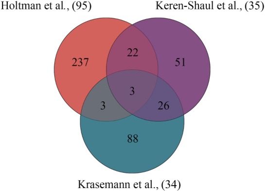
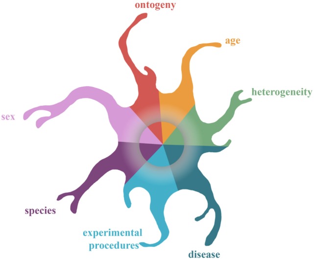
Verification
Claims the paper makes
The claims and statistics of this paper haven’t been checked yet.
Run the check — free, no account needed. It reads the open-access text and checks the sources it cites.
Claims other papers make about this one
Each is checked against this paper’s own text: does it say what the citing paper says it does?
In the citing paper
“In response to injury, the functional phenotype of microglia switches from quiescent to activated, and activated microglia are further divided into two distinct states, M1 and M2 (Dubbelaar et al. 2018).”
From MiR-124 and MiR-155 as Therapeutic Targets in Microglia-Mediated Inflammation in Multiple Sclerosis (2025) · cites this paper as Dubbelaar et al. 2018
Self-check
Percentages, sums and reported differences re-computed from the paper’s own numbers.
The self-check needs the paper’s full text.
Citations
Cites
Szklarczyk, D., Morris, J. H., Cook, H., Kuhn, M., Wyder, S., Simonovic, M., Santos, A., Doncheva, N. T., Roth, A., Bork, P., et al. (2017) · Nucleic Acids Research · doi:10.1093/nar/gkw937 · Cited by 6,980
Resting microglial cells are highly dynamic surveillants of brain parenchyma in vivo
Nimmerjahn, A., Kirchhoff, F., & Helmchen, F. (2005) · Science · doi:10.1126/science.1110647 · Cited by 5,827
A Unique Microglia Type Associated with Restricting Development of Alzheimer’s Disease
Keren-Shaul, H., Spinrad, A., Weiner, A., Matcovitch-Natan, O., Dvir-Szternfeld, R., Ulland, T. K., David, E., Baruch, K., Lara-Astaiso, D., Toth, B., et al. (2017) · Cell · doi:10.1016/j.cell.2017.05.018 · Cited by 5,493
Fate Mapping Analysis Reveals That Adult Microglia Derive from Primitive Macrophages
Ginhoux, F., Greter, M., Leboeuf, M., Nandi, S., See, P., Gokhan, S., Mehler, M. F., Conway, S. J., Ng, L. G., Stanley, E. R., et al. (2010) · Science · doi:10.1126/science.1194637 · Cited by 5,175
Macrophage biology in development, homeostasis and disease
Wynn, T. A., Chawla, A., & Pollard, J. W. (2013) · Nature · doi:10.1038/nature12034 · Cited by 4,684 · cited 4×
“Two major transcriptional changes are observed in MGnD: (1) the downregulation of microglial homeostatic genes [including Tgfb(r), Hexb, P2ry12, and Cx3cr1] and TFs (including Mef2a, Mafb, and Sall1) and (2) the upregulation of inflammatory genes (including Axl, Itgax, Clec7a, and Apoe), leading to a switch from a hom…”
InfluentialSynaptic Pruning by Microglia Is Necessary for Normal Brain Development
Paolicelli, R. C., Bolasco, G., Pagani, F., Maggi, L., Scianni, M., Panzanelli, P., Giustetto, M., Ferreira, T. A., Guiducci, E., Dumas, L., et al. (2011) · Science · doi:10.1126/science.1202529 · Cited by 4,244
Microglia-mediated neurotoxicity: uncovering the molecular mechanisms
Block, M. L., Zecca, L., & Hong, J.-S. (2007) · Nature Reviews Neuroscience · doi:10.1038/nrn2038 · Cited by 4,110
Microglia Sculpt Postnatal Neural Circuits in an Activity and Complement-Dependent Manner
Schafer, D. P., Lehrman, E. K., Kautzman, A. G., Koyama, R., Mardinly, A. R., Yamasaki, R., Ransohoff, R. M., Greenberg, M. E., Barres, B. A., & Stevens, B. (2012) · Neuron · doi:10.1016/j.neuron.2012.03.026 · Cited by 4,089
ATP mediates rapid microglial response to local brain injury in vivo
Davalos, D., Grutzendler, J., Yang, G., Kim, J. V., Zuo, Y., Jung, S., Littman, D. R., Dustin, M. L., & Gan, W.-B. (2005) · Nature Neuroscience · doi:10.1038/nn1472 · Cited by 3,913
Kettenmann, H., Hanisch, U.-K., Noda, M., & Verkhratsky, A. (2011) · Physiological Reviews · doi:10.1152/physrev.00011.2010 · Cited by 3,547
Fate Mapping Reveals Origins and Dynamics of Monocytes and Tissue Macrophages under Homeostasis
Yona, S., Kim, K.-W., Wolf, Y., Mildner, A., Varol, D., Breker, M., Strauss-Ayali, D., Viukov, S., Guilliams, M., Misharin, A., et al. (2013) · Immunity · doi:10.1016/j.immuni.2012.12.001 · Cited by 3,245
Krasemann, S., Madore, C., Cialic, R., Baufeld, C., Calcagno, N., El Fatimy, R., Beckers, L., O’Loughlin, E., Xu, Y., Fanek, Z., et al. (2017) · Immunity · doi:10.1016/j.immuni.2017.08.008 · Cited by 2,979
Identification of a unique TGF-β–dependent molecular and functional signature in microglia
Butovsky, O., Jedrychowski, M. P., Moore, C. S., Cialic, R., Lanser, A. J., Gabriely, G., Koeglsperger, T., Dake, B., Wu, P. M., Doykan, C. E., et al. (2014) · Nature Neuroscience · doi:10.1038/nn.3599 · Cited by 2,543
Hashimoto, D., Chow, A., Noizat, C., Teo, P., Beasley, M. B., Leboeuf, M., Becker, C. D., See, P., Price, J., Lucas, D., et al. (2013) · Immunity · doi:10.1016/j.immuni.2013.04.004 · Cited by 2,311 · cited 1×
“The Lifetime of Microglia Microglia, as well as other tissue-resident macrophages (46, 47), are stable, self-renewing cell populations over the entire lifespan of an animal.”
the Immunological Genome Consortium, Gautier, E. L., Shay, T., Miller, J., Greter, M., Jakubzick, C., Ivanov, S., Helft, J., Chow, A., Elpek, K. G., et al. (2012) · Nature Immunology · doi:10.1038/ni.2419 · Cited by 1,972
Elmore, M. R. P., Najafi, A. R., Koike, M. A., Dagher, N. N., Spangenberg, E. E., Rice, R. A., Kitazawa, M., Matusow, B., Nguyen, H., West, B. L., et al. (2014) · Neuron · doi:10.1016/j.neuron.2014.02.040 · Cited by 1,968
Neuroinflammation and M2 Microglia: The Good, the Bad, and the Inflamed
Cherry, J. D., Olschowka, J. A., & O’Banion, M. K. (2014) · Journal of Neuroinflammation · doi:10.1186/1742-2094-11-98 · Cited by 1,748
Integrated Systems Approach Identifies Genetic Nodes and Networks in Late-Onset Alzheimer’s Disease
Zhang, B., Gaiteri, C., Bodea, L.-G., Wang, Z., McElwee, J., Podtelezhnikov, A. A., Zhang, C., Xie, T., Tran, L., Dobrin, R., et al. (2013) · Cell · doi:10.1016/j.cell.2013.03.030 · Cited by 1,657
Microglia emerge from erythromyeloid precursors via Pu.1- and Irf8-dependent pathways
Kierdorf, K., Erny, D., Goldmann, T., Sander, V., Schulz, C., Perdiguero, E. G., Wieghofer, P., Heinrich, A., Riemke, P., Hölscher, C., et al. (2013) · Nature Neuroscience · doi:10.1038/nn.3318 · Cited by 1,457
An environment-dependent transcriptional network specifies human microglia identity
Gosselin, D., Skola, D., Coufal, N. G., Holtman, I. R., Schlachetzki, J. C. M., Sajti, E., Jaeger, B. N., O’Connor, C., Fitzpatrick, C., Pasillas, M. P., et al. (2017) · Science · doi:10.1126/science.aal3222 · Cited by 1,220 · cited 13×
“1) and IRF8, which are also crucial during murine microglia ontogeny and development (19) are highly expressed in both datasets (39, 43).”
InfluentialMicroglia development follows a stepwise program to regulate brain homeostasis
Matcovitch-Natan, O., Winter, D. R., Giladi, A., Vargas Aguilar, S., Spinrad, A., Sarrazin, S., Ben-Yehuda, H., David, E., Zelada González, F., Perrin, P., et al. (2016) · Science · doi:10.1126/science.aad8670 · Cited by 1,180 · cited 4×
“Consecutive Stages A recent study combining transcriptome with epigenome analysis identified genes and chromatin modulators that regu late different stages of microglia development in mice (19).”
InfluentialHoeffel, G., Chen, J., Lavin, Y., Low, D., Almeida, F. F., See, P., Beaudin, A. E., Lum, J., Low, I., Forsberg, E. C., et al. (2015) · Immunity · doi:10.1016/j.immuni.2015.03.011 · Cited by 1,135 · cited 5×
“5) in the YS, a process dependent on the transcription factors (TFs) Spf1 (Pu.1) and Irf8 (Kierdorf et al. , 2013; Hoeffel et al. , 2015).”
InfluentialYang, J., Zhang, L., Yu, C., Yang, X.-F., & Wang, H. (2014) · Biomarker Research · doi:10.1186/2050-7771-2-1 · Cited by 1,099
Microglia-derived ASC specks cross-seed amyloid-β in Alzheimer’s disease
Venegas, C., Kumar, S., Franklin, B. S., Dierkes, T., Brinkschulte, R., Tejera, D., Vieira-Saecker, A., Schwartz, S., Santarelli, F., Kummer, M. P., et al. (2017) · Nature · doi:10.1038/nature25158 · Cited by 983
Differential roles of microglia and monocytes in the inflamed central nervous system
Yamasaki, R., Lu, H., Butovsky, O., Ohno, N., Rietsch, A. M., Cialic, R., Wu, P. M., Doykan, C. E., Lin, J., Cotleur, A. C., et al. (2014) · Journal of Experimental Medicine · doi:10.1084/jem.20132477 · Cited by 856
Transcriptomic analysis of purified human cortical microglia reveals age-associated changes
Galatro, T. F., Holtman, I. R., Lerario, A. M., Vainchtein, I. D., Brouwer, N., Sola, P. R., Veras, M. M., Pereira, T. F., Leite, R. E. P., Möller, T., et al. (2017) · Nature Neuroscience · doi:10.1038/nn.4597 · Cited by 741
Self-citationAn updated histological classification system for multiple sclerosis lesions
Kuhlmann, T., Ludwin, S., Prat, A., Antel, J., Brück, W., & Lassmann, H. (2017) · Acta Neuropathologica · doi:10.1007/s00401-016-1653-y · Cited by 692
Loss of ‘homeostatic’ microglia and patterns of their activation in active multiple sclerosis
Zrzavy, T., Hametner, S., Wimmer, I., Butovsky, O., Weiner, H. L., & Lassmann, H. (2017) · Brain · doi:10.1093/brain/awx113 · Cited by 684
Bohlen, C. J., Bennett, F. C., Tucker, A. F., Collins, H. Y., Mulinyawe, S. B., & Barres, B. A. (2017) · Neuron · doi:10.1016/j.neuron.2017.04.043 · Cited by 658
Bruttger, J., Karram, K., Wörtge, S., Regen, T., Marini, F., Hoppmann, N., Klein, M., Blank, T., Yona, S., Wolf, Y., et al. (2015) · Immunity · doi:10.1016/j.immuni.2015.06.012 · Cited by 634
Holtman, I. R., Raj, D. D., Miller, J. A., Schaafsma, W., Yin, Z., Brouwer, N., Wes, P. D., Möller, T., Orre, M., Kamphuis, W., et al. (2015) · Acta Neuropathologica Communications · doi:10.1186/s40478-015-0203-5 · Cited by 619
Self-citationA new fate mapping system reveals context-dependent random or clonal expansion of microglia
Tay, T. L., Mai, D., Dautzenberg, J., Fernández-Klett, F., Lin, G., Sagar, Datta, M., Drougard, A., Stempfl, T., Ardura-Fabregat, A., et al. (2017) · Nature Neuroscience · doi:10.1038/nn.4547 · Cited by 597
Haenseler, W., Sansom, S. N., Buchrieser, J., Newey, S. E., Moore, C. S., Nicholls, F. J., Chintawar, S., Schnell, C., Antel, J. P., Allen, N. D., et al. (2017) · Stem Cell Reports · doi:10.1016/j.stemcr.2017.05.017 · Cited by 593
Local cues establish and maintain region-specific phenotypes of basal ganglia microglia
De Biase, L. M., Schuebel, K. E., Fusfeld, Z. H., Jair, K., Hawes, I. A., Cimbro, R., Zhang, H.-Y., Liu, Q.-R., Shen, H., Xi, Z.-X., et al. (2017) · Neuron · doi:10.1016/j.neuron.2017.06.020 · Cited by 432
Hanamsagar, R., Alter, M. D., Block, C. S., Sullivan, H., Bolton, J. L., & Bilbo, S. D. (2017) · Glia · doi:10.1002/glia.23176 · Cited by 384
Targeting mi R‐155 restores abnormal microglia and attenuates disease in SOD 1 mice
Butovsky, O., Jedrychowski, M. P., Cialic, R., Krasemann, S., Murugaiyan, G., Fanek, Z., Greco, D. J., Wu, P. M., Doykan, C. E., Kiner, O., et al. (2015) · Annals of Neurology · doi:10.1002/ana.24304 · Cited by 358
PU.1 and C/EBPα/β convert fibroblasts into macrophage-like cells
Feng, R., Desbordes, S. C., Xie, H., Tillo, E. S., Pixley, F., Stanley, E. R., & Graf, T. (2008) · Proceedings of the National Academy of Sciences · doi:10.1073/pnas.0711961105 · Cited by 348
Single-cell epigenomics: powerful new methods for understanding gene regulation and cell identity
Clark, S. J., Lee, H. J., Smallwood, S. A., Kelsey, G., & Reik, W. (2016) · Genome Biology · doi:10.1186/s13059-016-0944-x · Cited by 310
Microglia Versus Myeloid Cell Nomenclature during Brain Inflammation
Greter, M., Lelios, I., & Croxford, A. L. (2015) · Frontiers in Immunology · doi:10.3389/fimmu.2015.00249 · Cited by 273 · cited 2×
“brain tissue (6, 8), resulting in issues regarding the distinction of microglia and”
Hellwig, S., Brioschi, S., Dieni, S., Frings, L., Masuch, A., Blank, T., & Biber, K. (2016) · Brain, Behavior, and Immunity · doi:10.1016/j.bbi.2015.11.008 · Cited by 252
Kamphuis, W., Kooijman, L., Schetters, S., Orre, M., & Hol, E. M. (2016) · Biochimica et Biophysica Acta (BBA) - Molecular Basis of Disease · doi:10.1016/j.bbadis.2016.07.007 · Cited by 209
Increased White Matter Inflammation in Aging- and Alzheimer’s Disease Brain
Raj, D., Yin, Z., Breur, M., Doorduin, J., Holtman, I. R., Olah, M., Mantingh-Otter, I. J., Van Dam, D., De Deyn, P. P., den Dunnen, W., et al. (2017) · Frontiers in Molecular Neuroscience · doi:10.3389/fnmol.2017.00206 · Cited by 180 · cited 4×
“In contrast to the general notion that microglia obtain a primed profile (40, 86, 87) and are neurotoxic during aging and age-related diseases (88), Hickman and coworkers identified a neuroprotective gene expression profile of microglia derived from the entire brain in aged mice (24 months) due to upregulation of gene…”
InfluentialSelf-citationTREM2 in CNS homeostasis and neurodegenerative disease
Painter, M. M., Atagi, Y., Liu, C.-C., Rademakers, R., Xu, H., Fryer, J. D., & Bu, G. (2015) · Molecular Neurodegeneration · doi:10.1186/s13024-015-0040-9 · Cited by 169
The transcription factor PU.1 is critical for viability and function of human brain microglia
Smith, A. M., Gibbons, H. M., Oldfield, R. L., Bergin, P. M., Mee, E. W., Faull, R. L. M., & Dragunow, M. (2013) · Glia · doi:10.1002/glia.22486 · Cited by 141
Hendrickx, D. A. E., van Scheppingen, J., van der Poel, M., Bossers, K., Schuurman, K. G., van Eden, C. G., Hol, E. M., Hamann, J., & Huitinga, I. (2017) · Frontiers in Immunology · doi:10.3389/fimmu.2017.01810 · Cited by 139
Macrophage development from HSCs requires PU.1-coordinated microRNA expression
Ghani, S., Riemke, P., Schönheit, J., Lenze, D., Stumm, J., Hoogenkamp, M., Lagendijk, A., Heinz, S., Bonifer, C., Bakkers, J., et al. (2011) · Blood · doi:10.1182/blood-2011-02-335141 · Cited by 123
Hwang, H.-W., Saito, Y., Park, C. Y., Blachère, N. E., Tajima, Y., Fak, J. J., Zucker-Scharff, I., & Darnell, R. B. (2017) · Neuron · doi:10.1016/j.neuron.2017.08.024 · Cited by 74
Research works printed but not linked: 61
- Gosselin, 2014, Environment drives selection and function of enhancers controlling tissue-specific macrophage identities, Cell, 159, 1327, 10.1016/j.cell.2014.11.023 · DOI · Paper page
- Lavin, 2014, Tissue-resident macrophage enhancer landscapes are shaped by the local microenvironment, Cell, 159, 1312, 10.1016/j.cell.2014.11.018 · DOI · Paper page
- E Hirbec, 2017, Microglia responses in acute and chronic neurological diseases: what microglia-specific transcriptomic studies taught (and did not teach) us, Front Aging Neurosci, 9, 227, 10.3389/fnagi.2017.00227 · DOI
- Eggen, 2017, Regulation of microglia identity from an epigenetic and transcriptomic point of view, Neuroscience, 10.1016/j.neuroscience.2017.12.010 · DOI · Paper page
- Koeniger, 2017, Splitting the “unsplittable”: dissecting resident and infiltrating macrophages in experimental autoimmune encephalomyelitis, Int J Mol Sci, 18, 2072, 10.3390/ijms18102072 · DOI
- Goldmann, 2016, Origin, fate and dynamics of macrophages at central nervous system interfaces, Nat Immunol, 17, 797, 10.1038/ni.3423 · DOI · Paper page
- Holtman, 2015, Glia open access database (GOAD): a comprehensive gene expression encyclopedia of glia cells in health and disease, Glia, 63, 1495, 10.1002/glia.22810 · DOI
- Sierra, 2016, The “Big-Bang” for modern glial biology: translation and comments on Pío del Río-Hortega 1919 series of papers on microglia, Glia, 64, 1801, 10.1002/glia.23046 · DOI · Paper page
- Stark, 2014, Vertebrate neurogenic placode development: historical highlights that have shaped our current understanding, Dev Dyn, 243, 1167, 10.1002/dvdy.24152 · DOI
- Hoeffel, 2015, Ontogeny of tissue-resident macrophages, Front Immunol, 6, 486, 10.3389/fimmu.2015.00486 · DOI · Paper page
- Monier, 2006, Distribution and differentiation of microglia in the human encephalon during the first two trimesters of gestation, J Comp Neurol, 499, 565, 10.1002/cne.21123 · DOI
- Forsberg, 2010, Efficient reprogramming of adult neural stem cells to monocytes by ectopic expression of a single gene, Proc Natl Acad Sci U S A, 107, 14657, 10.1073/pnas.1009412107 · DOI
- Shiau, 2015, Differential requirement for irf8 in formation of embryonic and adult macrophages in zebrafish, PLoS One, 10, e0117513, 10.1371/journal.pone.0117513 · DOI
- Thion, 2018, Microbiome influences prenatal and adult microglia in a sex-specific manner, Cell, 172, 500, 10.1016/j.cell.2017.11.042 · DOI · Paper page
- Bennett, 2016, New tools for studying microglia in the mouse and human CNS, Proc Natl Acad Sci U S A, 113, E1738, 10.1073/pnas.1525528113 · DOI · Paper page
- Chiu, 2013, A neurodegeneration-specific gene-expression signature of acutely isolated microglia from an amyotrophic lateral sclerosis mouse model, Cell Rep, 4, 385, 10.1016/j.celrep.2013.06.018 · DOI · Paper page
- Hickman, 2013, The microglial sensome revealed by direct RNA sequencing, Nat Neurosci, 16, 1896, 10.1038/nn.3554 · DOI · Paper page
- Galatro, 2017, Isolation of microglia and immune infiltrates from mouse and primate central nervous system, Methods Mol Biol, 1559, 333, 10.1007/978-1-4939-6786-5_23 · DOI
- Grabert, 2016, Microglial brain region-dependent diversity and selective regional sensitivities to aging, Nat Neurosci, 19, 504, 10.1038/nn.4222 · DOI · Paper page
- David, 2011, Repertoire of microglial and macrophage responses after spinal cord injury, Nat Rev Neurosci, 12, 388, 10.1038/nrn3053 · DOI · Paper page
- Mastroeni, 2018, Laser-captured microglia in the Alzheimer’s and Parkinson’s brain reveal unique regional expression profiles and suggest a potential role for hepatitis B in the Alzheimer’s brain, Neurobiol Aging, 63, 12, 10.1016/j.neurobiolaging.2017.10.019 · DOI
- Askew, 2017, Coupled proliferation and apoptosis maintain the rapid turnover of microglia in the adult brain, Cell Rep, 18, 391, 10.1016/j.celrep.2016.12.041 · DOI · Paper page
- Füger, 2017, Microglia turnover with aging and in an Alzheimer’s model via long-term in vivo single-cell imaging, Nat Neurosci, 20, 1371, 10.1038/nn.4631 · DOI · Paper page
- Réu, 2017, The lifespan and turnover of microglia in the human brain, Cell Rep, 20, 779, 10.1016/j.celrep.2017.07.004 · DOI · Paper page
- Ajami, 2011, Infiltrating monocytes trigger EAE progression, but do not contribute to the resident microglia pool, Nat Neurosci, 14, 1142, 10.1038/nn.2887 · DOI · Paper page
- van Ham, 2014, Intravital correlated microscopy reveals differential macrophage and microglial dynamics during resolution of neuroinflammation, Dis Model Mech, 7, 857, 10.1242/dmm.014886 · DOI
- Schaafsma, 2017, Maternal inflammation induces immune activation of fetal microglia and leads to disrupted microglia immune responses, behavior, and learning performance in adulthood, Neurobiol Dis, 106, 291, 10.1016/j.nbd.2017.07.017 · DOI
- Abud, 2017, iPSC-derived human microglia-like cells to study neurological diseases, Neuron, 94, 278, 10.1016/j.neuron.2017.03.042 · DOI · Paper page
- Douvaras, 2017, Directed differentiation of human pluripotent stem cells to microglia, Stem Cell Reports, 8, 1516, 10.1016/j.stemcr.2017.04.023 · DOI · Paper page
- Muffat, 2016, Efficient derivation of microglia-like cells from human pluripotent stem cells, Nat Med, 22, 1358, 10.1038/nm.4189 · DOI · Paper page
- Pandya, 2017, Differentiation of human and murine induced pluripotent stem cells to microglia-like cells, Nat Neurosci, 20, 753, 10.1038/nn.4534 · DOI · Paper page
- Beutner, 2013, Unique transcriptome signature of mouse microglia, Glia, 61, 1429, 10.1002/glia.22524 · DOI · Paper page
- Mizee, 2017, Isolation of primary microglia from the human post-mortem brain: effects of ante- and post-mortem variables, Acta Neuropathol Commun, 5, 16, 10.1186/s40478-017-0418-8 · DOI
- Buttgereit, 2016, Sall1 is a transcriptional regulator defining microglia identity and function, Nat Immunol, 17, 1397, 10.1038/ni.3585 · DOI · Paper page
- Spaethling, 2017, Primary cell culture of live neurosurgically resected aged adult human brain cells and single cell transcriptomics, Cell Rep, 18, 791, 10.1016/j.celrep.2016.12.066 · DOI
- Walker, 2014, Dynamic structural remodelling of microglia in health and disease: a review of the models, the signals and the mechanisms, Brain Behav Immun, 37, 1, 10.1016/j.bbi.2013.12.010 · DOI
- Beynon, 2012, Microglial activation in the injured and healthy brain: what are we really talking about? Practical and theoretical issues associated with the measurement of changes in microglial morphology, Neuroscience, 225, 162, 10.1016/j.neuroscience.2012.07.029 · DOI
- Raj, 2014, Priming of microglia in a DNA-repair deficient model of accelerated aging, Neurobiol Aging, 35, 2147, 10.1016/j.neurobiolaging.2014.03.025 · DOI
- Kluge, 2017, Impaired microglia process dynamics post-stroke are specific to sites of secondary neurodegeneration, Glia, 65, 1885, 10.1002/glia.23201 · DOI
- Mills, 2000, M-1/M-2 macrophages and the Th1/Th2 paradigm, J Immunol, 164, 6166, 10.4049/jimmunol.164.12.6166 · DOI · Paper page
- Murray, 2014, Macrophage activation and polarization: nomenclature and experimental guidelines, Immunity, 41, 14, 10.1016/j.immuni.2014.06.008 · DOI · Paper page
- Colton, 2009, Heterogeneity of microglial activation in the innate immune response in the brain, J Neuroimmune Pharmacol, 4, 399, 10.1007/s11481-009-9164-4 · DOI
- Kim, 2016, Brain trauma elicits non-canonical macrophage activation states, J Neuroinflammation, 13, 117, 10.1186/s12974-016-0581-z · DOI · Paper page
- Xue, 2014, Transcriptome-based network analysis reveals a spectrum model of human macrophage activation, Immunity, 40, 274, 10.1016/j.immuni.2014.01.006 · DOI · Paper page
- Ransohoff, 2016, A polarizing question: do M1 and M2 microglia exist?, Nat Neurosci, 19, 987, 10.1038/nn.4338 · DOI · Paper page
- Martinez, 2014, The M1 and M2 paradigm of macrophage activation: time for reassessment, F1000Prime Rep, 6, 13, 10.12703/P6-13 · DOI · Paper page
- Ginhoux, 2016, New insights into the multidimensional concept of macrophage ontogeny, activation and function, Nat Immunol, 17, 34, 10.1038/ni.3324 · DOI
- Wes, 2016, Next generation transcriptomics and genomics elucidate biological complexity of microglia in health and disease, Glia, 64, 197, 10.1002/glia.22866 · DOI · Paper page
- Spittau, 2017, Aging microglia—phenotypes, functions and implications for age-related neurodegenerative diseases, Front Aging Neurosci, 9, 194, 10.3389/fnagi.2017.00194 · DOI
- Orre, 2014, Acute isolation and transcriptome characterization of cortical astrocytes and microglia from young and aged mice, Neurobiol Aging, 35, 1, 10.1016/j.neurobiolaging.2013.07.008 · DOI
- Collier, 2017, Aging and Parkinson’s disease: different sides of the same coin?, Mov Disord, 32, 983, 10.1002/mds.27037 · DOI
- Janssen, 2017, Aging, microglia and cytoskeletal regulation are key factors in the pathological evolution of the APP23 mouse model for Alzheimer’s disease, Biochim Biophys Acta, 1863, 395, 10.1016/j.bbadis.2016.11.014 · DOI
- Tang, 2016, Differential roles of M1 and M2 microglia in neurodegenerative diseases, Mol Neurobiol, 53, 1181, 10.1007/s12035-014-9070-5 · DOI · Paper page
- Hanisch, 2007, Microglia: active sensor and versatile effector cells in the normal and pathologic brain, Nat Neurosci, 10, 1387, 10.1038/nn1997 · DOI · Paper page
- Yin, 2017, Immune hyperreactivity of Aβ plaque-associated microglia in Alzheimer’s disease, Neurobiol Aging, 55, 115, 10.1016/j.neurobiolaging.2017.03.021 · DOI · Paper page
- Mathys, 2017, Temporal tracking of microglia activation in neurodegeneration at single-cell resolution, Cell Rep, 21, 366, 10.1016/j.celrep.2017.09.039 · DOI · Paper page
- Lewis, 2014, RNA sequencing of microglia and monocyte-derived macrophages from mice with experimental autoimmune encephalomyelitis illustrates a changing phenotype with disease course, J Neuroimmunol, 277, 26, 10.1016/j.jneuroim.2014.09.014 · DOI
- Rojanathammanee, 2011, Expression of mutant alpha-synuclein modulates microglial phenotype in vitro, J Neuroinflammation, 8, 44, 10.1186/1742-2094-8-44 · DOI · Paper page
- Hoss, 2016, microRNA profiles in Parkinson’s disease prefrontal cortex, Front Aging Neurosci, 8, 36, 10.3389/fnagi.2016.00036 · DOI · Paper page
- Yerbury, 2016, Walking the tightrope: proteostasis and neurodegenerative disease, J Neurochem, 137, 489, 10.1111/jnc.13575 · DOI
- Cuevas-Diaz Duran, 2017, Single-cell RNA-sequencing of the brain, Clin Transl Med, 6, 20, 10.1186/s40169-017-0150-9 · DOI
Cited by
Sorted by citations
Inflammation in Parkinson’s Disease: Mechanisms and Therapeutic Implications
Pajares, M., I. Rojo, A., Manda, G., Boscá, L., & Cuadrado, A. (2020) · Cells · doi:10.3390/cells9071687 · Cited by 779
Wang, M., Pan, W., Xu, Y., Zhang, J., Wan, J., & Jiang, H. (2022) · Journal of Inflammation Research · doi:10.2147/jir.s350109 · Cited by 461
Uncovering sex differences of rodent microglia
Han, J., Fan, Y., Zhou, K., Blomgren, K., & Harris, R. A. (2021) · Journal of Neuroinflammation · doi:10.1186/s12974-021-02124-z · Cited by 224
A new genetic strategy for targeting microglia in development and disease
McKinsey, G. L., Lizama, C. O., Keown-Lang, A. E., Niu, A., Santander, N., Larpthaveesarp, A., Chee, E., Gonzalez, F. F., & Arnold, T. D. (2020) · eLife · doi:10.7554/elife.54590 · Cited by 176
Abnormal synaptic pruning during adolescence underlying the development of psychotic disorders
Germann, M., Brederoo, S. G., & Sommer, I. E. C. (2021) · Current Opinion in Psychiatry · doi:10.1097/yco.0000000000000696 · Cited by 102
Metadata
- Authors
- Dubbelaar, M. L., Kracht, L., Eggen, B. J. L., & Boddeke, E. W. G. M.
- Venue
- Frontiers in Immunology
- Publisher
- Frontiers Media SA
- Published
- 2018-07-31
- Volume
- 9
- ISSN
- 1664-3224
- PMC
- PMC6079257
- Cited by
- 260
- References
- 47
About these checks
Registry, retraction, Cites and statistics results are computed, not judged, with one exception: a printed reference that no index lists is read once by a model before it counts as a citation error. Claims are extracted by a model and kept only when their quote appears verbatim in the text; each cited source is then checked, with a model’s help, for whether it says what the claim relies on. Those verdicts can be wrong — read the claim’s quote and the cited source before relying on one.
Statistics and arithmetic are recomputed, not judged.
Confidence bands are shown only for fields calibrated on our annotated test set. None is calibrated yet, so no band is shown.
What counts as a claim ›How we measure confidence ›How we check this ›
Are you an author, or do you see a problem with this page? Request a review
