

The DOI matches its Crossref record; 206 of the 206 references it cites were found in a registry or an index.
Axonal Degeneration in Tauopathies: Disease Relevance and Underlying Mechanisms
Open accessAbstract
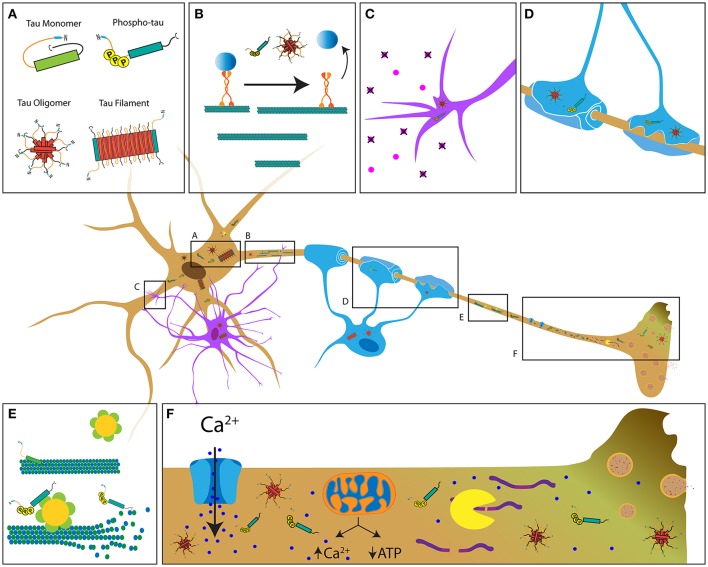
Tauopathies are a diverse group of diseases featuring progressive dying back neurodegeneration of specific neuronal populations in association with accumulation of abnormal forms of the microtubule-associated protein tau. It is well established that the clinical symptoms characteristic of tauopathies correlate with deficits in synaptic function and neuritic connectivity early in the course of disease, but mechanisms underlying these critical pathogenic events remain largely unknown. Biochemical in vitro evidence fueled the widespread notion that microtubule stabilization represents tau’s primary biological role and that the marked atrophy of neurites observed in tauopathies results from loss of microtubule stability. However, this notion contrasts with the mild phenotype associated with tau deletion. Instead, an analysis of cellular hallmarks common to different tauopathies, including aberrant patterns of protein phosphorylation and early degeneration of axons, suggests that alterations in kinase-based signaling pathways and deficits in axonal transport associated with such alterations contribute to the loss of neuronal connectivity triggered by pathogenic forms of tau. Here, we review a body of literature providing evidence that axonal pathology represents an early and common pathogenic event among human tauopathies. Observations of axonal degeneration in animal models of specific tauopathies are discussed and similarities to human disease highlighted. Finally, we discuss potential mechanistic pathways other than microtubule destabilization by which disease-related forms of tau may promote axonopathy.
Overview
The claims and statistics of this paper haven’t been checked yet.
Run the check — free, no account needed. It reads the open-access text and checks the sources it cites.
Integrity signals
Registry
Cites
Statistics
Figures
Verification
Claims the paper makes
The claims and statistics of this paper haven’t been checked yet.
Run the check — free, no account needed. It reads the open-access text and checks the sources it cites.
Claims other papers make about this one
Each is checked against this paper’s own text: does it say what the citing paper says it does?
The check wasn’t confident either way, so it gives no verdict — never disputed.
In the citing paper
“The dose for this experiment was chosen based upon previous work by Brunden et. al. and Zhang et. al. in which EpoD at 1mg/kg was sufficient to reduce measures of tauopathy, neurodegeneration and axonal dystrophy in PS19 tau mice 30, 31.”
From Amyloid-beta induced retrograde axonal degeneration in a mouse tauopathy model (2019) · cites this paper as [31]
Experimental — accuracy not yet measuredThe check wasn’t confident either way, so it gives no verdict — never disputed.
In the citing paper
“Additionally, previous studies utilizing EpoD as well as microtubule-stabilizing peptide NAP have both shown ability to reduce p-tau pathology in AD model mice over several months of treatment 31,66,67”
From Amyloid-beta induced retrograde axonal degeneration in a mouse tauopathy model (2019) · cites this paper as [31]
Experimental — accuracy not yet measuredThe check wasn’t confident either way, so it gives no verdict — never disputed.
In the citing paper
“Data from previous studies have found evidence that EpoD can promote axonal health by reducing axonal dystrophy, increasing ON microtubule density and normalizing fast axonal transport 30, 31.”
From Amyloid-beta induced retrograde axonal degeneration in a mouse tauopathy model (2019) · cites this paper as [31]
Experimental — accuracy not yet measured
Self-check
Percentages, sums and reported differences re-computed from the paper’s own numbers.
The self-check needs the paper’s full text.
Citations
Cites
Amyloid oligomers exacerbate tau pathology in a mouse model of tauopathy
Selenica, M.-L. B., Brownlow, M., Jimenez, J. P., Lee, D. C., Pena, G., Dickey, C. A., Gordon, M. N., & Morgan, D. (2013) · Neurodegenerative Diseases · doi:10.1159/000337230 · Cited by 10,359
Terry, R. D., Masliah, E., Salmon, D. P., Butters, N., DeTeresa, R., Hill, R., Hansen, L. A., & Katzman, R. (1991) · Annals of Neurology · doi:10.1002/ana.410300410 · Cited by 4,397
Association of missense and 5′-splice-site mutations in tau with the inherited dementia FTDP-17
Hutton, M., Lendon, C. L., Rizzu, P., Baker, M., Froelich, S., Houlden, H., Pickering-Brown, S., Chakraverty, S., Isaacs, A., Grover, A., et al. (1998) · Nature · doi:10.1038/31508 · Cited by 3,498
A protein factor essential for microtubule assembly
Weingarten, M. D., Lockwood, A. H., Hwo, S. Y., & Kirschner, M. W. (1975) · Proceedings of the National Academy of Sciences · doi:10.1073/pnas.72.5.1858 · Cited by 3,089
Synapse loss in frontal cortex biopsies in Alzheimer's disease: Correlation with cognitive severity
DeKosky, S. T., & Scheff, S. W. (1990) · Annals of Neurology · doi:10.1002/ana.410270502 · Cited by 2,376
Chronic traumatic encephalopathy in athletes: Progressive tauopathy after repetitive head injury
McKee, A. C., Cantu, R. C., Nowinski, C. J., Hedley-Whyte, E. T., Gavett, B. E., Budson, A. E., Santini, V. E., Lee, H.-S., Kubilus, C. A., & Stern, R. A. (2009) · Journal of Neuropathology & Experimental Neurology · doi:10.1097/nen.0b013e3181a9d503 · Cited by 2,295
Tau-mediated neurodegeneration in Alzheimer’s disease and related disorders
Ballatore, C., Lee, V. M.-Y., & Trojanowski, J. Q. (2007) · Nature Reviews Neuroscience · doi:10.1038/nrn2194 · Cited by 2,174
The spectrum of disease in chronic traumatic encephalopathy
McKee, A. C., Stein, T. D., Nowinski, C. J., Stern, R. A., Daneshvar, D. H., Alvarez, V. E., Lee, H.-S., Hall, G., Wojtowicz, S. M., Baugh, C. M., et al. (2013) · Brain · doi:10.1093/brain/aws307 · Cited by 2,098
Tau Suppression in a Neurodegenerative Mouse Model Improves Memory Function
SantaCruz, K., Lewis, J., Spires, T., Paulson, J., Kotilinek, L., Ingelsson, M., Guimaraes, A., DeTure, M., Ramsden, M., McGowan, E., et al. (2005) · Science · doi:10.1126/science.1113694 · Cited by 2,014
Masters, C. L., Bateman, R., Blennow, K., Rowe, C. C., Sperling, R. A., & Cummings, J. L. (2015) · Nature Reviews Disease Primers · doi:10.1038/nrdp.2015.56 · Cited by 1,853
Dendritic Function of Tau Mediates Amyloid-β Toxicity in Alzheimer's Disease Mouse Models
Ittner, L. M., Ke, Y. D., Delerue, F., Bi, M., Gladbach, A., van Eersel, J., Wölfing, H., Chieng, B. C., Christie, M. J., Napier, I. A., et al. (2010) · Cell · doi:10.1016/j.cell.2010.06.036 · Cited by 1,821
Control of memory formation through regulated expression of a CaMKII transgene
Mayford, M., Bach, M. E., Huang, Y.-Y., Wang, L., Hawkins, R. D., & Kandel, E. R. (1996) · Science · doi:10.1126/science.274.5293.1678 · Cited by 1,550
Formation of Neurofibrillary Tangles in P301L Tau Transgenic Mice Induced by Aβ42 Fibrils
Götz, J., Chen, F., van Dorpe, J., & Nitsch, R. M. (2001) · Science · doi:10.1126/science.1062097 · Cited by 1,526
Clinical and Pathological Diagnosis of Frontotemporal Dementia
McKhann, G. M. (2001) · Archives of Neurology · doi:10.1001/archneur.58.11.1803 · Cited by 1,478
Propagation of Tau Pathology in a Model of Early Alzheimer's Disease
de Calignon, A., Polydoro, M., Suárez-Calvet, M., William, C., Adamowicz, D. H., Kopeikina, K. J., Pitstick, R., Sahara, N., Ashe, K. H., Carlson, G. A., et al. (2012) · Neuron · doi:10.1016/j.neuron.2011.11.033 · Cited by 1,427
Lewis, J., McGowan, E., Rockwood, J., Melrose, H., Nacharaju, P., Van Slegtenhorst, M., Gwinn-Hardy, K., Murphy, M. P., Baker, M., Yu, X., et al. (2000) · Nature Genetics · doi:10.1038/78078 · Cited by 1,371
The Neurometabolic Cascade of Concussion.
Giza, C. C., & Hovda, D. A. (2001) · PubMed Central · Cited by 1,342
Microtubule assembly, organization and dynamics in axons and dendrites
Conde, C., & Cáceres, A. (2009) · Nature Reviews Neuroscience · doi:10.1038/nrn2631 · Cited by 1,086
Clinical progression in Parkinson disease and the neurobiology of axons
Cheng, H., Ulane, C. M., & Burke, R. E. (2010) · Annals of Neurology · doi:10.1002/ana.21995 · Cited by 1,012 · cited 1×
“Based on this knowledge, interventions focused on maintenance of neuronal connectivity, rather than prevention of cell death, may represent a more pressing therapeutic need for tauopathies (Cheng et al., 2010; Lingor et al., 2012).”
BackgroundRoles of tau protein in health and disease
Guo, T., Noble, W., & Hanger, D. P. (2017) · Acta Neuropathologica · doi:10.1007/s00401-017-1707-9 · Cited by 1,003
Kraus, M. F., Susmaras, T. M., Caughlin, B., Walker, C. J., Sweeney, J. A., & Little, D. M. (2007) · Brain · doi:10.1093/brain/awm216 · Cited by 994
Cleveland, D. W., Hwo, S.-Y., & Kirschner, M. W. (1977) · Journal of Molecular Biology · doi:10.1016/0022-2836(77)90213-3 · Cited by 915 · cited 2×
“The initial discovery of tau protein was based on biochemical procedures that revealed its ability to associate with purified microtubules and modulate their dynamic growth behavior in vitro (Weingarten et al., 1975; Cleveland et al., 1977a,b).”
BackgroundStamer, K., Vogel, R., Thies, E., Mandelkow, E., & Mandelkow, E.-M. (2002) · The Journal of Cell Biology · doi:10.1083/jcb.200108057 · Cited by 885
Physical and chemical properties of purified tau factor and the role of tau in microtubule assembly
Cleveland, D. W., Hwo, S. Y., & Kirschner, M. W. (1977) · Journal of Molecular Biology · doi:10.1016/0022-2836(77)90214-5 · Cited by 881
Hauw, J., Daniel, S. E., Dickson, D. W., Horoupian, D. S., Jellinger, K. A., Lantos, P. L., McKee, A. C., Tabaton, M., & Litvan, I. (1994) · Neurology · doi:10.1212/wnl.44.11.2015 · Cited by 854
Altered microtubule organization in small-calibre axons of mice lacking tau protein
Harada, A., Oguchi, K., Okabe, S., Kuno, J., Terada, S., Ohshima, T., Sato-Yoshitake, R., Takei, Y., Noda, T., & Hirokawa, N. (1994) · Nature · doi:10.1038/369488a0 · Cited by 802
Allen, B., Ingram, E., Takao, M., Smith, M. J., Jakes, R., Virdee, K., Yoshida, H., Holzer, M., Craxton, M., Emson, P. C., et al. (2002) · Journal of Neuroscience · doi:10.1523/jneurosci.22-21-09340.2002 · Cited by 763
Hyperphosphorylation and aggregation of tau in mice expressing normal human tau isoforms
Andorfer, C., Kress, Y., Espinoza, M., De Silva, R., Tucker, K. L., Barde, Y., Duff, K., & Davies, P. (2003) · Journal of Neurochemistry · doi:10.1046/j.1471-4159.2003.01879.x · Cited by 722
Blennow, K., Brody, D. L., Kochanek, P. M., Levin, H. S., McKee, A. C., Ribbers, G. M., Yaffe, K. S., & Zetterberg, H. (2016) · Nature Reviews Disease Primers · doi:10.1038/nrdp.2016.84 · Cited by 693
Axonal Self-Destruction and Neurodegeneration
Raff, M., Whitmore, A., & Finn, J. (2002) · Science · doi:10.1126/science.1068613 · Cited by 670
Frontotemporal dementia and parkinsonism linked to chromosome 17: A consensus conference
Foster, N. L., Wilhelmsen, K., Sima, A. A. F., Jones, M. Z., D'Amato, C., & Gilman, S. (1997) · Annals of Neurology · doi:10.1002/ana.410410606 · Cited by 664
Brandt, R., Léger, J. G., & Lee, G. S. (1995) · The Journal of Cell Biology · doi:10.1083/jcb.131.5.1327 · Cited by 663
Progressive supranuclear palsy: clinicopathological concepts and diagnostic challenges
Williams, D. R., & Lees, A. J. (2009) · The Lancet Neurology · doi:10.1016/s1474-4422(09)70042-0 · Cited by 650
Corticodentatonigral Degeneration With Neuronal Achromasia
Rebeiz, J. J., Kolodny, E. H., & Richardson, E. P. (1968) · Archives of Neurology · doi:10.1001/archneur.1968.00470310034003 · Cited by 627
Arendt, T., Stieler, J. T., & Holzer, M. (2016) · Brain Research Bulletin · doi:10.1016/j.brainresbull.2016.08.018 · Cited by 609
Bozzali, M. (2002) · Journal of Neurology, Neurosurgery & Psychiatry · doi:10.1136/jnnp.72.6.742 · Cited by 568
Ishihara, T., Hong, M., Zhang, B., Nakagawa, Y., Lee, M. K., Trojanowski, J. Q., & Lee, V. M.-Y. (1999) · Neuron · doi:10.1016/s0896-6273(00)81127-7 · Cited by 564
Inhibition of neuronal maturation in primary hippocampal neurons from tau deficient mice
Dawson, H. N., Ferreira, A., Eyster, M. V., Ghoshal, N., Binder, L. I., & Vitek, M. P. (2001) · Journal of Cell Science · doi:10.1242/jcs.114.6.1179 · Cited by 527
Mitochondria buffer physiological calcium loads in cultured rat dorsal root ganglion neurons
Werth, J. L., & Thayer, S. (1994) · Journal of Neuroscience · doi:10.1523/jneurosci.14-01-00348.1994 · Cited by 479
Axonal Transport Defects in Neurodegenerative Diseases
Morfini, G. A., Burns, M. R., Binder, L. I., Kanaan, N. M., LaPointe, N. E., Bosco, D. A., Brown, R. H., Brown, H. E., Tiwari, A., Hayward, L. J., et al. (2009) · Journal of Neuroscience · doi:10.1523/jneurosci.3463-09.2009 · Cited by 478
Self-citationNeurotrophins are required for nerve growth during development
Tucker, K. L., Meyer, M., & Barde, Y.-A. (2001) · Nature Neuroscience · doi:10.1038/82868 · Cited by 477
Tau interacts with src-family non-receptor tyrosine kinases
Lee, G. S., Newman, S. T., Gard, D. L., Band, H., & Panchamoorthy, G. (1998) · Journal of Cell Science · doi:10.1242/jcs.111.21.3167 · Cited by 471
Ghetti, B. F., Oblak, A. L., Boeve, B. F., Johnson, K. A., Dickerson, B. C., & Goedert, M. (2014) · Neuropathology and Applied Neurobiology · doi:10.1111/nan.12213 · Cited by 458
Stalning af amyloid percursor protein to study axonal damage in mild head Injury
Blumbergs, P. C., Scott, G., Manavis, J., Wainwright, H., Simpson, D. A., & McLean, A. J. (1994) · The Lancet · doi:10.1016/s0140-6736(94)91712-4 · Cited by 454
Memory‐related neural systems in Alzheimer's disease: An anatomic study
Hyman, B. T., Hoesen, G. W. V., & Damasio, A. R. (1990) · Neurology · doi:10.1212/wnl.40.11.1721 · Cited by 449 · cited 1×
“Despite this diversity, an examinations of human brains and animal models of tauopathies reveal axonopathy as a common feature of these diverse diseases (Hyman et al., 1990; Ahmed et al., 2008; Kovacs et al., 2008; Ling et al., 2015).”
BackgroundThe axon initial segment and the maintenance of neuronal polarity
Rasband, M. N. (2010) · Nature Reviews Neuroscience · doi:10.1038/nrn2852 · Cited by 442
Götz, J., Probst, A., Spillantini, M. G., Schäfer, T., Jakes, R., Bürki, K., & Goedert, M. (1995) · The EMBO Journal · doi:10.1002/j.1460-2075.1995.tb07116.x · Cited by 441
Global Hairpin Folding of Tau in Solution
Jeganathan, S., von Bergen, M., Brutlach, H., Steinhoff, H.-J., & Mandelkow, E. (2006) · Biochemistry · doi:10.1021/bi0521543 · Cited by 432
Rizzu, P., Swieten, J. C. V., Joosse, M., Hasegawa, M., Stevens, M., Tibben, A., Niermeijer, M. F., Hillebrand, M., Ravid, R., Oostra, B. A., et al. (1999) · The American Journal of Human Genetics · doi:10.1086/302256 · Cited by 432
Neuropathologic differentiation of progressive supranuclear palsy and corticobasal degeneration
Dickson, D. W. (1999) · Journal of Neurology · doi:10.1007/bf03161076 · Cited by 424
Nuclear Tau, a Key Player in Neuronal DNA Protection
Sultan, A., Nesslany, F., Violet, M., Bégard, S., Loyens, A., Talahari, S., Mansuroglu, Z., Marzin, D., Sergeant, N., Humez, S., et al. (2011) · Journal of Biological Chemistry · doi:10.1074/jbc.m110.199976 · Cited by 421 · cited 2×
“…regulation of AT (LaPointe et al., 2009; Kanaan et al., 2011), nuclear and nucleolar functions (Papasozomenos and Binder, 1987; Loomis et al., 1990; Sultan et al., 2011), end-binding protein regulation (RamirezRios et al., 2016), cytoskeleton-plasma membrane interactions (Brandt et al., 1995),…”
BackgroundPhosphorylation determines two distinct species of tau in the central nervous system
Papasozomenos, S. C., & Binder, L. I. (1987) · Cell Motility · doi:10.1002/cm.970080303 · Cited by 420
Animal Models of Alzheimer Disease
LaFerla, F. M., & Green, K. N. (2012) · Cold Spring Harbor Perspectives in Medicine · doi:10.1101/cshperspect.a006320 · Cited by 419
The Structural Basis of Monoclonal Antibody Alz50's Selectivity for Alzheimer's Disease Pathology
Carmel, G., Mager, E. M., Binder, L. I., & Kuret, J. (1996) · Journal of Biological Chemistry · doi:10.1074/jbc.271.51.32789 · Cited by 413
Clogging of axons by tau, inhibition of axonal traffic and starvation of synapses
Mandelkow�, E. (2003) · Neurobiology of Aging · doi:10.1016/j.neurobiolaging.2003.04.007 · Cited by 412
Defects in Axonal Elongation and Neuronal Migration in Mice with Disrupted tau and map1b Genes
Takei, Y., Teng, J., Harada, A., & Hirokawa, N. (2000) · The Journal of Cell Biology · doi:10.1083/jcb.150.5.989 · Cited by 408
Morfini, G. A., Szebenyi, G., Elluru, R. G., Ratner, N., & Brady, S. T. (2002) · The EMBO Journal · doi:10.1093/emboj/21.3.281 · Cited by 406
Self-citationSpires, T. L., Orne, J. D., SantaCruz, K., Pitstick, R., Carlson, G. A., Ashe, K. H., & Hyman, B. T. (2006) · The American Journal of Pathology · doi:10.2353/ajpath.2006.050840 · Cited by 401
Spittaels, K., den Haute, C. V., Dorpe, J. V., Bruynseels, K., Vandezande, K., Laenen, I., Geerts, H., Mercken, M. H., Sciot, R., Lommel, A. V., et al. (1999) · American Journal Of Pathology · doi:10.1016/s0002-9440(10)65533-2 · Cited by 390
Post-translational modifications of tubulin: pathways to functional diversity of microtubules
Song, Y., & Brady, S. T. (2014) · Trends in Cell Biology · doi:10.1016/j.tcb.2014.10.004 · Cited by 377
Axonopathy and amyotrophy in mice transgenic for human four-repeat tau protein
Probst, A., Götz, J., Wiederhold, K., Tolnay, M., Mistl, C., Jaton, A., Hong, M., Ishihara, T., Lee, V., Trojanowski, J., et al. (2000) · Acta Neuropathologica · doi:10.1007/s004010051148 · Cited by 362
Uryu, K., Chen, X.-H., Martinez, D., Browne, K. D., Johnson, V. E., Graham, D. I., Lee, V. M.-Y., Trojanowski, J. Q., & Smith, D. H. (2007) · Experimental Neurology · doi:10.1016/j.expneurol.2007.06.018 · Cited by 360
Kowall, N. W., & Kosik, K. S. (1987) · Annals of Neurology · doi:10.1002/ana.410220514 · Cited by 353
Interaction of tau with the RNA-Binding Protein TIA1 Regulates tau Pathophysiology and Toxicity
Vanderweyde, T. E., Apicco, D. J., Youmans-Kidder, K., Ash, P. E. A., Cook, C. N., da Rocha, E. L., Jansen‐West, K. R., Frame, A. A., Citro, A., Leszyk, J., et al. (2016) · Cell Reports · doi:10.1016/j.celrep.2016.04.045 · Cited by 346
Stability properties of neuronal microtubules
Baas, P. W., Rao, A. N., Matamoros, A. J., & Leo, L. (2016) · Cytoskeleton · doi:10.1002/cm.21286 · Cited by 315
Does oligodendrocyte survival depend on axons?
Barres, B. A., Jacobson, M. D., Schmid, R., Sendtner, M., & RAFF, M. C. (1993) · Current Biology · doi:10.1016/0960-9822(93)90039-q · Cited by 310
A critical appraisal of the pathogenic protein spread hypothesis of neurodegeneration
Walsh, D. M., & Selkoe, D. J. (2016) · Nature reviews. Neuroscience · doi:10.1038/nrn.2016.13 · Cited by 301
Kanaan, N. M., Morfini, G. A., LaPointe, N. E., Pigino, G. F., Patterson, K. R., Song, Y., Andreadis, A., Fu, Y., Brady, S. T., & Binder, L. I. (2011) · Journal of Neuroscience · doi:10.1523/jneurosci.0560-11.2011 · Cited by 292
Self-citationExtracellular Tau Oligomers Produce An Immediate Impairment of LTP and Memory
Fà, M., Puzzo, D., Piacentini, R., Staniszewski, A., Zhang, H., Baltrons, M. A., Puma, D. D. L., Chatterjee, I., Li, J., Saeed, F., et al. (2016) · Scientific Reports · doi:10.1038/srep19393 · Cited by 290
Self-citationJeganathan, S., Hascher, A., Chinnathambi, S., Biernat, J., Mandelkow, E.-M., & Mandelkow, E. (2008) · Journal of Biological Chemistry · doi:10.1074/jbc.m805300200 · Cited by 278
Widespread cytoskeletal pathology characterizes corticobasal degeneration.
Feany, M. Β., & Dickson, D. W. (1995) · PubMed Central · Cited by 275
Duff, K., Knight, H., Refolo, L. M., Sanders, S., Yu, X., Picciano, M., Malester, B., Hutton, M., Adamson, J., Goedert, M., et al. (2000) · Neurobiology of Disease · doi:10.1006/nbdi.1999.0279 · Cited by 269
A novel CDK5‐dependent pathway for regulating GSK3 activity and kinesin‐driven motility in neurons
Morfini, G. A., Szebenyi, G., Brown, H. J., Pant, H. C., Pigino, G. F., DeBoer, S. R., Beffert, U., & Brady, S. T. (2004) · The EMBO Journal · doi:10.1038/sj.emboj.7600237 · Cited by 268
Self-citationSingle-molecule investigation of the interference between kinesin, tau and MAP2c
Seitz, A., Kojima, H., Oiwa, K., Mandelkow, E.-M., Song, Y.-H., & Mandelkow, E. (2002) · The EMBO Journal · doi:10.1093/emboj/cdf503 · Cited by 257 · cited 1×
“In vitro studies using purified components of the AT system further supported a model where tau would elicit this effect by competing with conventional kinesin heavy chain subunits for microtubule binding (Seitz et al., 2002; Mandelkow et al., 2003).”
BackgroundLaPointe, N. E., Morfini, G. A., Pigino, G. F., Gaisina, I. N., Kozikowski, A. P., Binder, L. I., & Brady, S. T. (2008) · Journal of Neuroscience Research · doi:10.1002/jnr.21850 · Cited by 251
Self-citationIdentification of nuclear tau isoforms in human neuroblastoma cells.
Loomis, P. A., Howard, T. H., Castleberry, R. P., & Binder, L. I. (1990) · Proceedings of the National Academy of Sciences · doi:10.1073/pnas.87.21.8422 · Cited by 251
Ihara, M., Polvikoski, T. M., Hall, R., Slade, J. Y., Perry, R. H., Oakley, A. E., Englund, E. M., O'Brien, J. T., Ince, P. G., & Kalaria, R. N. (2010) · Acta Neuropathologica · doi:10.1007/s00401-009-0635-8 · Cited by 251
Effect of head impacts on diffusivity measures in a cohort of collegiate contact sport athletes
McAllister, T. W., Ford, J., Flashman, L. A., Maerlender, A., Greenwald, R. M., Beckwith, J. G., Bolander, R. P., Tosteson, T. D., Turco, J. H., Raman, R., et al. (2013) · Neurology · doi:10.1212/01.wnl.0000438220.16190.42 · Cited by 250
Corticobasal degeneration: a pathologically distinct 4R tauopathy
Kouri, N., Whitwell, J. L., Josephs, K. A., Rademakers, R., & Dickson, D. W. (2011) · Nature Reviews Neurology · doi:10.1038/nrneurol.2011.43 · Cited by 246
White Matter Integrity in the Brains of Professional Soccer Players Without a Symptomatic Concussion
Koerte, I. K., Ertl-Wagner, B. B., Reiser, M. F., ZAFONTE, R. D., & Shenton, M. E. (2012) · JAMA · doi:10.1001/jama.2012.13735 · Cited by 245
Dumanchin‐Njock, C., Camuzat, A., Campion, D., Verpillat, P., Hannequin, D., Dubois, B., Saugier-Véber, P., Martin, C., Penet, C., Charbonnier, F., et al. (1998) · Human Molecular Genetics · doi:10.1093/hmg/7.11.1825 · Cited by 244
Rohrer, J. D., Ridgway, G. R., Marc Modat, A., Sebastien Ourselin, A., Mead, S., Fox, N. C., Rossor, M. N., & Warren, J. D. (2010) · NeuroImage · doi:10.1016/j.neuroimage.2009.12.088 · Cited by 241
Tau Protects Microtubules in the Axon from Severing by Katanin
Qiang, L., Yu, W., Andreadis, A., Luo, M., & Baas, P. W. (2006) · Journal of Neuroscience · doi:10.1523/jneurosci.5392-05.2006 · Cited by 235
Muscle weakness, hyperactivity, and impairment in fear conditioning in tau-deficient mice
Ikegami, S. (2000) · Neuroscience Letters · doi:10.1016/s0304-3940(99)00964-7 · Cited by 221
Decker, H., Lo, K. Y., Unger, S. M., Ferreira, S. T., & Silverman, M. A. (2010) · Journal of Neuroscience · doi:10.1523/jneurosci.1074-10.2010 · Cited by 221
Hauw, J., Verny, M., Delaère, P., Cervera, P., He, Y., & Duyckaerts, C. J. (1990) · Neuroscience Letters · doi:10.1016/0304-3940(90)90829-x · Cited by 216
Kanaan, N. M., Pigino, G. F., Brady, S. T., Lazarov, O., Binder, L. I., & Morfini, G. A. (2012) · Experimental Neurology · doi:10.1016/j.expneurol.2012.06.003 · Cited by 215
Self-citationPetrie, E. C., Cross, D. J., Yarnykh, V. L., Richards, T. L., Martin, N., Pagulayan, K. F., Hoff, D., Hart, K. L., Mayer, C. L., Tarabochia, M. A., et al. (2013) · Journal of Neurotrauma · doi:10.1089/neu.2013.2952 · Cited by 213
Intrinsically disordered proteins and their (disordered) proteomes in neurodegenerative disorders
Uversky, V. N. (2015) · Frontiers in Aging Neuroscience · doi:10.3389/fnagi.2015.00018 · Cited by 211
In vivo characterization of chronic traumatic encephalopathy using [F-18]FDDNP PET brain imaging
Barrio, J. R., Small, G. W., Wong, K. P., Huang, S., Liu, J., Merrill, D. A., Giza, C. C., Fitzsimmons, R. P., Omalu, B. I., Bailes, J. E., et al. (2015) · Proceedings of the National Academy of Sciences · doi:10.1073/pnas.1409952112 · Cited by 211
Glial Inclusions in CNS Degenerative Diseases
Chin, S., & Goldman, J. E. (1996) · Journal of Neuropathology & Experimental Neurology · doi:10.1097/00005072-199605000-00001 · Cited by 210
Stoub, T. R., deToledo‐Morrell, L., Stebbins, G. T., Leurgans, S. E., Bennett, D. A., & Shah, R. C. (2006) · Proceedings of the National Academy of Sciences · doi:10.1073/pnas.0603414103 · Cited by 207
Pick's Disease: A Modern Approach
Dickson, D. W. (1998) · Brain Pathology · doi:10.1111/j.1750-3639.1998.tb00158.x · Cited by 205
Murrell, J. R., Spillantini, M. G., Zolo, P., Guazzelli, M., Smith, M. J., Hasegawa, M., Redi, F., Crowther, R. A., Pietrini, P., Ghetti, B. F., et al. (1999) · Journal of Neuropathology & Experimental Neurology · doi:10.1097/00005072-199912000-00002 · Cited by 205
Brion, J., Tremp, G. L., & Octave, J. (1999) · American Journal Of Pathology · doi:10.1016/s0002-9440(10)65272-8 · Cited by 203
Tokuda, T., Ikeda, S., Yanagisawa, N., Ihara, Y., & Glenner, G. G. (1991) · Acta Neuropathologica · doi:10.1007/bf00308813 · Cited by 202
Hampel, H. J., Teipel, S. J., Alexander, G. E., Horwitz, B., Teichberg, D., Schapiro, M. B., & Rapoport, S. I. (1998) · Archives of Neurology · doi:10.1001/archneur.55.2.193 · Cited by 200
Structural and functional brain connectivity in presymptomatic familial frontotemporal dementia
Dopper, E. G. P., Rombouts, S. A. R. B., Jiskoot, L. C., den Heijer, T., de Graaf, J. R. A., de Koning, I., Hammerschlag, A. R., Seelaar, H., Seeley, W. W., Veer, I. M., et al. (2013) · Neurology · doi:10.1212/wnl.0b013e31828407bc · Cited by 200
Feinstein, S. C., & Wilson, L. (2004) · Biochimica et Biophysica Acta (BBA) - Molecular Basis of Disease · doi:10.1016/j.bbadis.2004.07.002 · Cited by 200
Neurological consequences of traumatic brain injuries in sports
Ling, H., Hardy, J., & Zetterberg, H. (2015) · Molecular and Cellular Neuroscience · doi:10.1016/j.mcn.2015.03.012 · Cited by 197
Pick's disease: hyperphosphorylated tau protein segregates to the somatoaxonal compartment
Probst, A., Tolnay, M., Langui, D., Goedert, M., & Spillantini, M. G. (1996) · Acta Neuropathologica · doi:10.1007/s004010050565 · Cited by 192
Ghoshal, N. (2002) · Experimental Neurology · doi:10.1006/exnr.2002.8014 · Cited by 192
Axonal Transport RatesIn VivoAre Unaffected by Tau Deletion or Overexpression in Mice
Yuan, A., Kumar, A. R., Peterhoff, C. M., Duff, K., & Nixon, R. A. (2008) · Journal of Neuroscience · doi:10.1523/jneurosci.5242-07.2008 · Cited by 187
Wszołek, Z. K., Pfeiffer, R. F., Bhatt, M. H., Schelper, R. L., Cordes, M., Snow, B. J., Rodnitzky, R. L., Wolters, E. C., Arwert, F., & Calne, D. B. (1992) · Annals of Neurology · doi:10.1002/ana.410320303 · Cited by 186
Janning, D., Igaev, M., Sündermann, F., Brühmann, J., Beutel, O., Heinisch, J. J., Bakota, L., Piehler, J., Junge, W., & Brandt, R. (2014) · Molecular Biology of the Cell · doi:10.1091/mbc.e14-06-1099 · Cited by 181
A Decade of Tau Transgenic Animal Models and Beyond
Götz, J., Deters, N. A., Doldissen, A., Bokhari, L., Ke, Y. D., Wiesner, A., Schönrock, N., & Ittner, L. M. (2007) · Brain Pathology · doi:10.1111/j.1750-3639.2007.00051.x · Cited by 180
Diffusion tensor imaging and voxel based morphometry study in early progressive supranuclear palsy
Padovani, A. (2005) · Journal of Neurology Neurosurgery & Psychiatry · doi:10.1136/jnnp.2005.075713 · Cited by 180
Delisle, M., Murrell, J. R., Richardson, R., Trofatter, J. A., Rascol, O., Soulages, X., Mohr, M., Calvas, P., & Ghetti, B. F. (1999) · Acta Neuropathologica · doi:10.1007/s004010051052 · Cited by 171
Tau reduction prevents Aβ-induced axonal transport deficits by blocking activation of GSK3β
Vossel, K. A., Xu, J. C., Fomenko, V., Miyamoto, T., Suberbielle, E., Knox, J. A., Ho, K., Kim, D. H., Yu, G.-Q., & Mucke, L. (2015) · Journal of Cell Biology · doi:10.1083/jcb.201407065 · Cited by 167
Liu, C., Song, X., Nisbet, R. M., & Götz, J. (2016) · Journal of Biological Chemistry · doi:10.1074/jbc.m115.641902 · Cited by 156
Structural and functional brain connectivity in presymptomatic familial frontotemporal dementia
Dopper, E. G. P., Rombouts, S. A. R. B., Jiskoot, L. C., den Heijer, T., de Graaf, J. R. A., de Koning, I., Hammerschlag, A. R., Seelaar, H., Seeley, W. W., Veer, I. M., et al. (2014) · Neurology · doi:10.1212/wnl.0000000000000583 · Cited by 152
Regulation of motor proteins, axonal transport deficits and adult-onset neurodegenerative diseases
Brady, S. T., & Morfini, G. A. (2017) · Neurobiology of Disease · doi:10.1016/j.nbd.2017.04.010 · Cited by 150
Self-citationPollock, N., Mirra, S. S., Binder, L. I., Hansen, L., & Wood, J. (1986) · The Lancet · doi:10.1016/s0140-6736(86)92212-9 · Cited by 147
Protein Phosphatase 1 Is Targeted to Microtubules by the Microtubule-associated Protein Tau
Liao, H., Li, Y., Brautigan, D. L., & Gundersen, G. G. (1998) · Journal of Biological Chemistry · doi:10.1074/jbc.273.34.21901 · Cited by 145
Motor and cognitive deficits in aged tau knockout mice in two background strains
Lei, P., Ayton, S., Moon, S., Zhang, Q., Volitakis, I., Finkelstein, D. I., & Bush, A. I. (2014) · Molecular Neurodegeneration · doi:10.1186/1750-1326-9-29 · Cited by 145
Lessons from Tau-Deficient Mice
Ke, Y. D., Suchowerska, A. K., van der Hoven, J., Silva, D. M. D., Wu, C. W., van Eersel, J., Ittner, A., & Ittner, L. M. (2012) · International Journal of Alzheimer s Disease · doi:10.1155/2012/873270 · Cited by 142
Clinical Correlates of White Matter Tract Degeneration in Progressive Supranuclear Palsy
Whitwell, J. L., Master, A. V., Avula, R. T. V., Kantarci, K., Eggers, S. D. Z., Edmonson, H. A., Jack, C. R., & Josephs, K. A. (2011) · Archives of Neurology · doi:10.1001/archneurol.2011.107 · Cited by 140
Li, T., Hawkes, C. A., Qureshi, H. Y., Kar, S., & Paudel, H. K. (2006) · Biochemistry · doi:10.1021/bi051635j · Cited by 139
Microtubules in health and degenerative disease of the nervous system
Matamoros, A. J., & Baas, P. W. (2016) · Brain Research Bulletin · doi:10.1016/j.brainresbull.2016.06.016 · Cited by 137
Phosphorylation in the amino terminus of tau prevents inhibition of anterograde axonal transport
Kanaan, N. M., Morfini, G., Pigino, G., LaPointe, N. E., Andreadis, A., Song, Y., Leitman, E., Binder, L. I., & Brady, S. T. (2012) · Neurobiology of Aging · doi:10.1016/j.neurobiolaging.2011.06.006 · Cited by 134
Self-citationTau and Axonopathy in Neurodegenerative Disorders
Higuchi, M., Lee, V. M.-Y., & Trojanowski, J. Q. (2002) · NeuroMolecular Medicine · doi:10.1385/nmm:2:2:131 · Cited by 134
Higuchi, M., Zhang, B., Forman, M. S., Yoshiyama, Y., Trojanowski, J. Q., & Lee, V. M. ‐. (2005) · Journal of Neuroscience · doi:10.1523/jneurosci.2691-05.2005 · Cited by 133
Eckert, A., Nisbet, R. M., Grimm, A., & Götz, J. (2013) · Biochimica et Biophysica Acta (BBA) - Molecular Basis of Disease · doi:10.1016/j.bbadis.2013.08.013 · Cited by 133
Kovács, G. G., Majtényi, K., Spina, S., Murrell, J. R., Gelpí, E., Höftberger, R., Fraser, G. M., Crowther, R. A., Goedert, M., Budka, H., et al. (2008) · Journal of Neuropathology & Experimental Neurology · doi:10.1097/nen.0b013e318187a80f · Cited by 130
Tau interaction with microtubules in vivo
Samsonov, A., Yu, J.-Z., Rasenick, M., & Popov, S. V. (2004) · Journal of Cell Science · doi:10.1242/jcs.01531 · Cited by 129
Regulation of Axonal Transport by Protein Kinases
Gibbs, K. L., Greensmith, L., & Schiavo, G. (2015) · Trends in Biochemical Sciences · doi:10.1016/j.tibs.2015.08.003 · Cited by 126
Ultrastructural neuronal pathology in transgenic mice expressing mutant (P301L) human tau
Lin, W.-L., Lewis, J. M., Yen, S.-H. C., Hutton, M. L., & Dickson, D. W. (2003) · Journal of Neurocytology · doi:10.1023/b:neur.0000021904.61387.95 · Cited by 125
Siman, R., Cocca, R., & Dong, Y. (2015) · PLoS ONE · doi:10.1371/journal.pone.0142340 · Cited by 122
Jaworski, T., Lechat, B., Demedts, D., Gielis, L., Devijver, H., Borghgraef, P., Duimel, H., Verheyen, F., Kügler, S., & LEUVEN, F. V. (2011) · American Journal Of Pathology · doi:10.1016/j.ajpath.2011.06.025 · Cited by 121
Götz, J., Ittner, L. M., & Kins, S. (2006) · Journal of Neurochemistry · doi:10.1111/j.1471-4159.2006.03955.x · Cited by 119
Glial Tau Pathology in Tauopathies: Functional Consequences
Kahlson, M. A., & Colodner, K. J. (2015) · Journal of Experimental Neuroscience · doi:10.4137/jen.s25515 · Cited by 119
Axonal degeneration as a therapeutic target in the CNS
Lingor, P., Koch, J. C., Tönges, L., & Bähr, M. (2012) · Cell and Tissue Research · doi:10.1007/s00441-012-1362-3 · Cited by 119
Progressive supranuclear palsy: extensive neuropil threads in addition to neurofibrillary tangles
Probst, A., Langui, D., Lautenschläger, C., ULRICH, J., Brion, J., & Anderton, B. H. (1988) · Acta Neuropathologica · doi:10.1007/bf00688244 · Cited by 115
Maxwell, W. L., McCreath, B. J., Graham, D. I., & Gennarelli, T. A. (1995) · Journal of Neurocytology · doi:10.1007/bf01215643 · Cited by 114
Koerte, I. K., Kaufmann, D., Härtl, E., Sylvain Bouix, A., Pasternak, O., Kubicki, M. R., Rauscher, A., Li, D. K. B., Dadachanji, S. B., Taunton, J. A., et al. (2012) · Neurosurgical FOCUS · doi:10.3171/2012.10.focus12303 · Cited by 112
Glycogen Synthase Kinase-3β Is Complexed with Tau Protein in Brain Microtubules
Sun, W., Qureshi, H. Y., Cafferty, P. W., Sobue, K., Agarwal-Mawal, A., Neufield, K. D., & Paudel, H. K. (2002) · Journal of Biological Chemistry · doi:10.1074/jbc.m107182200 · Cited by 110
Kanaan, N. M., Cox, K. K., Alvarez, V. E., Stein, T. D., Poncil, S., & McKee, A. C. (2015) · Journal of Neuropathology & Experimental Neurology · doi:10.1093/jnen/nlv001 · Cited by 110
Self-citationCaMKII Activation in the Entorhinal Cortex Disrupts Previously Encoded Spatial Memory
Yasuda, M., & Mayford, M. R. (2006) · Neuron · doi:10.1016/j.neuron.2006.03.035 · Cited by 104
(2003) · PubMed Central · Cited by 103
Huang, J., & Auchus, A. P. (2007) · Annals of the New York Academy of Sciences · doi:10.1196/annals.1379.021 · Cited by 102
Rao, M. V., McBrayer, M. K., Campbell, J., Kumar, A., Hashim, A. T., Sershen, H. W., Stavrides, P. H., Ohno, M., Hutton, M., & Nixon, R. A. (2014) · Journal of Neuroscience · doi:10.1523/jneurosci.1132-14.2014 · Cited by 101
Ahmed, Z., Josephs, K. A., Gonzalez, J., DelleDonne, A., & Dickson, D. W. (2007) · Brain · doi:10.1093/brain/awm301 · Cited by 101
Adamec, E., Mohan, P. S., Vonsattel, J. P., & Nixon, R. A. (2002) · Acta Neuropathologica · doi:10.1007/s00401-002-0528-6 · Cited by 100
Praprotnik, D., Smith, M. A., Richey, P. L., Vinters, H. V., & Perry, G. L. (1996) · Acta Neuropathologica · doi:10.1007/s004010050420 · Cited by 98
Frontotemporal dementia: Neuropil spheroids and presynaptic terminal degeneration
Zhou, L., Miller, B. L., McDaniel, C. H., Kelly, L. K., Kim, O. J., & Miller, C. A. (1998) · Annals of Neurology · doi:10.1002/ana.410440116 · Cited by 98
Jaworski, T., Dewachter, I., Lechat, B., Croes, S., Termont, A., Demedts, D., Borghgraef, P., Devijver, H., Filipkowski, R. K., Kaczmarek, L., et al. (2009) · PLoS ONE · doi:10.1371/journal.pone.0007280 · Cited by 96
Tau is required for neurite outgrowth and growth cone motility of chick sensory neurons
Liu, C.-W. A., Lee, G. S., & Jay, D. G. (1999) · Cell Motility and the Cytoskeleton · doi:10.1002/(sici)1097-0169(1999)43:3 · Cited by 95
Sjöbeck, M., Haglund, M., & Englund, E. M. (2005) · International Journal of Geriatric Psychiatry · doi:10.1002/gps.1384 · Cited by 95
Fast Axonal Transport Misregulation and Alzheimer's Disease
Morfini, G. A., Pigino, G. F., Beffert, U., Busciglio, J. A., & Brady, S. T. (2002) · NeuroMolecular Medicine · doi:10.1385/nmm:2:2:089 · Cited by 95
Self-citationArgyrophilic thread-like structure in corticobasal degeneration and supranuclear palsy
Ikeda, K., Akiyama, H., Haga, C., Kondo, H., Arima, K., & Oda, T. (1994) · Neuroscience Letters · doi:10.1016/0304-3940(94)90010-8 · Cited by 95
Lau, D. H. W., Hogseth, M., Phillips, E. C., O’Neill, M. J., Pooler, A. M., Noble, W. J., & Hanger, D. P. (2016) · Acta Neuropathologica Communications · doi:10.1186/s40478-016-0317-4 · Cited by 93
Combs, B., Hamel, C., & Kanaan, N. M. (2016) · Neurobiology of Disease · doi:10.1016/j.nbd.2016.05.016 · Cited by 93
Self-citationWhite Matter Changes in Corticobasal Degeneration Syndrome and Correlation With Limb Apraxia
Borroni, B., Garibotto, V., Agosti, C., Brambati, S. M., Bellelli, G., Gasparotti, R., Padovani, A., & Perani, D. (2008) · Archives of Neurology · doi:10.1001/archneur.65.6.796 · Cited by 92
Xia, D., Li, C., & Götz, J. (2015) · Biochimica et Biophysica Acta (BBA) - Molecular Basis of Disease · doi:10.1016/j.bbadis.2014.12.017 · Cited by 91
Novel mutation in MAPT exon 13 (p.N410H) causes corticobasal degeneration
Kouri, N., Carlomagno, Y., Baker, M., Liesinger, A. M., Caselli, R. J., Wszołek, Z. K., Petrucelli, L., Boeve, B. F., Parisi, J. E., Josephs, K. A., et al. (2013) · Acta Neuropathologica · doi:10.1007/s00401-013-1193-7 · Cited by 89
Calcium dysregulation contributes to neurodegeneration in FTLD patient iPSC-derived neurons
Imamura, K., Sahara, N., Kanaan, N. M., Tsukita, K., Kondo, T., Kutoku, Y., Ohsawa, Y., Sunada, Y., Kawakami, K., Hotta, A., et al. (2016) · Scientific Reports · doi:10.1038/srep34904 · Cited by 88
Self-citationAD-linked, toxic NH2 human tau affects the quality control of mitochondria in neurons
Amadoro, G., Corsetti, V., Florenzano, F., Atlante, A., Ciotti, M. T., Mongiardi, M. P., Bussani, R., Nicolin, V., Nori, S. L., Campanella, M., et al. (2013) · Neurobiology of Disease · doi:10.1016/j.nbd.2013.10.018 · Cited by 86
Pro-aggregant Tau impairs mossy fiber plasticity due to structural changes and Ca++ dysregulation
Decker, J. M., Krüger, L., Sydow, A., Zhao, S., Frotscher, M., Mandelkow�, E., & Mandelkow, E. (2015) · Acta Neuropathologica Communications · doi:10.1186/s40478-015-0193-3 · Cited by 86
Tau binding to microtubules does not directly affect microtubule‐based vesicle motility
Morfini, G. A., Pigino, G. F., Mizuno, N., Kikkawa, M., & Brady, S. T. (2007) · Journal of Neuroscience Research · doi:10.1002/jnr.21154 · Cited by 84
Self-citationTint, I. S., Slaughter, T., Fischer, I., & Black, M. M. (1998) · Journal of Neuroscience · doi:10.1523/jneurosci.18-21-08660.1998 · Cited by 84
Sudo, H., & Baas, P. W. (2010) · Human Molecular Genetics · doi:10.1093/hmg/ddq521 · Cited by 77
Knake, S., Belke, M., Menzler, K., Pilatus, U., Eggert, K. M., Oertel, W. H., Stamelou, M., & Höglinger, G. U. (2010) · Movement Disorders · doi:10.1002/mds.23054 · Cited by 76
Swanson, E., Breckenridge, L., McMahon, L. P., Som, S., McConnell, I., & Bloom, G. S. (2017) · Journal of Alzheimer s Disease · doi:10.3233/jad-170168 · Cited by 76
Reduced number of axonal mitochondria and tau hypophosphorylation in mouse P301L tau knockin neurons
Rodríguez-Martín, T., Pooler, A. M., Lau, D. H. W., Mórotz, G. M., Vos, K. J. D., Gilley, J., Coleman, M. P., & Hanger, D. P. (2015) · Neurobiology of Disease · doi:10.1016/j.nbd.2015.10.007 · Cited by 75
Sjöbeck, M., & Englund, E. M. (2003) · Neuropathology and Applied Neurobiology · doi:10.1046/j.1365-2990.2003.00456.x · Cited by 73
Yoshiyama, Y., Zhang, B., Bruce, J., Trojanowski, J. Q., & Lee, V. M.-Y. (2003) · The Journal of Neuroscience · doi:10.1523/jneurosci.23-33-10662.2003 · Cited by 72
Phosphoregulation of Tau modulates inhibition of kinesin-1 motility
Stern, J. L., Lessard, D. V., Hoeprich, G. J., Morfini, G. A., & Berger, C. L. (2017) · Molecular Biology of the Cell · doi:10.1091/mbc.e16-10-0728 · Cited by 69
Self-citationRamírez‐Ríos, S., Denarier, É., Prezel, E., Vinit, A., Stoppin‐Mellet, V., Devred, F., Barbier, P., Peyrot, V., Sayas, C. L., Ávila, J., et al. (2016) · Molecular Biology of the Cell · doi:10.1091/mbc.e16-01-0029 · Cited by 68
Selden, S. C., & Pollard, T. D. (1986) · Annals of the New York Academy of Sciences · doi:10.1111/j.1749-6632.1986.tb38464.x · Cited by 65
Tau pathology-mediated presynaptic dysfunction
Moreno, H., Morfini, G. A., Buitrago, L., Ujlaki, G., Choi, S., Yu, E., Moreira, J. E., Ávila, J., Brady, S. T., Pant, H., et al. (2016) · Neuroscience · doi:10.1016/j.neuroscience.2016.03.044 · Cited by 65
Self-citationDickson, D. W., Feany, M. Β., Yen, S. H., Mattiace, L. A., & Davies, P. (1996) · Journal of neural transmission. Supplementum · doi:10.1007/978-3-7091-6892-9_2 · Cited by 65
Conventional kinesin: Biochemical heterogeneity and functional implications in health and disease
Morfini, G. A., Schmidt, N., Weissmann, C., Pigino, G. F., & Kins, S. (2016) · Brain Research Bulletin · doi:10.1016/j.brainresbull.2016.06.009 · Cited by 63
Self-citationCox, K. K., Combs, B., Abdelmesih, B., Morfini, G. A., Brady, S. T., & Kanaan, N. M. (2016) · Neurobiology of Aging · doi:10.1016/j.neurobiolaging.2016.07.015 · Cited by 62
Self-citationNiewidok, B., Igaev, M., Sündermann, F., Janning, D., Bakota, L., & Brandt, R. (2016) · Molecular Biology of the Cell · doi:10.1091/mbc.e16-06-0402 · Cited by 62
Regulation of Kinesin: Implications for Neuronal Development
Morfini, G. A., Szebenyi, G., Richards, B. W., & Brady, S. T. (2001) · Developmental Neuroscience · doi:10.1159/000048720 · Cited by 59
Self-citationTau Reduction Does Not Prevent Motor Deficits in Two Mouse Models of Parkinson's Disease
Morris, M., Koyama, A., Masliah, E., & Mucke, L. (2011) · PLoS ONE · doi:10.1371/journal.pone.0029257 · Cited by 56
Zhang, Y., Walter, R., Ng, P., Luong, P. N., Dutt, S., Heuer, H. W., Rojas-Rodriguez, J. C., Tsai, R. M., Litvan, I., Dickerson, B. C., et al. (2016) · PLoS ONE · doi:10.1371/journal.pone.0157218 · Cited by 54
Gatto, R. G., Chu, Y., Ye, A. Q., Price, S. D., Tavassoli, E., Buenaventura, A., Brady, S. T., Magin, R. L., Kordower, J. H., & Morfini, G. A. (2015) · Human Molecular Genetics · doi:10.1093/hmg/ddv248 · Cited by 52
Self-citationA Qualitative and Quantitative Study of Grumose Degeneration in Progressive Supranuclear Palsy
Ishizawa, K., Lin, W.-L., Tiseo, P. J., Honer, W. G., Davies, P. J., & Dickson, D. W. (2000) · Journal of Neuropathology & Experimental Neurology · doi:10.1093/jnen/59.6.513 · Cited by 52
Tiernan, C. T., Combs, B., Cox, K. K., Morfini, G. A., Brady, S. T., Counts, S., & Kanaan, N. M. (2016) · Experimental Neurology · doi:10.1016/j.expneurol.2016.06.030 · Cited by 46
Self-citationCaillierez, R., Bégard, S., Lécolle, K., Deramecourt, V., Zommer, N., Dujardin, S., Loyens, A., Dufour, N., Aurégan, G., Winderickx, J., et al. (2013) · Molecular Therapy · doi:10.1038/mt.2013.66 · Cited by 43
Black, M. M. (2015) · Methods in cell biology · doi:10.1016/bs.mcb.2015.06.001 · Cited by 42
Vermersch, P., Roche, J., Hamon, M., Daems‐Monpeurt, C., Pruvo, J., Dewailly, P., & Petit, H. (1996) · Journal of Neurology · doi:10.1007/bf00868519 · Cited by 42
Lin, W.-L., Zehr, C., Lewis, J. M., Hutton, M. L., Yen, S.-H. C., & Dickson, D. W. (2005) · Journal of Neurocytology · doi:10.1007/s11068-006-8726-0 · Cited by 41
No Overt Deficits in Aged Tau-Deficient C57Bl/6.Mapttm1(EGFP)Kit GFP Knockin Mice
van Hummel, A., Bi, M., Ippati, S., van der Hoven, J., Volkerling, A. M., Lee, W. S., Tan, D. C. S., Bongers, A., Ittner, A., Ke, Y. D., et al. (2016) · PLoS ONE · doi:10.1371/journal.pone.0163236 · Cited by 40
Kiernan, P. T., Montenigro, P. H., Solomon, T. M., & McKee, A. C. (2015) · Seminars in Neurology · doi:10.1055/s-0035-1545080 · Cited by 39
Accumulation of SNAP-25 immunoreactive material in axons of Alzheimerʼs disease
Dessi, F., Colle, M.-A., Hauw, J., & Duyckaerts, C. J. (1997) · Neuroreport · doi:10.1097/00001756-199712010-00006 · Cited by 39
Anisotropic fractional diffusion tensor imaging
Meerschaert, M. M., Magin, R. L., & Ye, A. Q. (2015) · Journal of Vibration and Control · doi:10.1177/1077546314568696 · Cited by 36
Gene Therapy Models of Alzheimer’s Disease and Other Dementias
Combs, B., Kneynsberg, A., & Kanaan, N. M. (2015) · Methods in molecular biology · doi:10.1007/978-1-4939-3271-9_25 · Cited by 33
Self-citationBakota, L., Ussif, A. M., Jeserich, G., & Brandt, R. (2017) · Molecular and Cellular Neuroscience · doi:10.1016/j.mcn.2017.03.003 · Cited by 33
Mufson, E. J., Perez, S. E., Nadeem, M., Mahady, L. J., Kanaan, N. M., Abrahamson, E. E., Ikonomović, M. D., Crawford, F., Alvarez, V. E., Stein, T. D., et al. (2016) · Brain Injury · doi:10.1080/02699052.2016.1219058 · Cited by 31
Self-citationKneynsberg, A., Collier, T. J., Manfredsson, F. P., & Kanaan, N. M. (2016) · Journal of Neuroscience Methods · doi:10.1016/j.jneumeth.2016.03.004 · Cited by 31
Self-citationYin, Y., Wang, Y., Gao, D., Ye, J., Wang, X., Fang, L., Wu, D., Pi, G., Lu, C., Zhou, X., et al. (2016) · Scientific Reports · doi:10.1038/srep27283 · Cited by 30
Ludvigson, A. E., Luebke, J. I., Lewis, J. M., & Peters, A. (2010) · Brain Structure and Function · doi:10.1007/s00429-010-0295-4 · Cited by 27
A Rapid Gene Delivery-Based Mouse Model for Early-Stage Alzheimer Disease-Type Tauopathy
Siman, R., Lin, Y., Malthankar‐Phatak, G. H., & Dong, Y. (2013) · Journal of Neuropathology & Experimental Neurology · doi:10.1097/nen.0000000000000006 · Cited by 27
Lippa, C. F. (2004) · American Journal of Alzheimer s Disease & Other Dementias® · doi:10.1177/153331750401900606 · Cited by 27
Cochran, E. J., Fox, J. H., & Mutson, E. J. (1994) · Acta Neuropathologica · doi:10.1007/bf00389503 · Cited by 25
Tau phosphorylation in the mouse brain during aversive conditioning
Fujio, J., Hosono, H., Ishiguro, K., Ikegami, S., & Fujita, S. C. (2007) · Neurochemistry International · doi:10.1016/j.neuint.2007.04.024 · Cited by 24
Phospholipids and calmodulin modulate the inhibition of PMCA activity by tau
Berrocal, M., Corbacho‐Cuello, I., Sepúlveda, M. R., Gutiérrez‐Merino, C., & Mata, A. M. (2016) · Biochimica et Biophysica Acta (BBA) - Molecular Cell Research · doi:10.1016/j.bbamcr.2016.10.023 · Cited by 23
White matter hyperintensities on MRI in a patient with corticobasal degeneration
Doi, T., Iwasa, K., Makifuchi, T., & Takamori, M. (1999) · Acta Neurologica Scandinavica · doi:10.1111/j.1600-0404.1999.tb07345.x · Cited by 22
Cited by
Sorted by citations
Kinetically distinct phases of tau on microtubules regulate kinesin motors and severing enzymes
Siahaan, V., Krattenmacher, J., Hyman, A. A., Diez, S., Hernández-Vega, A., Lansky, Z., & Braun, M. (2019) · Nature Cell Biology · doi:10.1038/s41556-019-0374-6 · Cited by 169
GSK3β and tau protein in Alzheimer's disease and epilepsy
Toral-Rios, D., Pichardo-Rojas, P. S., Alonso-Vanegas, M., & Campos-Peña, V. (2020) · Frontiers in Cellular Neuroscience · doi:10.3389/fncel.2020.00019 · Cited by 158
New Insights Into Drug Discovery Targeting Tau Protein
Soeda, Y., & Takashima, A. (2020) · Frontiers in Molecular Neuroscience · doi:10.3389/fnmol.2020.590896 · Cited by 136
Zavaliangos-Petropulu, A., Nir, T. M., Thomopoulos, S. I., Reid, R. I., Bernstein, M. A., Borowski, B., Jack Jr., C. R., Weiner, M. W., Jahanshad, N., & Thompson, P. M. (2019) · Frontiers in Neuroinformatics · doi:10.3389/fninf.2019.00002 · Cited by 101
“…of amyloid plaques and neurofibrillary tangles (NFTs) in the brain (Braak and Braak, 1991, 1996; Frank et al., 2003; Shaw et al., 2007) can directly impact WM (Lee et al., 2004; Roth et al., 2005), promoting myelin degeneration and axonal loss (Braak and Braak, 1996; Kneynsberg et al., 2017).”
Akbar, M., Shabbir, A., Rehman, K., Akash, M. S. H., & Shah, M. A. (2021) · Journal of Food Biochemistry · doi:10.1111/jfbc.13936 · Cited by 97
Metadata
- Authors
- Kneynsberg, A., Combs, B., Christensen, K., Morfini, G., & Kanaan, N. M.
- Affiliations
- Michigan State University (US)
- University of Illinois Chicago (US)
- Trinity Health Grand Rapids (US)
- Corresponding author
- Morfini, Gerardo
- Kanaan, Nicholas M.
- Venue
- Frontiers in Neuroscience
- Publisher
- Frontiers Media SA
- Published
- 2017-10-17
- Volume
- 11
- Pages
- 572-572
- ISSN
- 1662-453X (ISSN-L)
- PMC
- PMC5651019
- OpenAlex
- W2763441547
- Cited by
- 102
- References
- 206
- Citations by year
- 2026: 6 · 2025: 7 · 2024: 8 · 2023: 6 · 2022: 15 · 2021: 18 · 2020: 18 · 2019: 16 · 2018: 8
- FWCI
- 1.41
- Type
- journal article
- Language
- en
- Open access
- gold
- doi.org
- Licence
- cc-by
- Locations
- Frontiers in Neuroscience (publishedVersion, cc-by)
- PubMed Central (publishedVersion, cc-by)
- PubMed (publishedVersion)
- DOAJ (DOAJ: Directory of Open Access Journals) (submittedVersion, cc-by-sa)
- Funders
- National Institute on Aging (R01 AG044372)
- National Institute of Neurological Disorders and Stroke (R01 NS082730, R21 NS096642, R01 NS066942)
- BrightFocus Foundation (A2013364S)
- Topics
- Life Sciences › Neuroscience › Cellular and Molecular Neuroscience › Alzheimer's disease research and treatments
- Health Sciences › Medicine › Clinical Neurology › Amyotrophic Lateral Sclerosis Research
- Life Sciences › Neuroscience › Neurology › Neuroinflammation and Neurodegeneration Mechanisms
- Keywords
- tauopathy, axonal degeneration, axonopathy, tau protein, microtubule stabilization, microtubule destabilization, tau phosphorylation, kinase signaling pathways, axonal transport, neuronal connectivity, dying-back degeneration
About these checks
Registry, retraction, Cites and statistics results are computed, not judged, with one exception: a printed reference that no index lists is read once by a model before it counts as a citation error. Claims are extracted by a model and kept only when their quote appears verbatim in the text; each cited source is then checked, with a model’s help, for whether it says what the claim relies on. Those verdicts can be wrong — read the claim’s quote and the cited source before relying on one.
Statistics and arithmetic are recomputed, not judged.
Confidence bands are shown only for fields calibrated on our annotated test set. None is calibrated yet, so no band is shown.
What counts as a claim ›How we measure confidence ›How we check this ›
Are you an author, or do you see a problem with this page? Request a review
