

The DOI matches its Crossref record; 7 of the 7 references it cites were found in a registry or an index.
Differentially Expressed miRNAs in Age-Related Neurodegenerative Diseases: A Meta-Analysis
Abstract
To date, no neurodegenerative diseases (NDDs) have cures, and the underlying mechanism of their pathogenesis is undetermined. As miRNAs extensively regulate all biological processes and are crucial regulators of healthy brain function, miRNAs differentially expressed in NDDs may provide insight into the factors that contribute to the emergence of protein inclusions and the propagation of deleterious cellular environments. A meta-analysis of miRNAs dysregulated in Alzheimer’s disease, Parkinson’s disease, multiple system atrophy, progressive supranuclear palsy, corticobasal degeneration, dementia with Lewy bodies and frontotemporal lobar degeneration (TDP43 variant) was performed to determine if diseases within a proteinopathy have distinct or shared mechanisms of action leading to neuronal death, and if proteinopathies can be classified on the basis of their miRNA profiles. Our results identified both miRNAs distinct to the anatomy, disease type and pathology, and miRNAs consistently dysregulated within single proteinopathies and across neurodegeneration in general. Our results also highlight the necessity to minimize the variability between studies. These findings showcase the need for more transcriptomic research on infrequently occurring NDDs, and the need for the standardization of research techniques and platforms utilized across labs and diseases.
Overview
The claims and statistics of this paper haven’t been checked yet.
Run the check — free, no account needed. It reads the open-access text and checks the sources it cites.
Integrity signals
Registry
Cites
Statistics
Figures
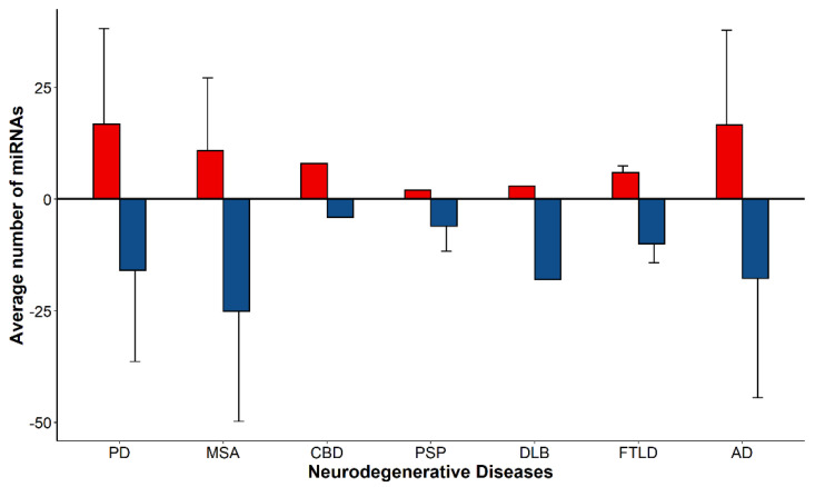
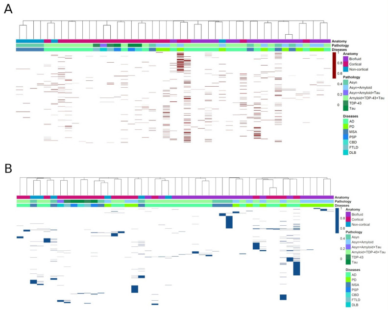
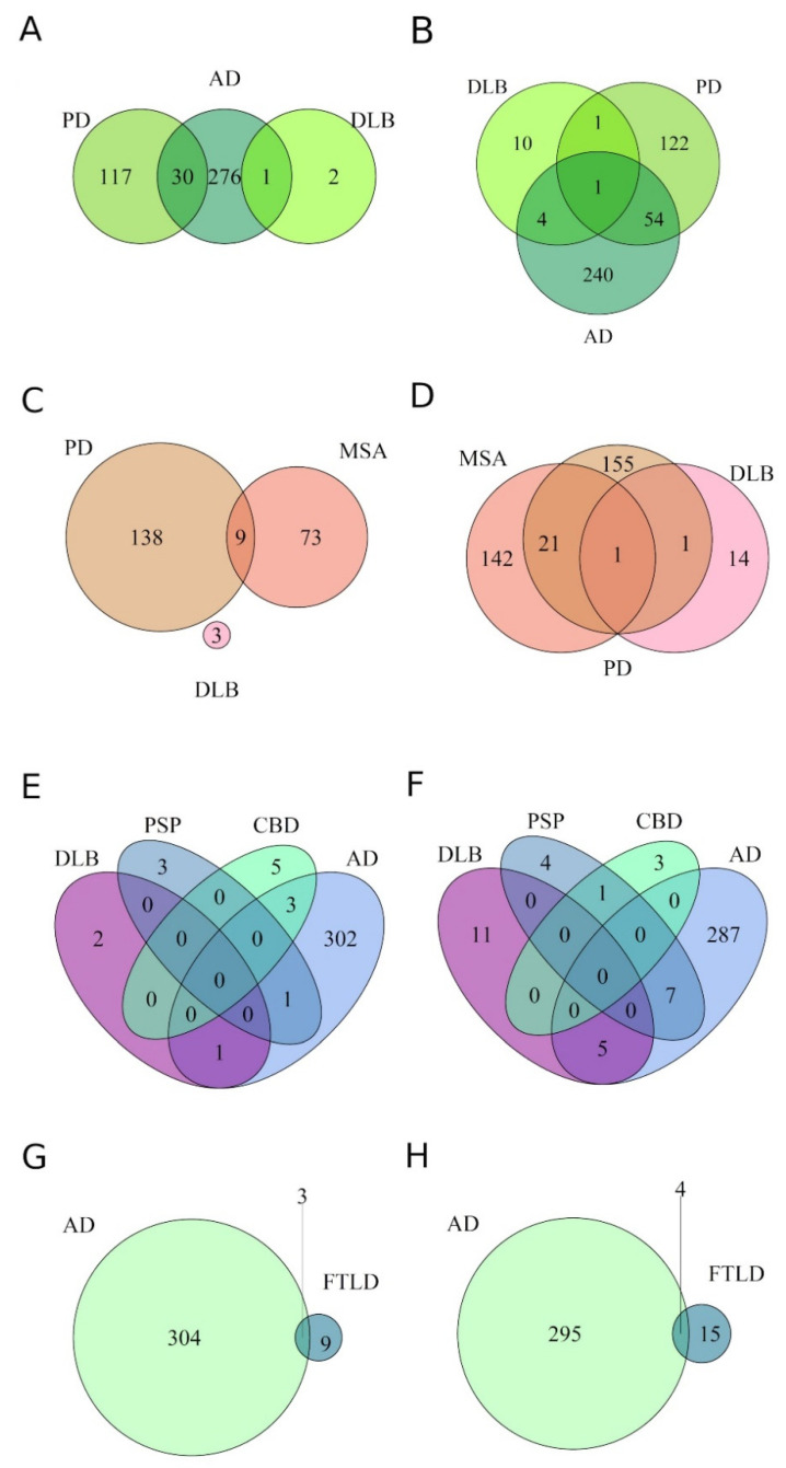
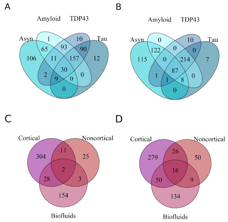
Verification
Claims the paper makes
The claims and statistics of this paper haven’t been checked yet.
Run the check — free, no account needed. It reads the open-access text and checks the sources it cites.
Claims other papers make about this one
Each is checked against this paper’s own text: does it say what the citing paper says it does?
In the citing paper
“Multiple studies have identified microRNA-132 (miR-132) as one of the most significantly downregulated microRNAs in AD.”
From microRNA-132 attenuates inflammation in induced pluripotent stem cell-derived microglia from Alzheimer’s disease patients (2026) · cites this paper as [19]
Self-check
Percentages, sums and reported differences re-computed from the paper’s own numbers.
The self-check needs the paper’s full text.
Citations
Cites
Subramanian, A., Tamayo, P., Mootha, V. K., Mukherjee, S., Ebert, B. L., Gillette, M. A., Paulovich, A., Pomeroy, S. L., Golub, T. R., Lander, E. S., et al. (2005) · Proceedings of the National Academy of Sciences · doi:10.1073/pnas.0506580102 · Cited by 53,625
Stages of the Pathologic Process in Alzheimer Disease: Age Categories From 1 to 100 Years
Braak, H., Thal, D. R., Ghebremedhin, E., & Del Tredici, K. (2011) · Journal of Neuropathology & Experimental Neurology · doi:10.1097/nen.0b013e318232a379 · Cited by 2,194
Pathology of Neurodegenerative Diseases
Dugger, B. N., & Dickson, D. W. (2017) · Cold Spring Harbor Perspectives in Biology · doi:10.1101/cshperspect.a028035 · Cited by 1,665
Cogswell, J. P., Ward, J., Taylor, I. A., Waters, M., Shi, Y., Cannon, B., Kelnar, K., Kemppainen, J., Brown, D., Chen, C., et al. (2008) · Journal of Alzheimer s Disease · doi:10.3233/jad-2008-14103 · Cited by 943
Burden of Neurological Disorders Across the US From 1990-2017: A Global Burden of Disease Study
Collaborators, G. 2. U. N. D., Feigin, V. L., Vos, T., Alahdab, F., Amit, A. M. L., Bärnighausen, T. W., Beghi, E., Beheshti, M., Chavan, P. P., Criqui, M. H., et al. (2021) · JAMA Neurology · doi:10.1001/jamaneurol.2020.4152 · Cited by 622
Pairing beyond the Seed Supports MicroRNA Targeting Specificity
Broughton, J. P., Lovci, M. T., Huang, J. L., Yeo, G. W., & Pasquinelli, A. E. (2016) · Molecular Cell · doi:10.1016/j.molcel.2016.09.004 · Cited by 487
Lekka, E., & Hall, J. (2018) · FEBS Letters · doi:10.1002/1873-3468.13182 · Cited by 251
Research works printed but not linked: 85
- Lee, 2001, Neurodegenerative Tauopathies, Annu. Rev. Neurosci., 24, 1121, 10.1146/annurev.neuro.24.1.1121 · DOI · Paper page
- Gomperts, 2016, Tau Positron Emission Tomographic Imaging in the Lewy Body Diseases, JAMA Neurol., 73, 1334, 10.1001/jamaneurol.2016.3338 · DOI
- Wenning, 2016, Glia and α-synuclein in neurodegeneration: A complex interaction, Neurobiol. Dis., 85, 262, 10.1016/j.nbd.2015.03.003 · DOI
- Prasansuklab, 2013, Amyloidosis in Alzheimer’s Disease: The Toxicity of Amyloid β (Aβ), Mechanisms of Its Accumulation and Implications of Medicinal Plants for Therapy, Evid.-Based Complement. Altern. Med., 2013, 413808, 10.1155/2013/413808 · DOI
- Lim, 2019, Amyloid-β and Parkinson’s disease, J. Neurol., 266, 2605, 10.1007/s00415-018-9100-8 · DOI
- Gomperts, 2008, Imaging amyloid deposition in Lewy body diseases, Neurology, 71, 903, 10.1212/01.wnl.0000326146.60732.d6 · DOI · Paper page
- Scheltens, 2019, Amyloid-β peptides in cerebrospinal fluid of patients with dementia with Lewy bodies, Alzheimers Res. Ther., 11, 83, 10.1186/s13195-019-0537-5 · DOI
- Hasegawa, 2008, Phosphorylated TDP-43 in frontotemporal lobar degeneration and amyotrophic lateral sclerosis, Ann. Neurol., 64, 60, 10.1002/ana.21425 · DOI · Paper page
- Josephs, 2016, Updated TDP-43 in Alzheimer’s disease staging scheme, Acta Neuropathol., 131, 571, 10.1007/s00401-016-1537-1 · DOI
- Thal, 2002, Phases of Aβ-deposition in the human brain and its relevance for the development of AD, Neurology, 58, 1791, 10.1212/WNL.58.12.1791 · DOI · Paper page
- Valera, 2018, The neuropathology of multiple system atrophy and its therapeutic implications, Auton. Neurosci., 211, 1, 10.1016/j.autneu.2017.11.002 · DOI
- Ahmed, 2012, The neuropathology, pathophysiology and genetics of multiple system atrophy, Neuropathol. Appl. Neurobiol., 38, 4, 10.1111/j.1365-2990.2011.01234.x · DOI
- Braak, 2003, Staging of brain pathology related to sporadic Parkinson’s disease, Neurobiol. Aging, 24, 197, 10.1016/S0197-4580(02)00065-9 · DOI · Paper page
- Ebashi, 2019, How to demix Alzheimer-type and PSP-type tau lesions out of their mixture -hybrid approach to dissect comorbidity-, Acta Neuropathol. Commun., 7, 71, 10.1186/s40478-019-0708-4 · DOI
- Ling, 2016, Astrogliopathy predominates the earliest stage of corticobasal degeneration pathology, Brain, 139, 3237, 10.1093/brain/aww256 · DOI
- MacKenzie, 2016, Molecular neuropathology of frontotemporal dementia: Insights into disease mechanisms from postmortem studies, J. Neurochem., 138, 54, 10.1111/jnc.13588 · DOI
- Marui, 2002, Progression and staging of Lewy pathology in brains from patients with dementia with Lewy bodies, J. Neurol. Sci., 195, 153, 10.1016/S0022-510X(02)00006-0 · DOI
- Allen, 2016, Human whole genome genotype and transcriptome data for Alzheimer’s and other neurodegenerative diseases, Sci. Data, 3, 160089, 10.1038/sdata.2016.89 · DOI · Paper page
- Fatemi, 2016, Transcriptomic Profiling of Extracellular RNAs Present in Cerebrospinal Fluid Identifies Differentially Expressed Transcripts in Parkinson’s Disease, J. Park. Dis., 6, 109
- Jiao, 2017, Expression signatures of long non-coding RNA in the substantia nigra of pre-symptomatic mouse model of Parkinson’s disease, Behav. Brain Res., 331, 123, 10.1016/j.bbr.2017.04.044 · DOI
- Karim, 2014, Transcriptomics Study of Neurodegenerative Disease: Emphasis on Synaptic Dysfunction Mechanism in Alzheimer’s Disease, CNS Neurol. Disord.-Drug Targets, 13, 1202, 10.2174/1871527313666140917113446 · DOI
- Majidinia, 2016, The roles of non-coding RNAs in Parkinson’s disease, Mol. Biol. Rep., 43, 1193, 10.1007/s11033-016-4054-3 · DOI
- Watson, 2019, Small Non-coding RNAs: New Class of Biomarkers and Potential Therapeutic Targets in Neurodegenerative Disease, Front. Genet., 10, 364, 10.3389/fgene.2019.00364 · DOI
- Ha, 2014, Regulation of microRNA biogenesis, Nat. Rev. Mol. Cell Biol., 15, 509, 10.1038/nrm3838 · DOI · Paper page
- Vasudevan, 2012, Posttranscriptional Upregulation by MicroRNAs, Wiley Interdiscip. Rev. RNA, 3, 311, 10.1002/wrna.121 · DOI
- Krol, 2010, The widespread regulation of microRNA biogenesis, function and decay, Nat. Rev. Genet., 11, 597, 10.1038/nrg2843 · DOI · Paper page
- Friedman, 2009, Most mammalian mRNAs are conserved targets of microRNAs, Genome Res., 19, 92, 10.1101/gr.082701.108 · DOI · Paper page
- Hu, 2017, Corrigendum: Diagnostic Value of microRNA for Alzheimer’s Disease: A Systematic Review and Meta-Analysis, Front. Aging Neurosci., 9, 35, 10.3389/fnagi.2017.00035 · DOI
- Moradifard, 2018, Analysis of microRNA and Gene Expression Profiles in Alzheimer’s Disease: A Meta-Analysis Approach, Sci. Rep., 8, 4767, 10.1038/s41598-018-20959-0 · DOI
- Su, 2019, Meta-Analysis of Gene Expression and Identification of Biological Regulatory Mechanisms in Alzheimer’s Disease, Front. Neurosci., 13, 633, 10.3389/fnins.2019.00633 · DOI
- Takousis, 2019, Differential expression of microRNAs in Alzheimer’s disease brain, blood, and cerebrospinal fluid, Alzheimer’s Dement., 15, 1468, 10.1016/j.jalz.2019.06.4952 · DOI · Paper page
- Zhang, 2019, Blood circulating miRNAs as biomarkers of Alzheimer’s disease: A systematic review and meta-analysis, Biomark. Med., 13, 1045, 10.2217/bmm-2018-0341 · DOI
- Chi, 2018, Integrated Analysis and Identification of Novel Biomarkers in Parkinson’s Disease, Front. Aging Neurosci., 10, 178, 10.3389/fnagi.2018.00178 · DOI
- Schulz, 2019, Meta-analyses identify differentially expressed microRNAs in Parkinson’s disease, Ann. Neurol., 85, 835, 10.1002/ana.25490 · DOI
- Su, L., Wang, C., Zheng, C., Wei, H., and Song, X. (2018). A meta-analysis of public microarray data identifies biological regulatory networks in Parkinson’s disease. BMC Med. Genom., 11. · DOI
- Wang, 2019, The landscape of multiscale transcriptomic networks and key regulators in Parkinson’s disease, Nat. Commun., 10, 5234, 10.1038/s41467-019-13144-y · DOI · Paper page
- Durrenberger, 2015, Common mechanisms in neurodegeneration and neuroinflammation: A BrainNet Europe gene expression microarray study, J. Neural Transm., 122, 1055, 10.1007/s00702-014-1293-0 · DOI
- Labadorf, 2018, Evidence for a Pan-Neurodegenerative Disease Response in Huntington’s and Parkinson’s Disease Expression Profiles, Front. Mol. Neurosci., 10, 430, 10.3389/fnmol.2017.00430 · DOI
- Noori, 2021, Systematic review and meta-analysis of human transcriptomics reveals neuroinflammation, deficient energy metabolism, and proteostasis failure across neurodegeneration, Neurobiol. Dis., 149, 105225, 10.1016/j.nbd.2020.105225 · DOI · Paper page
- Li, 2014, Integrated multi-cohort transcriptional meta-analysis of neurodegenerative diseases, Acta Neuropathol. Commun., 2, 93, 10.1186/s40478-014-0093-y · DOI
- Soldatov, 2018, RNA velocity of single cells, Nature, 560, 494, 10.1038/s41586-018-0414-6 · DOI · Paper page
- Zeisel, 2015, Cell types in the mouse cortex and hippocampus revealed by single-cell RNA-seq, Science (80-), 347, 1138, 10.1126/science.aaa1934 · DOI · Paper page
- Kolde, 2012, Robust rank aggregation for gene list integration and meta-analysis, Bioinformatics, 28, 573, 10.1093/bioinformatics/btr709 · DOI
- Sticht, C., de la Torre, C., Parveen, A., and Gretz, N. (2018). miRWalk: An online resource for prediction of microRNA binding sites. PLoS ONE, 13. · DOI · Paper page
- Mootha, 2003, PGC-1α-responsive genes involved in oxidative phosphorylation are coordinately downregulated in human diabetes, Nat. Genet., 34, 267, 10.1038/ng1180 · DOI · Paper page
- Schulze, 2018, Sporadic Parkinson’s disease derived neuronal cells show disease-specific mRNA and small RNA signatures with abundant deregulation of piRNAs, Acta Neuropathol. Commun., 6, 58, 10.1186/s40478-018-0561-x · DOI
- Hoss, 2016, microRNA Profiles in Parkinson’s Disease Prefrontal Cortex, Front. Aging Neurosci., 8, 36, 10.3389/fnagi.2016.00036 · DOI · Paper page
- Wake, C., Labadorf, A., Dumitriu, A., Hoss, A.G., Bregu, J., Albrecht, K.H., DeStefano, A.L., and Myers, R.H. (2016). Novel microRNA discovery using small RNA sequencing in post-mortem human brain. BMC Genom., 17. · DOI
- Ding, 2016, Identification of a panel of five serum miRNAs as a biomarker for Parkinson’s disease, Park. Relat. Disord., 22, 68, 10.1016/j.parkreldis.2015.11.014 · DOI · Paper page
- Dong, 2016, A panel of four decreased serum microRNAs as a novel biomarker for early Parkinson’s disease, Biomarkers, 21, 129, 10.3109/1354750X.2015.1118544 · DOI
- Compta, 2014, Identification of blood serum micro-RNAs associated with idiopathic and LRRK2 Parkinson’s disease, J. Neurosci. Res., 92, 1071, 10.1002/jnr.23377 · DOI
- Annese, 2018, Whole transcriptome profiling of Late-Onset Alzheimer’s Disease patients provides insights into the molecular changes involved in the disease, Sci. Rep., 8, 4282, 10.1038/s41598-018-22701-2 · DOI
- Burgos, K., Malenica, I., Metpally, R., Courtright, A., Rakela, B., Beach, T., Shill, H., Adler, C., Sabbagh, M., and Villa, S. (2014). Profiles of Extracellular miRNA in Cerebrospinal Fluid and Serum from Patients with Alzheimer’s and Parkinson’s Diseases Correlate with Disease Status and Features of Pathology. PLoS ONE, 9, Corrigendum in PLoS ONE 2014, 9, e106174. · DOI · Paper page
- Vallelunga, 2014, Identification of circulating microRNAs for the differential diagnosis of Parkinson’s disease and Multiple System Atrophy, Front. Cell. Neurosci., 8, 156, 10.3389/fncel.2014.00156 · DOI
- Gui, 2015, Altered microRNA profiles in cerebrospinal fluid exosome in Parkinson disease and Alzheimer disease, Oncotarget, 6, 37043, 10.18632/oncotarget.6158 · DOI · Paper page
- Kume, 2018, Serum MicroRNA expression profiling in patients with multiple system atrophy, Mol. Med. Rep., 17, 852
- Lee, 2014, Altered Expression of miR-202 in Cerebellum of Multiple-System Atrophy, Mol. Neurobiol., 51, 180, 10.1007/s12035-014-8788-4 · DOI
- Kim, 2019, Alterations in Striatal microRNA-mRNA Networks Contribute to Neuroinflammation in Multiple System Atrophy, Mol. Neurobiol., 56, 7003, 10.1007/s12035-019-1577-3 · DOI
- Wakabayashi, 2016, MicroRNA expression profiles of multiple system atrophy from formalin-fixed paraffin-embedded samples, Neurosci. Lett., 635, 117, 10.1016/j.neulet.2016.10.034 · DOI
- Ubhi, 2013, Widespread micro RNA dysregulation in multiple system atrophy—Disease-related alteration in miR-96, Eur. J. Neurosci., 39, 1026, 10.1111/ejn.12444 · DOI
- Uwatoko, 2019, Identification of plasma microRNA expression changes in multiple system atrophy and Parkinson’s disease, Mol. Brain, 12, 49, 10.1186/s13041-019-0471-2 · DOI
- Tatura, 2016, microRNA profiling: Increased expression of miR-147a and miR-518e in progressive supranuclear palsy (PSP), Neurogenetics, 17, 165, 10.1007/s10048-016-0480-6 · DOI
- Wang, 2013, A study of small RNAs from cerebral neocortex of pathology-verified Alzheimer’s disease, dementia with lewy bodies, hippocampal sclerosis, frontotemporal lobar dementia, and non-demented human controls, J. Alzheimer’s Dis., 35, 335, 10.3233/JAD-122350 · DOI · Paper page
- Unger, 2012, TMEM106B, the Risk Gene for Frontotemporal Dementia, Is Regulated by the microRNA-132/212 Cluster and Affects Progranulin Pathways, J. Neurosci., 32, 11213, 10.1523/JNEUROSCI.0521-12.2012 · DOI · Paper page
- Kumar, 2017, MicroRNA-455-3p as a potential peripheral biomarker for Alzheimer’s disease, Hum. Mol. Genet., 26, 3808, 10.1093/hmg/ddx267 · DOI · Paper page
- Lugli, G., Cohen, A.M., Bennett, D.A., Shah, R.C., Fields, C.J., Hernandez, A.G., and Smalheiser, N.R. (2015). Plasma Exosomal miRNAs in Persons with and without Alzheimer Disease: Altered Expression and Prospects for Biomarkers. PLoS ONE, 10. · DOI · Paper page
- Wu, 2017, Lower Serum Levels of miR-29c-3p and miR-19b-3p as Biomarkers for Alzheimer’s Disease, Tohoku J. Exp. Med., 242, 129, 10.1620/tjem.242.129 · DOI
- Hara, 2017, Serum microRNA miR-501-3p as a potential biomarker related to the progression of Alzheimer’s disease, Acta Neuropathol. Commun., 5, 10, 10.1186/s40478-017-0414-z · DOI · Paper page
- Nunez-Iglesias, J., Liu, C.-C., Morgan, T.E., Finch, C.E., and Zhou, X.J. (2010). Joint Genome-Wide Profiling of miRNA and mRNA Expression in Alzheimer’s Disease Cortex Reveals Altered miRNA Regulation. PLoS ONE, 5. · DOI
- Patrick, 2017, Dissecting the role of non-coding RNAs in the accumulation of amyloid and tau neuropathologies in Alzheimer’s disease, Mol. Neurodegener., 12, 51, 10.1186/s13024-017-0191-y · DOI · Paper page
- Wang, 2010, Patterns of microRNA expression in normal and early Alzheimer’s disease human temporal cortex: White matter versus gray matter, Acta Neuropathol., 121, 193, 10.1007/s00401-010-0756-0 · DOI · Paper page
- Papadopoulou, 2008, Loss of microRNA cluster miR-29a/b-1 in sporadic Alzheimer’s disease correlates with increased BACE1/β-secretase expression, Proc. Natl. Acad. Sci. USA, 105, 6415, 10.1073/pnas.0710263105 · DOI · Paper page
- Mulders, 2015, Differential Expression of microRNA in Cerebrospinal Fluid as a Potential Novel Biomarker for Alzheimer’s Disease, J. Alzheimer’s Dis., 47, 243, 10.3233/JAD-140075 · DOI · Paper page
- Dong, 2015, Serum MicroRNA Profiles Serve as Novel Biomarkers for the Diagnosis of Alzheimer’s Disease, Dis. Markers, 2015, 1
- Lv, 2018, Comparative study of microRNA profiling in one Chinese Family with PSEN1 G378E mutation, Metab. Brain Dis., 33, 1711, 10.1007/s11011-018-0279-2 · DOI
- Lau, 2013, Alteration of the micro RNA network during the progression of Alzheimer’s disease, EMBO Mol. Med., 5, 1613, 10.1002/emmm.201201974 · DOI · Paper page
- Li, 2018, MicroRNA-132 provides neuroprotection for tauopathies via multiple signaling pathways, Acta Neuropathol., 136, 537, 10.1007/s00401-018-1880-5 · DOI · Paper page
- Li, 2019, MiR-100 suppresses inflammatory activation of microglia and neuronal apoptosis following spinal cord injury via TLR4/NF-κB pathway, Eur. Rev. Med. Pharmacol. Sci., 23, 8713
- Qian, 2017, Advances in Roles of miR-132 in the Nervous System, Front. Pharmacol., 8, 770, 10.3389/fphar.2017.00770 · DOI · Paper page
- Zhang, 2021, Alzheimer’s Disease and microRNA-132: A Widespread Pathological Factor and Potential Therapeutic Target, Front. Neurosci., 15, 687973, 10.3389/fnins.2021.687973 · DOI
- Bhinge, 2016, MiR-375 is Essential for Human Spinal Motor Neuron Development and May Be Involved in Motor Neuron Degeneration, Stem Cells, 34, 124, 10.1002/stem.2233 · DOI
- Shen, 2016, Downregulation of miR-199b is associated with distant metastasis in colorectal cancer via activation of SIRT1 and inhibition of CREB/KISS1 signaling, Oncotarget, 7, 35092, 10.18632/oncotarget.9042 · DOI
- Wang, 2008, Variation in the miRNA-433 Binding Site of FGF20 Confers Risk for Parkinson Disease by Overexpression of α-Synuclein, Am. J. Hum. Genet., 82, 283, 10.1016/j.ajhg.2007.09.021 · DOI
- Zhang, 2021, MiR-127-3p targeting CISD1 regulates autophagy in hypoxic–ischemic cortex, Cell Death Dis., 12, 279, 10.1038/s41419-021-03541-x · DOI
- Nguyen, 2018, Role of miR-146a in neural stem cell differentiation and neural lineage determination: Relevance for neu-rodevelopmental disorders, Mol. Autism, 9, 38, 10.1186/s13229-018-0219-3 · DOI · Paper page
Cited by
Sorted by citations
Penning, A., Snoeck, S., Ormaechea, O. R., Ayyildiz, D., Polzer, O., Buitrago-Arango, M., Capobianco, R., de Winter, F., Balusu, S., Verhaagen, J., et al. (2026) · Acta Neuropathologica Communications · doi:10.1186/s40478-026-02228-8 · Cited by 6
Metadata
- Authors
- Noronha, O., Mesarosovo, L., Anink, J. J., Iyer, A., Aronica, E., & Mills, J. D.
- Affiliations
- Department of (Neuro)Pathology, Amsterdam UMC, University of Amsterdam, Amsterdam Neuroscience, 1105 AZ Amsterdam, The Netherlands
- Department of Internal Medicine, Erasmus Medicine Center, 3015 GD Rotterdam, The Netherlands
- ORCID
- Iyer, Anand
- Venue
- Genes
- Publisher
- MDPI AG
- Published
- 2022-06-09
- Volume
- 13
- Issue
- 6
- Pages
- 1034
- ISSN
- 2073-4425
- PMC
- PMC9222420
- Cited by
- 21
- References
- 7
- Language
- en
- Funders
- Stichting ParkinsonFonds (1862)
About these checks
Registry, retraction, Cites and statistics results are computed, not judged, with one exception: a printed reference that no index lists is read once by a model before it counts as a citation error. Claims are extracted by a model and kept only when their quote appears verbatim in the text; each cited source is then checked, with a model’s help, for whether it says what the claim relies on. Those verdicts can be wrong — read the claim’s quote and the cited source before relying on one.
Statistics and arithmetic are recomputed, not judged.
Confidence bands are shown only for fields calibrated on our annotated test set. None is calibrated yet, so no band is shown.
What counts as a claim ›How we measure confidence ›How we check this ›
Are you an author, or do you see a problem with this page? Request a review
How to cite
Cite CoChat for the checks on this page, and the paper for its own findings.
- This page
- CoChat. (2026). Citation check: Differentially Expressed miRNAs in Age-Related Neurodegenerative Diseases: A Meta-Analysis. https://app.cochat.ai/papers/10.3390/genes13061034
- The paper
- Noronha, O., Mesarosovo, L., Anink, J. J., Iyer, A., Aronica, E., & Mills, J. D. (2022). Differentially Expressed miRNAs in Age-Related Neurodegenerative Diseases: A Meta-Analysis. Genes. https://doi.org/10.3390/genes13061034
