

The DOI matches its Crossref record; 8 of the 8 references it cites were found in a registry or an index.
Applications and Efficacy of Iron Oxide Nanoparticles in the Treatment of Brain Tumors
Abstract
Cancers of the central nervous system are particularly difficult to treat due to a variety of factors. Surgical approaches are impeded by the skull—an issue which is compounded by the severity of possible harm that can result from damage to the parenchymal tissue. As a result, chemotherapeutic agents have been the standard of care for brain tumors. While some drugs can be effective on a case-by-case basis, there remains a critical need to improve the efficacy of chemotherapeutic agents for neurological cancers. Recently, advances in iron oxide nanoparticle research have highlighted how their unique properties could be leveraged to address the shortcomings of conventional therapeutics. Iron oxide nanoparticles combine the advantages of good biocompatibility, magnetic susceptibility, and functionalization via a range of coating techniques. Thus, iron oxide nanoparticles could be used in both the imaging of brain cancers with magnetic resonance imaging, as well as acting as trafficking vehicles across the blood–brain barrier for targeted drug delivery. Moreover, their ability to support minimally invasive therapies such as magnetic hyperthermia makes them particularly appealing for neuro-oncological applications, where precision and safety are paramount. In this review, we will outline the application of iron oxide nanoparticles in various clinical settings including imaging and drug delivery paradigms. Importantly, this review presents a novel approach of combining surface engineering and internal magnetic targeting for deep-seated brain tumors, proposing the surgical implantation of internal magnets as a next-generation strategy to overcome the limitations of external magnetic fields.
Overview
The claims and statistics of this paper haven’t been checked yet.
Run the check — free, no account needed. It reads the open-access text and checks the sources it cites.
Integrity signals
Registry
Cites
Statistics
Figures
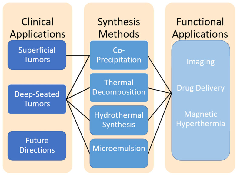
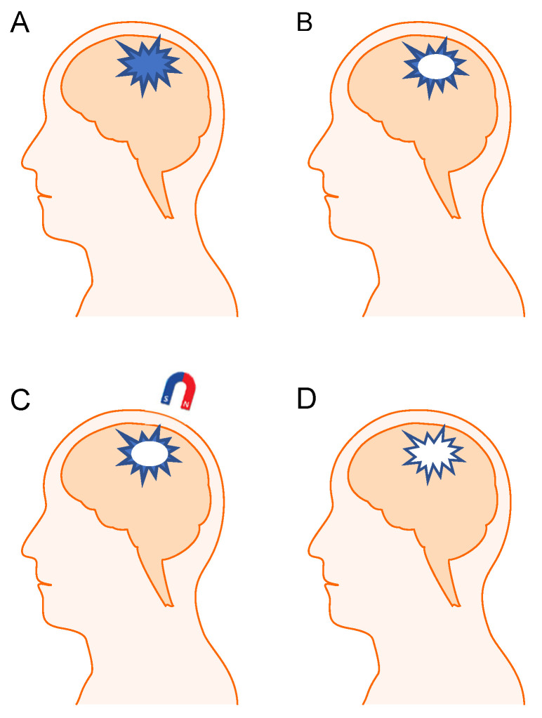
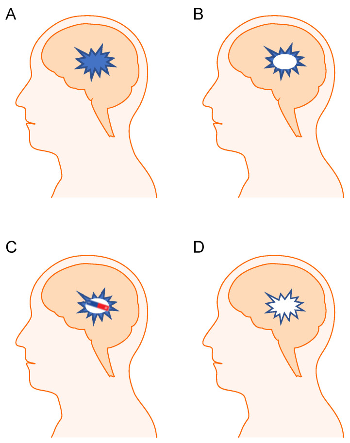
Verification
Claims the paper makes
The claims and statistics of this paper haven’t been checked yet.
Run the check — free, no account needed. It reads the open-access text and checks the sources it cites.
Claims other papers make about this one
Each is checked against this paper’s own text: does it say what the citing paper says it does?
The source backs part of the claim, not all of it — or backs it for a different population, condition or comparison.
In the citing paper
“MNPs have proven to be adaptable, heterogeneous drug delivery vectors that can cross multiple barriers for traditional drug carriers [84].”
From Advances in green-synthesized magnetic nanoparticles for targeted cancer therapy: mechanisms, applications, and future perspectives (2026) · cites this paper as [84]
Experimental — accuracy not yet measured
Self-check
Percentages, sums and reported differences re-computed from the paper’s own numbers.
The self-check needs the paper’s full text.
Citations
Cites
Magnetic iron oxide nanoparticles: synthesis and surface functionalization strategies
Wu, W., He, Q., & Jiang, C. (2008) · Nanoscale Research Letters · doi:10.1007/s11671-008-9174-9 · Cited by 2,300
The blood–brain barrier: Structure, regulation and drug delivery
Wu, D., Chen, Q., Chen, X., Han, F., Chen, Z., & Wang, Y. (2023) · Signal Transduction and Targeted Therapy · doi:10.1038/s41392-023-01481-w · Cited by 1,906 · cited 1×
“…the existence of the BBB allows for excellent protection of the central nervous system from various toxins and pathogens that could be detrimental to maintaining its homeostasis, the BBB causes diminished levels of therapeutics to reach neural cells, resulting in ineffective treatment [110,111].”
Wu, W., Wu, Z., Yu, T., Jiang, C., & Kim, W.-S. (2015) · Science and Technology of Advanced Materials · doi:10.1088/1468-6996/16/2/023501 · Cited by 1,574
Lee, N., & Hyeon, T. (2012) · Chem. Soc. Rev. · doi:10.1039/c1cs15248c · Cited by 995
Synthesis of phase-pure and monodisperse iron oxide nanoparticles by thermal decomposition
Hufschmid, R., Arami, H., Ferguson, R. M., Gonzales, M., Teeman, E., Brush, L. N., Browning, N. D., & Krishnan, K. M. (2015) · Nanoscale · doi:10.1039/c5nr01651g · Cited by 341
Cole, A. J., David, A. E., Wang, J., Galbán, C. J., Hill, H. L., & Yang, V. C. (2011) · Biomaterials · doi:10.1016/j.biomaterials.2010.11.040 · Cited by 308
Polyak, B., Fishbein, I., Chorny, M., Alferiev, I., Williams, D., Yellen, B., Friedman, G., & Levy, R. J. (2008) · Proceedings of the National Academy of Sciences · doi:10.1073/pnas.0708338105 · Cited by 257
Chorny, M., Fishbein, I., Yellen, B. B., Alferiev, I. S., Bakay, M., Ganta, S., Adamo, R., Amiji, M., Friedman, G., & Levy, R. J. (2010) · Proceedings of the National Academy of Sciences · doi:10.1073/pnas.0909506107 · Cited by 197 · cited 2×
“Such a strategy has shown promise in other fields, including using magnetic stents to attract Such a strategy has shown promise in other fi elds, including using magnetic stents to a tt ract therapeutic nanoparticles [177–181].”
Research works printed but not linked: 173
- Ostrom, 2019, CBTRUS Statistical Report: Primary Brain and Other Central Nervous System Tumors Diagnosed in the United States in 2012–2016, Neuro Oncol., 21, v1, 10.1093/neuonc/noz150 · DOI
- Ostrom, 2021, CBTRUS Statistical Report: Primary Brain and Other Central Nervous System Tumors Diagnosed in the United States in 2014–2018, Neuro Oncol., 23, iii1, 10.1093/neuonc/noab200 · DOI
- De Vleeschouwer, S. (2017). Current Standards of Care in Glioblastoma Therapy. Glioblastoma, Codon Publications. · DOI
- Maroon, 1995, Cryoprobe-assisted removal of spinal cord tumors: Technical note, Surg. Neurol., 43, 265, 10.1016/0090-3019(95)80011-5 · DOI
- Chang, 1983, Comparison of postoperative radiotherapy and combined postoperative radiotherapy and chemotherapy in the multidisciplinary management of malignant gliomas. A joint Radiation Therapy Oncology Group and Eastern Cooperative Oncology Group study, Cancer, 52, 997, 10.1002/1097-0142(19830915)52:6<997::AID-CNCR2820520612>3.0.CO;2-2 · DOI
- Gilbert, 2013, Dose-dense temozolomide for newly diagnosed glioblastoma: A randomized phase III clinical trial, J. Clin. Oncol., 31, 4085, 10.1200/JCO.2013.49.6968 · DOI · Paper page
- Balana, 2020, A phase II randomized, multicenter, open-label trial of continuing adjuvant temozolomide beyond 6 cycles in patients with glioblastoma (GEINO 14-01), Neuro Oncol., 22, 1851, 10.1093/neuonc/noaa107 · DOI
- Zanganeh, 2016, Iron oxide nanoparticles inhibit tumour growth by inducing pro-inflammatory macrophage polarization in tumour tissues, Nat. Nanotechnol., 11, 986, 10.1038/nnano.2016.168 · DOI · Paper page
- Ling, 2013, Chemical Design of Biocompatible Iron Oxide Nanoparticles for Medical Applications, Small, 9, 1450, 10.1002/smll.201202111 · DOI
- LaGrow, 2019, Unravelling the growth mechanism of the co-precipitation of iron oxide nanoparticles with the aid of synchrotron X-Ray diffraction in solution, Nanoscale, 11, 6620, 10.1039/C9NR00531E · DOI
- Ahn, 2012, Formation Pathways of Magnetite Nanoparticles by Coprecipitation Method, J. Phys. Chem. C, 116, 6069, 10.1021/jp211843g · DOI
- Khalil, 2015, Co-precipitation in aqueous solution synthesis of magnetite nanoparticles using iron (III) salts as precursors, Arab. J. Chem., 85, 279, 10.1016/j.arabjc.2015.02.008 · DOI
- Benamour, 2022, Synthesis of Fe3O4 Nanoparticles with Different Shapes Through a Co-Precipitation Method and Their Application, JOM, 74, 3531, 10.1007/s11837-022-05380-3 · DOI
- de Souza, T.C., Costa, A.F.S., Vinhas, G.M., and Sarubbo, L.A. (2023). Synthesis of Iron Oxides and Influence on Final Sizes and Distribution in Bacterial Cellulose Applications. Polymers, 15. · DOI
- Essyed, 2019, The effect of basic pH on the elaboration of ZnFe2O4 nanoparticles by co-precipitation method: Structural, magnetic and hyperthermia characterization, J. Magn. Magn. Mater., 478, 239, 10.1016/j.jmmm.2019.01.081 · DOI
- Rechtenbach, 2008, Synthesis and physical characterization of magnetite nanoparticles for biomedical applications, Mater. Chem. Phys., 110, 426, 10.1016/j.matchemphys.2008.02.037 · DOI
- Hauser, 2015, The effects of synthesis method on the physical and chemical properties of dextran coated iron oxide nanoparticles, Mater. Chem. Phys., 160, 177, 10.1016/j.matchemphys.2015.04.022 · DOI
- Basuki, 2014, A block copolymer-stabilized co-precipitation approach to magnetic iron oxide nanoparticles for potential use as MRI contrast agents, Polym. Chem., 5, 2611, 10.1039/C3PY01778H · DOI
- Chen, 2020, Moderate cooling coprecipitation for extremely small iron oxide as a pH dependent T1-MRI contrast agent, Nanoscale, 12, 5521, 10.1039/C9NR10397J · DOI
- Kayal, 2010, Doxorubicin loaded PVA coated iron oxide nanoparticles for targeted drug delivery, Mater. Sci. Eng. C, 30, 484, 10.1016/j.msec.2010.01.006 · DOI
- Predoi, 2008, Costache. Study of iron oxide nanoparticles coated with dextrin obtained by coprecipitation, J. Optoelectron. Adv. Mater., 10, 693
- Ebadi, M., Bullo, S., Buskara, K., Hussein, M.Z., Fakurazi, S., and Pastorin, G. (2020). Release of a liver anticancer drug, sorafenib from its PVA/LDH- and PEG/LDH-coated iron oxide nanoparticles for drug delivery applications. Sci. Rep., 10. · DOI
- Darwish, 2022, Functionalized iron oxide nanoparticles: Synthesis through ultrasonic-assisted co-precipitation and performance as hyperthermic agents for biomedical applications, Heliyon, 8, e09654, 10.1016/j.heliyon.2022.e09654 · DOI
- Darwish, M.S.A., Kim, H., Lee, H., Ryu, C., Lee, J.Y., and Yoon, J. (2019). Synthesis of Magnetic Ferrite Nanoparticles with High Hyperthermia Performance via a Controlled Co-Precipitation Method. Nanomaterials, 9. · DOI
- Ali, 2016, Synthesis, characterization, applications, and challenges of iron oxide nanoparticles, Nanotechnol. Sci. Appl., 9, 49, 10.2147/NSA.S99986 · DOI · Paper page
- Samrot, 2021, A review on synthesis, characterization and potential biological applications of superparamagnetic iron oxide nanoparticles, Curr. Res. Green Sustain. Chem., 4, 100042, 10.1016/j.crgsc.2020.100042 · DOI
- Dadfar, 2019, Iron oxide nanoparticles: Diagnostic, therapeutic and theranostic applications, Adv. Drug Deliv. Rev., 138, 302, 10.1016/j.addr.2019.01.005 · DOI · Paper page
- Kwon, 2011, Formation Mechanisms of Uniform Nanocrystals via Hot-Injection and Heat-Up Methods, Small, 7, 2685, 10.1002/smll.201002022 · DOI
- Turcheniuk, 2013, Recent advances in surface chemistry strategies for the fabrication of functional iron oxide based magnetic nanoparticles, Nanoscale, 5, 10729, 10.1039/c3nr04131j · DOI
- Hao, 2010, Synthesis, Functionalization, and Biomedical Applications of Multifunctional Magnetic Nanoparticles, Adv. Mater., 22, 2729, 10.1002/adma.201000260 · DOI
- Rezaei, 2024, Magnetic Nanoparticles: A Review on Synthesis, Characterization, Functionalization, and Biomedical Applications, Small, 20, 2304848, 10.1002/smll.202304848 · DOI
- Montiel Schneider, M.G., Martín, M.J., Otarola, J., Vakarelska, E., Simeonov, V., Lassalle, V., and Nedyalkova, M. (2022). Biomedical Applications of Iron Oxide Nanoparticles: Current Insights Progress and Perspectives. Pharmaceutics, 14. · DOI
- Meng, Y.Q., Shi, Y.N., Zhu, Y.P., Liu, Y.Q., Gu, L.W., Liu, D.D., Ma, A., Xia, F., Guo, Q.Y., and Xu, C.C. (2024). Recent trends in preparation and biomedical applications of iron oxide nanoparticles. J. Nanobiotechnol., 22. · DOI
- Lu, 2007, Magnetic nanoparticles: Synthesis, protection, functionalization, and application, Angew. Chem. Int. Ed., 46, 1222, 10.1002/anie.200602866 · DOI
- Okeil, 2024, Tuning the Properties of Iron Oxide Nanoparticles in Thermal Decomposition Synthesis: A Comparative Study of the Influence of Temperature, Ligand Length and Ligand Concentration, Part. Part. Syst. Charact., 41, 2400059, 10.1002/ppsc.202400059 · DOI
- Park, 2004, Ultra-large-scale syntheses of monodisperse nanocrystals, Nat. Mater., 3, 891, 10.1038/nmat1251 · DOI · Paper page
- Unni, 2017, Thermal Decomposition Synthesis of Iron Oxide Nanoparticles with Diminished Magnetic Dead Layer by Controlled Addition of Oxygen, ACS Nano, 11, 2284, 10.1021/acsnano.7b00609 · DOI
- Ramimoghadam, 2014, Progress in electrochemical synthesis of magnetic iron oxide nanoparticles, J. Magn. Magn. Mater., 368, 207, 10.1016/j.jmmm.2014.05.015 · DOI · Paper page
- Sodipo, 2016, Recent advances in synthesis and surface modification of superparamagnetic iron oxide nanoparticles with silica, J. Magn. Magn. Mater., 416, 275, 10.1016/j.jmmm.2016.05.019 · DOI · Paper page
- Sathya, 2017, One-step microwave-assisted synthesis of water-dispersible Fe3O4 magnetic nanoclusters for hyperthermia applications, J. Magn. Magn. Mater., 439, 107, 10.1016/j.jmmm.2017.05.018 · DOI
- Aivazoglou, 2017, Microwave-assisted synthesis of iron oxide nanoparticles in biocompatible organic environment, AIP Adv., 8, 048201, 10.1063/1.4994057 · DOI
- Laurent, 2008, Magnetic iron oxide nanoparticles: Synthesis, stabilization, vectorization, physicochemical characterizations, and biological applications, Chem. Rev., 108, 2064, 10.1021/cr068445e · DOI · Paper page
- Bilecka, 2010, Microwave chemistry for inorganic nanomaterials synthesis, Nanoscale, 2, 1358, 10.1039/b9nr00377k · DOI
- 2009, Practical Microwave Synthesis for Organic Chemists.Strategies, Instruments, and Protocols. Edited by C. Oliver Kappe, Doris Dallinger and Shaun Murphree, Angew. Chem. Int. Ed., 48, 2828, 10.1002/anie.200900791 · DOI
- Xiao, 2018, Microwave-assisted synthesis of nanocrystalline binary and ternary metal oxides, Int. Mater. Rev., 63, 341, 10.1080/09506608.2017.1402158 · DOI
- Ozel, 2015, Growth of Iron Oxide Nanoparticles by Hydrothermal Process: Effect of Reaction Parameters on the Nanoparticle Size, J. Supercond. Nov. Magn., 28, 823, 10.1007/s10948-014-2707-9 · DOI
- Ozel, 2015, Growth and characterizations of magnetic nanoparticles under hydrothermal conditions: Reaction time and temperature, J. Magn. Magn. Mater., 373, 213, 10.1016/j.jmmm.2014.02.072 · DOI
- Wegmann, M., and Scharr, M. (2018). Synthesis of Magnetic Iron Oxide Nanoparticles. Precision Medicine, Academic Press. · DOI
- Salvador, M., Gutiérrez, G., Noriega, S., Moyano, A., Blanco-López, M.C., and Matos, M. (2021). Microemulsion Synthesis of Superparamagnetic Nanoparticles for Bioapplications. Int. J. Mol. Sci., 22. · DOI
- Makovec, 2005, The synthesis of spinel–ferrite nanoparticles using precipitation in microemulsions for ferrofluid applications, J. Magn. Magn. Mater., 289, 32, 10.1016/j.jmmm.2004.11.010 · DOI
- Rivas, 2006, Synthesis of monodisperse maghemite nanoparticles by the microemulsion method, Colloids Surf. A Physicochem. Eng. Asp., 288, 44, 10.1016/j.colsurfa.2006.04.027 · DOI
- Darbandi, 2012, Nanoscale size effect on surface spin canting in iron oxide nanoparticles synthesized by the microemulsion method, J. Phys. D Appl. Phys., 45, 195001, 10.1088/0022-3727/45/19/195001 · DOI
- Overcast, 2021, Advanced imaging techniques for neuro-oncologic tumor diagnosis, with an emphasis on PET-MRI imaging of malignant brain tumors, Curr. Oncol. Rep., 23, 34, 10.1007/s11912-021-01020-2 · DOI
- Wagner, 2016, Pathophysiology of gadolinium-associated systemic fibrosis, Am. J. Physiol. Ren. Physiol., 311, F1, 10.1152/ajprenal.00166.2016 · DOI
- Quattrocchi, 2024, Ten years of gadolinium retention and deposition: ESMRMB-GREC looks backward and forward, Eur. Radiol., 34, 600
- Ringler, 2021, Gadolinium retention within multiple rat organs after intra-articular administration of gadolinium-based contrast agents, Skelet. Radiol., 50, 1419, 10.1007/s00256-020-03695-3 · DOI
- McDonald, 2015, Intracranial Gadolinium Deposition after Contrast-enhanced MR Imaging, Radiology, 275, 772, 10.1148/radiol.15150025 · DOI · Paper page
- Akai, 2021, Effects of Gadolinium Deposition in the Brain on Motor or Behavioral Function: A Mouse Model, Radiology, 301, 409, 10.1148/radiol.2021210892 · DOI
- Abujudeh, 2010, Acute adverse reactions to gadopentetate dimeglumine and gadobenate dimeglumine: Experience with 32,659 injections, AJR Am. J. Roentgenol., 194, 430, 10.2214/AJR.09.3099 · DOI
- Davies, J., Marino, M., Smith, A.P.L., Crowder, J.M., Larsen, M., Lowery, L., Castle, J., Hibberd, M.G., and Evans, P.M. (2021). Repeat and single dose administration of gadodiamide to rats to investigate concentration and location of gadolinium and the cell ultrastructure. Sci. Rep., 11. · DOI
- Kasten, A., Grüttner, C., Kühn, J.P., Bader, R., Pasold, J., and Frerich, B. (2014). Comparative in vitro study on magnetic iron oxide nanoparticles for MRI tracking of adipose tissue-derived progenitor cells. PLoS ONE, 9. · DOI
- McDonald, 2022, Neurologic Effects of Gadolinium Retention in the Brain after Gadolinium-based Contrast Agent Administration, Radiology, 302, 676, 10.1148/radiol.210559 · DOI
- Strzeminska, 2020, Long-Term Evaluation of Gadolinium Retention in Rat Brain After Single Injection of a Clinically Relevant Dose of Gadolinium-Based Contrast Agents, Investig. Radiol., 55, 138, 10.1097/RLI.0000000000000623 · DOI
- Marcus, M., Karni, M., Baranes, K., Levy, I., Alon, N., Margel, S., and Shefi, O. (2016). Iron oxide nanoparticles for neuronal cell applications: Uptake study and magnetic manipulations. J. Nanobiotechnol., 14. · DOI
- Cao, 2016, Effect of Renal Function on Gadolinium-Related Signal Increases on Unenhanced T1-Weighted Brain Magnetic Resonance Imaging, Investig. Radiol., 51, 677, 10.1097/RLI.0000000000000294 · DOI
- Foo, 2022, Alcohol solution strength preference predicts compulsive-like drinking behavior in rats, Alcohol. Clin. Exp. Res., 46, 1710, 10.1111/acer.14910 · DOI
- Roch, 1999, Theory of proton relaxation induced by superparamagnetic particles, J. Chem. Phys., 110, 5403, 10.1063/1.478435 · DOI
- Song, 2007, Labeling efficacy of superparamagnetic iron oxide nanoparticles to human neural stem cells: Comparison of ferumoxides, monocrystalline iron oxide, cross-linked iron oxide (CLIO)-NH2 and tat-CLIO, Korean J. Radiol., 8, 365, 10.3348/kjr.2007.8.5.365 · DOI
- Irrsack, 2021, Effects of Local Administration of Iron Oxide Nanoparticles in the Prefrontal Cortex, Striatum, and Hippocampus of Rats, Neurotox. Res., 39, 2056, 10.1007/s12640-021-00432-z · DOI
- Chen, 2015, Specific detection of CD133-positive tumor cells with iron oxide nanoparticles labeling using noninvasive molecular magnetic resonance imaging, Int. J. Nanomed., 10, 6997
- Unterweger, 2017, Non-immunogenic dextran-coated superparamagnetic iron oxide nanoparticles: A biocompatible, size-tunable contrast agent for magnetic resonance imaging, Int. J. Nanomed., 12, 5223, 10.2147/IJN.S138108 · DOI
- Hachani, R., Birchall, M.A., Lowdell, M.W., Kasparis, G., Tung, L.D., Manshian, B.B., Soenen, S.J., Gsell, W., Himmelreich, U., and Gharagouzloo, C.A. (2017). Assessing cell-nanoparticle interactions by high content imaging of biocompatible iron oxide nanoparticles as potential contrast agents for magnetic resonance imaging. Sci. Rep., 7. · DOI
- Calero, M., Chiappi, M., Lazaro-Carrillo, A., Rodríguez, M.J., Chichón, F.J., Crosbie-Staunton, K., Prina-Mello, A., Volkov, Y., Villanueva, A., and Carrascosa, J.L. (2015). Characterization of interaction of magnetic nanoparticles with breast cancer cells. J. Nanobiotechnol., 13. · DOI
- Coricovac, D.E., Moacă, E.A., Pinzaru, I., Cîtu, C., Soica, C., Mihali, C.V., Păcurariu, C., Tutelyan, V.A., Tsatsakis, A., and Dehelean, C.A. (2017). Biocompatible Colloidal Suspensions Based on Magnetic Iron Oxide Nanoparticles: Synthesis, Characterization and Toxicological Profile. Front. Pharmacol., 8. · DOI
- Neuwelt, 2007, The potential of ferumoxytol nanoparticle magnetic resonance imaging, perfusion, and angiography in central nervous system malignancy: A pilot study, Neurosurgery, 60, 601, 10.1227/01.NEU.0000255350.71700.37 · DOI · Paper page
- Landry, 2005, Pharmacokinetic Study of Ferumoxytol: A New Iron Replacement Therapy in Normal Subjects and Hemodialysis Patients, Am. J. Nephrol., 25, 400, 10.1159/000087212 · DOI
- Ersoy, 2007, Biochemical safety profiles of gadolinium-based extracellular contrast agents and nephrogenic systemic fibrosis, J. Magn. Reson. Imaging, 26, 1190, 10.1002/jmri.21135 · DOI
- Uchiyama, 2015, Ultrasmall cationic superparamagnetic iron oxide nanoparticles as nontoxic and efficient MRI contrast agent and magnetic-targeting tool, Int. J. Nanomed., 10, 4731
- Ali, 2020, Efficient Labeling Of Mesenchymal Stem Cells For High Sensitivity Long-Term MRI Monitoring In Live Mice Brains, Int. J. Nanomed., 15, 97, 10.2147/IJN.S211205 · DOI
- Wu, 2022, Ultrasmall iron oxide nanoparticles cause significant toxicity by specifically inducing acute oxidative stress to multiple organs, Part. Fibre Toxicol., 19, 24, 10.1186/s12989-022-00465-y · DOI
- Kenzaoui, 2012, Evaluation of uptake and transport of ultrasmall superparamagnetic iron oxide nanoparticles by human brain-derived endothelial cells, Nanomedicine, 7, 39, 10.2217/nnm.11.85 · DOI
- Petters, 2016, Lysosomal iron liberation is responsible for the vulnerability of brain microglial cells to iron oxide nanoparticles: Comparison with neurons and astrocytes, Nanotoxicology, 10, 332, 10.3109/17435390.2015.1071445 · DOI · Paper page
- Bourrinet, 2006, Preclinical safety and pharmacokinetic profile of ferumoxtran-10, an ultrasmall superparamagnetic iron oxide magnetic resonance contrast agent, Investig. Radiol., 41, 313, 10.1097/01.rli.0000197669.80475.dd · DOI
- Zou, 2015, Iron nanoparticles significantly affect the in vitro and in vivo expression of Id genes, Chem. Res. Toxicol., 28, 373, 10.1021/tx500333q · DOI
- Královec, K., Melounková, L., Slováková, M., Mannová, N., Sedlák, M., Bartáček, J., and Havelek, R. (2020). Disruption of Cell Adhesion and Cytoskeletal Networks by Thiol-Functionalized Silica-Coated Iron Oxide Nanoparticles. Int. J. Mol. Sci., 21. · DOI
- Mai, 2017, Magnetic nanoparticles: Reactive oxygen species generation and potential therapeutic applications, J. Nanoparticle Res., 19, 253, 10.1007/s11051-017-3943-2 · DOI
- Ahamed, 2012, Selective killing of cancer cells by iron oxide nanoparticles mediated through reactive oxygen species via p53 pathway, J. Nanoparticle Res., 15, 1225, 10.1007/s11051-012-1225-6 · DOI
- Shi, Y., Wang, J., Liu, J., Lin, G., Xie, F., Pang, X., Pei, Y., Cheng, Y., Zhang, Y., and Lin, Z. (2020). Oxidative stress-driven DR5 upregulation restores TRAIL/Apo2L sensitivity induced by iron oxide nanoparticles in colorectal cancer. Biomaterials, 233. · DOI
- Shokrollahi, 2023, Evaluation of the effect of iron oxide nanoparticles functionalized by glucose and conjugated with coumarin (Fe3O4@Glu-Coumarin NPs) on the expression of CASP8, CASP9, p53, mTOR1, and MAPK1 genes in liver cancer cell line, Gene Rep., 33, 101818, 10.1016/j.genrep.2023.101818 · DOI
- Kanda, 2016, Gadolinium deposition in the brain, Magn. Reson. Imaging, 34, 1346, 10.1016/j.mri.2016.08.024 · DOI
- Habermeyer, J., Boyken, J., Harrer, J., Canneva, F., Ratz, V., Moceri, S., Admard, J., Casadei, N., Jost, G., and Bäuerle, T. (2020). Comprehensive phenotyping revealed transient startle response reduction and histopathological gadolinium localization to perineuronal nets after gadodiamide administration in rats. Sci. Rep., 10. · DOI
- Irrsack, 2023, Local Administrations of Iron Oxide Nanoparticles in the Prefrontal Cortex and Caudate Putamen of Rats Do Not Compromise Working Memory and Motor Activity, Neurotox. Res., 42, 6, 10.1007/s12640-023-00684-x · DOI
- Taboada, 2007, Relaxometric and Magnetic Characterization of Ultrasmall Iron Oxide Nanoparticles with High Magnetization. Evaluation as Potential T1 Magnetic Resonance Imaging Contrast Agents for Molecular Imaging, Langmuir, 23, 4583, 10.1021/la063415s · DOI
- Xiao, 2016, MRI contrast agents: Classification and application (Review), Int. J. Mol. Med., 38, 1319, 10.3892/ijmm.2016.2744 · DOI
- Edelman, 2024, Dark Blood Contrast-Enhanced Brain MRI Using Echo-uT(1)RESS, J. Magn. Reson. Imaging, 60, 789, 10.1002/jmri.29124 · DOI
- Tao, C., Zheng, Q., An, L., He, M., Lin, J., Tian, Q., and Yang, S. (2019). T₁-Weight Magnetic Resonance Imaging Performances of Iron Oxide Nanoparticles Modified with a Natural Protein Macromolecule and an Artificial Macromolecule. Nanomaterials, 9. · DOI
- Wei, 2021, Single-nanometer iron oxide nanoparticles as tissue-permeable MRI contrast agents, Proc. Natl. Acad. Sci. USA, 118, e2102340118, 10.1073/pnas.2102340118 · DOI
- Yang, L., Afshari, M.J., Ge, J., Kou, D., Chen, L., Zhou, D., Li, C., Wu, S., Zhang, L., and Zeng, J. (2022). Functionalized Ultrasmall Iron Oxide Nanoparticles for T(1)-Weighted Magnetic Resonance Imaging of Tumor Hypoxia. Molecules, 27. · DOI
- Christoforidis, 2009, High resolution ultra high field magnetic resonance imaging of glioma microvascularity and hypoxia using ultra-small particles of iron oxide, Investig. Radiol., 44, 375, 10.1097/RLI.0b013e3181a8afea · DOI
- Toth, 2017, Current and potential imaging applications of ferumoxytol for magnetic resonance imaging, Kidney Int., 92, 47, 10.1016/j.kint.2016.12.037 · DOI · Paper page
- Rad, 2007, Quantification of superparamagnetic iron oxide (SPIO)-labeled cells using MRI, J. Magn. Reson. Imaging, 26, 366, 10.1002/jmri.20978 · DOI
- Zhang, 2009, Superparamagnetic iron oxide nanoparticle-labeled cells as an effective vehicle for tracking the GFP gene marker using magnetic resonance imaging, Cytotherapy, 11, 43, 10.1080/14653240802420243 · DOI
- Kratz, 2016, Labeling of mesenchymal stem cells for MRI with single-cell sensitivity, Int. J. Nanomed., 11, 1517
- Jarzyna, 2012, Tumor angiogenesis phenotyping by nanoparticle-facilitated magnetic resonance and near-infrared fluorescence molecular imaging, Neoplasia, 14, 964, 10.1593/neo.121148 · DOI
- Melemenidis, 2015, Molecular Magnetic Resonance Imaging of Angiogenesis In Vivo using Polyvalent Cyclic RGD-Iron Oxide Microparticle Conjugates, Theranostics, 5, 515, 10.7150/thno.10319 · DOI
- Daneman, R., and Prat, A. (2015). The blood-brain barrier. Cold Spring Harb. Perspect. Biol., 7. · DOI · Paper page
- Aksenov, D.P., and Doubovikov, E.D. (2025). Diffusion constraints in neuroprotection: Implications for clinical trials. Front. Pharmacol., 16. · DOI
- Enteshari Najafabadi, R., Kazemipour, N., Esmaeili, A., Beheshti, S., and Nazifi, S. (2018). Using superparamagnetic iron oxide nanoparticles to enhance bioavailability of quercetin in the intact rat brain. BMC Pharmacol. Toxicol., 19. · DOI
- Shi, 2016, Optimizing superparamagnetic iron oxide nanoparticles as drug carriers using an in vitro blood-brain barrier model, Int. J. Nanomed., 11, 5371, 10.2147/IJN.S108333 · DOI
- Shevtsov, 2014, Superparamagnetic iron oxide nanoparticles conjugated with epidermal growth factor (SPION-EGF) for targeting brain tumors, Int. J. Nanomed., 9, 273, 10.2147/IJN.S55118 · DOI
- Salehnia, 2019, Synthesis and characterisation of iron oxide nanoparticles conjugated with epidermal growth factor receptor (EGFR) monoclonal antibody as MRI contrast agent for cancer detection, IET Nanobiotechnol., 13, 400, 10.1049/iet-nbt.2018.5285 · DOI
- Shevtsov, 2014, Tumor targeting using magnetic nanoparticle Hsp70 conjugate in a model of C6 glioma, Neuro Oncol., 16, 38, 10.1093/neuonc/not141 · DOI
- Bach, 2004, Novel RGD lipopeptides for the targeting of liposomes to integrin-expressing endothelial and melanoma cells, Protein Eng. Des. Sel., 17, 433, 10.1093/protein/gzh055 · DOI
- Wu, 2018, Magnetic resonance imaging of tumor angiogenesis using dual-targeting RGD10-NGR9 ultrasmall superparamagnetic iron oxide nanoparticles, Clin. Transl. Oncol., 20, 599, 10.1007/s12094-017-1753-8 · DOI
- Neshastehriz, 2022, Folate functionalized gold-coated magnetic nanoparticles effect in combined electroporation and radiation treatment of HPV-positive oropharyngeal cancer, Med. Oncol., 39, 196, 10.1007/s12032-022-01780-2 · DOI
- Liang, 2016, Doxorubicin-modified magnetic nanoparticles as a drug delivery system for magnetic resonance imaging-monitoring magnet-enhancing tumor chemotherapy, Int. J. Nanomed., 11, 2021
- Patel, 2023, Development of a Personalised Device for Systemic Magnetic Drug Targeting to Brain Tumours, Nanotheranostics, 7, 102, 10.7150/ntno.76559 · DOI
- Sun, 2008, Tumor-targeted drug delivery and MRI contrast enhancement by chlorotoxin-conjugated iron oxide nanoparticles, Nanomedicine, 3, 495, 10.2217/17435889.3.4.495 · DOI
- Dutta, 2007, Adsorbed proteins influence the biological activity and molecular targeting of nanomaterials, Toxicol. Sci., 100, 303, 10.1093/toxsci/kfm217 · DOI
- Moghimi, 2001, Long-circulating and target-specific nanoparticles: Theory to practice, Pharmacol. Rev., 53, 283, 10.1016/S0031-6997(24)01494-7 · DOI
- Nagayama, 2007, Time-dependent changes in opsonin amount associated on nanoparticles alter their hepatic uptake characteristics, Int. J. Pharm., 342, 215, 10.1016/j.ijpharm.2007.04.036 · DOI
- Camner, 2002, Experimental and calculated parameters on particle phagocytosis by alveolar macrophages, J. Appl. Physiol., 92, 2608, 10.1152/japplphysiol.01067.2001 · DOI
- Chen, 2010, Reducing non-specific binding and uptake of nanoparticles and improving cell targeting with an antifouling PEO-b-PgammaMPS copolymer coating, Biomaterials, 31, 5397, 10.1016/j.biomaterials.2010.03.036 · DOI
- Foy, 2010, Optical imaging and magnetic field targeting of magnetic nanoparticles in tumors, ACS Nano, 4, 5217, 10.1021/nn101427t · DOI
- Aggarwal, 2009, Nanoparticle interaction with plasma proteins as it relates to particle biodistribution, biocompatibility and therapeutic efficacy, Adv. Drug Deliv. Rev., 61, 428, 10.1016/j.addr.2009.03.009 · DOI · Paper page
- Ikeda, 2018, Activatable fluorescence imaging of macrophages in atherosclerotic plaques using iron oxide nanoparticles conjugated with indocyanine green, Atherosclerosis, 275, 1, 10.1016/j.atherosclerosis.2018.05.028 · DOI
- Wang, 2018, Iron oxide-carbon core-shell nanoparticles for dual-modal imaging-guided photothermal therapy, J. Control. Release, 289, 70, 10.1016/j.jconrel.2018.09.022 · DOI
- Gneveckow, 2004, Description and characterization of the novel hyperthermia- and thermoablation-system for clinical magnetic fluid hyperthermia, Med. Phys., 31, 1444, 10.1118/1.1748629 · DOI
- Pu, 2000, Apoptosis induced by hyperthermia in human glioblastoma cell line and murine glioblastoma, Chin. J. Cancer Res., 12, 257, 10.1007/BF02983501 · DOI
- Rothe, 2007, Intracranial Thermotherapy using Magnetic Nanoparticles Combined with External Beam Radiotherapy: Results of a Feasibility Study on Patients with Glioblastoma Multiforme, J. Neuro-Oncol., 81, 53, 10.1007/s11060-006-9195-0 · DOI
- Usov, 2019, Iron Oxide Nanoparticles for Magnetic Hyperthermia, SPIN, 09, 1940001, 10.1142/S2010324719400010 · DOI
- Etheridge, 2014, Accounting for biological aggregation in heating and imaging of magnetic nanoparticles, Technology, 02, 214, 10.1142/S2339547814500198 · DOI · Paper page
- Nemati, 2016, Enhanced Magnetic Hyperthermia in Iron Oxide Nano-Octopods: Size and Anisotropy Effects, J. Phys. Chem. C, 120, 8370, 10.1021/acs.jpcc.6b01426 · DOI
- Pericchi, 2015, Optimization of synthesis and peptization steps to obtain iron oxide nanoparticles with high energy dissipation rates, J. Magn. Magn. Mater., 394, 361, 10.1016/j.jmmm.2015.06.076 · DOI
- Nemati, 2018, Improving the Heating Efficiency of Iron Oxide Nanoparticles by Tuning Their Shape and Size, J. Phys. Chem. C, 122, 2367, 10.1021/acs.jpcc.7b10528 · DOI
- Singh, 2023, A threefold increase in SAR performance for magnetic hyperthermia by compositional tuning in zinc-substituted iron oxide superparamagnetic nanoparticles with superior biocompatibility, J. Alloys Compd., 968, 171868, 10.1016/j.jallcom.2023.171868 · DOI
- Myrovali, E., Maniotis, N., Makridis, A., Terzopoulou, A., Ntomprougkidis, V., Simeonidis, K., Sakellari, D., Kalogirou, O., Samaras, T., and Salikhov, R. (2016). Arrangement at the nanoscale: Effect on magnetic particle hyperthermia. Sci. Rep., 6. · DOI
- Mehdaoui, 2010, Large specific absorption rates in the magnetic hyperthermia properties of metallic iron nanocubes, J. Magn. Magn. Mater., 322, L49, 10.1016/j.jmmm.2010.05.012 · DOI
- Fatima, H., Charinpanitkul, T., and Kim, K.-S. (2021). Fundamentals to Apply Magnetic Nanoparticles for Hyperthermia Therapy. Nanomaterials, 11. · DOI
- Kahil, H., Faramawy, A., El-Sayed, H., and Abdel-Sattar, A. (2021). Magnetic Properties and SAR for Gadolinium-Doped Iron Oxide Nanoparticles Prepared by Hydrothermal Method. Crystals, 11. · DOI
- Balakrishnan, 2020, Exploiting Unique Alignment of Cobalt Ferrite Nanoparticles, Mild Hyperthermia, and Controlled Intrinsic Cobalt Toxicity for Cancer Therapy, Adv. Mater., 32, e2003712, 10.1002/adma.202003712 · DOI
- Khurshid, 2015, Anisotropy effects in magnetic hyperthermia: A comparison between spherical and cubic exchange-coupled FeO/Fe3O4 nanoparticles, J. Appl. Phys., 117, 17A337, 10.1063/1.4919250 · DOI
- Thanh, 2025, Iron oxide nanoparticles synthesized by coprecipitation method: Impacts of zinc doping and surface functionalization by polyvinylpyrrolidone on the magnetic properties and heating efficiency, Ceram. Int., 51, 1448, 10.1016/j.ceramint.2024.11.118 · DOI
- Oroskhani, N., Amini, S.M., Shirvalilou, S., Khodaie, M., and Mahdavi, S.R. (2024). Anti-Proliferative Activity of Poloxamer Cobalt Ferrite Nanoparticles against Human Prostate Cancer (DU-145) Cells: In-Vitro Study. IET Nanobiotechnol., 2024. · DOI
- Yang, Y., Wang, F., Zheng, K., Deng, L., Yang, L., Zhang, N., Xu, C., Ran, H., Wang, Z., and Wang, Z. (2017). Injectable PLGA/Fe3O4 implants carrying cisplatin for synergistic magnetic hyperthermal ablation of rabbit VX2 tumor. PLoS ONE, 12. · DOI
- Shirvalilou, 2021, Magnetic Hyperthermia as an adjuvant cancer therapy in combination with radiotherapy versus radiotherapy alone for recurrent/progressive glioblastoma: A systematic review, J. Neuro-Oncol., 152, 419, 10.1007/s11060-021-03729-3 · DOI
- Kampinga, 2001, Hyperthermic radiosensitization: Mode of action and clinical relevance, Int. J. Radiat. Biol., 77, 399, 10.1080/09553000010024687 · DOI
- Krawczyk, 2011, Mild hyperthermia inhibits homologous recombination, induces BRCA2 degradation, and sensitizes cancer cells to poly (ADP-ribose) polymerase-1 inhibition, Proc. Natl. Acad. Sci. USA, 108, 9851, 10.1073/pnas.1101053108 · DOI
- Karakatsanis, 2017, Superparamagnetic iron oxide nanoparticles as the sole method for sentinel node biopsy detection in patients with breast cancer, Br. J. Surg., 104, 1675, 10.1002/bjs.10606 · DOI
- Lee, D., Sohn, J., and Kirichenko, A. (2022). Quantifying Liver Heterogeneity via R2*-MRI with Super-Paramagnetic Iron Oxide Nanoparticles (SPION) to Characterize Liver Function and Tumor. Cancers, 14. · DOI
- Colombo, 2012, Biological applications of magnetic nanoparticles, Chem. Soc. Rev., 41, 4306, 10.1039/c2cs15337h · DOI
- Leiner, 2003, Multicenter phase-II trial of safety and efficacy of NC100150 for steady-state contrast-enhanced peripheral magnetic resonance angiography, Eur. Radiol., 13, 1620, 10.1007/s00330-002-1791-6 · DOI
- Weishaupt, 2000, Equilibrium-Phase MR Angiography of the Aortoiliac and Renal Arteries Using a Blood Pool Contrast Agent, Am. J. Roentgenol., 175, 189, 10.2214/ajr.175.1.1750189 · DOI
- Lassalle, 2017, Magnetic iron oxide nanoparticles as novel and efficient tools for atherosclerosis diagnosis, Biomed. Pharmacother., 93, 1098, 10.1016/j.biopha.2017.07.012 · DOI
- Taupitz, 2004, Phase I Clinical Evaluation of Citrate-coated Monocrystalline Very Small Superparamagnetic Iron Oxide Particles as a New Contrast Medium for Magnetic Resonance Imaging, Investig. Radiol., 39, 394, 10.1097/01.rli.0000129472.45832.b0 · DOI
- Wagner, 2011, Coronary MR angiography using citrate-coated very small superparamagnetic iron oxide particles as blood-pool contrast agent: Initial experience in humans, J. Magn. Reson. Imaging, 34, 816, 10.1002/jmri.22683 · DOI
- Teshome, 2016, Use of a Magnetic Tracer for Sentinel Lymph Node Detection in Early-Stage Breast Cancer Patients: A Meta-analysis, Ann. Surg. Oncol., 23, 1508, 10.1245/s10434-016-5135-1 · DOI
- Engberink, 2010, Magnetic Resonance Imaging of Monocytes Labeled with Ultrasmall Superparamagnetic Particles of Iron Oxide Using Magnetoelectroporation in an Animal Model of Multiple Sclerosis, Mol. Imaging, 9, 268, 10.2310/7290.2010.00016 · DOI
- Blumenstein, 2021, Newer formulations of intravenous iron: A review of their chemistry and key safety aspects–hypersensitivity, hypophosphatemia, and cardiovascular safety, Expert. Opin. Drug Saf., 20, 757, 10.1080/14740338.2021.1912010 · DOI
- Tassa, 2011, Dextran-Coated Iron Oxide Nanoparticles: A Versatile Platform for Targeted Molecular Imaging, Molecular Diagnostics, and Therapy, Acc. Chem. Res., 44, 842, 10.1021/ar200084x · DOI · Paper page
- Jin, 2014, Superparamagnetic iron oxide nanoparticles for MR imaging and therapy: Design considerations and clinical applications, Curr. Opin. Pharmacol., 18, 18, 10.1016/j.coph.2014.08.002 · DOI · Paper page
- Ahmad, 2015, Magnetic nanoparticles: In vivo cancer diagnosis and therapy, Int. J. Pharm., 493, 313, 10.1016/j.ijpharm.2015.07.059 · DOI
- Nardecchia, S., Sánchez-Moreno, P., de Vicente, J., Marchal, J.A., and Boulaiz, H. (2019). Clinical Trials of Thermosensitive Nanomaterials: An Overview. Nanomaterials, 9. · DOI
- Lapusan, 2024, Advancing MRI with magnetic nanoparticles: A comprehensive review of translational research and clinical trials, Nanoscale Adv., 6, 2234, 10.1039/D3NA01064C · DOI
- Egea-Benavente, D., Ovejero, J.G., Morales, M.D.P., and Barber, D.F. (2021). Understanding MNPs Behaviour in Response to AMF in Biological Milieus and the Effects at the Cellular Level: Implications for a Rational Design That Drives Magnetic Hyperthermia Therapy toward Clinical Implementation. Cancers, 13. · DOI
- Grauer, 2016, RTHP-22. inflammatory response after modified nanotherm and radiotherapy of recurrent glioblastoma, Jpn. Soc. Neuro-Oncol., 18, vi178, 10.1093/neuonc/now212.748 · DOI
- Marcus, M., Smith, A., Maswadeh, A., Shemesh, Z., Zak, I., Motiei, M., Schori, H., Margel, S., Sharoni, A., and Shefi, O. (2018). Magnetic Targeting of Growth Factors Using Iron Oxide Nanoparticles. Nanomaterials, 8. · DOI
- Yellen, 2005, Targeted drug delivery to magnetic implants for therapeutic applications, J. Magn. Magn. Mater., 293, 647, 10.1016/j.jmmm.2005.01.083 · DOI
- Doubovikov, 2024, Brain Tissue Oxygen Dynamics Under Localised Hypoxia in the Awake State and the Physical Neuroprotective Effects of General Anaesthesia, Oxygen Transport to Tissue XLV, Volume 1463, 35, 10.1007/978-3-031-67458-7_6 · DOI
- Pislaru, 2006, Magnetic forces enable rapid endothelialization of synthetic vascular grafts, Circulation, 114, I314, 10.1161/CIRCULATIONAHA.105.001446 · DOI
- Pislaru, 2006, Magnetically Targeted Endothelial Cell Localization in Stented Vessels, J. Am. Coll. Cardiol., 48, 1839, 10.1016/j.jacc.2006.06.069 · DOI
- Consigny, 1999, Use of Endothelial Cells Containing Superparamagnetic Microspheres to Improve Endothelial Cell Delivery to Arterial Surfaces after Angioplasty, J. Vasc. Interv. Radiol., 10, 155, 10.1016/S1051-0443(99)70458-6 · DOI
- Westphal, 2003, A phase 3 trial of local chemotherapy with biodegradable carmustine (BCNU) wafers (Gliadel wafers) in patients with primary malignant glioma, Neuro Oncol., 5, 79, 10.1093/neuonc/5.2.79 · DOI
Cited by
Sorted by citations
El-khawaga, A. M., Youssef, O. I., El-dydamoni, O. A., Ragab, W. M., & Mohammed, R. A. E. (2026) · European Journal of Medical Research · doi:10.1186/s40001-025-03804-9 · Cited by 14
Metadata
- Authors
- Varalli, L., Berlet, R., Abenojar, E., McDaid, J., Gascoigne, D. A., Bailes, J., & Aksenov, D. P.
- Venue
- Pharmaceutics
- Publisher
- MDPI AG
- Published
- 2025-04-09
- Volume
- 17
- Issue
- 4
- Pages
- 499
- ISSN
- 1999-4923
- PMC
- PMC12030199
- Cited by
- 23
- References
- 8
- Language
- en
About these checks
Registry, retraction, Cites and statistics results are computed, not judged, with one exception: a printed reference that no index lists is read once by a model before it counts as a citation error. Claims are extracted by a model and kept only when their quote appears verbatim in the text; each cited source is then checked, with a model’s help, for whether it says what the claim relies on. Those verdicts can be wrong — read the claim’s quote and the cited source before relying on one.
Statistics and arithmetic are recomputed, not judged.
Confidence bands are shown only for fields calibrated on our annotated test set. None is calibrated yet, so no band is shown.
What counts as a claim ›How we measure confidence ›How we check this ›
Are you an author, or do you see a problem with this page? Request a review
