

A Chest‐Conformable, Wireless Electro‐Mechanical E‐Tattoo for Measuring Multiple Cardiac Time Intervals
Open accessThe DOI matches its Crossref record; 42 of the 42 references it cites were found in a registry or an index; 23 of the 54 sources its claims cite were checked for support.
Abstract
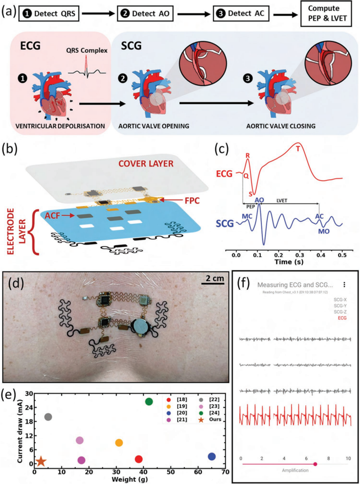
Abstract Cardiovascular diseases are the leading cause of death globally. Noninvasive, accurate, and continuous cardiovascular monitoring can enable the preemptive detection of heart diseases and timely intervention to prevent serious cardiac complications. However, unobtrusive, ambulatory, and comprehensive cardiac monitoring is still a challenge as conventional electronics are rigid, heavy, or consume too much power for long‐term measurement. This work presents a thin (200 µm), stretchable (20%), lightweight (2.5 g), wireless, and low‐power (<3 mW) cardiac monitoring device that conforms to the human chest like a temporary tattoo sticker, correspondingly known as an e‐tattoo. This chest e‐tattoo features dual‐mode electro‐mechanical sensing—bio‐electric cardiac signals via electrocardiography and mechanical cardiac rhythm via seismocardiography. A unique peripheral synchronization strategy between the two sensors enables the measurement of systolic time intervals like the pre‐ejection period and the left ventricular ejection time with high accuracy (error = −0.44 ± 8.74 ms) while consuming very low power. The e‐tattoo is validated against clinically approved gold‐standard instruments on five human subjects. The good wearability and low power consumption of this e‐tattoo permit 24‐h continuous ambulatory monitoring.
Overview
CoChat Summary
TL;DR
This paper presents a chest-conformable, wireless electro-mechanical e-tattoo that combines ECG and SCG for measuring cardiac time intervals such as PEP and LVET. It reports low-power, stretchable, reusable operation and validates STI measurement against a clinical monitor in five healthy subjects, with 24-h ambulatory feasibility in one subject.
Key findings
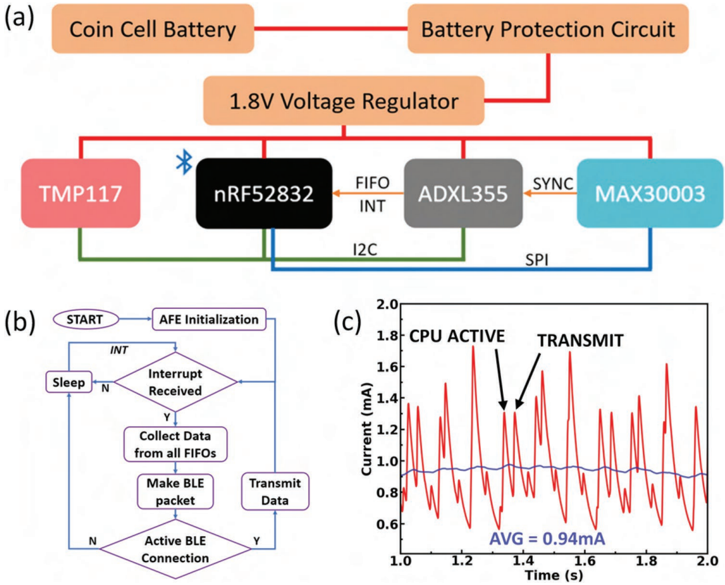
- The e-tattoo is thin (200 µm), stretchable (20%), lightweight (2.5 g including battery), wireless, and low-power (<3 mW; average current draw 940 µA), with a CR1220 coin cell supporting up to 40 h of operation.
- Dual ECG/SCG sensing with peripheral synchronization enabled LVET measurement against NICOM with a mean difference of −0.44 ± 8.74 ms and Pearson correlation of 0.955; HR differences were 0.07 ± 1.21 bpm during static poses and 0.02 ± 1.24 bpm during cycling.
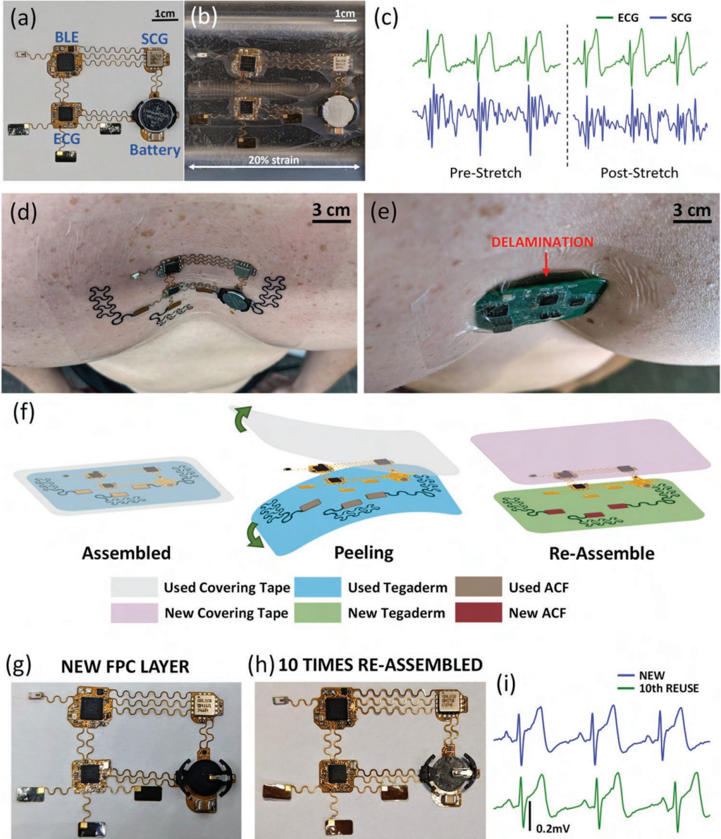
- The e-tattoo showed no significant signal degradation after 20% off-chest stretching and reapplication, and no perceptible damage after ten cycles of electrode-layer reuse.
- In posture transitions, statistically significant changes were observed in PEP (p < 0.05, paired t-test) and LVET (p < 0.005, paired t-test) from supine to upright/standing.
Methods
- Device design: a flexible printed circuit with serpentine interconnects, graphite polyurethane dry ECG electrodes on Tegaderm, an ADXL355 accelerometer for SCG, a MAX30003 ECG front end, and an nRF52832 BLE CPU; ECG and SCG were sampled at 125 Hz with hardware synchronization using an external 32.768 kHz crystal and ECG-generated sync pulses.
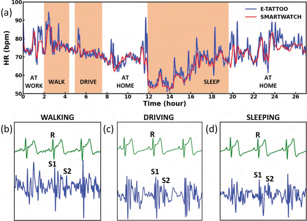
- Human testing: IRB-approved study at UT Austin; five healthy males aged 20–28 for static posture and incremental cycling experiments compared against NICOM, plus one 27-year-old male for >24 h ambulatory monitoring compared against a Samsung Galaxy Watch 3 for HR.
- Signal processing: ECG band-pass filtered at 0.5–40 Hz, SCG at 1–40 Hz, R-peak extraction, beat-by-beat SCG segmentation, signal quality indexing (λ = 25, threshold = 0.5), and AO/AC peak tracking to compute HR, PEP, and LVET.
Limitations
- The paper states that SCG was corrupted by motion during cycling, so STIs were computed only during rest segments after exercise; long-term LVET/PEP could not be compared against a reference device because NICOM is non-portable and no wearable LVET/PEP device was available.
- Evident from the text: the primary validation sample was small (five healthy males aged 20–28), the long-term ambulatory study used only one participant, and participants with known cardiovascular disease were excluded, limiting generalizability to patients, women, older adults, and higher-BMI or diseased populations.
- The paper states “Research data are not shared,” which limits independent verification; the text also notes that SCG is unusable during significant motion, restricting continuous STI extraction to restful segments.
Model-written summary. Check it against the paper before relying on it.
Integrity signals
Registry
Cites
Statistics
Figures







Verification
Claims the paper makes
13 Valid · 6 Not valid · 10 Not checked · 59 Own results and interpretations
The sources with a verdict support this claim; the rest weren’t checked yet, have no full text we could read, or their verdicts aren’t rated yet.
In the paper
“This form factor coupled with innovative firmware for sensor interfacing and transmission of data allows us to achieve one of the lowest total device weights (2.5 g including battery) and current draws (940 µ A) among wearable cardiac sensors developed in prior work as illustrated via Ashby plot in Figure 1e. [18-24]”
What the sources say
- Supportedcites [18] M. Etemadi, O. T. Inan, J. A. Heller, S. Hersek, L. Klein, S. Roy, IEEE Trans. Biomed. Circuits Syst. 2015 , 10 , 280. source
The citation is part of a group identifying prior wearable cardiac sensors used for comparison, not a source for the citing authors’ e-tattoo measurement of 940 μA. The Abstract and Introduction describe this paper’s low-power wearable patch for cardiac monitoring, so it qualifies as a prior device in that comparison. (LLM, conf=0.88)
- Not checked — no full text of the sourcecites [19] S. Preejith, R. Dhinesh, J. Joseph, M. Sivaprakasam, in 2016 38th Annual Int. Conf. of the IEEE Engineering in Medicine and Biology Society (EMBC) , IEEE, Piscataway, NJ 2016 , pp. 623-626. source
- Supportedcites [20] B. Semiz, A. M. Carek, J. C. Johnson, S. Ahmad, J. A. Heller, F. G. Vicente, S. Caron, C. W. Hogue, M. Etemadi, O. T. Inan, IEEE J. Biomed. Health Inf. 2020 , 25 , 1572. source
The Wearable Patch section (page unavailable) describes a chest-mounted patch that records ECG and seismocardiogram signals, confirming that this paper is an example of prior wearable cardiac-sensor work. The 940 μA draw is stated for the citing authors’ e-tattoo, not attributed to this source. (LLM, conf=0.84)
- Supportedcites [21] Y .-S. Hsu, T.-Y . Chen, D. Wu, C.-M. Lin, J.-N. Juang, W.-T. Liu, J. Clin. Sleep Med. 2020 , 16 , 1149. source
The Introduction describes a wireless single-lead ECG monitoring patch, and the Discussion describes it as a wearable patch suitable for multiday examination. This supports citing the paper as an example of prior wearable cardiac-sensor work. The 940 μA current draw is presented in the citing sentence as the e-tattoo authors’ own result, not as a measurement reported by this source. (LLM, conf=0.85)
- Not checked — no full text of the sourcecites [22] J. Li, J. Zhang, Y . Jiang, C. Ren, R. Guo, Y . Ma, Y . Qin, in 2022 44th Annual Int. Conf. of the IEEE Engineering in Medicine and Biology Society (EMBC) , IEEE, Piscataway, NJ 2022 , pp. 4312-4315. source
- Not checked — no full text of the sourcecites [23] Y . Zhong, Y . Pan, L. Zhang, K.-T. Cheng, in 2016 38th Annual Int. Conf. of the IEEE Engineering in Medicine and Biology Society (EMBC) , IEEE, Piscataway, NJ 2016 , pp. 603-606. source
- Not matchedcites [24] S. Lee, B. Grundlehner, R. G. van der Westen, S. Polito, C. Van Hoof, in 2019 41st Annual Int. Conf. of the IEEE Engineering in Medicine and Biology Society (EMBC) , IEEE, Piscataway, NJ 2019 , pp. 1290-1293.
Couldn’t be matched to a paper in Crossref, so it can’t be checked.
plain- Asserted: passes — The authors put it forward as true.
- Checkable: passes — It can be checked against evidence.
- Load-bearing: unclear — It is unclear whether the paper’s conclusions depend on it.
- Based: passes — The paper gives a reason why it holds.
- Supported
The cited sources support only part of this claim — read each source’s verdict below.
In the paper
“In healthy subjects with low body mass index (BMI), this technique can provide accurate measures of STIs; however, in broader populations with cardiovascular diseases and in patients with higher BMI, ICG has been shown to have high error compared to reference standard invasive measures [14,15]”
What the sources say
- Partly supportedcites [14] A. Quain, M. Hoyer, G. Ephrem, W. A. Kay, Int. J. Cardiol. Congenital Heart Dis. 2021 , 6 , 100287. sourceExperimental — accuracy not yet measured
The Results report poor agreement between noninvasive NICOM cardiac-output measurements and invasive right-heart catheterization in adults with Fontan palliation (ICC approximately 0.40). The paper studies thoracic bioreactance, not ICG, and this finding does not establish high error across broader cardiovascular populations or in patients with higher BMI. No page numbers were available in our copy. (LLM, conf=0.84)
- Partly supportedcites [15] A. Rali, T . Buechler, B. Van-Gotten, E. Burgen, J. Chen, Z. Shah, N. Haglund, A. Sauer, J. Am. Coll. Cardiol. 2019 , 73 , 819. sourceExperimental — accuracy not yet measured
The Results and Conclusion (p. 1) report that NICOM cardiac-output and cardiac-index measurements did not correlate with invasive Fick or thermodilution measurements in patients with acute decompensated heart failure. The paper identifies NICOM as a bioreactance technology, not ICG, and gives only a mean BMI; it does not establish high error for ICG or a specific finding in patients with higher BMI. (LLM, conf=0.85)
plainstates an association- Asserted: passes — The authors put it forward as true.
- Checkable: passes — It can be checked against evidence.
- Load-bearing: passes — The paper’s conclusions depend on it.
- Based: passes — The paper gives a reason why it holds.
- Partly supported
The cited sources that could be checked couldn’t confirm or dispute this claim.
In the paper
“In healthy subjects with low body mass index (BMI), this technique can provide accurate measures of STIs; however, in broader populations with cardiovascular diseases and in patients with higher BMI, ICG has been shown to have high error compared to reference standard invasive measures [14,15]”
What the sources say
- Could not verifycites [14] A. Quain, M. Hoyer, G. Ephrem, W. A. Kay, Int. J. Cardiol. Congenital Heart Dis. 2021 , 6 , 100287. sourceExperimental — accuracy not yet measured
The Methods and Results compare NICOM-derived cardiac output with invasive measurements in adults with Fontan palliation; they do not assess ICG measures of systolic time intervals in healthy, low-BMI subjects. Searches for healthy subjects, low BMI, ICG, and STIs found no passage addressing the attributed claim. Page numbers are unavailable in our copy. (LLM, conf=0.75)
- Could not verifycites [15] A. Rali, T . Buechler, B. Van-Gotten, E. Burgen, J. Chen, Z. Shah, N. Haglund, A. Sauer, J. Am. Coll. Cardiol. 2019 , 73 , 819. sourceExperimental — accuracy not yet measured
The Methods and Results (p. 1) concern NICOM measurements of cardiac output and cardiac index in patients with acute decompensated heart failure. Searches for healthy subjects, low BMI, ICG, and systolic time intervals found no passage assessing whether ICG accurately measures STIs in that population. (LLM, conf=0.76)
plainstates an association- Asserted: passes — The authors put it forward as true.
- Checkable: passes — It can be checked against evidence.
- Load-bearing: unclear — It is unclear whether the paper’s conclusions depend on it.
- Based: passes — The paper gives a reason why it holds.
- Could not verify
The cited sources support only part of this claim — read each source’s verdict below.
In the paper
“Systolic timing intervals (STI) which measure the duration of cardiac phases have been demonstrated as viable alternatives to these parameters for assessing cardiac performance.”
What the sources say
- Partly supportedcites [9] S. Hassan, P. Turner, Postgrad. Med. J. 1983 , 59 , 423. sourceExperimental — accuracy not yet measured
The Introduction defines STI as durations of systolic phases, and the Validation section reports correlations with stroke volume, cardiac output, and ejection fraction, including accuracy equivalent to ejection fraction in patients with coronary artery disease. But the Conclusions call STI a helpful measure of ventricular performance that is “not competitive with other invasive or non-invasive tests”; the paper does not establish STI as a general alternative to all three parameters. Page numbers were not supplied in our copy. (LLM, conf=0.87)
boosted- Asserted: passes — The authors put it forward as true.
- Checkable: passes — It can be checked against evidence.
- Load-bearing: passes — The paper’s conclusions depend on it.
- Based: passes — The paper gives a reason why it holds.
- Partly supported
Every source the paper cites for this claim was checked and supports it.
In the paper
“The PEP and LVET offer a convenient non-invasive method to assess LV performance.”
What the sources say
- Supportedcites [9] S. Hassan, P. Turner, Postgrad. Med. J. 1983 , 59 , 423. source
The Validation of systolic time intervals section reports that both PEP and LVET correlate significantly with left ventricular ejection fraction. The Place of systolic time intervals among other noninvasive techniques section describes the measurement as requiring relatively cheap equipment and being feasible in almost all subjects without discomfort. Page numbers were not available in our copy. (LLM, conf=0.92)
plain- Asserted: passes — The authors put it forward as true.
- Checkable: passes — It can be checked against evidence.
- Load-bearing: passes — The paper’s conclusions depend on it.
- Based: passes — The paper gives a reason why it holds.
- Supported
42 more claims · 6 with their cited sources checked
Create a free account to see all 88 claims and to verify the 1 reference still open.
- It may state two separate claims as one. 27
- Probably not a substantial claim. 21
Self-check
Percentages, sums and reported differences re-computed from the paper’s own numbers.
Each contradiction between two passages listed here was confirmed by an independent check.
Rated minor: it would not touch a result — a value no result rests on, a p-value that stays on the same side of the significance level, or a conflict that is clerical, resolved elsewhere in the paper, or outside its own findings.
In the paper
§ 3. Conclusion · p. 10
“STIs are important indicators of many different cardiovascular diseases but are only measurable through invasive means in the current medical practice.”
§ 2. Results and Discussion · p. 6
“Subjects simultaneously wore our e-tattoo along with a non-invasive cardiac output monitor (NICOM) (Starling Fluid Management Monitoring System, Baxter). The NICOM was used to measure multiple hemodynamic parameters including HR and LVET”
Citations
Cites
Monitoring of Vital Signs with Flexible and Wearable Medical Devices
Khan, Y., Ostfeld, A. E., Lochner, C. M., Pierre, A., & Arias, A. C. (2016) · Advanced Materials · doi:10.1002/adma.201504366 · Cited by 1,408
Systolic Time Intervals in Heart Failure in Man
WEISSLER, A. M., HARRIS, W. S., & SCHOENFELD, C. D. (1968) · Circulation · doi:10.1161/01.cir.37.2.149 · Cited by 1,217
Ballistocardiography and Seismocardiography: A Review of Recent Advances
Inan, O. T., Migeotte, P.-F., Park, K.-S., Etemadi, M., Tavakolian, K., Casanella, R., Zanetti, J., Tank, J., Funtova, I., Prisk, G. K., et al. (2015) · IEEE Journal of Biomedical and Health Informatics · doi:10.1109/jbhi.2014.2361732 · Cited by 775
Self-citationA critical review of the systolic time intervals.
Lewis, R. P., Rittogers, S. E., Froester, W. F., & Boudoulas, H. (1977) · Circulation · doi:10.1161/01.cir.56.2.146 · Cited by 624
Hong, Y. J., Jeong, H., Cho, K. W., Lu, N., & Kim, D. (2019) · Advanced Functional Materials · doi:10.1002/adfm.201808247 · Cited by 559
Self-citationDesign of metal interconnects for stretchable electronic circuits
Gonzalez, M., Axisa, F., Bulcke, M. V., Brosteaux, D., Vandevelde, B., & Vanfleteren, J. (2008) · Microelectronics Reliability · doi:10.1016/j.microrel.2008.03.025 · Cited by 405
“Cut‐and‐Paste” Manufacture of Multiparametric Epidermal Sensor Systems
Yang, S., Chen, Y., Nicolini, L., Pasupathy, P., Sacks, J., Su, B., Yang, R., Sanchez, D., Chang, Y., Wang, P., et al. (2015) · Advanced Materials · doi:10.1002/adma.201502386 · Cited by 304
Self-citationNovel dry electrodes for ECG monitoring
Gruetzmann, A., Hansen, S., & Müller, J. (2007) · Physiological Measurement · doi:10.1088/0967-3334/28/11/005 · Cited by 248
AHMED, S. S., LEVINSON, G. E., SCHWARTZ, C. J., & ETTINGER, P. O. (1972) · Circulation · doi:10.1161/01.cir.46.3.559 · Cited by 241
Ha, T., Tran, J., Liu, S., Jang, H., Jeong, H., Mitbander, R., Huh, H., Qiu, Y., Duong, J., Wang, R. L., et al. (2019) · Advanced Science · doi:10.1002/advs.201900290 · Cited by 220
Self-citation
Research works printed but not linked: 10
- Bruss Z. S., 2022, Physiology, Stroke Volume, StatPearls
- Preejith S., 2016, 2016 38th Annual Int. Conf. of the IEEE Engineering in Medicine and Biology Society (EMBC), 623
- Li J., 2022, 2022 44th Annual Int. Conf. of the IEEE Engineering in Medicine and Biology Society (EMBC), 4312
- Zhong Y., 2016, 2016 38th Annual Int. Conf. of the IEEE Engineering in Medicine and Biology Society (EMBC), 603
- Lee S., 2019, 2019 41st Annual Int. Conf. of the IEEE Engineering in Medicine and Biology Society (EMBC), 1290
- Di Rienzo M., 2014, 2014 36th Annual Int. Conf. of the IEEE Engineering in Medicine and Biology Society, 6089
- Tadi M. J., 2015, Sixth Int. Conf. on Graphic and Image Processing (ICGIP 2014), 571
- Hansen I. H., 2015, 2015 37th Annual Int. Conf. of the IEEE Engineering in Medicine and Biology Society (EMBC), 330
- Gazi A. H., 2021, 43rd Annual Int. Conf. of the IEEE Engineering in Medicine and Biology Society (EMBC), 1444
- Yang Y.‐F., 2019, Proc. of Int. Conf. on Biomedical and Health Informatics, 363
Cited by
E-Tattoos: Toward Functional but Imperceptible Interfacing with Human Skin
Li, H., Tan, P., Rao, Y., Bhattacharya, S., Wang, Z., Kim, S., Gangopadhyay, S., Shi, H., Jankovic, M., Huh, H., et al. (2024) · Chemical Reviews · doi:10.1021/acs.chemrev.3c00626 · Cited by 151
Self-citationHo, D. H., Hu, C., Li, L., & Bartlett, M. D. (2024) · Nature Electronics · doi:10.1038/s41928-024-01268-z · Cited by 78
“For soft electronics—which are of use in soft robotics 4–6 , wearable electronics 7–10 and flexible displays 11–13 —integration approaches and multilevel architectures are undeveloped compared with their rigid counterparts.”
An Ultra‐Conductive and Patternable 40 nm‐Thick Polymer Film for Reliable Emotion Recognition
Du, X., Wang, H., Wang, Y., Cao, Z., Yang, L., Shi, X., Zhang, X. H., He, C., Gu, X., & Liu, N. (2024) · Advanced Materials · doi:10.1002/adma.202403411 · Cited by 50
Chen, S., Ouyang, Q., Meng, X., Yang, Y., Li, C., Miao, X.-B., Chen, Z., Zhao, G., Lei, Y., Ghanem, B., et al. (2025) · Science Advances · doi:10.1126/sciadv.adv2406 · Cited by 58
“Simultaneous measurements of cardiac mechanical activities, such as seismocardiogram (SCG) and gyrocardiogram (GCG) that track the translational and rotational components of cardiac-induced chest vibrations, can offer critical information that ECG alone cannot capture ( 20 – 22 ).”
Atomically thin bioelectronics
Kireev, D., Kutagulla, S., Hong, J., Wilson, M. N., Ramezani, M., Kuzum, D., Ahn, J., & Akinwande, D. (2024) · Nature Reviews Materials · doi:10.1038/s41578-024-00728-4 · Cited by 50
Metadata
- Authors
- Bhattacharya, S., Nikbakht, M., Alden, A., Tan, P., Wang, J., Alhalimi, T. A., Kim, S., Wang, P., Tanaka, H., Tandon, A., Coyle, E. F., Inan, O. T., & Lu, N.
- Affiliations
- The University of Texas at Austin (US)
- Georgia Institute of Technology (US)
- Stretch Med Inc. Austin TX 78750 USA
- Cleveland Clinic (US)
- Texas Materials Institute (US)
- Corresponding author
- Lu, Nanshu
- ORCID
- Venue
- Advanced Electronic Materials
- Publisher
- Wiley
- Published
- 2023-04-18
- Volume
- 9
- Issue
- 9
- ISSN
- 2199-160X (ISSN-L)
- OpenAlex
- W4366245263
- Cited by
- 42
- References
- 42
- Citations by year
- 2026: 8 · 2025: 17 · 2024: 10 · 2023: 7
- FWCI
- 3.31
- Type
- journal article
- Language
- en
- Open access
- gold
- doi.org
- Locations
- Advanced Electronic Materials (publishedVersion, cc-by)
- DOAJ (DOAJ: Directory of Open Access Journals) (submittedVersion, cc-by-sa)
- Funders
- National Science Foundation (STTR‐1938254, ASCENT‐2133106)
- Office of Naval Research Global (N00014‐20‐1‐2112)
- Topics
- Physical Sciences › Engineering › Biomedical Engineering › Non-Invasive Vital Sign Monitoring
- Physical Sciences › Engineering › Biomedical Engineering › Advanced Sensor and Energy Harvesting Materials
- Physical Sciences › Engineering › Biomedical Engineering › Wireless Body Area Networks
- Keywords
- stretchable electronics, electrocardiogram, seismocardiography, cardiac time intervals, systolic time intervals, pre-ejection period, left ventricular ejection time, low-power sensors, sensor synchronization, ambulatory monitoring, wearable health monitoring
About these checks
Registry, retraction, Cites and statistics results are computed, not judged, with one exception: a printed reference that no index lists is read once by a model before it counts as a citation error. Claims are extracted by a model and kept only when their quote appears verbatim in the text; each cited source is then checked, with a model’s help, for whether it says what the claim relies on. Those verdicts can be wrong — read the claim’s quote and the cited source before relying on one.
The claim verdicts this report counts are experimental: their accuracy is not yet measured.
A language model drew the claims from the paper on 2026-10-12, under version 3 of the claim definition (model deepseek-v4.1-flash, prompt extract-v3).
A second model labelled each claim on 2026-10-12 (model jev-1.13-20260917, definition revision gold-guideline-v0.2).
Cited sources are checked with a language model’s help (gpt-6-sol).
Statistics and arithmetic are recomputed, not judged.
Confidence bands are shown only for fields calibrated on our annotated test set. None is calibrated yet, so no band is shown.
What counts as a claim ›How we measure confidence ›How we check this ›
Are you an author, or do you see a problem with this page? Request a review
Data sources
Bibliographic data: Semantic Scholar, OpenAlex, Crossref.
Contains information from the Semantic Scholar Academic Graph Datasets, made available under the ODC Attribution License (ODC-BY 1.0).
Semantic Scholar’s data is described in: Kinney, R., et al. (2023). The Semantic Scholar Open Data Platform. arXiv:2301.10140.
