

The DOI matches its Crossref record; 103 of the 105 references it cites were found in a registry or an index; 1 of the 81 sources its claims cite were checked for support.
Immunology of Wound Healing
Open accessAbstract
PURPOSE OF REVIEW: Chronic wounds are a tremendous burden on the healthcare system and lead to significant patient morbidity and mortality. Normal cutaneous wound healing occurs through an intricate and delicate interplay between the immune system, keratinocytes, and dermal cells. Each cell type contributes signals that drive the normal phases of wound healing: hemostasis, inflammation, proliferation, and remodeling. This paper reviews how various immunological cell types and signaling molecules influence the way wounds develop, persist, and heal. RECENT FINDINGS: Concurrent with the achievement of hemostasis, neutrophils are the first cells to migrate to the wound bed, brought in by pro-inflammatory signals including IL-8. Their apoptosis and engulfment by macrophages (efferocytosis) provides a key signal to the local immune milieu, including macrophages, to transition to an anti-inflammatory, pro-repair state, where angiogenesis occurs and granulation tissue is laid down. Myofibroblasts, activated through contractile forces and signaling molecules, then drive remodeling, where granulation tissue becomes scar. Unchecked inflammation at this stage can result in abnormal scar formation. SUMMARY: Although the derangement of immune signals at any stage can result in impaired wound healing, recent research has shown that the key transition point lies between the inflammatory and the proliferative phases. This review summarizes the events that facilitate this transition and discusses how this process can be disrupted, leading to chronic, non-healing wounds.
Overview
AI snapshot
Written from the paper’s full text.
TL;DR
This review summarizes immune mechanisms across hemostasis, inflammation, proliferation, and remodeling, arguing that the critical transition from the inflammatory to the proliferative phase often fails in chronic wounds. It highlights neutrophil apoptosis/efferocytosis and macrophage polarization as key drivers of repair.
Key findings
- Chronic wounds in the USA afflict approximately 6.5 million individuals and cost the healthcare system over 25 billion dollars annually; their mortality effect has been reported as comparable to cancer.
- Normal wound healing is divided into hemostasis (minutes to hours), inflammation (days 1–3), proliferation and repair (days 4–21), and remodeling (days 21–365); the key inflammatory-to-proliferative transition occurs around days 2–5 and often fails in chronic wounds.
- Neutrophils are early responders recruited by IL-8/CXCL8; their apoptosis and macrophage engulfment (efferocytosis) signal a shift to anti-inflammatory, pro-repair macrophages. Persistent neutrophils, NETs, proteases/MMPs, reactive oxygen species, and elevated CXCL8 contribute to chronic, non-healing wounds.
- Anti-inflammatory macrophages promote angiogenesis, granulation tissue, and re-epithelialization via VEGF, TGF-β, PDGF, EGF, KGF, and other signals; impaired pro- to anti-inflammatory macrophage transition—seen with iron overload, hyperglycemia, and hypoxia—stalls healing. Mature scars regain only 80% of initial tensile strength.
Methods
- Narrative review of the literature; no primary data, sample, or statistical analysis are reported.
- Synthesizes immune cell and signaling pathways across the four phases of wound healing, focusing on the inflammatory-to-proliferative transition and chronic wounds.
- Draws on cited animal models, in vitro studies, and human wound observations; no systematic search or meta-analysis is described.
Limitations
- The paper states no limitations.
- Evident from the text: it is a narrative review without systematic search, inclusion criteria, or quality appraisal.
- Evident from the text: many mechanistic conclusions rely on mouse models and in vitro findings, with limited direct human chronic wound data; no original experiments or quantitative synthesis are presented.
Model-written summary. Check it against the paper before relying on it.
Integrity signals
Registry
Cites
Statistics
Figures
Verification
Claims the paper makes
51 Not checked yet · 8 Own interpretation · 6 No basis given · 1 Partly supported · accuracy not yet measured
In the paper
“In impaired models of wound healing, such as diabetes, where infection risk is higher, neutrophils are clearly required [28].”
§ Immunology of Wound Healing > Four Stages of Wound Healing > Inflammation
What the sources say
- Not checked yetcites [28] Wilgus TA, Roy S, McDaniel JC. Neutrophils and wound repair: positive actions and negative reactions. Adv Wound Care. 2013;2(7):379–88. https://doi.org/10.1089/wound.2012.0383 . source
boosteddirect- Asserted: passes — The authors put it forward as true.
- Checkable: passes — It can be checked against evidence.
- Load-bearing: unclear — It is unclear whether the paper’s conclusions depend on it.
- Based: passes — The paper gives a reason why it holds.
- Rests on an assumption: Findings from mouse models of wound healing can be generalized to human wound healing and chronic wounds.
In the paper
“Their influx is mediated by a number of chemical signals, including IL-8, or CXCL8, as mentioned above, and neutrophils have over 30 different receptors that mitigate their migration and activation response [24].”
§ Immunology of Wound Healing > Four Stages of Wound Healing > Inflammation
What the sources say
- Not checked yetcites [24] Futosi K, Fodor S, Mocsai A. Neutrophil cell surface receptors and their intracellular signal transduction pathways. Int Immunopharmacol. 2013;17(3):638–50. https://doi.org/10.1016/j.intimp.2013.06.034 . source
plaindirect- Asserted: passes — The authors put it forward as true.
- Checkable: passes — It can be checked against evidence.
- Load-bearing: unclear — It is unclear whether the paper’s conclusions depend on it.
- Based: passes — The paper gives a reason why it holds.
None of the sources cited for this claim have been checked yet.
In the paper
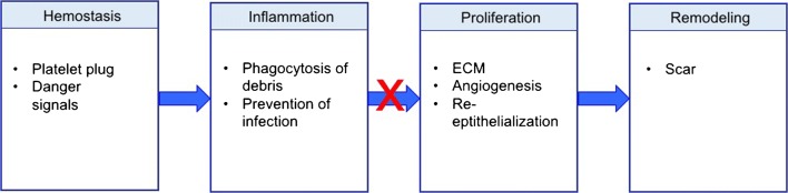
“normal wound healing can generally be broken down into four over-lapping but distinct steps (Fig. 1). These steps include hemostasis (minutes to hours after injury), inflammation (days 1–3), proliferation and repair (days 4–21), and lastly, wound remodeling (days 21–365) [8, 9].”
§ Immunology of Wound Healing > Introduction
What the sources say
- Not checked yetcites [8] Morton LM, Phillips TJ. Wound healing and treating wounds: differential diagnosis and evaluation of chronic wounds. J Am Acad Dermatol. 2016;74(4):589–605; quiz −6. https://doi.org/10.1016/j.jaad.2015.08.068 . source
- Not checked yetcites [9] Reinke JM, Sorg H. Wound repair and regeneration. Eur Surg Res. 2012;49(1):35–43. https://doi.org/10.1159/000339613 . source
plain- Asserted: passes — The authors put it forward as true.
- Checkable: passes — It can be checked against evidence.
- Load-bearing: passes — The paper’s conclusions depend on it.
- Based: passes — The paper gives a reason why it holds.
In the paper
“chronic wounds demonstrate significantly increased levels of the potent neutrophil chemoattractant, CXCL8 [58].”
§ Immunology of Wound Healing > Four Stages of Wound Healing > Inflammation
What the sources say
- Not checked yetcites [58] Fivenson DP, Faria DT, Nickoloff BJ, Poverini PJ, Kunkel S, Burdick M, et al. Chemokine and inflammatory cytokine changes during chronic wound healing. Wound Repair Regen. 1997;5(4):310–22. https://doi.org/10.1046/j.1524-475X.1997.50405.x . source
boostedcorrelational- Asserted: passes — The authors put it forward as true.
- Checkable: passes — It can be checked against evidence.
- Load-bearing: passes — The paper’s conclusions depend on it.
- Based: passes — The paper gives a reason why it holds.
The paper states this without a source or data behind it here, so there is nothing to check it against. The quote shows it is in the paper.
In the paper
“Platelet activation not only results in hemostasis, but also in the release of growth factors such as platelet-derived growth factor (PDGF) as well as immune mediators that are responsible for activation of the immune system and transition to the inflammatory phase of wound healing.”
§ Immunology of Wound Healing > Four Stages of Wound Healing > Hemostasis
plaindirect- Asserted: passes — The authors put it forward as true.
- Checkable: passes — It can be checked against evidence.
- Load-bearing: unclear — It is unclear whether the paper’s conclusions depend on it.
- Based: passes — The paper gives a reason why it holds.
None of the sources cited for this claim have been checked yet.
In the paper
“mouse models of wound healing have shown that in non-aged, non-impaired models, neutrophil depletion does not negatively affect wound healing as profoundly as macrophage deletion [25–27].”
§ Immunology of Wound Healing > Four Stages of Wound Healing > Inflammation
What the sources say
- Not checked yetcites [25] Leibovich SJ, Ross R. The role of the macrophage in wound repair. A study with hydrocortisone and antimacrophage serum. Am J Pathol. 1975;78(1):71–100.
- Not checked yetcites [26] Pinhal-Enfield G, Ramanathan M, Hasko G, Vogel SN, Salzman AL, Boons GJ, et al. An angiogenic switch in macrophages involving synergy between Toll-like receptors 2, 4, 7, and 9 and adenosine A(2A) receptors. Am J Pathol. 2003;163(2):711–21. https://doi.org/10.1016/s0002-9440(10)63698-x . source
- Not checked yetcites [27] Simpson DM, Ross R. The neutrophilic leukocyte in wound repair a study with antineutrophil serum. J Clin Invest. 1972;51(8):2009–23. https://doi.org/10.1172/jci107007 . source
plaindirect- Asserted: passes — The authors put it forward as true.
- Checkable: passes — It can be checked against evidence.
- Load-bearing: unclear — It is unclear whether the paper’s conclusions depend on it.
- Based: passes — The paper gives a reason why it holds.
- Rests on an assumption: Findings from mouse models of wound healing can be generalized to human wound healing and chronic wounds.
A conclusion the paper draws from its own results — it cites no other source for it, so there is none to check. The quote shows it is in the paper.
In the paper
“New research has illustrated that chronic wounds fail to shift from the inflammatory to the proliferative phase of wound healing”
§ Immunology of Wound Healing > Introduction
boosted- Asserted: passes — The authors put it forward as true.
- Checkable: passes — It can be checked against evidence.
- Load-bearing: passes — The paper’s conclusions depend on it.
- Based: passes — The paper gives a reason why it holds.
A conclusion the paper draws from its own results — it cites no other source for it, so there is none to check. The quote shows it is in the paper.
In the paper
“recent research has shown that the key transition point lies between the inflammatory and the proliferative phases.”
- Also stated in: § Conclusions · plain
“Recent research has shown that the key transition point in wound healing lies between the inflammatory and the proliferative phases.”
§ Immunology of Wound Healing > Abstract > Summary
plain- Asserted: passes — The authors put it forward as true.
- Checkable: passes — It can be checked against evidence.
- Load-bearing: passes — The paper’s conclusions depend on it.
- Based: unclear — It is unclear why the paper says it holds.
- Also stated in: § Conclusions · plain
None of the sources cited for this claim have been checked yet.
In the paper
“the immune system has been found to play a substantial role due to its impact on several repair mechanisms [6, 7].”
§ Immunology of Wound Healing > Introduction
What the sources say
- Not checked yetcites [6] MA S, MJ N. The innate immune system in acute and chronic wounds. Adv Wound Care. 2016;5(2):65–78. https://doi.org/10.1089/wound.2014.0608 . source
- Not checked yetcites [7] Strbo N, Yin N, Stojadinovic O. Innate and adaptive immune responses in wound epithelialization. Adv Wound Care. 2014;3(7):492–501. https://doi.org/10.1089/wound.2012.0435 . source
plaindirect- Asserted: passes — The authors put it forward as true.
- Checkable: passes — It can be checked against evidence.
- Load-bearing: passes — The paper’s conclusions depend on it.
- Based: passes — The paper gives a reason why it holds.
The paper states this without a source or data behind it here, so there is nothing to check it against. The quote shows it is in the paper.
In the paper
“Myofibroblasts, activated through contractile forces and signaling molecules, then drive remodeling, where granulation tissue becomes scar.”
§ Immunology of Wound Healing > Abstract > Recent Findings
plaindirect- Asserted: passes — The authors put it forward as true.
- Checkable: passes — It can be checked against evidence.
- Load-bearing: passes — The paper’s conclusions depend on it.
- Based: unclear — It is unclear why the paper says it holds.
- It may state two separate claims as one. 15
The cited sources support only part of this claim — read each source’s verdict below.
In the paper
“The inflammatory phase overlaps considerably with initial hemostasis, occurring during the first 72 h after tissue injury [16].”
§ Immunology of Wound Healing > Four Stages of Wound Healing > Inflammation
What the sources say
- Partly supportedcites [16] Eming SA, Martin P, Tomic-Canic M. Wound repair and regeneration: mechanisms, signaling, and translation. Sci Transl Med. 2014;6(265):265sr6. https://doi.org/10.1126/scitranslmed.3009337 . sourceExperimental — accuracy not yet measured
The ‘Inflammatory response’ section says that acute wound-healing phases largely overlap and that the early inflammatory response occurs in parallel with hemostasis. It does not specify that the inflammatory phase occurs during the first 72 hours after injury; searches for that timing found no supporting passage in our copy. Page numbers were unavailable. (LLM, conf=0.85)
plaincorrelational- Asserted: passes — The authors put it forward as true.
- Checkable: passes — It can be checked against evidence.
- Load-bearing: unclear — It is unclear whether the paper’s conclusions depend on it.
- Based: passes — The paper gives a reason why it holds.
It may state two separate claims as one.
- Partly supported
In the paper
“Concurrent with the achievement of hemostasis, neutrophils are the first cells to migrate to the wound bed, brought in by pro-inflammatory signals including IL-8.”
§ Immunology of Wound Healing > Abstract > Recent Findings
What the sources say
- Not checked yetcites [31] Baggiolini M, Loetscher P, Moser B. Interleukin-8 and the chemokine family. Int J Immunopharmacol. 1995;17(2):103–8. source
plaindirect- Asserted: passes — The authors put it forward as true.
- Checkable: passes — It can be checked against evidence.
- Load-bearing: unclear — It is unclear whether the paper’s conclusions depend on it.
- Based: passes — The paper gives a reason why it holds.
It may state two separate claims as one.
None of the sources cited for this claim have been checked yet.
In the paper
“Neutrophils represent the most abundant inflammatory cells to infiltrate a new wound and function mainly to remove debris and prevent infection [22, 23].”
§ Immunology of Wound Healing > Four Stages of Wound Healing > Inflammation
What the sources say
- Not checked yetcites [22] Engelhardt E, Toksoy A, Goebeler M, Debus S, Brocker EB, Gillitzer R. Chemokines IL-8, GROalpha, MCP-1, IP-10, and Mig are sequentially and differentially expressed during phase-specific infiltration of leukocyte subsets in human wound healing. Am J Pathol. 1998;153(6):1849–60. source
- Not checked yetcites [23] de Oliveira S, Rosowski EE, Huttenlocher A. Neutrophil migration in infection and wound repair: going forward in reverse. Nat Rev Immunol. 2016;16(6):378–91. https://doi.org/10.1038/nri.2016.49 . source
plaindirect- Asserted: passes — The authors put it forward as true.
- Checkable: passes — It can be checked against evidence.
- Load-bearing: unclear — It is unclear whether the paper’s conclusions depend on it.
- Based: passes — The paper gives a reason why it holds.
It may state two separate claims as one.
In the paper
“NETs have also been detected in excess in diabetic foot wounds and have been shown to delay healing, and inhibition of NETosis and NET function in mouse models of delayed wound healing improves outcomes [59•].”
§ Immunology of Wound Healing > Four Stages of Wound Healing > Inflammation
What the sources say
- Not checked yetcites [59•]
plaindirect- Asserted: passes — The authors put it forward as true.
- Checkable: passes — It can be checked against evidence.
- Load-bearing: passes — The paper’s conclusions depend on it.
- Based: passes — The paper gives a reason why it holds.
It may state two separate claims as one.
- Rests on an assumption: Findings from mouse models of wound healing can be generalized to human wound healing and chronic wounds.
None of the sources cited for this claim have been checked yet.
In the paper
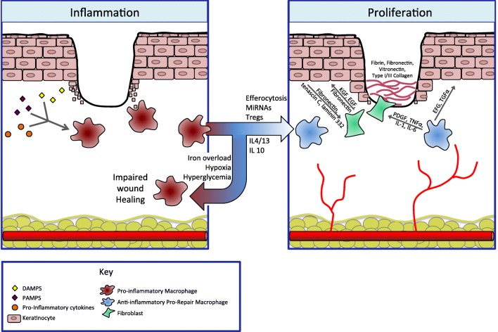
“Macrophages play a critical role in wound healing, and their roles in angiogenesis, fibroplasia, cell proliferation, and transition out of the inflammatory phase is clear (Fig. 2) [25–27, 60].”
§ Immunology of Wound Healing > Four Stages of Wound Healing > Inflammation
What the sources say
- Not checked yetcites [25] Leibovich SJ, Ross R. The role of the macrophage in wound repair. A study with hydrocortisone and antimacrophage serum. Am J Pathol. 1975;78(1):71–100.
- Not checked yetcites [26] Pinhal-Enfield G, Ramanathan M, Hasko G, Vogel SN, Salzman AL, Boons GJ, et al. An angiogenic switch in macrophages involving synergy between Toll-like receptors 2, 4, 7, and 9 and adenosine A(2A) receptors. Am J Pathol. 2003;163(2):711–21. https://doi.org/10.1016/s0002-9440(10)63698-x . source
- Not checked yetcites [27] Simpson DM, Ross R. The neutrophilic leukocyte in wound repair a study with antineutrophil serum. J Clin Invest. 1972;51(8):2009–23. https://doi.org/10.1172/jci107007 . source
- Not checked yetcites [60] Koh TJ, DiPietro LA. Inflammation and wound healing: the role of the macrophage. Expert Rev Mol Med. 2011;13:e23. https://doi.org/10.1017/s1462399411001943 . source
boosteddirect- Asserted: passes — The authors put it forward as true.
- Checkable: passes — It can be checked against evidence.
- Load-bearing: unclear — It is unclear whether the paper’s conclusions depend on it.
- Based: passes — The paper gives a reason why it holds.
It may state two separate claims as one.
In the paper
“After hemostasis is achieved, histamine released by the activated complement cascade causes capillary dilation and leakage, accelerating migration of inflammatory cells into the wound bed and full transition to the inflammatory phase of wound healing [15].”
§ Immunology of Wound Healing > Four Stages of Wound Healing > Hemostasis
What the sources say
- Not checked yetcites [15] Sinno H, Prakash S. Complements and the wound healing cascade: an updated review. Plast Surg Int. 2013;2013:146764–7. https://doi.org/10.1155/2013/146764 . source
plaindirect- Asserted: passes — The authors put it forward as true.
- Checkable: passes — It can be checked against evidence.
- Load-bearing: unclear — It is unclear whether the paper’s conclusions depend on it.
- Based: passes — The paper gives a reason why it holds.
It may state two separate claims as one.
None of the sources cited for this claim have been checked yet.
In the paper
“neutrophil-derived proteases, such as elastase and matrix metalloproteinases (MMPs), can degrade healthy extracellular matrix (ECM), and increased levels of these proteases have been repeatedly detected in chronic wounds [52–56].”
§ Immunology of Wound Healing > Four Stages of Wound Healing > Inflammation
What the sources say
- Not checked yetcites [52] Briggaman RA, Schechter NM, Fraki J, Lazarus GS. Degradation of the epidermal-dermal junction by proteolytic enzymes from human skin and human polymorphonuclear leukocytes. J Exp Med. 1984;160(4):1027–42. source
- Not checked yetcites [53] Grinnell F, Zhu M. Fibronectin degradation in chronic wounds depends on the relative levels of elastase, alpha1-proteinase inhibitor, and alpha2-macroglobulin. J Invest Dermatol. 1996;106(2):335–41. source
- Not checked yetcites [54] Herrick S, Ashcroft G, Ireland G, Horan M, McCollum C, Ferguson M. Up-regulation of elastase in acute wounds of healthy aged humans and chronic venous leg ulcers are associated with matrix degradation. Lab Invest. 1997;77(3):281–8.
- Not checked yetcites [55] Nwomeh BC, Liang HX, Cohen IK, Yager DR. MMP-8 is the predominant collagenase in healing wounds and nonhealing ulcers. J Surg Res. 1999;81(2):189–95. https://doi.org/10.1006/jsre.1998.5495 . source
- Not checked yetcites [56] Pirila E, Korpi JT, Korkiamaki T, Jahkola T, Gutierrez-Fernandez A, Lopez-Otin C, et al. Collagenase-2 (MMP-8) and matrilysin-2 (MMP-26) expression in human wounds of different etiologies. Wound Repair Regen. 2007;15(1):47–57. https://doi.org/10.1111/j.1524-475X.2006.00184.x . source
plaindirect- Asserted: passes — The authors put it forward as true.
- Checkable: passes — It can be checked against evidence.
- Load-bearing: passes — The paper’s conclusions depend on it.
- Based: passes — The paper gives a reason why it holds.
It may state two separate claims as one.
None of the sources cited for this claim have been checked yet.
In the paper
“Platelet degranulation also leads to the release of inflammatory mediators such as interleukin (IL)-8, or CXCL8 (a potent neutrophil chemoattractant), in addition to IL-1α, IL-1β, IL-6, and tumor necrosis factor (TNF)-α, and activates the complement cascade [9, 14].”
§ Immunology of Wound Healing > Four Stages of Wound Healing > Hemostasis
What the sources say
plaindirect- Asserted: passes — The authors put it forward as true.
- Checkable: passes — It can be checked against evidence.
- Load-bearing: unclear — It is unclear whether the paper’s conclusions depend on it.
- Based: passes — The paper gives a reason why it holds.
It may state two separate claims as one.
In the paper
“Dysregulation of any of these events can result in delayed wound healing and the potential to form chronic ulcers and/or excessive scarring [10••].”
§ Immunology of Wound Healing > Introduction
What the sources say
- Not checked yetcites [10••]
plaindirect- Asserted: passes — The authors put it forward as true.
- Checkable: passes — It can be checked against evidence.
- Load-bearing: passes — The paper’s conclusions depend on it.
- Based: passes — The paper gives a reason why it holds.
It may state two separate claims as one.
None of the sources cited for this claim have been checked yet.
In the paper
“Not only do anti-inflammatory, pro-repair macrophages produce VEGF, which promotes vessel sprouting, they also express two transmembrane proteins that have been shown to promote vascular anastomosis [80, 85, 86].”
§ Immunology of Wound Healing > Four Stages of Wound Healing > Proliferation
What the sources say
- Not checked yetcites [80] Brancato SK, Albina JE. Wound macrophages as key regulators of repair: origin, phenotype, and function. Am J Pathol. 2011;178(1):19–25. https://doi.org/10.1016/j.ajpath.2010.08.003 . source
- Not checked yetcites [85] Fantin A, Vieira JM, Gestri G, Denti L, Schwarz Q, Prykhozhij S, et al. Tissue macrophages act as cellular chaperones for vascular anastomosis downstream of VEGF-mediated endothelial tip cell induction. Blood. 2010;116(5):829–40. https://doi.org/10.1182/blood-2009-12-257832 . source
- Not checked yetcites [86] Affolter M, Zeller R, Caussinus E. Tissue remodelling through branching morphogenesis. Nat Rev Mol Cell Biol. 2009;10:831–42. https://doi.org/10.1038/nrm2797 . source
boosteddirect- Asserted: passes — The authors put it forward as true.
- Checkable: passes — It can be checked against evidence.
- Load-bearing: unclear — It is unclear whether the paper’s conclusions depend on it.
- Based: passes — The paper gives a reason why it holds.
It may state two separate claims as one.
In the paper
“Wounds without IL-6 lack an appropriate inflammatory response and demonstrate stunted angiogenesis, collagen accumulation, and re-epithelialization [89].”
§ Immunology of Wound Healing > Four Stages of Wound Healing > Proliferation
What the sources say
- Not checked yetcites [89] Lin ZQ, Kondo T, Ishida Y, Takayasu T, Mukaida N. Essential involvement of IL-6 in the skin wound-healing process as evidenced by delayed wound healing in IL-6-deficient mice. J Leukoc Biol. 2003;73(6):713–21. source
plaindirect- Asserted: passes — The authors put it forward as true.
- Checkable: passes — It can be checked against evidence.
- Load-bearing: unclear — It is unclear whether the paper’s conclusions depend on it.
- Based: passes — The paper gives a reason why it holds.
It may state two separate claims as one.
In the paper
“When the transition from a pro- to an anti-inflammatory macrophage phenotype is impaired, wound healing stalls in the inflammatory phase and a chronic wound results [10••].”
§ Immunology of Wound Healing > Four Stages of Wound Healing > Inflammation
What the sources say
- Not checked yetcites [10••]
plaindirect- Asserted: passes — The authors put it forward as true.
- Checkable: passes — It can be checked against evidence.
- Load-bearing: passes — The paper’s conclusions depend on it.
- Based: passes — The paper gives a reason why it holds.
It may state two separate claims as one.
The paper states this without a source or data behind it here, so there is nothing to check it against. The quote shows it is in the paper.
In the paper
“Additional signals, such as hyperglycemia in the setting of diabetes, hypoxia in the setting of arterial or venous insufficiency, or secondary infection likely also contribute to the persistence of pro-inflammatory macrophages.”
§ Immunology of Wound Healing > Four Stages of Wound Healing > Inflammation
hedgedconditional- Asserted: passes — The authors put it forward as true.
- Checkable: passes — It can be checked against evidence.
- Load-bearing: unclear — It is unclear whether the paper’s conclusions depend on it.
- Based: unclear — It is unclear why the paper says it holds.
It may state two separate claims as one.
A conclusion the paper draws from its own results — it cites no other source for it, so there is none to check. The quote shows it is in the paper.
In the paper
“If wounds fail to transition from the inflammatory phase, pro-inflammatory macrophages persist and non-healing wounds develop.”
§ Immunology of Wound Healing > Conclusions
plaindirect- Asserted: passes — The authors put it forward as true.
- Checkable: passes — It can be checked against evidence.
- Load-bearing: passes — The paper’s conclusions depend on it.
- Based: passes — The paper gives a reason why it holds.
It may state two separate claims as one.
A conclusion the paper draws from its own results — it cites no other source for it, so there is none to check. The quote shows it is in the paper.
In the paper
“Numerous signals are responsible for the transition, most notably the apoptosis and phagocytosis of wound-bed resident neutrophils (efferocytosis).”
§ Immunology of Wound Healing > Conclusions
plaindirect- Asserted: passes — The authors put it forward as true.
- Checkable: passes — It can be checked against evidence.
- Load-bearing: passes — The paper’s conclusions depend on it.
- Based: unclear — It is unclear why the paper says it holds.
It may state two separate claims as one.
The paper relies on this without support.
Findings from mouse models of wound healing can be generalized to human wound healing and chronic wounds.
“mouse models of wound healing have shown that in non-aged, non-impaired models, neutrophil depletion does not negatively affect wound healing as profoundly as macrophage deletion [25–27].”
“in a mouse model of iron overload, local wound macrophages persisted in a pro-inflammatory state with excess production of inducible nitric oxide synthase (iNOS), IL-12, and TNF-α, in addition to free radicals, leading to impaired wound healing [81].”
“inhibition of NETosis and NET function in mouse models of delayed wound healing improves outcomes [59•].”
Claims other papers make about this one
Each is checked against this paper’s own text: does it say what the citing paper says it does?
The source backs part of the claim, not all of it — or backs it for a different population, condition or comparison.
In the citing paper
“Immune regulators promote better surveillance and wound clearance in multiple tissues [75, 76].”
From Rejuvenation of three germ layers tissues by exchanging old blood plasma with saline-albumin (2020) · cites this paper as [75]
Experimental — accuracy not yet measuredIn the citing paper
“Indeed, these cells are known to be irreplaceable partners in a successful wound healing70.”
From Platelets induce increased estrogen production through NF-κB and TGF-β1 signaling pathways in endometriotic stromal cells (2020) · cites this paper as [70]
In the citing paper
“The absence or over-abundance of any of these cell types would derail the normal healing process, leading healing astray and causing pathological healing such as fibrosis70.”
From Platelets induce increased estrogen production through NF-κB and TGF-β1 signaling pathways in endometriotic stromal cells (2020) · cites this paper as [70]
Citations
Cites
Neutrophil Extracellular Traps Kill Bacteria
Brinkmann, V., Reichard, U., Goosmann, C., Fauler, B., Uhlemann, Y., Weiss, D. S., Weinrauch, Y., & Zychlinsky, A. (2004) · Science · doi:10.1126/science.1092385 · Cited by 10,268
Exploring the full spectrum of macrophage activation
Mosser, D. M., & Edwards, J. P. (2008) · Nature Reviews Immunology · doi:10.1038/nri2448 · Cited by 9,144
Pattern Recognition Receptors and Inflammation
Takeuchi, O., & Akira, S. (2010) · Cell · doi:10.1016/j.cell.2010.01.022 · Cited by 8,895
Mizgerd, J. P., Kubo, H., Kutkoski, G. J., Bhagwan, S. D., Scharffetter-Kochanek, K., Beaudet, A. L., & Doerschuk, C. M. (1997) · The Journal of Experimental Medicine · doi:10.1084/jem.186.8.1357 · Cited by 7,332
The chemokine system in diverse forms of macrophage activation and polarization
MANTOVANI, A., SICA, A., SOZZANI, S., ALLAVENA, P., VECCHI, A., & LOCATI, M. (2004) · Trends in Immunology · doi:10.1016/j.it.2004.09.015 · Cited by 6,534
Macrophage plasticity and polarization: in vivo veritas
Sica, A., & Mantovani, A. (2012) · Journal of Clinical Investigation · doi:10.1172/jci59643 · Cited by 6,335
MANTOVANI, A., SOZZANI, S., LOCATI, M., ALLAVENA, P., & SICA, A. (2002) · Trends in Immunology · doi:10.1016/s1471-4906(02)02302-5 · Cited by 5,528
Neutrophil recruitment and function in health and inflammation
Kolaczkowska, E., & Kubes, P. (2013) · Nature Reviews Immunology · doi:10.1038/nri3399 · Cited by 5,472
Monocyte and macrophage heterogeneity
Gordon, S., & Taylor, P. R. (2005) · Nature Reviews Immunology · doi:10.1038/nri1733 · Cited by 5,190
Wound repair and regeneration: Mechanisms, signaling, and translation
Eming, S. A., Martin, P., & Tomic-Canic, M. (2014) · Science Translational Medicine · doi:10.1126/scitranslmed.3009337 · Cited by 3,562
Cited by
Sorted by citations
Items 401–500 of 618
Current research progress of smart hydrogels in wound healing – a review
Sui, Y., Yu, J., Meng, F., & Sui, W. (2025) · International Journal of Polymeric Materials · doi:10.1080/00914037.2025.2495144 · Cited by 4
Preparation of Multilayer Platforms for Advanced Wound Care Management
Sharafi, A., Pakkhesal, S., Monajjemzadeh, F., Alipour, N., & Hamidi, S. (2025) · Polymers · doi:10.3390/polym17172393 · Cited by 4
“Whereby mentioned cell activities lead to the lysis of bacteria, engulf dead cells, liberate platelets, and form aggregates [36,38].”
Xiao, X., Wang, P., Zhu, Z., Gu, P., Jiang, L., & Sun, Y. (2025) · Small · doi:10.1002/smll.202507423 · Cited by 4
Research progress on scar-free healing
Yang, W., Liu, L., & Cheng, X. (2025) · Annals of Medicine · doi:10.1080/07853890.2025.2575109 · Cited by 6
“A rapid and mild inflammatory response can facilitate scarless healing [113].”
Regulation of macrophage transcriptional dynamics during acute and chronic wound repair
Raja, S. R., Sk, M. H., Wajeed, S., Yangdol, R., Yadav, A., Jindal, H., Fatima, A., Siddiquie, A., Pulakat, L., Subramanian, R., et al. (2025) · Inflammation Research · doi:10.1007/s00011-025-02133-1 · Cited by 6
“Later, during the proliferative phase, granulation tissue development, neovascularization, and re-epitheliali-zation occur [9, 52].”
Acupuncture accelerates wound healing via CGRP-RAMP1-TSP1-mediated macrophage M2 polarization
Liu, X., Chen, J., Zou, L., et al. (2025) · Chinese Medicine · doi:10.1186/s13020-025-01255-2 · Cited by 4
“Early inflammation enhances antimicrobial defense, whereas subsequent pro-reparative responses drive tissue regeneration [3].”
Immune Imbalance Drives Keloid Pathogenesis: Emerging Targets for Precision Immunotherapy
Chen, Q., Wan, S., Duan, Y., Yan, J., & Zhang, M. (2026) · Advances in Wound Care · doi:10.1177/21621918261417702 · Cited by 5
Advancing preclinical research with reconstructed in vitro skin models mimicking non-healing wounds
Daré, R. G., Lopes, L. B., Petri‐Fink, A., & Rothen‐Rutishauser, B. (2026) · International Journal of Pharmaceutics X · doi:10.1016/j.ijpx.2026.100518 · Cited by 6
Detection of Ebola virus from skin ulcers after clearance of viremia
Ericson, A. D., Claude, K. M., Vicky, K. M., et al. (2020) · Journal of Clinical Virology · doi:10.1016/j.jcv.2020.104595 · Cited by 4
Najimudeen, R. A., Amaldoss, C., Raghu, A., Daniel, A. A., Shanmugam, D. K., Pandurangan, P., & Subbaiya, R. (2025) · Biophysical Chemistry · doi:10.1016/j.bpc.2025.107458 · Cited by 4
Engineering Nanotrap Hydrogel for Immune Modulation in Wound Healing
Yang, X., Guo, D., Ji, X., et al. (2023) · Macromolecular Rapid Communications · doi:10.1002/marc.202300322 · Cited by 4
Utilization of Lyotropic Liquid Crystalline Gels for Chronic Wound Management
Luo, P., Shu, L., Huang, Z., et al. (2023) · Gels · doi:10.3390/gels9090738 · Cited by 4
“Hemostatic plug can also serve as a provisional matrix for many cell types involved in later stages of wound healing [6].”
BackgroundAriesanti, Y., Wahyudina, S. P., Poedjiastoeti, W., & Angraini, Y. (2023) · Padjadjaran Journal of Dentistry · doi:10.24198/pjd.vol35no3.50423 · Cited by 4
Yang, Y., Li, L., Fei, J., & Li, Z. (2024) · Gene · doi:10.1016/j.gene.2024.149048 · Cited by 4
Neutrophil granulocytes: participation in homeostatic and reparative processes. Part I
Dolgushin, I. I., & Анатольевна, М. Е. (2020) · Russian Journal of Infection and Immunity · doi:10.15789/2220-7619-ngp-1257 · Cited by 4
Huang, Z., Wei, P., Gan, L., Li, W., Zeng, T., Qin, C., Chen, Z., & Liu, G. (2021) · Experimental and Therapeutic Medicine · doi:10.3892/etm.2021.9872 · Cited by 5
“It has been reported that IL‐8 is an important pro‐inflam‐ matory signal that promotes the migration of neutrophils to the wound area at the early stage of wound healing, which alters the local immune environment and initiates the granulation tissue formation to repair the wound (48).”
BackgroundGonzalez, B. A., Gonzalez, M. P., Scholl, F. G., et al. (2021) · Bioengineering · doi:10.3390/bioengineering8070100 · Cited by 4
“This would be similar to a common wound healing response involving immune cells that migrate to the site of injury [46], except that in this case, the implanted PSIS mitral valve was the targeted destination.”
BackgroundGonzalez, B. A., Herrera, A., Ponce, C., et al. (2022) · Bioengineering · doi:10.3390/bioengineering9100587 · Cited by 4
“In particular, the augmented elastin in the engineered ECM deposited onto the valve could serve as a trigger for chemotaxis [32] and hence accelerate regeneration, which we previously reported needs to occur within 3 months following implantation in a non-human primate model [11].”
BackgroundSoliman, A. M., Haddad, F., & Barreda, D. R. (2023) · Fish & Shellfish Immunology · doi:10.1016/j.fsi.2023.109157 · Cited by 4
Perdzyński, W., & Adamek, M. (2020) · Editor-in-Chief s Voice List of Authors is an Important Element in a Scientific Publication · doi:10.5173/ceju.2020.0050 · Cited by 4
Inhibitors of corneal inflammation and angiogenesis : Prospectives and challenges
Mirabelli, P. (2019) · Linköping University medical dissertations · doi:10.3384/diss.diva-156415 · Cited by 5
El‐Din, M. I. G., Mantawy, E. M., Said, R. S., Fahmy, N. M., Fayez, S., Shahin, M. I., Nasr, M., Elissawy, A. M., & Singab, A. N. B. (2024) · Archiv der Pharmazie · doi:10.1002/ardp.202400392 · Cited by 3
Topical metformin in wound healing: a comprehensive systematic review of therapeutic outcomes
Maghsoodloo, D., Zartab, H., Alipour, M., Rukerd, M. R. Z., Mirkamali, H., Parsi-Moud, A., & Firooz, A. (2025) · Archives of Dermatological Research · doi:10.1007/s00403-025-04277-w · Cited by 4
O’Connor, K. A., Koscianski, C. A., Larson, N. B., Mangalaparthi, K. K., Hoffmann, C., Bedard, N. A., Elmenawi, K. A., Ducharme, M. T., Hohenstein, J. D., O’Brien, D. R., et al. (2025) · mBio · doi:10.1128/mbio.01700-25 · Cited by 4
“These changes prevent wound healing through runaway inflammation, leading to degradation of extracellular growth factors and the extracellular matrix necessary for healing (53, 54).”
Aftab, M., Mehreen, S., Ullah, M., Ikram, S., & Naeem, M. (2025) · Journal of Cluster Science · doi:10.1007/s10876-025-02932-w · Cited by 3
“Several traditional therapies, including wound dressings, antibacterial agents, debridement, and surgical treatments, have been employed to promote wound healing [42].”
Self-assembled hydrogels promote wound healing
Lin, D., Huang, Y.-Q., Qian, Y., Shen, J., & Wei, H. (2026) · Journal of Materials Chemistry B · doi:10.1039/d5tb02548f · Cited by 4
Li, Q., Li, S., Li, Z., et al. (2026) · International Journal of Biological Macromolecules · doi:10.1016/j.ijbiomac.2026.151161 · Cited by 3
Next-generation skin wound healing related disease models with integration of immune cells
Yuan, Y., Zhu, S., Liu, Y., Yao, Y., Zhang, J., Li, X. Y., Gao, Y., Zhang, Z., Song, B., Ouyang, J., et al. (2026) · Protein & Cell · doi:10.1093/procel/pwag013 · Cited by 3
Brites-Ferreira, A., Taïar, R., Cardoso, A. L. B. D., de Souza-Santos, D., da Costa-Borges, P. P., Torres-Nunes, L., Jaques-Albuquerque, L. T., de Oliveira, B. B. M., Boyer, F. C., de Sá-Caputo, D. D. C., et al. (2023) · International Journal of Environmental Research and Public Health · doi:10.3390/ijerph20064925 · Cited by 3
“Thus, an important interplay between many cellular skin agents, such as keratinocytes, fibroblasts, endothelial cells of vessels and recruited immune cells, and those associated with the extracellular matrix is observed [7].”
BackgroundHu, Y.-P., Xiao, K., Si, M.-C., et al. (2024) · Journal of Materials Chemistry C · doi:10.1039/d3tc04463g · Cited by 3
The role of Latin America medicinal plants in wound healing
Sánchez‒Ramos, M., Ruiz-Betancourt, A., Tadeo-Cuenca, S. A., Román‐Guerrero, A., Columba‐Palomares, M. C., Guerrero‐Alonso, A., Bernabé‐Antonio, A., Ojeda-Ramírez, D., & Cruz‐Sosa, F. (2025) · Frontiers in Chemical Engineering · doi:10.3389/fceng.2024.1514962 · Cited by 5
“It also plays a pivotal role in maintaining homeostasis and as a protective barrier against the external environment to prevent infections and fl uid losses (Díaz-García et al., 2021; Ellis et al., 2018).”
Shestakova, V. A., Smirnova, E. I., Atiakshin, D., Kisel, A. A., Koryakin, S. N., Litun, E. V., Saburov, V. O., Demyashkin, G. A., Lagoda, T. S., Yakimova, A. O., et al. (2025) · International Journal of Molecular Sciences · doi:10.3390/ijms26051994 · Cited by 3
“The initial influx of neutrophils is usually followed by a shift to macrophages, resulting in a predominance of proinflammatory signaling that prevents effective healing [41].”
Anti-Inflammatory and Regenerative Properties of Herbal Extracts: Wound Management in Equine Models
Fehrenbach, G. W., Shiels, K., Silva, M. J., Walshe, J., Madden, O., Major, I., Burke, N., Yeomans, T., Rezoagli, E., & Murphy, E. J. (2025) · Drug Target Insights · doi:10.33393/dti.2025.3499 · Cited by 4
“Finally, in the remodeling phase, scar tis - sue forms and immune cells are cleared from the epidermis through apoptosis or reintegration into the dermis, complet - ing the healing process (3,8).”
THE WAR UNDER THE SKIN - BIOFILM AND INFLAMMATION IN CHRONIC WOUNDS
Telenta, J. M., Dimova, M., Karajovanov, I. D., Simeonovski, V., Ahtarova, B., & PHI “PRIMADERMA” - Private Dermatology Practice, S. (2025) · Academic Medical Journal · doi:10.53582/amj255181mt · Cited by 3
In Vitro Analysis of the Dynamic Role of the Bacterial Virulence Factors in Skin Wound Healing
Hammad, A. S., Zahedy, S. H., Elqasass, S. S., Said, S. S., Elgamal, A., Mahmoud, N. N., & Al‐Asmakh, M. (2025) · International Journal of Molecular Sciences · doi:10.3390/ijms262110472 · Cited by 3
“This is followed by the inflammatory phase, wherein innate immune cells eliminate invading pathogens and prepare the wound bed for repair [8,9].”
Arkhipova, A. Y., Куликов, Д. А., Мойсенович, А. М., et al. (2019) · Bulletin of Experimental Biology and Medicine · doi:10.1007/s10517-019-04656-0 · Cited by 3
“In addition, the neutrophils secreted cytokines promoting angiogenesis (VEGF, CXCL3, and MCP-1), fibroblast and keratinocyte proliferation (IL-8, IL-1β, and MCP-1), keratinocyte adhesion to the dermal layer (laminin 5β3), and tissue remodeling (uPA) [4].”
BackgroundRole of Medicinal Plants in Wound Healing: An Ethnopharmacological Approach
Patel, F., Ankita, A. D., & Baxi, D. B. (2021) · Springer eBooks · doi:10.1007/978-981-16-2677-7_6 · Cited by 4
White-Dzuro, C. G., Pollins, A. C., Kalmar, C. L., Assi, P. E., Rector, J. A., Bellan, L. M., & Thayer, W. P. (2022) · Burns · doi:10.1016/j.burns.2022.02.003 · Cited by 3
Yoganandam, G. P., Sakthiganapathi, M., & Gopal, V. (2022) · Pharmaceutical Nanotechnology · doi:10.2174/2211738510666221003113247 · Cited by 3
Almasyan, R., Jafari, P., Farjanikish, G., et al. (2023) · Iranian Journal of Microbiology · doi:10.18502/ijm.v15i1.11924 · Cited by 3
“Neutrophils in the wound site produce IL-8, which leads to a pro-inflammatory feedback loop (48).”
BackgroundDamayanti, M. M., Rachmawati, M., Widiyastuti, E., et al. (2022) · RASAYAN Journal of Chemistry · doi:10.31788/rjc.2022.1558192 · Cited by 3
Iida, O., Takahara, M., Kodama, A., et al. (2023) · Vascular Medicine · doi:10.1177/1358863x231164659 · Cited by 3
Yaminy, R. T., Gunasekaran, R., Prithika, V. S., et al. (2023) · Journal of Natural Fibers · doi:10.1080/15440478.2023.2245142 · Cited by 3
“Wound healing is of 4 stages: Hemostasis, Inflammation, Proliferation, Maturation (Ellis, Lin, and Tartar 2018).”
BackgroundSayed, L. H., Badr, G., Omar, H. E.-D. M., Abd‐Elghaffar, S. K., & Sayed, A. A. (2023) · Scientific Reports · doi:10.1038/s41598-023-47206-5 · Cited by 4
Jura, G., Masiuk, H., Pruss, A., Kurzawski, M., Sienkiewicz, M., Wojciechowska‐Koszko, I., & Kwiatkowski, P. A. (2024) · Antibiotics · doi:10.3390/antibiotics13080730 · Cited by 3
“Initial exposure of S. aureus to host tissues, such as mucosal surfaces or skin, is thought to activate the process of phagocytosis to kill the pathogen, which, by producing virulence genes, increases the host’s immune response [32].”
BackgroundInhibitory effect of RGD peptide hydrogel on inflammation and angiogenesis in vitro
Chen, B., Liang, L., Jia, D., Qin, M., He, L., Liu, S., Lv, Y., Jiang, R., & Liang, L. (2024) · Journal of Biomaterials Applications · doi:10.1177/08853282241296520 · Cited by 3
“25 The most signi fi cant fi nding is that the combined action of TLR/NF-κ B and Smad can induce an in fl ammatory response and promote fi brosis.”
Radio Frequency-Based Implantable Glucose Biosensor
Tan, J., Chang, Y.-C., Tang, Q., Yang, Y.-L., & Tseng, T.-F. (2021) · International Journal of Electrochemical Science · doi:10.20964/2022.01.27 · Cited by 3
Dysregulated inflammation in diabetic wounds
denDekker, A. D., & Gallagher, K. A. (2020) · Elsevier eBooks · doi:10.1016/b978-0-12-816413-6.00005-8 · Cited by 5
Effects of Black Vinegar and Niacinamide on LPS-Induced Inflammation on Human Keratinocytes
Park, J. E., & Kim, Y. M. (2020) · SDRP Journal of Cellular and Molecular Physiology · doi:10.25177/jcmp.3.2.ra.10672 · Cited by 3
“The healing process consists of hemostasis, inflammation, proliferation, and remodeling [6].”
BackgroundSoundrarajan, N., Cho, H. S., Prathap, S., Kang, M., Choi, M., Lee, Y., Jeon, H., Song, H., Kim, J., & Park, C. (2021) · Amino Acids · doi:10.1007/s00726-021-02948-2 · Cited by 3
(2023) · International Research Journal of Modernization in Engineering Technology and Science · doi:10.56726/irjmets45714 · Cited by 3
Cancer cell phagocytosis induces an anti-inflammatory gene regulatory program in macrophagesPreprint
Zhang, C. R., Xiong, L., Gu, M., Yeh, C. Y., Jerby‐Arnon, L., Kundaje, A. B., Greenleaf, W. J., & Bassik, M. C. (2025) · bioRxiv (Cold Spring Harbor Laboratory) · doi:10.1101/2025.08.18.670953 · Cited by 3
Yang, J., & Li, S. (2024) · Journal of Biomaterials Applications · doi:10.1177/08853282241267253 · Cited by 3
Bioengineering strategies for regeneration of skin integrity: A literature review
Shiraishi, M., Sowa, Y., Sunaga, A., Yamamoto, K., & Okazaki, M. (2024) · Regenerative Therapy · doi:10.1016/j.reth.2024.12.006 · Cited by 2
Zhang, P., Pei, H., Zhou, G., Fu, Q., Bai, R., Lin, P., Wu, Q., Xu, X., & Chen, M. (2024) · Journal of Cosmetic Dermatology · doi:10.1111/jocd.16728 · Cited by 2
Yanis, M., Mirnasari Amirsyah, X., & Harsono, A. D. (2024) · Jurnal Penelitian Pendidikan IPA · doi:10.29303/jppipa.v10i12.9423 · Cited by 2
“The peak concentration of macrophages occurs 48 – 72 hours after wounding (Isnaini et al., 2019; Ellis et al., 2018).”
Unveiling the therapeutic journey of snail mucus in diabetic wound care
Mishra, R., Singh, T. G., Bhatia, R., & Awasthi, A. (2025) · Naunyn-Schmiedeberg s Archives of Pharmacology · doi:10.1007/s00210-024-03657-9 · Cited by 2
“This increased permeability causes serous fluid to seep through vessel walls into the wound and surrounding tissues, resulting in oedema (Ellis et al. 2018).”
Jaziri, A. A., Shapawi, R., Mokhtar, R. A. M., Noordin, W. N. M., Sukoso, & Huda, N. (2025) · Annals of Animal Science · doi:10.2478/aoas-2025-0026 · Cited by 4
“These components occur by overlapping one another (Ellis et al., 2018).”
Porous Sodium Carboxymethyl Starch Microspheres for Hemostasis and Skin Wound Healing
Zhou, Q. Z., Chen, W., Wang, H., et al. (2025) · ACS Applied Bio Materials · doi:10.1021/acsabm.4c01933 · Cited by 2
Yadav, S., Prakash, A., Saxena, P. S., & Srivastava, A. (2025) · Next research. · doi:10.1016/j.nexres.2025.100467 · Cited by 3
Overview of the application of nanogels in wound healing
Tayebi-Khorrami, V., Fadaei, M. S., Dabbaghi, M. M., Azimi, E., Kalalinia, F., Fadaei, M. R., & Hashemi, M. (2025) · Journal of Drug Delivery Science and Technology · doi:10.1016/j.jddst.2025.107268 · Cited by 3
Liu, L., Wang, L., Zhang, C., et al. (2025) · Advanced Functional Materials · doi:10.1002/adfm.202513316 · Cited by 2
Tri-layer hydrogel scaffold supporting co-culture paracrine signaling for accelerated skin repair
Kim, Y. H., Lee, G., Lee, G., et al. (2025) · Journal of Industrial and Engineering Chemistry · doi:10.1016/j.jiec.2025.08.021 · Cited by 2
Braun, N. R., Nussler, A. K., & Ehnert, S. (2025) · Journal of Functional Biomaterials · doi:10.3390/jfb16100385 · Cited by 2
“The local increase in active TGF-β within the wound or fracture then modulates macrophage phenotypes to resolve the inflammation [25,26].”
Bhattacharya, T., Bej, A., Das, A., Bartwal, S. S., & Nandi, A. (2025) · Regenerative Engineering and Translational Medicine · doi:10.1007/s40883-025-00499-6 · Cited by 2
Review of Polysaccharides/Proteins Combine With Metals: A New Strategy for Wound Healing Therapy
Mahdi, L., Martien, R., Murwanti, R., et al. (2025) · Biopolymers · doi:10.1002/bip.70058 · Cited by 2
“Additionally, neutrophils express chemokine genes such as TNF-α, IL-1β, IL-6, CXCL8, CXCL2, and MCP-1, which regulate the activity of macrophages, T-cells, and other neutrophils [23].”
Immune cells in diabetic wound repair: the key to better wound management
Ru, Y., Cai, Y., Cheng, G., Ma, X., Jiang, J., Song, J., Luo, Y., Zhang, Y., Zheng, Q., Wang, M., et al. (2025) · MedScience · doi:10.1007/s11684-025-1190-y · Cited by 2
Maranha, A., Leal, E. C., Alarico, S., Tiago, I., Pereira, S. G., Empadinhas, N., & Carvalho, E. J. H. P. (2026) · Scientific Reports · doi:10.1038/s41598-025-30723-w · Cited by 4
“These immune cells are typically elevated in chronic wounds and contribute to tissue damage and fibrosis 37,38 .”
Liu, W., Li, J., Liu, L., et al. (2026) · Frontiers in Materials · doi:10.3389/fmats.2026.1730249 · Cited by 2
Photodynamic therapy with porphyrins for skin wound healing and infection control
Bitsos, D. R., Ofrydopoulou, A., Τσούπρας, Α., et al. (2026) · Journal of Porphyrins and Phthalocyanines · doi:10.1142/s108842462630003x · Cited by 2
Perego, F., Ticozzi, R., Troia, A., Prato, M., Taramelli, D., & Basilico, N. (2022) · European Journal of Pharmacology · doi:10.1016/j.ejphar.2022.175161 · Cited by 3
Mahmoud, O., Mammdoh, J. K., & Saeed, M. G. (2022) · Egyptian Journal of Veterinary Science · doi:10.21608/ejvs.2022.147321.1358 · Cited by 2
“It promotes re-epithelialization, boosts the generation of IL6, and enhances blood vessel contraction[37].”
BackgroundA Review in the View on Wounds
Qasim, Z. (2022) · Al-Mağallaẗ al-ʻirāqiyyaẗ li-l-ṣaydalaẗ · doi:10.33899/iphr.2022.174809 · Cited by 3
Vasanthakumari, K., Sharmila, C., Priya, S. J., et al. (2023) · Natural Product Research · doi:10.1080/14786419.2023.2180636 · Cited by 2
“Inflammation plays a significant role in wound healing because it helps to clear cell debris and pathogens through macrophages and neutrophils (ellis et al. 2018) and alteration of this key phase can result in improper healing.”
BackgroundSingh, A., & Maheshwari, S. (2023) · Intelligent Pharmacy · doi:10.1016/j.ipha.2023.10.012 · Cited by 2
Silver nanoparticles in wound therapy and tissue regeneration
Thakare, A. P., Kar, A., Giri, L., et al. (2024) · Elsevier eBooks · doi:10.1016/b978-0-443-15343-3.00015-2 · Cited by 2
Can Carotenoid Encapsulated Hydrogel Be a Promising Medium for Repairing Skin Damage
Brahma, D., Dutta, D., Das, P., & Manvi, C. V. (2024) · Regenerative Engineering and Translational Medicine · doi:10.1007/s40883-024-00364-y · Cited by 2
“Tissue is prepared to restore anatomical integrity by forming granulation tissue, neovascularization, and re-epithe-lialization [17].”
Lu, J., Zhang, W., Zhu, Y., Luo, P., Tong, X., Xie, S., Jiang, L., Guo, X., Huang, J., Gu, M., et al. (2024) · Stem Cells International · doi:10.1155/2024/1914585 · Cited by 4
Poorkazem, H., Saber, M., Moradmand, A., Yakhkeshi, S., Seydi, H., Hajizadeh‐Saffar, E., Shekari, F., & Hassani, S. (2025) · The International Journal of Biochemistry & Cell Biology · doi:10.1016/j.biocel.2025.106737 · Cited by 2
Designer Engineered Exosomes: a New Regenerative Medicine Platform for Skin Wound Healing
Zahedi, P., Ghaderi, G., Shekarriz, H., Rahimi, P., Panahian, A., Salmani, M., Movahed, N. Y., Parsaei, H., & Jafari, D. (2025) · Regenerative Engineering and Translational Medicine · doi:10.1007/s40883-025-00385-1 · Cited by 2
Topische Anwendung von Heilpflanzen zur Unterstützung der Wundheilung
Embacher, K., Zilkowski, I., Turek, C., et al. (2021) · Zeitschrift für Phytotherapie · doi:10.1055/a-1406-4785 · Cited by 2
“Am Ende dieser Phase werden die Immunzellen von Makrophagen abgebaut, sodass sich die Granulationsphase anschließen kann [25, 84].”
BackgroundAdvances in Nanocellulose for Wound Healing Applications
Patel, K. N., Shaikh, J., & Khan, T. A. (2021) · Springer eBooks · doi:10.1007/978-3-030-62976-2_42-1 · Cited by 2
Jere, S. W., & Houreld, N. N. (2024) · Journal of Biomedical Photonics & Engineering · doi:10.18287/jbpe24.10.010306 · Cited by 2
Castellucci, L. C., de Almeida, L. D. S., Cherlin, S., Fakiola, M., Carvalho, E. M., Figueiredo, A. B., Cavalcanti, C. M., Alves, N. S., Dutra, W. O., Gollob, K. J., et al. (2020) · bioRxiv (Cold Spring Harbor Laboratory) · doi:10.1101/2020.01.13.903989 · Cited by 2
“healing [45], are potent producers of IL10 and TGF [54], and have been shown to change to”
BackgroundRare Gli1+ perivascular fibroblasts promote skin wound repairPreprint
Sun, X., Annusver, K., Dalessandri, T., & Kasper, M. (2022) · bioRxiv (Cold Spring Harbor Laboratory) · doi:10.1101/2022.05.16.491785 · Cited by 3
“Specifically, for better discrimination between different cell types based on markers in combination with anatomical location, manual quantification was performed for each cell type in (1) Gli1 healthy skin (Figure 1d), (2) Gli1 lineage-traced healthy skin (Figure 1e), (3) Gli1 lineage-traced wound (Figure 3e).”
MethodMATHEMATICAL INSIGHTS INTO THE DYNAMICS OF INNATE IMMUNE RESPONSE DURING INFLAMMATION
Trejo, I., Büyükkahraman, M. L., & Kojouharov, H. V. (2022) · Journal of Biological Systems · doi:10.1142/s0218339022500139 · Cited by 1
Wound Physiology and Experimental Wound Models: Traditional Review
Köprülü, R. E. P., Mutlu, G., İPEKÇİ, E., & Okur, M. E. (2022) · Journal of Literature Pharmacy Sciences · doi:10.5336/pharmsci.2022-88482 · Cited by 3
Soares, C. S., Carvalho, P. P., & Pires, M. A. (2024) · IntechOpen eBooks · doi:10.5772/intechopen.1004700 · Cited by 2
Thi, T. N.-N., Thi, T. N.-N., Phan, T. T. T., Doan, H. N., Nguyen, H. T., & Nguyen, H. T. (2024) · Fibers and Polymers · doi:10.1007/s12221-024-00649-7 · Cited by 1
“Once a dermal injury occurs, the body promptly initiates a series of defensive cascades known as the wound healing process, which consists of four sequential stages: hemostasis, inflammation, proliferation, and remodeling [4].”
Yang, J., & Li, S. (2024) · Journal of Biomaterials Applications · doi:10.1177/08853282241270963 · Cited by 1
RetractedA Review on Film Forming Spray Technology in Wound Management
Singh, S., Prajapati, B., & Dharamsi, A. T. (2024) · Current Drug Therapy · doi:10.2174/0115748855318430241010132428 · Cited by 2
Delivery of therapeutic proteins for wound healing
Duarte, S. O. D., Lima, A. F., Brandão, P., et al. (2024) · Elsevier eBooks · doi:10.1016/b978-0-323-85840-3.00008-x · Cited by 1
Cahyati, M., Salsabila, Z. D., Susilo, A., Pranowo, D., & Nurjannah, N. (2024) · International Journal of Design & Nature and Ecodynamics · doi:10.18280/ijdne.190612 · Cited by 1
“…peak concentration and ensuring that all bacteria and excess debris in the wound area have been cleared, the number of macrophages and TGF-β decrease on days 5 and 7 to continue the process of re-forming vascular channels, granulation tissue, and re-epithelialization of the wound surface [24].”
Surgical Excision and Radiotherapy for Giant Keloids in Auricula: A Case Report
Boesoirie, S. F., Winastuti, R. A., Permana, A., et al. (2025) · International Medical Case Reports Journal · doi:10.2147/imcrj.s496531 · Cited by 1
Misganaw, B., Pinto, D. N., Le, T. D., Pusateri, A. E., Gautam, A., Carney, B. C., Moffatt, L. T., SYSCOT Study Group, McLawhorn, M. M., Callcut, R. A., et al. (2025) · Journal of Burn Care & Research · doi:10.1093/jbcr/iraf012 · Cited by 2
Abritsova, M. V., & Torchua, N. R. (2025) · Ambulatornaya khirurgiya = Ambulatory Surgery (Russia) · doi:10.21518/akh2025-020 · Cited by 1
Supercritical Carbon Dioxide-Processed Acellular Dermal Matrix Patch for Enhanced Wound Healing
Zhang, X., Le, L. T. T., Jin, Y., Jin, C., Giang, N. N., Trinh, T. T., Lee, Y. H., Shin, Y. W., Bae, J. W., Chien, P. N., et al. (2025) · International Journal of Molecular Sciences · doi:10.3390/ijms26125715 · Cited by 1
“…molecular mediators stimulate keratinocyte proliferation and antimicrobial peptide synthesis, which facilitate acute wound repair; however, sustained or excessive cytokine production can disrupt the healing cascade by prolonging inflammatory responses and impeding tissue restoration [18,23,85–87].”
Engineering Drug‐Eluting Ocular Bioadhesive “OcuTAPE” via Tannic Acid‐Mediated Nanoparticle Bridging
Zheng, Y., Monu, M., Vo, S., Gupta, S., Kumar, L., Kumar, P., Nassiri, N., Singh, P. K., & Annabi, N. (2025) · Advanced Functional Materials · doi:10.1002/adfm.202516281 · Cited by 1
Eita, A. S., Makky, A. M. A., Anter, A., & Khalil, I. A. H. (2025) · International Journal of Pharmaceutics X · doi:10.1016/j.ijpx.2025.100406 · Cited by 2
“After that, re-epithelialization, lasting two to three weeks, consists of the mobilization and proliferation of keratinocytes from the wound margins to cover the injured area, constructing a new epithelial cell layer to restore the skin ’ s protective barrier (Ellis et al., 2018).”
Metadata
- Authors
- Ellis, S., Lin, E. J., & Tartar, D.
- Affiliations
- University of California, Davis (US)
- Corresponding author
- Tartar, Danielle
- ORCID
- Tartar, Danielle
- Venue
- Current Dermatology Reports
- Published
- 2018-09-28
- Volume
- 7
- Issue
- 4
- Pages
- 350-358
- ISSN
- 2162-4933 (ISSN-L)
- PMC
- PMC6244748
- OpenAlex
- W2892642834
- Semantic Scholar
- 2db5782e12aeaff45400ab4236b744f21a3178e0
- Cited by
- 625
- References
- 105
- Citations by year
- 2026: 65 · 2025: 114 · 2024: 109 · 2023: 90 · 2022: 96 · 2021: 86 · 2020: 44 · 2019: 21
- FWCI
- 4.74
- Type
- journal article
- Language
- en
- Open access
- hybrid
- doi.org
- Licence
- Locations
- Current Dermatology Reports (publishedVersion, cc-by)
- Europe PMC (PubMed Central) (publishedVersion, other-oa)
- PubMed Central (publishedVersion, cc-by)
- PubMed (publishedVersion)
- eScholarship (California Digital Library) (submittedVersion, cc-by)
- Topics
- Health Sciences › Medicine › Surgery › Wound Healing and Treatments
- Health Sciences › Medicine › Dermatology › Dermatologic Treatments and Research
- Health Sciences › Medicine › Hepatology › Liver physiology and pathology
- Keywords
- wound healing, chronic wounds, skin wound healing, neutrophils, efferocytosis, macrophages, interleukin-8, inflammation, angiogenesis, granulation tissue, myofibroblasts, scar formation, keratinocytes, dermal cells, immune signaling
About these checks
Registry, retraction, Cites and statistics results are computed, not judged, with one exception: a printed reference that no index lists is read once by a model before it counts as a citation error. Claims are extracted by a model and kept only when their quote appears verbatim in the text; each cited source is then checked, with a model’s help, for whether it says what the claim relies on. Those verdicts can be wrong — read the claim’s quote and the cited source before relying on one.
A language model drew the claims from the paper on 2026-10-10, under version 3 of the claim definition (model deepseek-v4.1-flash, prompt extract-v3).
A second model labelled each claim on 2026-10-10 (model jev-1.13-20260917, definition revision gold-guideline-v0.2).
Cited sources are checked with a language model’s help (gpt-6-sol).
Statistics and arithmetic are recomputed, not judged.
Confidence bands are shown only for fields calibrated on our annotated test set. None is calibrated yet, so no band is shown.
What counts as a claim ›How we measure confidence ›How we check this ›
Are you an author, or do you see a problem with this page? Request a review
