

The DOI matches its Crossref record; 103 of the 105 references it cites were found in a registry or an index; 1 of the 81 sources its claims cite were checked for support.
Immunology of Wound Healing
Open accessAbstract
PURPOSE OF REVIEW: Chronic wounds are a tremendous burden on the healthcare system and lead to significant patient morbidity and mortality. Normal cutaneous wound healing occurs through an intricate and delicate interplay between the immune system, keratinocytes, and dermal cells. Each cell type contributes signals that drive the normal phases of wound healing: hemostasis, inflammation, proliferation, and remodeling. This paper reviews how various immunological cell types and signaling molecules influence the way wounds develop, persist, and heal. RECENT FINDINGS: Concurrent with the achievement of hemostasis, neutrophils are the first cells to migrate to the wound bed, brought in by pro-inflammatory signals including IL-8. Their apoptosis and engulfment by macrophages (efferocytosis) provides a key signal to the local immune milieu, including macrophages, to transition to an anti-inflammatory, pro-repair state, where angiogenesis occurs and granulation tissue is laid down. Myofibroblasts, activated through contractile forces and signaling molecules, then drive remodeling, where granulation tissue becomes scar. Unchecked inflammation at this stage can result in abnormal scar formation. SUMMARY: Although the derangement of immune signals at any stage can result in impaired wound healing, recent research has shown that the key transition point lies between the inflammatory and the proliferative phases. This review summarizes the events that facilitate this transition and discusses how this process can be disrupted, leading to chronic, non-healing wounds.
Overview
AI snapshot
Written from the paper’s full text.
TL;DR
This review summarizes immune mechanisms across hemostasis, inflammation, proliferation, and remodeling, arguing that the critical transition from the inflammatory to the proliferative phase often fails in chronic wounds. It highlights neutrophil apoptosis/efferocytosis and macrophage polarization as key drivers of repair.
Key findings
- Chronic wounds in the USA afflict approximately 6.5 million individuals and cost the healthcare system over 25 billion dollars annually; their mortality effect has been reported as comparable to cancer.
- Normal wound healing is divided into hemostasis (minutes to hours), inflammation (days 1–3), proliferation and repair (days 4–21), and remodeling (days 21–365); the key inflammatory-to-proliferative transition occurs around days 2–5 and often fails in chronic wounds.
- Neutrophils are early responders recruited by IL-8/CXCL8; their apoptosis and macrophage engulfment (efferocytosis) signal a shift to anti-inflammatory, pro-repair macrophages. Persistent neutrophils, NETs, proteases/MMPs, reactive oxygen species, and elevated CXCL8 contribute to chronic, non-healing wounds.
- Anti-inflammatory macrophages promote angiogenesis, granulation tissue, and re-epithelialization via VEGF, TGF-β, PDGF, EGF, KGF, and other signals; impaired pro- to anti-inflammatory macrophage transition—seen with iron overload, hyperglycemia, and hypoxia—stalls healing. Mature scars regain only 80% of initial tensile strength.
Methods
- Narrative review of the literature; no primary data, sample, or statistical analysis are reported.
- Synthesizes immune cell and signaling pathways across the four phases of wound healing, focusing on the inflammatory-to-proliferative transition and chronic wounds.
- Draws on cited animal models, in vitro studies, and human wound observations; no systematic search or meta-analysis is described.
Limitations
- The paper states no limitations.
- Evident from the text: it is a narrative review without systematic search, inclusion criteria, or quality appraisal.
- Evident from the text: many mechanistic conclusions rely on mouse models and in vitro findings, with limited direct human chronic wound data; no original experiments or quantitative synthesis are presented.
Model-written summary. Check it against the paper before relying on it.
Integrity signals
Registry
Cites
Statistics
Figures
Verification
Claims the paper makes
51 Not checked yet · 8 Own interpretation · 6 No basis given · 1 Partly supported · accuracy not yet measured
In the paper
“In impaired models of wound healing, such as diabetes, where infection risk is higher, neutrophils are clearly required [28].”
§ Immunology of Wound Healing > Four Stages of Wound Healing > Inflammation
What the sources say
- Not checked yetcites [28] Wilgus TA, Roy S, McDaniel JC. Neutrophils and wound repair: positive actions and negative reactions. Adv Wound Care. 2013;2(7):379–88. https://doi.org/10.1089/wound.2012.0383 . source
boosteddirect- Asserted: passes — The authors put it forward as true.
- Checkable: passes — It can be checked against evidence.
- Load-bearing: unclear — It is unclear whether the paper’s conclusions depend on it.
- Based: passes — The paper gives a reason why it holds.
- Rests on an assumption: Findings from mouse models of wound healing can be generalized to human wound healing and chronic wounds.
In the paper
“Their influx is mediated by a number of chemical signals, including IL-8, or CXCL8, as mentioned above, and neutrophils have over 30 different receptors that mitigate their migration and activation response [24].”
§ Immunology of Wound Healing > Four Stages of Wound Healing > Inflammation
What the sources say
- Not checked yetcites [24] Futosi K, Fodor S, Mocsai A. Neutrophil cell surface receptors and their intracellular signal transduction pathways. Int Immunopharmacol. 2013;17(3):638–50. https://doi.org/10.1016/j.intimp.2013.06.034 . source
plaindirect- Asserted: passes — The authors put it forward as true.
- Checkable: passes — It can be checked against evidence.
- Load-bearing: unclear — It is unclear whether the paper’s conclusions depend on it.
- Based: passes — The paper gives a reason why it holds.
None of the sources cited for this claim have been checked yet.
In the paper
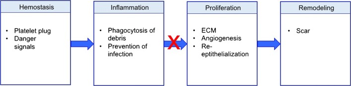
“normal wound healing can generally be broken down into four over-lapping but distinct steps (Fig. 1). These steps include hemostasis (minutes to hours after injury), inflammation (days 1–3), proliferation and repair (days 4–21), and lastly, wound remodeling (days 21–365) [8, 9].”
§ Immunology of Wound Healing > Introduction
What the sources say
- Not checked yetcites [8] Morton LM, Phillips TJ. Wound healing and treating wounds: differential diagnosis and evaluation of chronic wounds. J Am Acad Dermatol. 2016;74(4):589–605; quiz −6. https://doi.org/10.1016/j.jaad.2015.08.068 . source
- Not checked yetcites [9] Reinke JM, Sorg H. Wound repair and regeneration. Eur Surg Res. 2012;49(1):35–43. https://doi.org/10.1159/000339613 . source
plain- Asserted: passes — The authors put it forward as true.
- Checkable: passes — It can be checked against evidence.
- Load-bearing: passes — The paper’s conclusions depend on it.
- Based: passes — The paper gives a reason why it holds.
In the paper
“chronic wounds demonstrate significantly increased levels of the potent neutrophil chemoattractant, CXCL8 [58].”
§ Immunology of Wound Healing > Four Stages of Wound Healing > Inflammation
What the sources say
- Not checked yetcites [58] Fivenson DP, Faria DT, Nickoloff BJ, Poverini PJ, Kunkel S, Burdick M, et al. Chemokine and inflammatory cytokine changes during chronic wound healing. Wound Repair Regen. 1997;5(4):310–22. https://doi.org/10.1046/j.1524-475X.1997.50405.x . source
boostedcorrelational- Asserted: passes — The authors put it forward as true.
- Checkable: passes — It can be checked against evidence.
- Load-bearing: passes — The paper’s conclusions depend on it.
- Based: passes — The paper gives a reason why it holds.
The paper states this without a source or data behind it here, so there is nothing to check it against. The quote shows it is in the paper.
In the paper
“Platelet activation not only results in hemostasis, but also in the release of growth factors such as platelet-derived growth factor (PDGF) as well as immune mediators that are responsible for activation of the immune system and transition to the inflammatory phase of wound healing.”
§ Immunology of Wound Healing > Four Stages of Wound Healing > Hemostasis
plaindirect- Asserted: passes — The authors put it forward as true.
- Checkable: passes — It can be checked against evidence.
- Load-bearing: unclear — It is unclear whether the paper’s conclusions depend on it.
- Based: passes — The paper gives a reason why it holds.
None of the sources cited for this claim have been checked yet.
In the paper
“mouse models of wound healing have shown that in non-aged, non-impaired models, neutrophil depletion does not negatively affect wound healing as profoundly as macrophage deletion [25–27].”
§ Immunology of Wound Healing > Four Stages of Wound Healing > Inflammation
What the sources say
- Not checked yetcites [25] Leibovich SJ, Ross R. The role of the macrophage in wound repair. A study with hydrocortisone and antimacrophage serum. Am J Pathol. 1975;78(1):71–100.
- Not checked yetcites [26] Pinhal-Enfield G, Ramanathan M, Hasko G, Vogel SN, Salzman AL, Boons GJ, et al. An angiogenic switch in macrophages involving synergy between Toll-like receptors 2, 4, 7, and 9 and adenosine A(2A) receptors. Am J Pathol. 2003;163(2):711–21. https://doi.org/10.1016/s0002-9440(10)63698-x . source
- Not checked yetcites [27] Simpson DM, Ross R. The neutrophilic leukocyte in wound repair a study with antineutrophil serum. J Clin Invest. 1972;51(8):2009–23. https://doi.org/10.1172/jci107007 . source
plaindirect- Asserted: passes — The authors put it forward as true.
- Checkable: passes — It can be checked against evidence.
- Load-bearing: unclear — It is unclear whether the paper’s conclusions depend on it.
- Based: passes — The paper gives a reason why it holds.
- Rests on an assumption: Findings from mouse models of wound healing can be generalized to human wound healing and chronic wounds.
A conclusion the paper draws from its own results — it cites no other source for it, so there is none to check. The quote shows it is in the paper.
In the paper
“New research has illustrated that chronic wounds fail to shift from the inflammatory to the proliferative phase of wound healing”
§ Immunology of Wound Healing > Introduction
boosted- Asserted: passes — The authors put it forward as true.
- Checkable: passes — It can be checked against evidence.
- Load-bearing: passes — The paper’s conclusions depend on it.
- Based: passes — The paper gives a reason why it holds.
A conclusion the paper draws from its own results — it cites no other source for it, so there is none to check. The quote shows it is in the paper.
In the paper
“recent research has shown that the key transition point lies between the inflammatory and the proliferative phases.”
- Also stated in: § Conclusions · plain
“Recent research has shown that the key transition point in wound healing lies between the inflammatory and the proliferative phases.”
§ Immunology of Wound Healing > Abstract > Summary
plain- Asserted: passes — The authors put it forward as true.
- Checkable: passes — It can be checked against evidence.
- Load-bearing: passes — The paper’s conclusions depend on it.
- Based: unclear — It is unclear why the paper says it holds.
- Also stated in: § Conclusions · plain
None of the sources cited for this claim have been checked yet.
In the paper
“the immune system has been found to play a substantial role due to its impact on several repair mechanisms [6, 7].”
§ Immunology of Wound Healing > Introduction
What the sources say
- Not checked yetcites [6] MA S, MJ N. The innate immune system in acute and chronic wounds. Adv Wound Care. 2016;5(2):65–78. https://doi.org/10.1089/wound.2014.0608 . source
- Not checked yetcites [7] Strbo N, Yin N, Stojadinovic O. Innate and adaptive immune responses in wound epithelialization. Adv Wound Care. 2014;3(7):492–501. https://doi.org/10.1089/wound.2012.0435 . source
plaindirect- Asserted: passes — The authors put it forward as true.
- Checkable: passes — It can be checked against evidence.
- Load-bearing: passes — The paper’s conclusions depend on it.
- Based: passes — The paper gives a reason why it holds.
The paper states this without a source or data behind it here, so there is nothing to check it against. The quote shows it is in the paper.
In the paper
“Myofibroblasts, activated through contractile forces and signaling molecules, then drive remodeling, where granulation tissue becomes scar.”
§ Immunology of Wound Healing > Abstract > Recent Findings
plaindirect- Asserted: passes — The authors put it forward as true.
- Checkable: passes — It can be checked against evidence.
- Load-bearing: passes — The paper’s conclusions depend on it.
- Based: unclear — It is unclear why the paper says it holds.
- It may state two separate claims as one. 15
The cited sources support only part of this claim — read each source’s verdict below.
In the paper
“The inflammatory phase overlaps considerably with initial hemostasis, occurring during the first 72 h after tissue injury [16].”
§ Immunology of Wound Healing > Four Stages of Wound Healing > Inflammation
What the sources say
- Partly supportedcites [16] Eming SA, Martin P, Tomic-Canic M. Wound repair and regeneration: mechanisms, signaling, and translation. Sci Transl Med. 2014;6(265):265sr6. https://doi.org/10.1126/scitranslmed.3009337 . sourceExperimental — accuracy not yet measured
The ‘Inflammatory response’ section says that acute wound-healing phases largely overlap and that the early inflammatory response occurs in parallel with hemostasis. It does not specify that the inflammatory phase occurs during the first 72 hours after injury; searches for that timing found no supporting passage in our copy. Page numbers were unavailable. (LLM, conf=0.85)
plaincorrelational- Asserted: passes — The authors put it forward as true.
- Checkable: passes — It can be checked against evidence.
- Load-bearing: unclear — It is unclear whether the paper’s conclusions depend on it.
- Based: passes — The paper gives a reason why it holds.
It may state two separate claims as one.
- Partly supported
In the paper
“Concurrent with the achievement of hemostasis, neutrophils are the first cells to migrate to the wound bed, brought in by pro-inflammatory signals including IL-8.”
§ Immunology of Wound Healing > Abstract > Recent Findings
What the sources say
- Not checked yetcites [31] Baggiolini M, Loetscher P, Moser B. Interleukin-8 and the chemokine family. Int J Immunopharmacol. 1995;17(2):103–8. source
plaindirect- Asserted: passes — The authors put it forward as true.
- Checkable: passes — It can be checked against evidence.
- Load-bearing: unclear — It is unclear whether the paper’s conclusions depend on it.
- Based: passes — The paper gives a reason why it holds.
It may state two separate claims as one.
None of the sources cited for this claim have been checked yet.
In the paper
“Neutrophils represent the most abundant inflammatory cells to infiltrate a new wound and function mainly to remove debris and prevent infection [22, 23].”
§ Immunology of Wound Healing > Four Stages of Wound Healing > Inflammation
What the sources say
- Not checked yetcites [22] Engelhardt E, Toksoy A, Goebeler M, Debus S, Brocker EB, Gillitzer R. Chemokines IL-8, GROalpha, MCP-1, IP-10, and Mig are sequentially and differentially expressed during phase-specific infiltration of leukocyte subsets in human wound healing. Am J Pathol. 1998;153(6):1849–60. source
- Not checked yetcites [23] de Oliveira S, Rosowski EE, Huttenlocher A. Neutrophil migration in infection and wound repair: going forward in reverse. Nat Rev Immunol. 2016;16(6):378–91. https://doi.org/10.1038/nri.2016.49 . source
plaindirect- Asserted: passes — The authors put it forward as true.
- Checkable: passes — It can be checked against evidence.
- Load-bearing: unclear — It is unclear whether the paper’s conclusions depend on it.
- Based: passes — The paper gives a reason why it holds.
It may state two separate claims as one.
In the paper
“NETs have also been detected in excess in diabetic foot wounds and have been shown to delay healing, and inhibition of NETosis and NET function in mouse models of delayed wound healing improves outcomes [59•].”
§ Immunology of Wound Healing > Four Stages of Wound Healing > Inflammation
What the sources say
- Not checked yetcites [59•]
plaindirect- Asserted: passes — The authors put it forward as true.
- Checkable: passes — It can be checked against evidence.
- Load-bearing: passes — The paper’s conclusions depend on it.
- Based: passes — The paper gives a reason why it holds.
It may state two separate claims as one.
- Rests on an assumption: Findings from mouse models of wound healing can be generalized to human wound healing and chronic wounds.
None of the sources cited for this claim have been checked yet.
In the paper
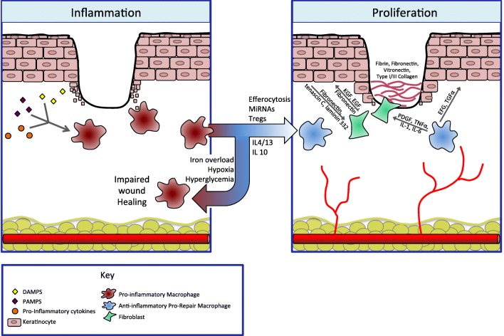
“Macrophages play a critical role in wound healing, and their roles in angiogenesis, fibroplasia, cell proliferation, and transition out of the inflammatory phase is clear (Fig. 2) [25–27, 60].”
§ Immunology of Wound Healing > Four Stages of Wound Healing > Inflammation
What the sources say
- Not checked yetcites [25] Leibovich SJ, Ross R. The role of the macrophage in wound repair. A study with hydrocortisone and antimacrophage serum. Am J Pathol. 1975;78(1):71–100.
- Not checked yetcites [26] Pinhal-Enfield G, Ramanathan M, Hasko G, Vogel SN, Salzman AL, Boons GJ, et al. An angiogenic switch in macrophages involving synergy between Toll-like receptors 2, 4, 7, and 9 and adenosine A(2A) receptors. Am J Pathol. 2003;163(2):711–21. https://doi.org/10.1016/s0002-9440(10)63698-x . source
- Not checked yetcites [27] Simpson DM, Ross R. The neutrophilic leukocyte in wound repair a study with antineutrophil serum. J Clin Invest. 1972;51(8):2009–23. https://doi.org/10.1172/jci107007 . source
- Not checked yetcites [60] Koh TJ, DiPietro LA. Inflammation and wound healing: the role of the macrophage. Expert Rev Mol Med. 2011;13:e23. https://doi.org/10.1017/s1462399411001943 . source
boosteddirect- Asserted: passes — The authors put it forward as true.
- Checkable: passes — It can be checked against evidence.
- Load-bearing: unclear — It is unclear whether the paper’s conclusions depend on it.
- Based: passes — The paper gives a reason why it holds.
It may state two separate claims as one.
In the paper
“After hemostasis is achieved, histamine released by the activated complement cascade causes capillary dilation and leakage, accelerating migration of inflammatory cells into the wound bed and full transition to the inflammatory phase of wound healing [15].”
§ Immunology of Wound Healing > Four Stages of Wound Healing > Hemostasis
What the sources say
- Not checked yetcites [15] Sinno H, Prakash S. Complements and the wound healing cascade: an updated review. Plast Surg Int. 2013;2013:146764–7. https://doi.org/10.1155/2013/146764 . source
plaindirect- Asserted: passes — The authors put it forward as true.
- Checkable: passes — It can be checked against evidence.
- Load-bearing: unclear — It is unclear whether the paper’s conclusions depend on it.
- Based: passes — The paper gives a reason why it holds.
It may state two separate claims as one.
None of the sources cited for this claim have been checked yet.
In the paper
“neutrophil-derived proteases, such as elastase and matrix metalloproteinases (MMPs), can degrade healthy extracellular matrix (ECM), and increased levels of these proteases have been repeatedly detected in chronic wounds [52–56].”
§ Immunology of Wound Healing > Four Stages of Wound Healing > Inflammation
What the sources say
- Not checked yetcites [52] Briggaman RA, Schechter NM, Fraki J, Lazarus GS. Degradation of the epidermal-dermal junction by proteolytic enzymes from human skin and human polymorphonuclear leukocytes. J Exp Med. 1984;160(4):1027–42. source
- Not checked yetcites [53] Grinnell F, Zhu M. Fibronectin degradation in chronic wounds depends on the relative levels of elastase, alpha1-proteinase inhibitor, and alpha2-macroglobulin. J Invest Dermatol. 1996;106(2):335–41. source
- Not checked yetcites [54] Herrick S, Ashcroft G, Ireland G, Horan M, McCollum C, Ferguson M. Up-regulation of elastase in acute wounds of healthy aged humans and chronic venous leg ulcers are associated with matrix degradation. Lab Invest. 1997;77(3):281–8.
- Not checked yetcites [55] Nwomeh BC, Liang HX, Cohen IK, Yager DR. MMP-8 is the predominant collagenase in healing wounds and nonhealing ulcers. J Surg Res. 1999;81(2):189–95. https://doi.org/10.1006/jsre.1998.5495 . source
- Not checked yetcites [56] Pirila E, Korpi JT, Korkiamaki T, Jahkola T, Gutierrez-Fernandez A, Lopez-Otin C, et al. Collagenase-2 (MMP-8) and matrilysin-2 (MMP-26) expression in human wounds of different etiologies. Wound Repair Regen. 2007;15(1):47–57. https://doi.org/10.1111/j.1524-475X.2006.00184.x . source
plaindirect- Asserted: passes — The authors put it forward as true.
- Checkable: passes — It can be checked against evidence.
- Load-bearing: passes — The paper’s conclusions depend on it.
- Based: passes — The paper gives a reason why it holds.
It may state two separate claims as one.
None of the sources cited for this claim have been checked yet.
In the paper
“Platelet degranulation also leads to the release of inflammatory mediators such as interleukin (IL)-8, or CXCL8 (a potent neutrophil chemoattractant), in addition to IL-1α, IL-1β, IL-6, and tumor necrosis factor (TNF)-α, and activates the complement cascade [9, 14].”
§ Immunology of Wound Healing > Four Stages of Wound Healing > Hemostasis
What the sources say
plaindirect- Asserted: passes — The authors put it forward as true.
- Checkable: passes — It can be checked against evidence.
- Load-bearing: unclear — It is unclear whether the paper’s conclusions depend on it.
- Based: passes — The paper gives a reason why it holds.
It may state two separate claims as one.
In the paper
“Dysregulation of any of these events can result in delayed wound healing and the potential to form chronic ulcers and/or excessive scarring [10••].”
§ Immunology of Wound Healing > Introduction
What the sources say
- Not checked yetcites [10••]
plaindirect- Asserted: passes — The authors put it forward as true.
- Checkable: passes — It can be checked against evidence.
- Load-bearing: passes — The paper’s conclusions depend on it.
- Based: passes — The paper gives a reason why it holds.
It may state two separate claims as one.
None of the sources cited for this claim have been checked yet.
In the paper
“Not only do anti-inflammatory, pro-repair macrophages produce VEGF, which promotes vessel sprouting, they also express two transmembrane proteins that have been shown to promote vascular anastomosis [80, 85, 86].”
§ Immunology of Wound Healing > Four Stages of Wound Healing > Proliferation
What the sources say
- Not checked yetcites [80] Brancato SK, Albina JE. Wound macrophages as key regulators of repair: origin, phenotype, and function. Am J Pathol. 2011;178(1):19–25. https://doi.org/10.1016/j.ajpath.2010.08.003 . source
- Not checked yetcites [85] Fantin A, Vieira JM, Gestri G, Denti L, Schwarz Q, Prykhozhij S, et al. Tissue macrophages act as cellular chaperones for vascular anastomosis downstream of VEGF-mediated endothelial tip cell induction. Blood. 2010;116(5):829–40. https://doi.org/10.1182/blood-2009-12-257832 . source
- Not checked yetcites [86] Affolter M, Zeller R, Caussinus E. Tissue remodelling through branching morphogenesis. Nat Rev Mol Cell Biol. 2009;10:831–42. https://doi.org/10.1038/nrm2797 . source
boosteddirect- Asserted: passes — The authors put it forward as true.
- Checkable: passes — It can be checked against evidence.
- Load-bearing: unclear — It is unclear whether the paper’s conclusions depend on it.
- Based: passes — The paper gives a reason why it holds.
It may state two separate claims as one.
In the paper
“Wounds without IL-6 lack an appropriate inflammatory response and demonstrate stunted angiogenesis, collagen accumulation, and re-epithelialization [89].”
§ Immunology of Wound Healing > Four Stages of Wound Healing > Proliferation
What the sources say
- Not checked yetcites [89] Lin ZQ, Kondo T, Ishida Y, Takayasu T, Mukaida N. Essential involvement of IL-6 in the skin wound-healing process as evidenced by delayed wound healing in IL-6-deficient mice. J Leukoc Biol. 2003;73(6):713–21. source
plaindirect- Asserted: passes — The authors put it forward as true.
- Checkable: passes — It can be checked against evidence.
- Load-bearing: unclear — It is unclear whether the paper’s conclusions depend on it.
- Based: passes — The paper gives a reason why it holds.
It may state two separate claims as one.
In the paper
“When the transition from a pro- to an anti-inflammatory macrophage phenotype is impaired, wound healing stalls in the inflammatory phase and a chronic wound results [10••].”
§ Immunology of Wound Healing > Four Stages of Wound Healing > Inflammation
What the sources say
- Not checked yetcites [10••]
plaindirect- Asserted: passes — The authors put it forward as true.
- Checkable: passes — It can be checked against evidence.
- Load-bearing: passes — The paper’s conclusions depend on it.
- Based: passes — The paper gives a reason why it holds.
It may state two separate claims as one.
The paper states this without a source or data behind it here, so there is nothing to check it against. The quote shows it is in the paper.
In the paper
“Additional signals, such as hyperglycemia in the setting of diabetes, hypoxia in the setting of arterial or venous insufficiency, or secondary infection likely also contribute to the persistence of pro-inflammatory macrophages.”
§ Immunology of Wound Healing > Four Stages of Wound Healing > Inflammation
hedgedconditional- Asserted: passes — The authors put it forward as true.
- Checkable: passes — It can be checked against evidence.
- Load-bearing: unclear — It is unclear whether the paper’s conclusions depend on it.
- Based: unclear — It is unclear why the paper says it holds.
It may state two separate claims as one.
A conclusion the paper draws from its own results — it cites no other source for it, so there is none to check. The quote shows it is in the paper.
In the paper
“If wounds fail to transition from the inflammatory phase, pro-inflammatory macrophages persist and non-healing wounds develop.”
§ Immunology of Wound Healing > Conclusions
plaindirect- Asserted: passes — The authors put it forward as true.
- Checkable: passes — It can be checked against evidence.
- Load-bearing: passes — The paper’s conclusions depend on it.
- Based: passes — The paper gives a reason why it holds.
It may state two separate claims as one.
A conclusion the paper draws from its own results — it cites no other source for it, so there is none to check. The quote shows it is in the paper.
In the paper
“Numerous signals are responsible for the transition, most notably the apoptosis and phagocytosis of wound-bed resident neutrophils (efferocytosis).”
§ Immunology of Wound Healing > Conclusions
plaindirect- Asserted: passes — The authors put it forward as true.
- Checkable: passes — It can be checked against evidence.
- Load-bearing: passes — The paper’s conclusions depend on it.
- Based: unclear — It is unclear why the paper says it holds.
It may state two separate claims as one.
The paper relies on this without support.
Findings from mouse models of wound healing can be generalized to human wound healing and chronic wounds.
“mouse models of wound healing have shown that in non-aged, non-impaired models, neutrophil depletion does not negatively affect wound healing as profoundly as macrophage deletion [25–27].”
“in a mouse model of iron overload, local wound macrophages persisted in a pro-inflammatory state with excess production of inducible nitric oxide synthase (iNOS), IL-12, and TNF-α, in addition to free radicals, leading to impaired wound healing [81].”
“inhibition of NETosis and NET function in mouse models of delayed wound healing improves outcomes [59•].”
Claims other papers make about this one
Each is checked against this paper’s own text: does it say what the citing paper says it does?
The source backs part of the claim, not all of it — or backs it for a different population, condition or comparison.
In the citing paper
“Immune regulators promote better surveillance and wound clearance in multiple tissues [75, 76].”
From Rejuvenation of three germ layers tissues by exchanging old blood plasma with saline-albumin (2020) · cites this paper as [75]
Experimental — accuracy not yet measuredIn the citing paper
“Indeed, these cells are known to be irreplaceable partners in a successful wound healing70.”
From Platelets induce increased estrogen production through NF-κB and TGF-β1 signaling pathways in endometriotic stromal cells (2020) · cites this paper as [70]
In the citing paper
“The absence or over-abundance of any of these cell types would derail the normal healing process, leading healing astray and causing pathological healing such as fibrosis70.”
From Platelets induce increased estrogen production through NF-κB and TGF-β1 signaling pathways in endometriotic stromal cells (2020) · cites this paper as [70]
Citations
Cites
Neutrophil Extracellular Traps Kill Bacteria
Brinkmann, V., Reichard, U., Goosmann, C., Fauler, B., Uhlemann, Y., Weiss, D. S., Weinrauch, Y., & Zychlinsky, A. (2004) · Science · doi:10.1126/science.1092385 · Cited by 10,268
Exploring the full spectrum of macrophage activation
Mosser, D. M., & Edwards, J. P. (2008) · Nature Reviews Immunology · doi:10.1038/nri2448 · Cited by 9,144
Pattern Recognition Receptors and Inflammation
Takeuchi, O., & Akira, S. (2010) · Cell · doi:10.1016/j.cell.2010.01.022 · Cited by 8,895
Mizgerd, J. P., Kubo, H., Kutkoski, G. J., Bhagwan, S. D., Scharffetter-Kochanek, K., Beaudet, A. L., & Doerschuk, C. M. (1997) · The Journal of Experimental Medicine · doi:10.1084/jem.186.8.1357 · Cited by 7,332
The chemokine system in diverse forms of macrophage activation and polarization
MANTOVANI, A., SICA, A., SOZZANI, S., ALLAVENA, P., VECCHI, A., & LOCATI, M. (2004) · Trends in Immunology · doi:10.1016/j.it.2004.09.015 · Cited by 6,534
Macrophage plasticity and polarization: in vivo veritas
Sica, A., & Mantovani, A. (2012) · Journal of Clinical Investigation · doi:10.1172/jci59643 · Cited by 6,335
MANTOVANI, A., SOZZANI, S., LOCATI, M., ALLAVENA, P., & SICA, A. (2002) · Trends in Immunology · doi:10.1016/s1471-4906(02)02302-5 · Cited by 5,528
Neutrophil recruitment and function in health and inflammation
Kolaczkowska, E., & Kubes, P. (2013) · Nature Reviews Immunology · doi:10.1038/nri3399 · Cited by 5,472
Monocyte and macrophage heterogeneity
Gordon, S., & Taylor, P. R. (2005) · Nature Reviews Immunology · doi:10.1038/nri1733 · Cited by 5,190
Wound repair and regeneration: Mechanisms, signaling, and translation
Eming, S. A., Martin, P., & Tomic-Canic, M. (2014) · Science Translational Medicine · doi:10.1126/scitranslmed.3009337 · Cited by 3,562
Cited by
Sorted by citations
Immunology of Acute and Chronic Wound Healing
Raziyeva, K., Kim, Y., Zharkinbekov, Z., Kassymbek, K., Jimi, S., & Saparov, A. (2021) · Biomolecules · doi:10.3390/biom11050700 · Cited by 1,152
Immunoregulation in Diabetic Wound Repair with a Photoenhanced Glycyrrhizic Acid Hydrogel Scaffold
Qian, Y., Zheng, Y., Jin, J., Wu, X., Xu, K., Dai, M., Niu, Q., Zheng, H., He, X., & Shen, J. (2022) · Advanced Materials · doi:10.1002/adma.202200521 · Cited by 769
Rational Design of Immunomodulatory Hydrogels for Chronic Wound Healing
Kharaziha, M., Baidya, A., & Annabi, N. (2021) · Advanced Materials · doi:10.1002/adma.202100176 · Cited by 735
The Dynamics of the Skin’s Immune System
Nguyen, A. V., & Soulika, A. M. (2019) · International Journal of Molecular Sciences · doi:10.3390/ijms20081811 · Cited by 759
“Once initiated, the remodeling phase can continue for at least one year post-injury [81,366].”
BackgroundChouhan, D., Dey, N., Bhardwaj, N., & Mandal, B. B. (2019) · Biomaterials · doi:10.1016/j.biomaterials.2019.119267 · Cited by 537
The Role of the Extracellular Matrix (ECM) in Wound Healing: A Review
Diller, R. B., & Tabor, A. J. (2022) · Biomimetics · doi:10.3390/biomimetics7030087 · Cited by 550
“, days to weeks), while the remodeling phase of the wound healing responses can take up to months (12 months) for completion [45,47].”
BackgroundMarconi, G. D., Fonticoli, L., Rajan, T. S., Pierdomenico, S. D., Trubiani, O., Pizzicannella, J., & Diomede, F. (2021) · Cells · doi:10.3390/cells10071587 · Cited by 449
“The haemostasis phase occurs immediately after injury and results in the formation of a provisional wound matrix [22].”
BackgroundThe Healing Power of Neutrophils
Phillipson, M., & Kubes, P. (2019) · Trends in Immunology · doi:10.1016/j.it.2019.05.001 · Cited by 398
Cheng, H., Shi, Z., Yue, K., Huang, X., Xu, Y., Gao, C., Yao, Z., Zhang, Y. S., & Wang, J. (2021) · Acta Biomaterialia · doi:10.1016/j.actbio.2021.02.002 · Cited by 372
Bian, D., Wu, Y., Song, G., Azizi, R., & Zamani, A. (2022) · Stem Cell Research & Therapy · doi:10.1186/s13287-021-02697-9 · Cited by 392
“bed for the founding of novel tissue [48, 49].”
BackgroundMicroenvironment‐Based Diabetic Foot Ulcer Nanomedicine
Huang, F., Lu, X., Yang, Y., Yang, Y., Li, Y., Kuai, L., Li, B., Dong, H., & Shi, J. (2022) · Advanced Science · doi:10.1002/advs.202203308 · Cited by 388
Hydrogels and Wound Healing: Current and Future Prospects
Gounden, V., & Singh, M. (2024) · Gels · doi:10.3390/gels10010043 · Cited by 382
“The granulation tissue undergoes a slow remodeling process, forming scar tissue [21].”
BackgroundChronic Inflammation in Non-Healing Skin Wounds and Promising Natural Bioactive Compounds Treatment
Schilrreff, P., & Alexiev, U. (2022) · International Journal of Molecular Sciences · doi:10.3390/ijms23094928 · Cited by 389
“Besides removing pathogens, neutrophils can upregulate and secrete various pro-inflammatory cytokines such as IL-1α, IL-1β, IL-6, and TNF-α to stimulate monocytes to differentiate into M1 macrophages [31].”
BackgroundRecent advances in molecular mechanisms of skin wound healing and its treatments
Mamun, A. A., Shao, C., Geng, P., Wang, S., & Xiao, J. (2024) · Frontiers in Immunology · doi:10.3389/fimmu.2024.1395479 · Cited by 338
“Apoptosis, a regulated form of cell death, serves a critical function in the stages of tissue healing, as shown by its complex involvement in the resolution of these phases (112).”
BackgroundPlasma membrane integrity: implications for health and disease
Ammendolia, D. A., Bement, W. M., & Brumell, J. H. (2021) · BMC Biology · doi:10.1186/s12915-021-00972-y · Cited by 279
“It is convenient to conceptualize the requirements for cell repair by paralleling those of tissue healing, encompassing four complementary stages [115]: hemostasis (sealing off the breach), inflammation (removal of dangerous material that entered the cell), proliferation and migration (replacement of lost or damaged c…”
BackgroundAcute Inflammation in Tissue Healing
Soliman, A. M., & Barreda, D. R. (2022) · International Journal of Molecular Sciences · doi:10.3390/ijms24010641 · Cited by 301
Biofilm-Innate Immune Interface: Contribution to Chronic Wound Formation
Versey, Z., Nizer, W. S. D. C., Russell, E., Zigic, S., DeZeeuw, K. G., Marek, J. E., Overhage, J., & Cassol, E. (2021) · Frontiers in Immunology · doi:10.3389/fimmu.2021.648554 · Cited by 273
“During the proliferation phase they also release growth factors, such as vascular endothelial growth factor (VEGF), platelet-derived growth factor (PDGF), and fibroblast growth factor-2 (FGF-2), which promote angiogenesis and keratinocyte and fibroblast proliferation (86, 88, 89, 91, 111).”
BackgroundInfluentialElectrospun nanofibers promote wound healing: theories, techniques, and perspectives
Gao, C., Zhang, L., Wang, J., Jin, M., Tang, Q., Chen, Z., Cheng, Y., Yang, R., & Zhao, G. (2021) · Journal of Materials Chemistry B · doi:10.1039/d1tb00067e · Cited by 247
Applications of Mesenchymal Stem Cells in Skin Regeneration and Rejuvenation
Jo, H., Brito, S., Kwak, B.-M., Park, S., Lee, M., & Bin, B. (2021) · International Journal of Molecular Sciences · doi:10.3390/ijms22052410 · Cited by 242
“Fibroblasts increase the migration and proliferation of keratinocytes via EGF, fibronectin, and keratinocyte growth factor (KGF) [77].”
BackgroundInfluentialWang, T., Bai, J., Lu, M., Huang, C., Geng, D., Chen, G., Wang, L., Qi, J., Cui, W., & Deng, L. (2022) · Nature Communications · doi:10.1038/s41467-021-27816-1 · Cited by 221
“Tissue regeneration involves three indispensable stages: (i) immune action, (ii) cell proliferation and new tissue formation, and (iii) remodeling and maturation 7,8 .”
Mahmoud, N. N., Hamad, K. M., Shibitini, A. A., Juma, S., Sharifi, S., Gould, L., & Mahmoudi, M. (2024) · ACS Pharmacology & Translational Science · doi:10.1021/acsptsci.3c00336 · Cited by 210
Films for Wound Healing Fabricated Using a Solvent Casting Technique
Borbolla‐Jiménez, F. V., Peña‐Corona, S. I., Farah, S. J., Jiménez-Valdés, M. T., Pineda-Pérez, E., Romero‐Montero, A., Prado-Audelo, M. L. D., Bernal-Chávez, S. A., Magaña, J. J., & Leyva‐Gómez, G. (2023) · Pharmaceutics · doi:10.3390/pharmaceutics15071914 · Cited by 227
“Furthermore, this leads to the formation of a fibrin clot, referred to as an eschar, which acts as a plug for the wound, preventing blood loss, providing a scaffold for incoming immune cells, serving as a reservoir for cytokines and growth factors during the early stages of repair, and offering protection against bact…”
BackgroundInfluentialCannabinoid Signaling in the Skin: Therapeutic Potential of the “C(ut)annabinoid” System
Tóth, K. F., Ádám, D., Bı́ró, T., & Oláh, A. (2019) · Molecules · doi:10.3390/molecules24050918 · Cited by 214
Therapeutic Potential of Cannabidiol (CBD) for Skin Health and Disorders
Baswan, S. M., Klosner, A. E., Glynn, K. M., Rajgopal, A., Malik, K. A., Yim, S., & Stern, N. (2020) · Clinical Cosmetic and Investigational Dermatology · doi:10.2147/ccid.s286411 · Cited by 208
Advances in bioactive glass-containing injectable hydrogel biomaterials for tissue regeneration
Zeimaran, E., Pourshahrestani, S., Fathi, A., Razak, N. A. A., Kadri, N. A., Sheikhi, A., & Baino, F. (2021) · Acta Biomaterialia · doi:10.1016/j.actbio.2021.09.034 · Cited by 171
The Colorectal Cancer Tumor Microenvironment and Its Impact on Liver and Lung Metastasis
Chandra, R., Karalis, J. D., Liu, C., Murimwa, G. Z., Park, J. V., Heid, C. A., Reznik, S. I., Huang, E. H., Minna, J. D., & Brekken, R. A. (2021) · Cancers · doi:10.3390/cancers13246206 · Cited by 168
Natural polymers used in the dressing materials for wound healing: Past, present and future
Sheokand, B., Vats, M., Kumar, A., Srivastava, C. M., Bahadur, I., & Pathak, S. R. (2023) · Journal of Polymer Science · doi:10.1002/pol.20220734 · Cited by 168
Cai, Y., Chen, K., Liu, C., & Qu, X. (2023) · Bioactive Materials · doi:10.1016/j.bioactmat.2023.04.019 · Cited by 112
“247 initiating re-epithelialization by producing epidermal growth factor (EGF) and keratinocyte growth factor (KGF), M1 macrophages hinder keratinocyte migration with over-expressed TNF-α [60,61].”
BackgroundInfluentialAn Updated Review of Hypertrophic Scarring
Mony, M. P., Harmon, K. A., Hess, R., Dorafshar, A. H., & Shafikhani, S. H. (2023) · Cells · doi:10.3390/cells12050678 · Cited by 183
“The primary functions of the inflammation phase during wound healing are to protect wounds against invading pathogens and to jumpstart the subsequent inflammatory and non-inflammatory responses needed for proper healing [26,27].”
BackgroundThe Insights of Microbes’ Roles in Wound Healing: A Comprehensive Review
Thambirajoo, M., Nurul, A. A., & Fauzi, M. B. (2021) · Pharmaceutics · doi:10.3390/pharmaceutics13070981 · Cited by 166
“Granulation tissue Normal production [40] Neoangiogeneis, less fibroblasts, low oxygen→ tissue hypoxia Tissue hypoxia—low production of granulation tissue [40]”
BackgroundTopical gel-based biomaterials for the treatment of diabetic foot ulcers
Bardill, J. R., Laughter, M. R., Stager, M. A., Liechty, K. W., Krebs, M. D., & Zgheib, C. (2021) · Acta Biomaterialia · doi:10.1016/j.actbio.2021.10.045 · Cited by 171
Anti-inflammation biomaterial platforms for chronic wound healing
Xu, Z., Liang, B., Tian, J., & Wu, J. (2021) · Biomaterials Science · doi:10.1039/d1bm00637a · Cited by 151
Mohsin, F., Javaid, S., Tariq, M., & Mustafa, M. (2024) · International Immunopharmacology · doi:10.1016/j.intimp.2024.112713 · Cited by 153
Heras, K. L., García-Orue, I., Rancan, F., Igartúa, M., Santos‐Vizcaíno, E., & Hernández, R. M. (2024) · Advanced Drug Delivery Reviews · doi:10.1016/j.addr.2024.115342 · Cited by 143
Bacteria and Antibiotics in Wound Healing
Caldwell, M. D. (2020) · Surgical Clinics of North America · doi:10.1016/j.suc.2020.05.007 · Cited by 136
Chittasupho, C., Manthaisong, A., Okonogi, S., Tadtong, S., & Samee, W. (2021) · International Journal of Molecular Sciences · doi:10.3390/ijms23010142 · Cited by 131
“The inflammation occurs where white blood cells such as phagocytic cells, neutrophils and macrophages migrate to the injured site [5].”
BackgroundApplication of stem cells in regeneration medicine
Jin, Y. H., Li, S., Yu, Q., Chen, T., & Liu, D. (2023) · MedComm · doi:10.1002/mco2.291 · Cited by 149
Healing Mechanisms in Cutaneous Wounds: Tipping the Balance
Singer, A. J. (2021) · Tissue Engineering Part B Reviews · doi:10.1089/ten.teb.2021.0114 · Cited by 133
“For more on the role of the immune response in wound healing, see the recent reviews by CañedoDorantes and Cañedo-Ayala and Ellis et al.(45,60) Compared with reparative healing, the inflammatory response in regenerative healing is attenuated.”
BackgroundWound healing and microbiome, an unexpected relationship
Canchy, L., Kérob, D., Demessant‐Flavigny, A., & Amici, J. M. (2023) · Journal of the European Academy of Dermatology and Venereology · doi:10.1111/jdv.18854 · Cited by 131
“Moreover, excessive inf lammation is commonly associated with chronicity as it further delays the complete transition to the proliferative phase.(9,10) Besides, it can provide an ideal environment for bacterial infiltration and proliferation, increasing the risk of persistent infection.”
BackgroundDiban, F., Lodovico, S. D., Fermo, P. D., D’Ercole, S., D’Arcangelo, S., Giulio, M. D., & Cellini, L. (2023) · International Journal of Molecular Sciences · doi:10.3390/ijms24021004 · Cited by 138
Hydrogel-Based Skin Regeneration
Arabpour, Z., Abedi, F., Salehi, M., Baharnoori, S. M., Soleimani, M., & Djalilian, A. R. (2024) · International Journal of Molecular Sciences · doi:10.3390/ijms25041982 · Cited by 130
“These stages involve the interaction of various cell types and matrix components, including hemostasis, inflammation, proliferation, and remodeling, as illustrated in Figure 1 [11].”
BackgroundEpigenetic regulation of macrophage polarization in wound healing
Chen, C., Liu, T., Tang, Y., Luo, G., Liang, G., & He, W. (2023) · Burns & Trauma · doi:10.1093/burnst/tkac057 · Cited by 130
“At an early stage of wound healing, a proinflammatory reaction is conducive to anti-infection activity in injured tissue and a late pro-healing response is beneficial to wound repair [9].”
BackgroundXing, C., Zhu, H., Dou, X., Gao, L., Baddi, S., Zou, Y., Zhao, C., Peng, Y., Fang, Y., & Feng, C. (2023) · ACS Nano · doi:10.1021/acsnano.2c10039 · Cited by 121
Distinct inflammatory and wound healing responses to complex caudal fin injuries of larval zebrafish
Miskolci, V., Squirrell, J. M., Rindy, J., Vincent, W. J. B., Sauer, J., Gibson, A. L. F., Eliceiri, K. W., & Anna Huttenlocher, A. (2019) · eLife · doi:10.7554/elife.45976 · Cited by 114
“We evaluated three aspects of these injuries (Figure 1A), representing key stages of wound healing – new tissue formation, inflammation and remodeling (Ellis et al., 2018).”
MethodBeneficial Effects of Green Tea EGCG on Skin Wound Healing: A Comprehensive Review
Xu, F.-W., Lv, Y.-L., Zhong, Y., Xue, Y., Wang, Y., Zhang, L., Hu, X. M., & Tan, W. (2021) · Molecules · doi:10.3390/molecules26206123 · Cited by 127
“The skin wound healing process has four sequential and overlapping stages, including hemostasis, inflammation, proliferation and tissue remodeling [52], which involves various types of cells (e.”
BackgroundInfluentialPalani, N., Vijayakumar, P., Monisha, P., Ayyadurai, S., & Rajadesingu, S. (2024) · Journal of Nanobiotechnology · doi:10.1186/s12951-024-02491-8 · Cited by 125
Rejuvenation of three germ layers tissues by exchanging old blood plasma with saline-albumin
Mehdipour, M., Skinner, C., Wong, N., Lieb, M., Liu, C., Etienne, J., Kato, C., Kiprov, D., Conboy, M. J., & Conboy, I. M. (2020) · Aging · doi:10.18632/aging.103418 · Cited by 115
“Immune regulators promote better surveillance and wound clearance in multiple tissues [75, 76].”
BackgroundČoma, M., Fröhlichová, L., Urban, L., Zajíček, R., Urban, T., Szabó, P., Novak, S., Fetissov, V., Dvořánková, B., Smetana, K., et al. (2021) · International Journal of Molecular Sciences · doi:10.3390/ijms22020897 · Cited by 124
Bacteriophages for Chronic Wound Treatment: From Traditional to Novel Delivery Systems
Pinto, A. M., Cerqueira, M. Â., Bañobre‐López, M., Pastrana, L. M., & Sillankorva, S. (2020) · Viruses · doi:10.3390/v12020235 · Cited by 115
“Fibroblasts that acquire contractile properties can contract the wound edges and the migration and proliferation of keratinocytes, which are responsible for the closure of the lesion [17,27].”
BackgroundInfluentialBacterial Cellulose as a Versatile Biomaterial for Wound Dressing Application
de Amorim, J. D. P., da Silva, C. J. G., de Medeiros, A. D. M., do Nascimento, H. A., Sarubbo, M., de Medeiros, T. P. M., Costa, A. F. D. S., & Sarubbo, L. A. (2022) · Molecules · doi:10.3390/molecules27175580 · Cited by 113
Harrell, C. R., Markovic, B. S., Fellabaum, C., Arsenijevic, N., Djonov, V., & Volarevic, V. (2020) · BioFactors · doi:10.1002/biof.1587 · Cited by 106
“inflammatory M1 macrophages and neutrophils in IFN-γ, IL-17, and IL-22-dependent manner, creating an “autocrine inflammatory loop” that results in a persistent inflammation within the nonhealing wounds.(25) Vander Beken and colleagues recently demonstrated that within the ATP-binding cassette Subfamily B Member 5 (ABC…”
BackgroundLiu, B., Fu, R., Duan, Z., Zhu, C., Deng, J., & Fan, D. (2022) · Composites Part B Engineering · doi:10.1016/j.compositesb.2022.109804 · Cited by 103
Sadat, Z., Farrokhi-Hajiabad, F., Lalebeigi, F., et al. (2022) · Biomaterials Science · doi:10.1039/d2bm01308h · Cited by 101
Kalasin, S., Sangnuang, P., & Surareungchai, W. (2022) · Analytical Chemistry · doi:10.1021/acs.analchem.2c00782 · Cited by 102
Flynn, K., Mahmoud, N. N., Sharifi, S., Gould, L. J., & Mahmoudi, M. (2023) · ACS Pharmacology & Translational Science · doi:10.1021/acsptsci.3c00030 · Cited by 101
Applications of Nanosized-Lipid-Based Drug Delivery Systems in Wound Care
Matei, A.-M., Căruntu, C., Tampa, M. Ş., Georgescu, S. R., Matei, C., Constantin, M. M., Constantin, T. V., Călina, D. C., Ciubotaru, D. A., Bădărău, I. A., et al. (2021) · Applied Sciences · doi:10.3390/app11114915 · Cited by 101
“It involves activation of the immune system’s cellular components, the coagulation cascade, and the inflammatory pathways, leading to a massive remodeling of all skin compartments [1,2].”
BackgroundMacrophage Phenotypes in Normal and Diabetic Wound Healing and Therapeutic Interventions
Sadoun, H. A. (2022) · Cells · doi:10.3390/cells11152430 · Cited by 96
“C3b mediates bacterial opsonization, and this further stimulates neutrophil recruitment to the wound site [1,16,17] (Figure 1).”
BackgroundKeratinocyte-Macrophage Crosstalk by the Nrf2/Ccl2/EGF Signaling Axis Orchestrates Tissue Repair
VILLARREAL-PONCE, A. P., Tiruneh, M. W., Lee, J., Guerrero‐Juarez, C. F., Kuhn, J. F., David, J. A., Dammeyer, K. L., Kell, R. M., Kwong, J., Rabbani, P. S., et al. (2020) · Cell Reports · doi:10.1016/j.celrep.2020.108417 · Cited by 91
“Granulation tissue formation describes an essential component of wound repair that encompasses the production of new connective tissue and blood vessels within the wound bed (Ellis et al., 2018).”
BackgroundZhao, Y., & Jalili, S. (2022) · International Journal of Biological Macromolecules · doi:10.1016/j.ijbiomac.2022.02.114 · Cited by 89
Multifunctional lipid-based nanoparticles for wound healing and antibacterial applications: A review
Motsoene, F., Abrahamse, H., & Kumar, S. S. D. (2023) · Advances in Colloid and Interface Science · doi:10.1016/j.cis.2023.103002 · Cited by 86
Myofibroblast progeny in wound biology and wound healing studies
Monika, P., Waiker, P. V., Chandraprabha, M. N., Rangarajan, A., & Murthy, K. N. C. (2021) · Wound Repair and Regeneration · doi:10.1111/wrr.12937 · Cited by 90
“hospitalization, loss of mobility, compromising quality of life, requirement of support and sometime increase the risk of nosocomial infection causing burden to health care workers and other patients visiting hospital.(2) Considering the number of diabetic patients, accidents and other events associated with wound, th…”
BackgroundCerium oxide nanoparticles in wound care: a review of mechanisms and therapeutic applications
Chen, S., Wang, Y., Bao, S., Li, Y., Fu, X., Yu, Y., Lyu, H., Pang, H., Guo, S., Zhang, H., et al. (2024) · Frontiers in Bioengineering and Biotechnology · doi:10.3389/fbioe.2024.1404651 · Cited by 88
“These factors play a catalytic role in the wound healing cascade by activating neutrophils, and subsequently macrophages, endothelial cells and fi broblasts (Ellis et al., 2018).”
BackgroundAn Update on Molecular Mechanisms of Scarring—A Narrative Review
Kohlhauser, M., Mayrhofer, M., Kamolz, L., & Smolle, C. (2024) · International Journal of Molecular Sciences · doi:10.3390/ijms252111579 · Cited by 91
“These cells in turn not only lead to expression of cytokines but also chemokines, which subsequently attract inflammatory cells [6,21,22].”
BackgroundAdvanced Wound Diagnostics: Toward Transforming Wound Care into Precision Medicine
Weigelt, M. A., Lev‐Tov, H. A., Tomic‐Canic, M., Lee, W. D., Williams, R., Strasfeld, D. B., Kirsner, R. S., & Herman, I. M. (2021) · Advances in Wound Care · doi:10.1089/wound.2020.1319 · Cited by 85
“77,78 MMP-2, neutrophil elastase, and CG have highest activity at pH 7–8, which fits in with our understanding of the increased protease activity seen in chronic wounds.”
Recent advancements in bioadhesive self-healing hydrogels for effective chronic wound care
Nezhad‐Mokhtari, P., Hasany, M., Kohestanian, M., Dolatshahi‐Pirouz, A., Milani, M., & Mehrali, M. (2024) · Advances in Colloid and Interface Science · doi:10.1016/j.cis.2024.103306 · Cited by 88
Khalili‐Tanha, G., Radisky, E. S., Radisky, D. C., & Shoari, A. (2025) · Journal of Translational Medicine · doi:10.1186/s12967-025-06447-w · Cited by 92
“Following injury, platelets create a provisional clot containing fibrinogen and fibronectin, and inflammatory cells clear debris, setting the stage for epithelial cells to migrate over the wound bed [2, 112].”
The role of the host—Neutrophil biology
Chapple, I. L. C., Hirschfeld, J., Kantarci, A., Wilensky, A., & Shapira, L. (2023) · Periodontology 2000 · doi:10.1111/prd.12490 · Cited by 83
Enhancing wound healing dressing development through interdisciplinary collaboration
Hawthorne, B., Simmons, J. K., Stuart, B., Tung, R., Zamierowski, D. S., & Mellott, A. J. (2021) · Journal of Biomedical Materials Research Part B Applied Biomaterials · doi:10.1002/jbm.b.34861 · Cited by 80
“as well as secrete proinflammatory cytokines and chemokines that increase the number of natural killer cells, helper T cells, and macrophages at the site of injury.(13) Some macrophages will differentiate into antiinflammatory and pro-regenerative M2 macrophages.”
BackgroundInfluentialdel Olmo, J. A., Pérez‐Álvarez, L., Sáez‐Martínez, V., Cid, S. B., Ruiz‐Rubio, L., Pérez-González, R., Vilas‐Vilela, J. L., & Alonso, J. M. (2022) · International Journal of Biological Macromolecules · doi:10.1016/j.ijbiomac.2022.01.193 · Cited by 80
Cutaneous Wound Healing: A Review about Innate Immune Response and Current Therapeutic Applications
Adib, Y., Bensussan, A., & Michel, L. (2022) · Mediators of Inflammation · doi:10.1155/2022/5344085 · Cited by 85
“Different signals can induce the activation of resident innate cells and the recruitment of circulating inflammatory cells into the wound site; they include the release of alarmins, also known as damage-associated molecular patterns or DAMPs by damaged host cells (e.g., uric acid, DNA, RNA, and extracellular matrix co…”
BackgroundInfluentialHybrid-Based Wound Dressings: Combination of Synthetic and Biopolymers
Aderibigbe, B. A. (2022) · Polymers · doi:10.3390/polym14183806 · Cited by 78
“In this phase, neutrophils and monocyte infiltration in the wound bed prevent the invasion of microbes, foreign debris, and tissue damage [27,28].”
BackgroundTumors vs. Chronic Wounds: An Immune Cell's Perspective
Hua, Y., & Bergers, G. (2019) · Frontiers in Immunology · doi:10.3389/fimmu.2019.02178 · Cited by 82
“During this inflammation period, neutrophils secrete reactive oxygen species (ROS), nitric oxide (NO), and antimicrobial proteins (AMPs) and deploy web-like extracellular traps (NETs) in order to phagocytose and kill contaminating microorganisms (12, 13).”
BackgroundBacterial cellulose/chitosan composite materials for biomedical applications
Liu, K., Wang, Y., Liu, W., et al. (2024) · Chemical Engineering Journal · doi:10.1016/j.cej.2024.153014 · Cited by 75
MSCs and their exosomes: a rapidly evolving approach in the context of cutaneous wounds therapy
Marofi, F., Alexandrovna, K. I., Margiana, R., Bahramali, M., Suksatan, W., Abdelbasset, W. K., Chupradit, S., Nasimi, M., & Maashi, M. S. (2021) · Stem Cell Research & Therapy · doi:10.1186/s13287-021-02662-6 · Cited by 80
“The inflammatory phase, as the second step of wound healing, concentrates on deteriorating bacteria and eliminating debris, thereby providing the wound bed for the establishment of new tissue [27, 28].”
BackgroundQin, X., He, J., Wang, X., Wang, J., Yang, R., & Chen, X. (2023) · Frontiers in Immunology · doi:10.3389/fimmu.2023.1256687 · Cited by 80
Biofilms and Chronic Wounds: Pathogenesis and Treatment Options
Shen, A., Taha, M. R., Ghannoum, M. A., & Tyring, S. K. (2025) · Journal of Clinical Medicine · doi:10.3390/jcm14217784 · Cited by 99
“After this, proliferation begins where granulation tissue forms and the keratinocytes undergo epithelialization [30,31].”
Tang, Q., Chen, C., Jiang, Y., Huang, J., Liu, Y., Nthumba, P. M., Gu, G., Wu, X., Zhao, Y., & Ren, J. (2020) · Journal of Materials Chemistry B · doi:10.1039/d0tb00726a · Cited by 74
Current Progress and Outlook of Nano-Based Hydrogel Dressings for Wound Healing
Zhang, X., Wei, P., Yang, Z., Liu, Y., Yang, K., Cheng, Y., Yao, H., & Zhang, Z. (2022) · Pharmaceutics · doi:10.3390/pharmaceutics15010068 · Cited by 75
Constantino, V. R. L., Figueiredo, M. P., Magri, V. R., Eulálio, D., Cunha, V. R. R., Alcântara, A. C. S., & Perotti, G. F. (2023) · Pharmaceutics · doi:10.3390/pharmaceutics15020413 · Cited by 76
Stimuli-responsive hydrogels for skin wound healing and regeneration
Xin, H., Maruf, D. S. A. A., Akin-Ige, F., & Amin, S. (2024) · Emergent Materials · doi:10.1007/s42247-024-00930-8 · Cited by 86
“Granula - tion tissues are also converted to scar at this stage [ 41, 51].”
Tyavambiza, C., Meyer, M., & Meyer, S. (2022) · Bioengineering · doi:10.3390/bioengineering9110712 · Cited by 75
Yang, Y., Suo, D., Xu, T., Zhao, S., Xu, X., Bei, H., Wong, K. K. Y., Li, Q., Zheng, Z., Li, B., et al. (2024) · Science Advances · doi:10.1126/sciadv.ado9479 · Cited by 70
“During hemostasis, the activated platelets could secrete immune mediators (e.g., IL-8 and transforming growth factor– β ) to recruit immune cells (e.g., macrophages) and, thus, to evoke the immune response ( 53 ).”
Applications of mesenchymal stem cell-exosome components in wound infection healing: new insights
Fakouri, A., Razavi, Z.-S., Mohammed, A. T., Hussein, A. H. A., Afkhami, H., & Hooshiar, M. H. (2024) · Burns & Trauma · doi:10.1093/burnst/tkae021 · Cited by 69
“During the advanced phases of wound healing, it has been observed that effector cells undergo apoptosis, collagen III is replaced by collagen I, matrix metalloprotease degradation occurs and the synthesis of other ECM proteins takes place [146].”
BackgroundImmunomodulatory Properties of Host Defence Peptides in Skin Wound Healing
Petkovic, M., Mouritzen, M. V., Mojsoska, B., & Jenssen, H. (2021) · Biomolecules · doi:10.3390/biom11070952 · Cited by 69
“Hemostasis involves various protease cascades leading to the formation of a fibrin cloth to prevent blood loss and seal the wound [61].”
BackgroundDirect and Indirect Roles of Macrophages in Hypertrophic Scar Formation
Feng, Y., Sun, Z., Liu, S., Wu, J., Zhao, B.-H., Lv, G., Du, Y., Yu, S., Yang, M., Yuan, F., et al. (2019) · Frontiers in Physiology · doi:10.3389/fphys.2019.01101 · Cited by 72
Wójcik, M., Kazimierczak, P., Benko, A., Pałka, K., Vivcharenko, V., & Przekora, A. (2021) · Materials Science and Engineering C · doi:10.1016/j.msec.2021.112068 · Cited by 66
Hasannasab, M., Nourmohammadi, J., Dehghan, M. M., & Ghaee, A. (2021) · International Journal of Pharmaceutics · doi:10.1016/j.ijpharm.2021.121227 · Cited by 70
Clinically relevant experimental rodent models of diabetic foot ulcer
Rai, V., Moellmer, R., & Agrawal, D. K. (2022) · Molecular and Cellular Biochemistry · doi:10.1007/s11010-022-04372-w · Cited by 68
3D scaffolds in the treatment of diabetic foot ulcers: New trends vs conventional approaches
Glover, K., Stratakos, A. C., Váradi, A., & Lamprou, D. A. (2021) · International Journal of Pharmaceutics · doi:10.1016/j.ijpharm.2021.120423 · Cited by 66
Skin γδ T Cells and Their Function in Wound Healing
Hu, W., Shang, R., Yang, J., Chen, C., Liu, Z., Liang, G., He, W., & Luo, G. (2022) · Frontiers in Immunology · doi:10.3389/fimmu.2022.875076 · Cited by 76
Skin immunity in wound healing and cancer
Jakovija, A., & Chtanova, T. (2023) · Frontiers in Immunology · doi:10.3389/fimmu.2023.1060258 · Cited by 70
“After sterilizing the wound, neutrophils initiate an apoptotic celldeath pathway which leads to efferocytosis by macrophages (53).”
BackgroundMetal-organic framework-based dressings: Application and opportunities in wound healing
Huang, Q., Chen, Y., Ye, M., Zhuang, S., Zhong, A., Liu, J., Maduraiveeran, G., Peng, Y., & Huang, Y. (2024) · Materials Today Chemistry · doi:10.1016/j.mtchem.2024.102235 · Cited by 70
Eosinophils in wound healing and epithelial remodeling: Is coagulation a missing link?
Coden, M. E., & Berdnikovs, S. (2020) · Journal of Leukocyte Biology · doi:10.1002/jlb.3mr0120-390r · Cited by 62
Masri, S., Mazlan, Z., Zulkiflee, I., Salleh, A., Fadilah, N. I. M., Maarof, M., Wen, A. P. Y., Duman, F., Tabata, Y., Aziz, I. A., et al. (2022) · International Journal of Molecular Sciences · doi:10.3390/ijms23010476 · Cited by 63
Buck, E., Lee, S., Stone, L. S., & Cerruti, M. (2021) · ACS Applied Materials & Interfaces · doi:10.1021/acsami.0c16509 · Cited by 60
Ding, Y., Ding, X., Zhang, H., Li, S., Yang, P., & Tan, Q. (2022) · Oxidative Medicine and Cellular Longevity · doi:10.1155/2022/9687925 · Cited by 66
“Nonheal-ing wounds in patients with DM negatively a ff ect the quality of life, mortality, and morbidity, and hence, pose a critical clinical challenge and impose an economic burden worldwide [3].”
Lacina, L., Kolář, M., Pfeiferová, L., Gál, P., & Smetana, K. (2024) · Frontiers in Immunology · doi:10.3389/fimmu.2024.1403570 · Cited by 69
Zhang, S., Li, Y., Li, Y., et al. (2021) · Bioactive Materials · doi:10.1016/j.bioactmat.2021.03.009 · Cited by 59
Zhang, Y., Luo, J., Zhang, Q., & Deng, T. (2021) · International Journal of Biological Macromolecules · doi:10.1016/j.ijbiomac.2021.09.210 · Cited by 58
A Photo‐Responsive Hollow Manganese/Carbon Hybrid Nanosphere for Wound Disinfection and Healing
Lu, M., Li, S., Xiong, X., et al. (2022) · Advanced Functional Materials · doi:10.1002/adfm.202208061 · Cited by 58
Metadata
- Authors
- Ellis, S., Lin, E. J., & Tartar, D.
- Affiliations
- University of California, Davis (US)
- Corresponding author
- Tartar, Danielle
- ORCID
- Tartar, Danielle
- Venue
- Current Dermatology Reports
- Published
- 2018-09-28
- Volume
- 7
- Issue
- 4
- Pages
- 350-358
- ISSN
- 2162-4933 (ISSN-L)
- PMC
- PMC6244748
- OpenAlex
- W2892642834
- Semantic Scholar
- 2db5782e12aeaff45400ab4236b744f21a3178e0
- Cited by
- 625
- References
- 105
- Citations by year
- 2026: 65 · 2025: 114 · 2024: 109 · 2023: 90 · 2022: 96 · 2021: 86 · 2020: 44 · 2019: 21
- FWCI
- 4.74
- Type
- journal article
- Language
- en
- Open access
- hybrid
- doi.org
- Licence
- Locations
- Current Dermatology Reports (publishedVersion, cc-by)
- Europe PMC (PubMed Central) (publishedVersion, other-oa)
- PubMed Central (publishedVersion, cc-by)
- PubMed (publishedVersion)
- eScholarship (California Digital Library) (submittedVersion, cc-by)
- Topics
- Health Sciences › Medicine › Surgery › Wound Healing and Treatments
- Health Sciences › Medicine › Dermatology › Dermatologic Treatments and Research
- Health Sciences › Medicine › Hepatology › Liver physiology and pathology
- Keywords
- wound healing, chronic wounds, skin wound healing, neutrophils, efferocytosis, macrophages, interleukin-8, inflammation, angiogenesis, granulation tissue, myofibroblasts, scar formation, keratinocytes, dermal cells, immune signaling
About these checks
Registry, retraction, Cites and statistics results are computed, not judged, with one exception: a printed reference that no index lists is read once by a model before it counts as a citation error. Claims are extracted by a model and kept only when their quote appears verbatim in the text; each cited source is then checked, with a model’s help, for whether it says what the claim relies on. Those verdicts can be wrong — read the claim’s quote and the cited source before relying on one.
A language model drew the claims from the paper on 2026-10-10, under version 3 of the claim definition (model deepseek-v4.1-flash, prompt extract-v3).
A second model labelled each claim on 2026-10-10 (model jev-1.13-20260917, definition revision gold-guideline-v0.2).
Cited sources are checked with a language model’s help (gpt-6-sol).
Statistics and arithmetic are recomputed, not judged.
Confidence bands are shown only for fields calibrated on our annotated test set. None is calibrated yet, so no band is shown.
What counts as a claim ›How we measure confidence ›How we check this ›
Are you an author, or do you see a problem with this page? Request a review
