

Multimodal decoding of human liver regeneration
Open accessPublished version of a preprint
The DOI matches its Crossref record; 59 of the 59 references it cites were found in a registry or an index; 24 of the 41 sources its claims cite were checked for support.
Abstract
The liver has a unique ability to regenerate 1,2 ; however, in the setting of acute liver failure (ALF), this regenerative capacity is often overwhelmed, leaving emergency liver transplantation as the only curative option 3–5 . Here, to advance understanding of human liver regeneration, we use paired single-nucleus RNA sequencing combined with spatial profiling of healthy and ALF explant human livers to generate a single-cell, pan-lineage atlas of human liver regeneration. We uncover a novel ANXA2 + migratory hepatocyte subpopulation, which emerges during human liver regeneration, and a corollary subpopulation in a mouse model of acetaminophen (APAP)-induced liver regeneration. Interrogation of necrotic wound closure and hepatocyte proliferation across multiple timepoints following APAP-induced liver injury in mice demonstrates that wound closure precedes hepatocyte proliferation. Four-dimensional intravital imaging of APAP-induced mouse liver injury identifies motile hepatocytes at the edge of the necrotic area, enabling collective migration of the hepatocyte sheet to effect wound closure. Depletion of hepatocyte ANXA2 reduces hepatocyte growth factor-induced human and mouse hepatocyte migration in vitro, and abrogates necrotic wound closure following APAP-induced mouse liver injury. Together, our work dissects unanticipated aspects of liver regeneration, demonstrating an uncoupling of wound closure and hepatocyte proliferation and uncovering a novel migratory hepatocyte subpopulation that mediates wound closure following liver injury. Therapies designed to promote rapid reconstitution of normal hepatic microarchitecture and reparation of the gut–liver barrier may advance new areas of therapeutic discovery in regenerative medicine.
Overview
CoChat Summary
## TL;DR
Using paired snRNA-seq and spatial profiling of human ALF explants, the authors built a pan-lineage atlas of human liver regeneration and identified a novel ANXA2+ migratory hepatocyte state. In mouse APAP injury, they show wound closure happens before hepatocyte proliferation and depends on hepatocyte migration/ANXA2.
## Key findings
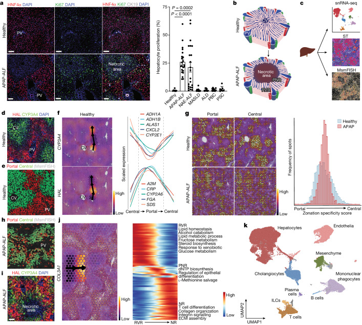
- In human APAP-ALF and NAE-ALF, hepatocyte proliferation was “markedly increased” versus healthy liver and all chronic liver diseases examined; the text says this increase had no correlation with patient age.
- The integrated human dataset contained 72,262 nuclei from healthy (n = 9), APAP-ALF (n = 10), and NAE-ALF (n = 12) livers; the mouse injury time course contained 59,051 nuclei.
- The authors identified a distinct ANXA2+ hepatocyte subpopulation in human APAP-ALF and NAE-ALF, with GO terms for ameboidal-type cell migration, epithelial cell migration, and regulation of cell shape.
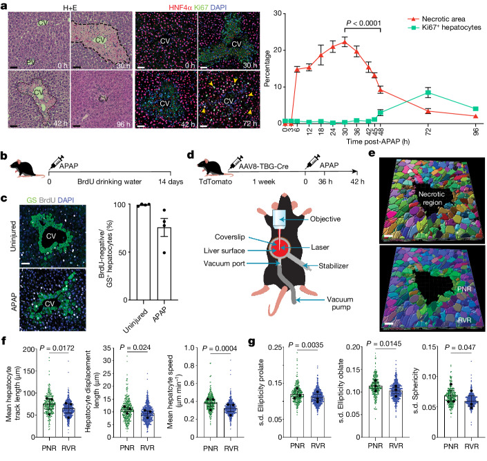
- In mice, peak hepatocyte necrosis occurred at 30 h after APAP (male: 22.3 ± 1.3% s.e.m.), while hepatocyte proliferation peaked at 72 h; wound closure therefore preceded proliferation. At 14 days, 75.6% ± 9.4% of glutamine synthetase+ hepatocytes in males and 86.9% ± 2.3% in females were BrdU−, supporting that most peri-central hepatocytes did not arise from proliferation.
- Hepatocyte-specific Anxa2 knockdown reduced wound closure in vitro and, in vivo, abrogated necrotic wound closure after APAP injury; 4D intravital imaging showed 19.4% ± 2.8% reduction in necrosis during the imaging period and greater mobility in peri-necrotic hepatocytes than in residual viable-region hepatocytes.
## Methods
- Multimodal design combining human snRNA-seq, spatial transcriptomics, multiplex smFISH, immunohistochemistry/immunofluorescence, mouse APAP injury time courses, 4D intravital microscopy, and in vitro ANXA2 knockdown/scratch-wound assays.
- Human samples came from explanted livers or biopsies across multiple centers; computational analyses covered healthy, APAP-ALF, NAE-ALF, and some chronic liver disease tissues. The paper also analyzed mouse APAP injury across multiple timepoints and included reporter-line lineage tracing.
- Main analyses included Seurat/Harmony-based snRNA-seq processing, spatial trajectory modeling with SPATA2, interactome analysis with CellChat, GO enrichment, and standard statistical testing in GraphPad Prism.
## Limitations
- The paper states that blinding to tissue origin was not performed for some mouse experiments, including IVM studies.
- Human ALF tissue was biobanked/explant tissue rather than prospective mechanistic sampling; this limits direct causal inference from human data (evident from the design).
- The study focuses primarily on APAP-ALF and non-A–E hepatitis ALF for deep mechanistic work, so generalization to all causes of ALF is not directly established by the text (evident from the text).
Model-written summary. Check it against the paper before relying on it.
Integrity signals
Registry
Cites
Statistics
Figures



Verification
Claims the paper makes
14 Valid · 0 Not valid · 4 Not checked · 117 Own results and interpretations
The cited sources support only part of this claim — read each source’s verdict below.
In the paper
“however, in the setting of acute liver failure (ALF), this regenerative capacity is often overwhelmed, leaving emergency liver transplantation as the only curative option3–5”
§ Multimodal decoding of human liver regeneration > Abstract The liver has a unique ability to regenerate1,2; however, in the setting of acute liver failure (ALF), this regenerative capacity is often overwhelmed, leaving emergency liver transplantation as the only curative option3–5. Here, to advance understanding of human liver regeneration, we use paired single-nucleus RNA sequencing combined with spatial profiling of healthy and ALF explant human livers to generate a single-cell, pan-lineage atlas of human liver regeneration. We uncover a novel ANXA2+ migratory hepatocyte subpopulation, which emerges during human liver regeneration, and a corollary subpopulation in a mouse model of acetaminophen (APAP)-induced liver regeneration. Interrogation of necrotic wound closure and hepatocyte proliferation across multiple timepoints following APAP-induced liver injury in mice demonstrates that wound closure precedes hepatocyte proliferation. Four-dimensional intravital imaging of APAP-induced mouse liver injury identifies motile hepatocytes at the edge of the necrotic area, enabling collective migration of the hepatocyte sheet to effect wound closure. Depletion of hepatocyte ANXA2 reduces hepatocyte growth factor-induced human and mouse hepatocyte migration in vitro, and abrogates necrotic wound closure following APAP-induced mouse liver injury. Together, our work dissects unanticipated aspects of liver regeneration, demonstrating an uncoupling of wound closure and hepatocyte proliferation and uncovering a novel migratory hepatocyte subpopulation that mediates wound closure following liver injury. Therapies designed to promote rapid reconstitution of normal hepatic microarchitecture and reparation of the gut–liver barrier may advance new areas of therapeutic discovery in regenerative medicine.
What the sources say
- Partly supportedcites [3] Stravitz, R. T. & Lee, W. M. Acute liver failure. Lancet 394, 869–881 (2019). sourceExperimental — accuracy not yet measured
The Pathogenesis section says failure of regeneration is an important feature of acute liver failure for at least some causes, and the Liver transplantation section describes transplantation as life-saving. But the Outcomes section reports that many patients recover with their native liver—about 75% after paracetamol-induced failure and 40% after other causes—so the paper does not support transplantation as the only curative option for acute liver failure generally. Page numbers were not available in our copy. (LLM, conf=0.88)
- Not checked — no full text of the sourcecites [4] Bernal, W. & Wendon, J. Acute liver failure. N. Engl. J. Med. 369, 2525–2534 (2013). source
- Partly supportedcites [5] Tujios, S., Todd Stravitz, R. & Lee, W. M. Management of acute liver failure: update 2022. Semin. Liver Dis. 42, 362–378 (2022). sourceExperimental — accuracy not yet measured
The Abstract and Section III describe urgent transplantation as important for selected patients with acute liver failure. But Section III also reports spontaneous recovery with supportive care, and the Conclusion attributes improved spontaneous survival to treatments and critical care. The paper therefore does not support transplantation as the only curative option, nor does it say that regeneration is often overwhelmed. (LLM, conf=0.86)
plainstates a cause- Asserted: passes — The authors put it forward as true.
- Checkable: passes — It can be checked against evidence.
- Load-bearing: passes — The paper’s conclusions depend on it.
- Based: passes — The paper gives a reason why it holds.
- Partly supported
Every source the paper cites for this claim was checked and supports it.
In the paper
“Hepatocyte replenishment can occur via conversion of cholangiocytes during severe liver injury12,13”
§ Multimodal decoding of human liver regeneration > Main
What the sources say
- Supportedcites [12] Raven, A. et al. Cholangiocytes act as facultative liver stem cells during impaired hepatocyte regeneration. Nature 547, 350–354 (2017). source
The Abstract (page not specified) reports that, when hepatocyte proliferation was impaired during liver injury, lineage tracing showed cholangiocyte-derived hepatocytes. It describes this as regeneration of functional hepatocytes from biliary cells, supporting the claim that replenishment can occur by this route during severe injury. (LLM, conf=0.93)
- Supportedcites [13] Deng, X. et al. Chronic liver injury induces conversion of biliary epithelial cells into hepatocytes. Cell Stem Cell 23, 114–122.e3 (2018). source
The Abstract and Introduction (p.1) of our copy state that, following severe chronic liver injury induced by long-term TAA or DDC treatment, NPC-derived hepatocytes replenish a large fraction of the parenchyma and that lineage tracing of biliary epithelial cells (cholangiocytes) shows BECs are a source of new hepatocytes via a bi-phenotypic intermediate, 'without genetic interventions'. The Discussion (p.121) repeats that BECs are an important cellular source of hepatocyte regeneration in 6–12 month chronic injury models, with equivalent bi-phenotypic cells found in cirrhotic human livers. The claim's terms ('conversion of cholangiocytes', 'severe liver injury') map directly onto the paper's BEC-to-hepatocyte conversion during severe/chronic injury. (LLM, conf=0.92)
plainstates a cause- Asserted: passes — The authors put it forward as true.
- Checkable: passes — It can be checked against evidence.
- Load-bearing: passes — The paper’s conclusions depend on it.
- Based: passes — The paper gives a reason why it holds.
- Supported
Every source the paper cites for this claim was checked and supports it.
In the paper
“Mice with knockout of the gene encoding plasminogen displayed persistent centrilobular damage and severe impairment of repair following CCl4-induced acute liver injury, despite a normal hepatocyte proliferative response compared with control mice26”
§ Multimodal decoding of human liver regeneration > Discussion
What the sources say
- Supportedcites [26] Bezerra, J. A. et al. Plasminogen deficiency leads to impaired remodeling after a toxic injury to the liver. Proc. Natl Acad. Sci. USA 96, 15143–15148 (1999). source
Split into 2 atoms — 2 confirmed, 0 partially supported, 0 disputed, 0 misattributed, 0 unverifiable. Expand the claim to see per-atom verdicts.
plainstates a cause- Asserted: passes — The authors put it forward as true.
- Checkable: passes — It can be checked against evidence.
- Load-bearing: passes — The paper’s conclusions depend on it.
- Based: passes — The paper gives a reason why it holds.
- Supported
Every source the paper cites for this claim was checked and supports it.
In the paper
“Conditional knockout of the HGF receptor (Met) in mouse hepatocytes followed by CCl4-induced acute liver injury impaired centrilobular wound closure and restitution of normal tissue architecture, despite similar levels of hepatocyte proliferation compared with controls27”
§ Multimodal decoding of human liver regeneration > Discussion
What the sources say
- Supportedcites [27] Huh, C.-G. et al. Hepatocyte growth factor/c-Met signaling pathway is required for efficient liver regeneration and repair. Proc. Natl Acad. Sci. USA 101, 4477–4482 (2004). source
Split into 2 atoms — 2 confirmed, 0 partially supported, 0 disputed, 0 misattributed, 0 unverifiable. Expand the claim to see per-atom verdicts.
plainstates a cause- Asserted: passes — The authors put it forward as true.
- Checkable: passes — It can be checked against evidence.
- Load-bearing: passes — The paper’s conclusions depend on it.
- Based: passes — The paper gives a reason why it holds.
- Supported
The cited sources support only part of this claim — read each source’s verdict below.
In the paper
“Expeditious wound closure in the liver, with restoration of the gut–liver barrier following acute, massive epithelial injury, may be a key determinant of patient outcome in ALF in which sepsis and multi-organ failure are the most common cause of death3–5.”
§ Multimodal decoding of human liver regeneration > Discussion
What the sources say
- Partly supportedcites [3] Stravitz, R. T. & Lee, W. M. Acute liver failure. Lancet 394, 869–881 (2019). sourceExperimental — accuracy not yet measured
The Management of systemic complications section (page unavailable) says systemic complications are the most common cause of death in acute liver failure and that infection often triggers multisystem organ failure. It does not identify sepsis specifically as a most common cause of death. The wound-closure proposal is the citing authors’ framing, not a finding attributed to this source. (LLM, conf=0.83)
- Not checked — no full text of the sourcecites [4] Bernal, W. & Wendon, J. Acute liver failure. N. Engl. J. Med. 369, 2525–2534 (2013). source
- Could not verifycites [5] Tujios, S., Todd Stravitz, R. & Lee, W. M. Management of acute liver failure: update 2022. Semin. Liver Dis. 42, 362–378 (2022). sourceExperimental — accuracy not yet measured
The Abstract links ALF outcomes to liver-injury severity and systemic complications, including sepsis, while the Conclusion mentions possible future treatments that promote liver regeneration. Neither passage says that rapid wound closure or restoration of the gut–liver barrier determines patient outcome; searches for those mechanisms found no supporting passage in our copy. (LLM, conf=0.72)
hedgedstates a possible cause- Asserted: passes — The authors put it forward as true.
- Checkable: passes — It can be checked against evidence.
- Load-bearing: passes — The paper’s conclusions depend on it.
- Based: passes — The paper gives a reason why it holds.
- Partly supported
84 more claims
Create a free account to see all 135 claims and to verify the 9 references still open.
- It may state two separate claims as one. 24
- Probably not a substantial claim. 28
- The quote only partly states this claim. 1
Claims other papers make about this one
Each is checked against this paper’s own text: does it say what the citing paper says it does?
In the citing paper
“Conversely, a recent study has identified a subset of hepatocytes that express annexin A2 that preferentially repopulate damaged tissue in acute liver injury (115) – the relevance of this finding to MASH injury is not clear yet.”
From Found in translation—Fibrosis in metabolic dysfunction–associated steatohepatitis (MASH) (2023) · cites this paper as [115]
Citations
Cites
Integrated analysis of multimodal single-cell data
Hao, Y., Hao, S., Andersen-Nissen, E., Mauck, W. M., Zheng, S., Butler, A., Lee, M. J., Wilk, A. J., Darby, C., Zagar, M., et al. (2020) · doi:10.1101/2020.10.12.335331 · Cited by 15,508
clusterProfiler 4.0: A universal enrichment tool for interpreting omics data
(2021) · doi:10.1016/j.xinn.2021.100141 · Cited by 12,455 · cited 1×
“Gene ontology analysis was performed using the clusterProfiler R package 41 .”
MethodFast, sensitive and accurate integration of single-cell data with Harmony
Korsunsky, I., Millard, N., Fan, J., Slowikowski, K., Zhang, F., Wei, K., Baglaenko, Y., Brenner, M., Loh, P.-R., & Raychaudhuri, S. (2019) · Nature Methods · doi:10.1038/s41592-019-0619-0 · Cited by 10,488
QuPath: Open source software for digital pathology image analysis
Bankhead, P., Loughrey, M. B., Fernández, J. A., Dombrowski, Y., McArt, D. G., Dunne, P. D., McQuaid, S., Gray, R. T., Murray, L. J., Coleman, H. G., et al. (2017) · Scientific Reports · doi:10.1038/s41598-017-17204-5 · Cited by 8,908
SCANPY: large-scale single-cell gene expression data analysis
Wolf, F. A., Angerer, P., & Theis, F. J. (2018) · Genome Biology · doi:10.1186/s13059-017-1382-0 · Cited by 8,881
Inference and analysis of cell-cell communication using CellChat
Jin, S., Guerrero-Juarez, C. F., Zhang, L., Chang, I., Ramos, R., Kuan, C.-H., Myung, P., Plikus, M. V., & Nie, Q. (2021) · Nature Communications · doi:10.1038/s41467-021-21246-9 · Cited by 8,409
A robust and high-throughput Cre reporting and characterization system for the whole mouse brain
Madisen, L., Zwingman, T. A., Sunkin, S. M., Oh, S. W., Zariwala, H. A., Gu, H., Ng, L. L., Palmiter, R. D., Hawrylycz, M. J., Jones, A. R., et al. (2010) · Nature Neuroscience · doi:10.1038/nn.2467 · Cited by 6,907
Cellpose: a generalist algorithm for cellular segmentation
Stringer, C., Wang, T., Michaelos, M., & Pachitariu, M. (2021) · Nature Methods · doi:10.1038/s41592-020-01018-x · Cited by 4,369
A global double-fluorescent Cre reporter mouse
Muzumdar, M. D., Tasic, B., Miyamichi, K., Li, L., & Luo, L. (2007) · genesis · doi:10.1002/dvg.20335 · Cited by 3,491
Scrublet: computational identification of cell doublets in single-cell transcriptomic data
Wolock, S. L., Lopez, R., & Klein, A. M. (2019) · Cell Systems · doi:10.1016/j.cels.2018.11.005 · Cited by 2,758
Scrublet: computational identification of cell doublets in single-cell transcriptomic data
Wolock, S. L., Lopez, R., & Klein, A. M. (2018) · doi:10.1101/357368 · Cited by 2,758
Cell Detection with Star-Convex Polygons
Schmidt, U., Weigert, M., Broaddus, C., & Myers, G. (2018) · Lecture Notes in Computer Science · doi:10.1007/978-3-030-00934-2_30 · Cited by 1,826
Resolving the fibrotic niche of human liver cirrhosis at single-cell level
Ramachandran, P., Dobie, R., Wilson-Kanamori, J. R., Dora, E. F., Henderson, B. E. P., Luu, N. T., Portman, J. R., Matchett, K. P., Brice, M., Marwick, J. A., et al. (2019) · Nature · doi:10.1038/s41586-019-1631-3 · Cited by 1,784
Self-citationSoupX removes ambient RNA contamination from droplet-based single-cell RNA sequencing data
Young, M. D., & Behjati, S. (2020) · GigaScience · doi:10.1093/gigascience/giaa151 · Cited by 1,739
SoupX removes ambient RNA contamination from droplet based single-cell RNA sequencing data
Young, M. D., & Behjati, S. (2018) · doi:10.1101/303727 · Cited by 1,648
Bernal, W., & Wendon, J. (2013) · New England Journal of Medicine · doi:10.1056/nejmra1208937 · Cited by 1,169
A human liver cell atlas reveals heterogeneity and epithelial progenitors
Aizarani, N., Saviano, A., Sagar, Mailly, L., Durand, S., Herman, J. S., Pessaux, P., Baumert, T. F., & Grün, D. (2019) · Nature · doi:10.1038/s41586-019-1373-2 · Cited by 1,147 · cited 1×
“5a).”
Single-cell spatial reconstruction reveals global division of labour in the mammalian liver
Halpern, K. B., Shenhav, R., Matcovitch-Natan, O., Tóth, B., Lemze, D., Golan, M., Massasa, E. E., Baydatch, S., Landen, S., Moor, A. E., et al. (2017) · Nature · doi:10.1038/nature21065 · Cited by 1,059
Liver regeneration: biological and pathological mechanisms and implications
Michalopoulos, G. K., & Bhushan, B. (2021) · Nature Reviews Gastroenterology & Hepatology · doi:10.1038/s41575-020-0342-4 · Cited by 998
Stravitz, R. T., & Lee, W. M. (2019) · The Lancet · doi:10.1016/s0140-6736(19)31894-x · Cited by 844
Self-citationQuPath: Open source software for digital pathology image analysisPreprint
Bankhead, P., Loughrey, M. B., Fernández, J. A., et al. (2017) · bioRxiv (Cold Spring Harbor Laboratory) · doi:10.1101/099796 · Cited by 808
Huh, C.-G., Factor, V. M., Sánchez, A., Uchida, K., Conner, E. A., & Thorgeirsson, S. S. (2004) · Proceedings of the National Academy of Sciences · doi:10.1073/pnas.0306068101 · Cited by 750
A single-cell and single-nucleus RNA-Seq toolbox for fresh and frozen human tumors
Slyper, M., Porter, C. B. M., Ashenberg, O., Waldman, J., Drokhlyansky, E., Wakiro, I., Smillie, C., Smith-Rosario, G., Wu, J., Dionne, D., et al. (2020) · Nature Medicine · doi:10.1038/s41591-020-0844-1 · Cited by 744
Self-renewing diploid Axin2+ cells fuel homeostatic renewal of the liver
Wang, B., Zhao, L., Fish, M., Logan, C. Y., & Nusse, R. (2015) · Nature · doi:10.1038/nature14863 · Cited by 674
Spatially resolved multi-omics deciphers bidirectional tumor-host interdependence in glioblastoma
Ravi, V. M., Will, P., Kueckelhaus, J., Sun, N., Joseph, K., Salié, H., Vollmer, L., Kuliesiute, U., von Ehr, J., Benotmane, J. K., et al. (2022) · Cancer Cell · doi:10.1016/j.ccell.2022.05.009 · Cited by 663
Inference and analysis of cell-cell communication using CellChatPreprint
Jin, S., Guerrero‐Juarez, C. F., Zhang, L., et al. (2020) · bioRxiv (Cold Spring Harbor Laboratory) · doi:10.1101/2020.07.21.214387 · Cited by 604
Cholangiocytes act as facultative liver stem cells during impaired hepatocyte regeneration
Raven, A., Lu, W.-Y., Man, T. Y., Ferreira-Gonzalez, S., O’Duibhir, E., Dwyer, B. J., Thomson, J. P., Meehan, R. R., Bogorad, R., Koteliansky, V., et al. (2017) · Nature · doi:10.1038/nature23015 · Cited by 533 · cited 1×
“In addition, this Figure shows peak proliferation at the same time as peak necrosis.”
Democratising deep learning for microscopy with ZeroCostDL4Mic
von Chamier, L., Laine, R. F., Jukkala, J., Spahn, C., Krentzel, D., Nehme, E., Lerche, M., Hernández-Pérez, S., Mattila, P. K., Karinou, E., et al. (2021) · Nature Communications · doi:10.1038/s41467-021-22518-0 · Cited by 521
Single-cell transcriptomics uncovers zonation of function in the mesenchyme during liver fibrosis
Dobie, R., Wilson-Kanamori, J. R., Henderson, B. E. P., Smith, J. R., Matchett, K. P., Portman, J. R., Wallenborg, K., Picelli, S., Zagorska, A., Pendem, S. V., et al. (2019) · Cell Reports · doi:10.1016/j.celrep.2019.10.024 · Cited by 420
Self-citationOpposing roles of hepatic stellate cell subpopulations in hepatocarcinogenesis
Filliol, A., Saito, Y., Nair, A., Dapito, D. H., Yu, L.-X., Ravichandra, A., Bhattacharjee, S., Affo, S., Fujiwara, N., Su, H., et al. (2022) · Nature · doi:10.1038/s41586-022-05289-6 · Cited by 349
Self-citationLiver homeostasis is maintained by midlobular zone 2 hepatocytes
Wei, Y., Wang, Y. G., Jia, Y., Li, L., Yoon, J., Zhang, S., Wang, Z., Zhang, Y., Zhu, M., Sharma, T., et al. (2021) · Science · doi:10.1126/science.abb1625 · Cited by 292 · cited 1×
“Referee #2 (Remarks to the Author): In this study, Matchett et al aim to shed further lights on the mechanisms of liver regeneration upon Acute Liver Failure (ALF).”
Chronic Liver Injury Induces Conversion of Biliary Epithelial Cells into Hepatocytes
Deng, X., Zhang, X., Li, W., Feng, R.-X., Li, L., Yi, G.-R., Zhang, X.-N., Yin, C., Yu, H.-Y., Zhang, J.-P., et al. (2018) · Cell Stem Cell · doi:10.1016/j.stem.2018.05.022 · Cited by 289 · cited 1×
“In addition, this Figure shows peak proliferation at the same time as peak necrosis.”
Proliferation tracing reveals regional hepatocyte generation in liver homeostasis and repair
He, L., Pu, W., Liu, X., Zhang, Z., Han, M., Li, Y., Huang, X., Han, X., Li, Y., Liu, K., et al. (2021) · Science · doi:10.1126/science.abc4346 · Cited by 261 · cited 1×
“22 Headon, D. Reversing stratification during wound healing. Nature Cell Biology 19, 595-597 (2017). 23 Chopyk, D. M. et al. Acetaminophen Intoxication Rapidly Induces Apoptosis of Intestinal Crypt Stem Cells and Enhances Intestinal Permeability. Hepatology Communications 3, 1435-1449 (2019). 24 Niu, M. et al. Intesti…”
Broad Distribution of Hepatocyte Proliferation in Liver Homeostasis and Regeneration
Chen, F., Jimenez, R. J., Sharma, K., Luu, H. Y., Hsu, B. Y., Ravindranathan, A., Stohr, B. A., & Willenbring, H. (2020) · Cell Stem Cell · doi:10.1016/j.stem.2019.11.001 · Cited by 236 · cited 3×
“This would fit the ANXA2 staining in the injury area with HNF4a-negative/presumably dead hepatocytes, as well as the abnormal morphology of ANXA2+ hepatocytes.”
The spatiotemporal program of zonal liver regeneration following acute injury
Ben-Moshe, S., Veg, T., Manco, R., Dan, S., Papinutti, D., Lifshitz, A., Kolodziejczyk, A. A., Bahar Halpern, K., Elinav, E., & Itzkovitz, S. (2022) · Cell Stem Cell · doi:10.1016/j.stem.2022.04.008 · Cited by 224 · cited 1×
“2) The roles of ANXA2 and ANXA2+ hepatocytes remain unclear.”
Matsumoto, T., Wakefield, L., Tarlow, B. D., & Grompe, M. (2020) · Cell Stem Cell · doi:10.1016/j.stem.2019.11.014 · Cited by 194
Epithelial plasticity, epithelial-mesenchymal transition, and the TGF-β family
Katsuno, Y., & Derynck, R. (2021) · Developmental Cell · doi:10.1016/j.devcel.2021.02.028 · Cited by 187
Critical role of c-jun (NH2) terminal kinase in paracetamol- induced acute liver failure
Henderson, N. C., Pollock, K. J., Frew, J., Mackinnon, A. C., Flavell, R. A., Davis, R. J., Sethi, T., & Simpson, K. J. (2007) · Gut · doi:10.1136/gut.2006.104372 · Cited by 182
Self-citationLiver Regeneration after Acetaminophen Hepatotoxicity
Bhushan, B., & Apte, U. (2019) · The American Journal of Pathology · doi:10.1016/j.ajpath.2018.12.006 · Cited by 179
Fast, sensitive, and accurate integration of single cell data with HarmonyPreprint
Korsunsky, I., Fan, J., Slowikowski, K., et al. (2018) · bioRxiv · doi:10.1101/461954 · Cited by 177
Wang, S., Li, K., Pickholz, E., Dobie, R., Matchett, K. P., Henderson, N. C., Carrico, C., Driver, I., Borch Jensen, M., Chen, L., et al. (2023) · Science Translational Medicine · doi:10.1126/scitranslmed.add3949 · Cited by 176 · cited 1×
“…the possible role of transdifferentiation (Reviewer #4) -Relate findings to clinical presentation (Reviewer #4) We thank the reviewers for their very helpful and thoughtful comments, and we feel that the new data we have generated in response to their comments has very much enhanced our manuscript.”
Self-citationAutomatic and unbiased segmentation and quantification of myofibers in skeletal muscle
Waisman, A., Norris, A. M., Costa, M. E., & Kopinke, D. (2021) · Scientific Reports · doi:10.1038/s41598-021-91191-6 · Cited by 168
AXIN2+ Pericentral Hepatocytes Have Limited Contributions to Liver Homeostasis and Regeneration
Sun, T., Pikiolek, M., Orsini, V., Bergling, S., Holwerda, S., Morelli, L., Hoppe, P. S., Planas-Paz, L., Yang, Y., Ruffner, H., et al. (2020) · Cell Stem Cell · doi:10.1016/j.stem.2019.10.011 · Cited by 166
Plasminogen deficiency leads to impaired remodeling after a toxic injury to the liver
Bezerra, J. A., Bugge, T. H., Melin-Aldana, H., Sabla, G., Kombrinck, K. W., Witte, D. P., & Degen, J. L. (1999) · Proceedings of the National Academy of Sciences · doi:10.1073/pnas.96.26.15143 · Cited by 137 · cited 1×
“• Line 168, Sfig1i, has too many colors, difficult to track any pattern, in particular for color-blinded readers!”
Management of Acute Liver Failure: Update 2022
Tujios, S., Stravitz, R. T., & Lee, W. M. (2022) · Seminars in Liver Disease · doi:10.1055/s-0042-1755274 · Cited by 117 · cited 2×
“2j were independently identified from the mouse ANXA2 + hepatocyte cluster, the marker genes of which were similar but not equivalent to the genes listed in the human migratory score.”
Self-citationZhao, L., Jin, Y., Donahue, K., Tsui, M., Fish, M., Logan, C. Y., Wang, B., & Nusse, R. (2019) · Hepatology · doi:10.1002/hep.30563 · Cited by 111 · cited 1×
“2) The roles of ANXA2 and ANXA2+ hepatocytes remain unclear.”
Intestinal Dysbiosis Amplifies Acetaminophen-Induced Acute Liver Injury
Schneider, K. M., Elfers, C., Ghallab, A., Schneider, C. V., Galvez, E. J. C., Mohs, A., Gui, W., Candels, L. S., Wirtz, T. H., Zuehlke, S., et al. (2021) · Cellular and Molecular Gastroenterology and Hepatology · doi:10.1016/j.jcmgh.2020.11.002 · Cited by 108 · cited 1×
“2j were independently identified from the mouse ANXA2 + hepatocyte cluster, the marker genes of which were similar but not equivalent to the genes listed in the human migratory score.”
Babbin, B. A., Parkos, C. A., Mandell, K. J., Winfree, L. M., Laur, O., Ivanov, A. I., & Nusrat, A. (2007) · The American Journal of Pathology · doi:10.2353/ajpath.2007.060647 · Cited by 89 · cited 1×
“Given the multifaceted nature of the following comment, we have partitioned our responses below: The finding that peri-wound hepatocytes promote liver regeneration following loss of zone3 by moving to the pericentral niche and acquiring a zone3 signature is not new.”
Kiourtis, C., Wilczynska, A., Nixon, C., Clark, W. A., May, S., & Bird, T. G. (2021) · Biology Open · doi:10.1242/bio.058678 · Cited by 75
Self-citationKpetemey, M., Dasgupta, S., Rajendiran, S., Das, S., Gibbs, L. D., Shetty, P., Gryczynski, Z., & Vishwanatha, J. K. (2015) · Molecular Cancer · doi:10.1186/s12943-015-0428-8 · Cited by 68
A single-cell and single-nucleus RNA-seq toolbox for fresh and frozen human tumors
Slyper, M., Porter, C. B. M., Ashenberg, O., Waldman, J., Drokhlyansky, E., Wakiro, I., Smillie, C., Smith-Rosario, G., Wu, J., Dionne, D., et al. (2019) · bioRxiv (Cold Spring Harbor Laboratory) · doi:10.1101/761429 · Cited by 66
Annexin A2 Regulates β1 Integrin Internalization and Intestinal Epithelial Cell Migration
Rankin, C. R., Hilgarth, R. S., Leoni, G., Kwon, M., Den Beste, K. A., Parkos, C. A., & Nusrat, A. (2013) · Journal of Biological Chemistry · doi:10.1074/jbc.m112.440909 · Cited by 55 · cited 1×
“ANXA2, which we have identified as a key marker in both human and mouse migratory hepatocytes, has previously been shown to regulate cell migration in other disease settings including carcinogenesis 16-18 .”
BackgroundSynergistic enhancement of EGF, but not HGF, stimulated hepatocyte motility by TGF-β1 in vitro
Stolz, D. B., & Michalopoulos, G. K. (1997) · Journal of Cellular Physiology · doi:10.1002/(sici)1097-4652(199701)170:1<57::aid-jcp7>3.0.co;2-k · Cited by 45
Spatially Resolved Multi-Omics Deciphers Bidirectional Tumor-Host Interdependence in GlioblastomaPreprint
Ravi, V. M., Will, P., Kueckelhaus, J., Sun, N., Joseph, K., Salié, H., Vollmer, L., Kuliešiūtė, U., von Ehr, J., Benotmane, J. K., et al. (2021) · SSRN Electronic Journal · doi:10.2139/ssrn.3865275 · Cited by 36
Chopyk, D. M., Stuart, J. D., Zimmerman, M. G., Wen, J., Gumber, S., Suthar, M. S., Thapa, M., Czaja, M. J., & Grakoui, A. (2019) · Hepatology Communications · doi:10.1002/hep4.1406 · Cited by 33 · cited 1×
“2j were independently identified from the mouse ANXA2 + hepatocyte cluster, the marker genes of which were similar but not equivalent to the genes listed in the human migratory score.”
Niu, M., Luo, Z., Gong, S., Win, S., Kaplowitz, N., Jiang, Y., & Chen, P. (2020) · The American Journal of Pathology · doi:10.1016/j.ajpath.2019.09.009 · Cited by 26
Loss of Integrin αvβ8 in Murine Hepatocytes Accelerates Liver Regeneration
Greenhalgh, S. N., Matchett, K. P., Taylor, R. S., Huang, K., Li, J. T., Saeteurn, K., Donnelly, M. C., Simpson, E. E. M., Pollack, J. L., Atakilit, A., et al. (2018) · American Journal Of Pathology · doi:10.1016/j.ajpath.2018.10.007 · Cited by 17
Self-citationReversing stratification during wound healing
Headon, D. (2017) · Nature Cell Biology · doi:10.1038/ncb3545 · Cited by 13 · cited 1×
“Generating Pearson correlations between patient age and the harmony components of the hepatocyte dataset demonstrated that age did not influence variability in the snRNA-seq data (Extended Data Fig.”
Tujios, S. R., & Lee, W. M. (2011) · Wiley eBooks · doi:10.1002/9781444341294.ch5 · Cited by 4
Self-citation
Cited by
Harrison, S. A., Frias, J. P., Neff, G., Abrams, G. A., Lucas, K. J., Sanchez, W., Gogia, S., Sheikh, M. Y., Behling, C., Bedossa, P., et al. (2023) · The Lancet Gastroenterology & Hepatology · doi:10.1016/s2468-1253(23)00272-8 · Cited by 276
Found in translation—Fibrosis in metabolic dysfunction–associated steatohepatitis (MASH)
Wang, S., & Friedman, S. L. (2023) · Science Translational Medicine · doi:10.1126/scitranslmed.adi0759 · Cited by 158
“Conversely, a recent study has identified a subset of hepatocytes that express annexin A2 that preferentially repopulate damaged tissue in acute liver injury ( 114 ) — the relevance of this finding to MASH injury is not clear yet.”
BackgroundECM formation and degradation during fibrosis, repair, and regeneration
Mayorca-Guiliani, A. E., Leeming, D. J., Henriksen, K., Mortensen, J. H., Nielsen, S. H., Anstee, Q. M., Sanyal, A. J., Karsdal, M. A., & Schuppan, D. (2025) · npj Metabolic Health and Disease · doi:10.1038/s44324-025-00063-4 · Cited by 176
“A subpopulation of annexin 2 (ANXA2) positive hepatocytes 139 has been shown to respond to HGF and drive the regenerative response to injury.”
Molecular mechanisms in liver repair and regeneration: from physiology to therapeutics
Ma, X., Huang, T., Chen, X., Li, Q., Liao, M., Fu, L., Huang, J., Yuan, K., Wang, Z., & Zeng, Y. (2025) · Signal Transduction and Targeted Therapy · doi:10.1038/s41392-024-02104-8 · Cited by 133
Hepatic stellate cells control liver zonation, size and functions via R-spondin 3
Sugimoto, A., Saito, Y., Wang, G., Sun, Q., Yin, C., Lee, K. H., Geng, Y., Rajbhandari, P., Hernandez, C., Steffani, M., et al. (2025) · Nature · doi:10.1038/s41586-025-08677-w · Cited by 133
Self-citation
Metadata
- Authors
- Matchett, K. P., Wilson-Kanamori, J. R., Portman, J. R., Kapourani, C. A., Fercoq, F., May, S., Zajdel, E., Beltran, M., Sutherland, E. F., Mackey, J. B. G., Brice, M., Wilson, G. C., Wallace, S. J., Kitto, L., Younger, N. T., Dobie, R., Mole, D. J., Oniscu, G. C., Wigmore, S. J., Ramachandran, P., Vallejos, C. A., Carragher, N. O., Saeidinejad, M. M., Quaglia, A., Jalan, R., Simpson, K. J., Kendall, T. J., Rule, J. A., Lee, W. M., Hoare, M., Weston, C. J., Marioni, J. C., Teichmann, S. A., Bird, T. G., Carlin, L. M., & Henderson, N. C.
- Affiliations
- Centre for Inflammation Research (GB)
- Cancer Research UK Scotland Institute (GB)
- Edinburgh Royal Infirmary (GB)
- The Alan Turing Institute (GB)
- Edinburgh Cancer Research (GB)
- University College London (GB)
- Royal Free London NHS Foundation Trust (GB)
- European Foundation for the Study of Chronic Liver Failure (ES)
- The University of Texas Southwestern Medical Center (US)
- University of Cambridge (GB)
- University Hospitals Birmingham NHS Foundation Trust (GB)
- European Bioinformatics Institute (GB)
- Corresponding author
- Henderson, N. C.
- ORCID
- Wilson-Kanamori, J. R.
- Portman, J. R.
- Kapourani, C. A.
- Fercoq, F.
- May, S.
- Beltran, M.
- Sutherland, E. F.
- Mackey, J. B. G.
- Brice, M.
- Wallace, S. J.
- Younger, N. T.
- Dobie, R.
- Mole, D. J.
- Oniscu, G. C.
- Wigmore, S. J.
- Ramachandran, P.
- Vallejos, C. A.
- Carragher, N. O.
- Saeidinejad, M. M.
- Quaglia, A.
- Jalan, R.
- Kendall, T. J.
- Hoare, M.
- Weston, C. J.
- Teichmann, S. A.
- Bird, T. G.
- Carlin, L. M.
- Henderson, N. C.
- Venue
- Nature
- Published
- 2024-05-01
- Volume
- 630
- Issue
- 8015
- Pages
- 158-165
- ISSN
- 0028-0836 (ISSN-L)
- 1476-4687
- PMC
- PMC11153152
- OpenAlex
- W4396544281
- Cited by
- 140
- References
- 58
- Citations by year
- 2026: 50 · 2025: 61 · 2024: 25
- FWCI
- 35.15
- Type
- journal article
- Language
- en
- Open access
- hybrid
- doi.org
- Licence
- cc-by
- creativecommons.org/licenses/by/4.0 (tdm)
- creativecommons.org/licenses/by/4.0 (vor)
- Locations
- Nature (publishedVersion, cc-by)
- bioRxiv (Cold Spring Harbor Laboratory) (acceptedVersion, cc-by)
- PubMed Central (publishedVersion, cc-by)
- PubMed (publishedVersion)
- ENLIGHTEN (Jurnal Bimbingan dan Konseling Islam) (acceptedVersion, cc-by-sa)
- HAL (submittedVersion)
- RePEc: Research Papers in Economics (submittedVersion)
- UCL Discovery (University College London) (submittedVersion)
- Figshare (submittedVersion, cc-by)
- University of Birmingham Research Portal (University of Birmingham) (submittedVersion, cc-by)
- Edinburgh Research Explorer (University of Edinburgh) (submittedVersion, other-oa)
- Apollo (University of Cambridge) (submittedVersion, cc-by)
- Figshare (cc-by)
- Funders
- Wellcome Trust (219542/Z/19/Z, WT107492Z)
- Cancer Research UK (A31287, A17196/, A23983)
- Tenovus
- University of Edinburgh (MC_UU_00009/2)
- University of Birmingham
- Medical Research Council (MR/X00970X/1, MR/N008340/1, MR/P008887/1, MR/M01245X/1, MC_UU_00009/2, MR/W015919/1)
- Beatson Institute for Cancer Research (A31287, A17196, A23983)
- Topics
- Health Sciences › Medicine › Hepatology › Liver physiology and pathology
- Life Sciences › Pharmacology, Toxicology and Pharmaceutics › Pharmacology › Drug-Induced Hepatotoxicity and Protection
- Health Sciences › Medicine › Surgery › Tissue Engineering and Regenerative Medicine
- Keywords
- liver regeneration, acute liver failure, hepatocyte migration, annexin A2, hepatocyte proliferation, acetaminophen hepatotoxicity, hepatocyte growth factor, single-nucleus RNA sequencing, spatial transcriptomics, intravital imaging, collective migration, liver explants
- MeSH terms
- Acetaminophen, Single-Cell Gene Expression Analysis, Animals, Cell Movement, Disease Models, Animal, Female, Humans, Liver, Liver Regeneration, Male, Mice, Inbred C57BL, Necrosis, Wound Healing, Liver Failure, Acute, Hepatocyte Growth Factor, Annexin A2, Cell Lineage, Hepatocytes, Regenerative Medicine, Cell Proliferation, Mice, Chemical and Drug Induced Liver Injury
About these checks
Registry, retraction, Cites and statistics results are computed, not judged, with one exception: a printed reference that no index lists is read once by a model before it counts as a citation error. Claims are extracted by a model and kept only when their quote appears verbatim in the text; each cited source is then checked, with a model’s help, for whether it says what the claim relies on. Those verdicts can be wrong — read the claim’s quote and the cited source before relying on one.
The claim verdicts this report counts are experimental: their accuracy is not yet measured.
A language model drew the claims from the paper on 2026-10-10, under version 3 of the claim definition (model deepseek-v4.1-flash, prompt extract-v3).
A second model labelled each claim on 2026-10-10 (model jev-1.13-20260917, definition revision gold-guideline-v0.2).
Cited sources are checked with a language model’s help (gpt-6-sol and qwen3.8-omni-flash).
Statistics and arithmetic are recomputed, not judged.
Confidence bands are shown only for fields calibrated on our annotated test set. None is calibrated yet, so no band is shown.
What counts as a claim ›How we measure confidence ›How we check this ›
Are you an author, or do you see a problem with this page? Request a review
Data sources
Bibliographic data: Semantic Scholar, OpenAlex, Crossref.
Contains information from the Semantic Scholar Academic Graph Datasets, made available under the ODC Attribution License (ODC-BY 1.0).
Semantic Scholar’s data is described in: Kinney, R., et al. (2023). The Semantic Scholar Open Data Platform. arXiv:2301.10140.
