

Dynamic Halbach array magnet integrated microfluidic system for the continuous-flow separation of rare tumor cells
Open accessThe DOI matches its Crossref record; 43 of the 43 references it cites were found in a registry or an index; 9 of the 14 sources its claims cite were checked for support.
Abstract
. Finally, the CTCs were successfully detected in 10 of 10 (100%) blood samples from patients with cancer. Therefore, the dHAMI microfluidic system could effectively isolate intact and heterogeneous CTCs for downstream cellular and molecular analyses, and this robust microfluidic platform with an excellent magnetic manipulation performance also has great application potential for the separation of other rare cells.
Overview
CoChat Summary
TL;DR
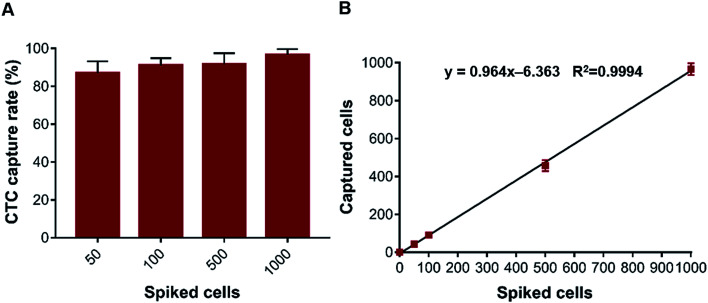
The authors developed a dynamic Halbach array magnet-integrated (dHAMI) microfluidic system for continuous-flow negative isolation of circulating tumor cells (CTCs) from whole blood. Under optimized conditions, the system achieved an average CTC capture rate of 91.6 ± 3.9% across 50–1000 spiked cancer cells per mL and detected CTCs in 10 of 10 gastric cancer patient blood samples.
Key findings
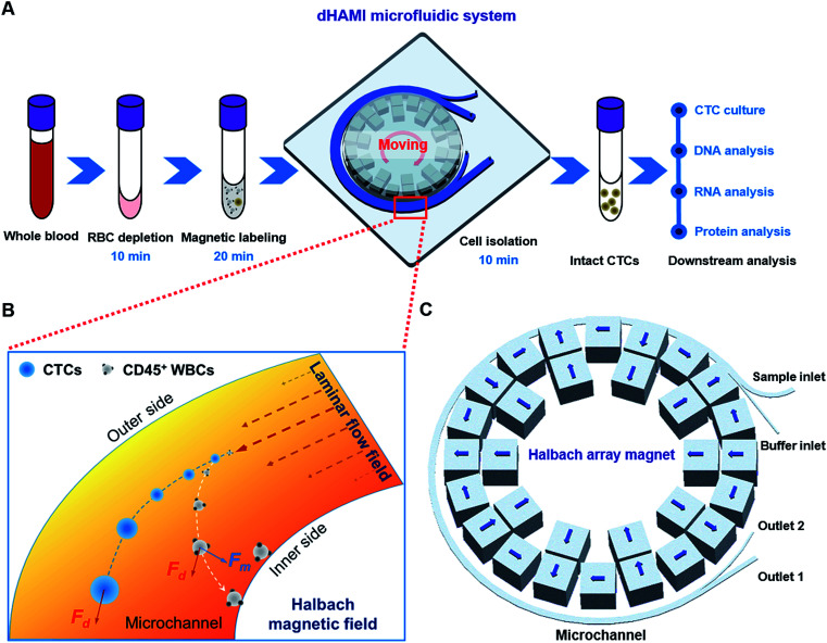
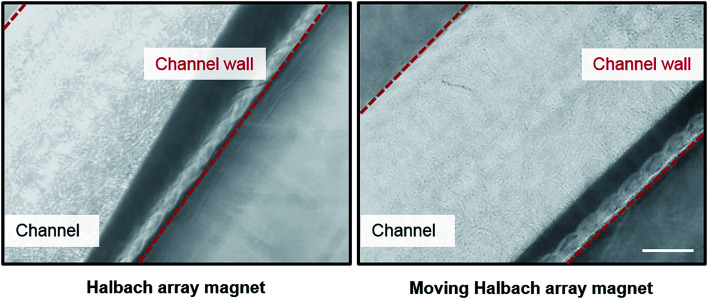
- The dHAMI system uses a continuously moving Halbach array magnet to reduce magnetic bead aggregation and enable continuous-flow negative CTC isolation without a sample loading volume limitation.
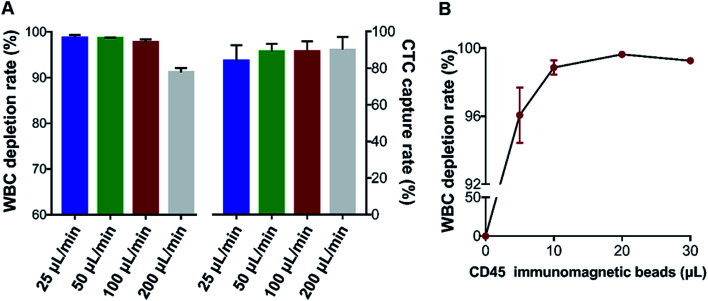
- At the optimal flow rate of 100 μL min−1, the average CTC capture rate over 50–1000 spiked AGS cells per mL was 91.6 ± 3.9%; capture increased from 87.0% at 50 cells per mL to 96.6% at 1000 cells per mL.
- The optimal CD45-labelled immunomagnetic bead ratio was 20:1, giving a WBC depletion rate of 99.6 ± 0.2%; at flow rates from 100 to 200 μL min−1, WBC depletion fell from 97.7% to 91.2%.
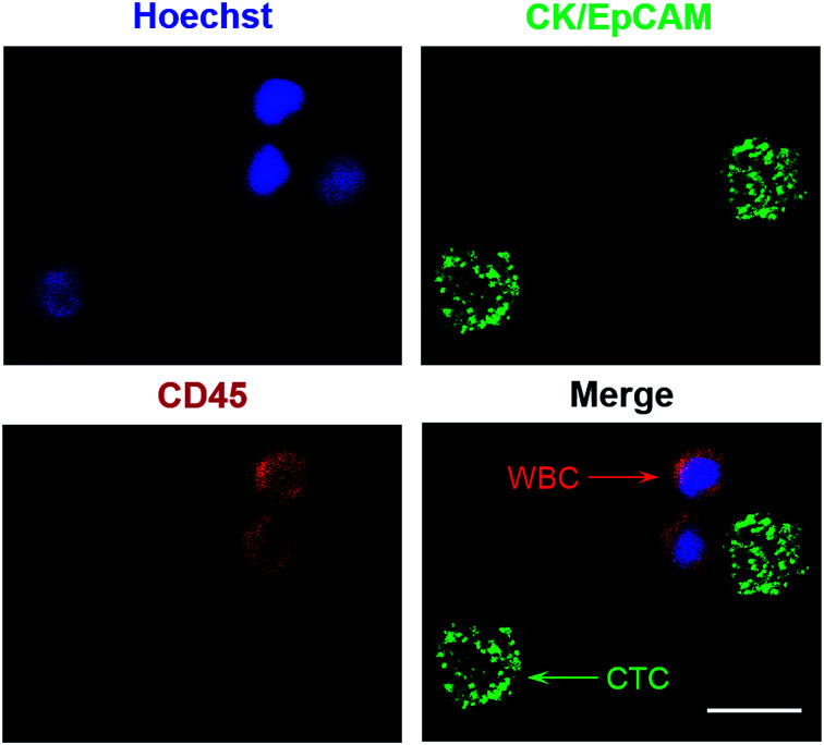
- In clinical samples from 10 patients with gastric cancer, CTCs were detected in 10 of 10 (100%) samples, ranging from 2 to 8 CTCs; the system processed 1 mL sample input in 10 min and had a throughput of 6 mL h−1.
Methods
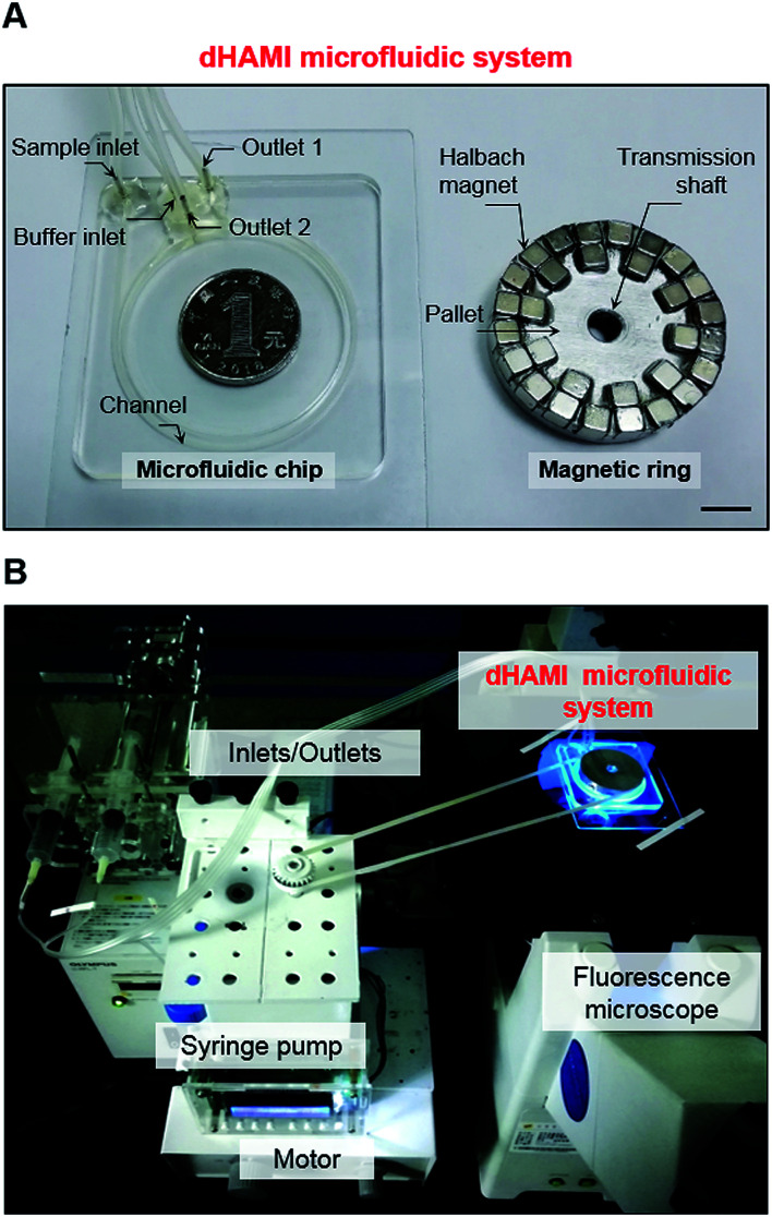
- Design: a ring-shaped PMMA microfluidic chip (channel 1 mm wide × 0.5 mm high) with two inlets and two outlets, integrated with a ring-shaped Halbach array magnet made of thirty NdFeB cubes (5 × 5 × 5 mm; ring diameter 45 mm) moved circularly at 120 rpm by a stepper motor.
- Samples: 15 μm fluorescent polystyrene particles and CellTracker Green-labelled human gastric cancer AGS cells were spiked into whole blood; clinical validation used 3 mL whole blood from 10 patients with gastric cancer. Blood was RBC-lysed, incubated with CD45-labelled immunomagnetic beads, diluted, and injected at flow rates of 25–200 μL min−1.
- Analysis: CTC capture rate and WBC depletion rate were calculated from fluorescence microscopy and automated cell counting; data are reported as mean ± SD from three independent experiments using GraphPad Prism 7.0.
Limitations
- The paper states that negative isolation generally has unsatisfactory purity of isolated CTCs, which can interfere with subsequent analysis, and that future work will focus on further miniaturization and optimization of channel sizes for different rare cells.
- Evident from the text: clinical validation was limited to 10 patients with gastric cancer, with no non-cancer clinical control cohort reported; spiking and capture-rate experiments used AGS gastric cancer cells only.
- Evident from the text: the study reports isolation and immunofluorescent identification but does not demonstrate downstream molecular, culture, or functional analyses of the isolated CTCs.
Model-written summary. Check it against the paper before relying on it.
Integrity signals
Registry
Cites
Statistics
Figures
Verification
Claims the paper makes
7 Valid · 1 Not valid · 5 Not checked · 37 Own results and interpretations
The cited sources support only part of this claim — read each source’s verdict below.
In the paper
“The most obvious limitation of the positive isolation method is that the subpopulations of cancer cells without or with a very low epithelial marker expression cannot be captured by this method.13”
§ Dynamic Halbach array magnet integrated microfluidic system for the continuous-flow separation of rare tumor cells† > Introduction
What the sources say
- Partly supportedcites [13] sourceExperimental — accuracy not yet measured
The passage on CTC analysis (indexed as Abstract; page not provided) says reliance on epithelial markers has hampered analysis, supporting the concern behind the claim. But the paper does not say that positive isolation categorically cannot capture cells with low epithelial-marker expression: its antibody-based method captured a mesenchymal cell line and identified exclusively mesenchymal CTCs. (LLM, conf=0.84)
plainstates a cause- Asserted: passes — The authors put it forward as true.
- Checkable: passes — It can be checked against evidence.
- Load-bearing: passes — The paper’s conclusions depend on it.
- Based: passes — The paper gives a reason why it holds.
- Partly supported
Every source the paper cites for this claim was checked and supports it.
In the paper
“Recent studies have also demonstrated that the microfluidic magnetic separation platforms enabled the efficient and continuous-flow separation of CTCs.22”
§ Dynamic Halbach array magnet integrated microfluidic system for the continuous-flow separation of rare tumor cells† > Introduction
What the sources say
- Supportedcites [22] source
In “Trends in CTCs separation microdevices” (page unavailable), the review describes the CTC-iChip separating cells in a microfluidic channel by magnetically deflecting them to different outlets, with approximately 97% capture efficiency in one tested mode. It also reports a magnetic-bead chip that operated at flow rates up to about 4 ml/min and achieved yields up to 90% under specified conditions. These examples support the claim about efficient, flow-based magnetic separation of CTCs. (LLM, conf=0.87)
boostedstates a cause- Asserted: passes — The authors put it forward as true.
- Checkable: passes — It can be checked against evidence.
- Load-bearing: passes — The paper’s conclusions depend on it.
- Based: passes — The paper gives a reason why it holds.
- Supported
The cited sources support only part of this claim — read each source’s verdict below.
In the paper
“In our microfluidic chip, the drag force and the magnetic force were dominant forces for the magnetic bead-labelled cells (Fig. 1B).”
§ Dynamic Halbach array magnet integrated microfluidic system for the continuous-flow separation of rare tumor cells† > Materials and methods > Theory
What the sources say
- Partly supportedcites [21] sourceExperimental — accuracy not yet measured
The “Significant forces and optimisation parameters for cell separation” section says drag and magnetic forces are the most dominant forces in relatively high magnetic fields, such as those generated by permanent magnets. It also says that in relatively weak fields, drag dominates while magnetic and gravitational forces are comparable. The source therefore supports the force comparison under a high-field condition, but does not establish it for the citing authors’ chip without that condition. (LLM, conf=0.84)
- Supportedcites [24] source
The Device model section identifies drag and magnetic force as the two important forces acting on a particle in the microchannel. The Cancer cell capture rate section applies that model to cancer cells with attached Ferrofluid particles, supporting the claim for magnetically labelled cells. (LLM, conf=0.90)
plain- Asserted: passes — The authors put it forward as true.
- Checkable: passes — It can be checked against evidence.
- Load-bearing: passes — The paper’s conclusions depend on it.
- Based: passes — The paper gives a reason why it holds.
- Partly supported
The cited sources support only part of this claim — read each source’s verdict below.
In the paper
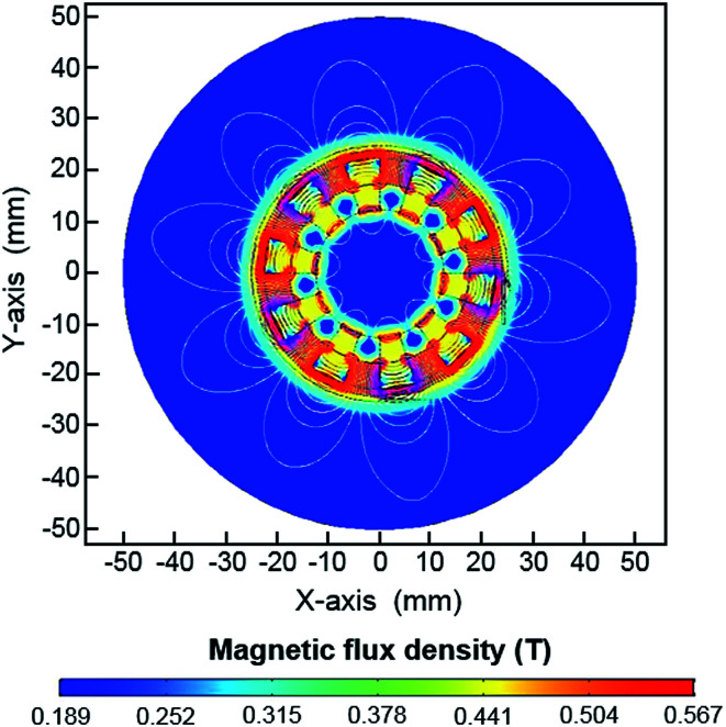
“A large magnetic field gradient created by the permanent magnet was required to enhance the separation efficiency of the magnetic cell separation.33”
§ Dynamic Halbach array magnet integrated microfluidic system for the continuous-flow separation of rare tumor cells† > Results and discussion > Design and separation principle of dHAMI microfluidic system
What the sources say
- Partly supportedcites [33] sourceExperimental — accuracy not yet measured
The Introduction (page unavailable) attributes high capture efficiency to extremely high field gradients at the sifter’s pore edges. The Experimental apparatus section (page unavailable) says the permanent magnet supplies an external field that magnetizes the sifter and nanoparticle labels; it does not say the magnet itself creates those large gradients. The claim captures the importance of gradients but misstates their location and source. (LLM, conf=0.86)
plainstates a cause- Asserted: passes — The authors put it forward as true.
- Checkable: passes — It can be checked against evidence.
- Load-bearing: passes — The paper’s conclusions depend on it.
- Based: passes — The paper gives a reason why it holds.
- Partly supported
A result the paper reports from its own data — it cites no other source for it, so there is none to check. The quote shows it is in the paper.
In the paper
“In this study, the dHAMI microfluidic system only required 10 min to process the 1 mL sample input (3 mL whole blood) (Fig. 1A).”
§ Dynamic Halbach array magnet integrated microfluidic system for the continuous-flow separation of rare tumor cells† > Results and discussion > Isolation of CTCs from blood samples spiked with cancer cells
plain- Asserted: passes — The authors put it forward as true.
- Checkable: passes — It can be checked against evidence.
- Load-bearing: passes — The paper’s conclusions depend on it.
- Based: passes — The paper gives a reason why it holds.
23 more claims
Create a free account to see all 50 claims and every source verdict.
- It may state two separate claims as one. 16
- Probably not a substantial claim. 6
- The quote only partly states this claim. 1
Citations
Cites
Circulating Tumor Cells, Disease Progression, and Survival in Metastatic Breast Cancer
Cristofanilli, M., Budd, G. T., Ellis, M. J., Stopeck, A., Matera, J., Miller, M. C., Reuben, J. M., Doyle, G. V., Allard, W. J., Terstappen, L. W. M. M., et al. (2004) · New England Journal of Medicine · doi:10.1056/nejmoa040766 · Cited by 4,593
Circulating Breast Tumor Cells Exhibit Dynamic Changes in Epithelial and Mesenchymal Composition
Yu, M., Bardia, A., Wittner, B. S., Stott, S. L., Smas, M. E., Ting, D. T., Isakoff, S. J., Ciciliano, J. C., Wells, M. N., Shah, A. M., et al. (2013) · Science · doi:10.1126/science.1228522 · Cited by 2,476
Metastatic colonization by circulating tumour cells
Massagué, J., & Obenauf, A. C. (2016) · Nature · doi:10.1038/nature17038 · Cited by 2,062
Current and future perspectives of liquid biopsies in genomics-driven oncology
Heitzer, E., Haque, I. S., Roberts, C. E. S., & Speicher, M. R. (2019) · Nature Reviews Genetics · doi:10.1038/s41576-018-0071-5 · Cited by 1,557
Design of permanent multipole magnets with oriented rare earth cobalt material
Halbach, K. (1980) · Nuclear Instruments and Methods · doi:10.1016/0029-554x(80)90094-4 · Cited by 1,532
Clinical Applications of Circulating Tumor Cells and Circulating Tumor DNA as Liquid Biopsy
Alix-Panabières, C., & Pantel, K. (2016) · Cancer Discovery · doi:10.1158/2159-8290.cd-15-1483 · Cited by 1,394
Riethdorf, S., Fritsche, H., Müller, V., Rau, T., Schindlbeck, C., Rack, B., Janni, W., Coith, C., Beck, K., Jänicke, F., et al. (2007) · Clinical Cancer Research · doi:10.1158/1078-0432.ccr-06-1695 · Cited by 1,338
Hayes, D. F., Cristofanilli, M., Budd, G. T., Ellis, M. J., Stopeck, A., Miller, M. C., Matera, J., Allard, W. J., Doyle, G. V., & Terstappen, L. W. W. M. (2006) · Clinical Cancer Research · doi:10.1158/1078-0432.ccr-05-2821 · Cited by 1,030
Liquid Biopsies, What We Do Not Know (Yet)
Bardelli, A., & Pantel, K. (2017) · Cancer Cell · doi:10.1016/j.ccell.2017.01.002 · Cited by 482
Klank, H., Kutter, J. P., & Geschke, O. (2002) · Lab on a Chip · doi:10.1039/b206409j · Cited by 477
Current detection technologies for circulating tumor cells
Shen, Z., Wu, A., & Chen, X. (2017) · Chemical Society Reviews · doi:10.1039/c6cs00803h · Cited by 433
Microchip-based immunomagnetic detection of circulating tumor cells
Hoshino, K., Huang, Y.-Y., Lane, N., Huebschman, M., Uhr, J. W., Frenkel, E. P., & Zhang, X. (2011) · Lab on a Chip · doi:10.1039/c1lc20270g · Cited by 400
Technologies for circulating tumor cell separation from whole blood
Bankó, P., Lee, S. Y., Nagygyörgy, V., Zrínyi, M., Chae, C. H., Cho, D. H., & Telekes, A. (2019) · Journal of Hematology & Oncology · doi:10.1186/s13045-019-0735-4 · Cited by 358
Lab on a chip for continuous-flow magnetic cell separation
Hejazian, M., Li, W., & Nguyen, N.-T. (2015) · Lab on a Chip · doi:10.1039/c4lc01422g · Cited by 347
Rare cell isolation and analysis in microfluidics
Chen, Y., Li, P., Huang, P.-H., Xie, Y., Mai, J. D., Wang, L., Nguyen, N.-T., & Huang, T. J. (2014) · Lab on a Chip · doi:10.1039/c3lc90136j · Cited by 307
Self-citationLiu, Z., Fusi, A., Klopocki, E., Schmittel, A., Tinhofer, I., Nonnenmacher, A., & Keilholz, U. (2011) · Journal of Translational Medicine · doi:10.1186/1479-5876-9-70 · Cited by 189
Antfolk, M., & Laurell, T. (2017) · Analytica Chimica Acta · doi:10.1016/j.aca.2017.02.017 · Cited by 184
Microfluidic technologies for circulating tumor cell isolation
Cho, H., Kim, J., Song, H., Sohn, K. Y., Jeon, M., & Han, K.-H. (2018) · The Analyst · doi:10.1039/c7an01979c · Cited by 167
Earhart, C. M., Hughes, C. E., Gaster, R. S., Ooi, C. C., Wilson, R. J., Zhou, L. Y., Humke, E. W., Xu, L., Wong, D. J., Willingham, S. B., et al. (2014) · Lab Chip · doi:10.1039/c3lc50580d · Cited by 165
Recent advances and current challenges in magnetophoresis based micro magnetofluidics
Munaz, A., Shiddiky, M. J. A., & Nguyen, N.-T. (2018) · Biomicrofluidics · doi:10.1063/1.5035388 · Cited by 164
Abdulla, A., Liu, W., Gholamipour-Shirazi, A., Sun, J., & Ding, X. (2018) · Analytical Chemistry · doi:10.1021/acs.analchem.7b04210 · Cited by 131
Circulating Tumor Cells: Fluid Surrogates of Solid Tumors
Thiele, J.-A., Bethel, K., Králíčková, M., & Kuhn, P. (2017) · Annual Review of Pathology: Mechanisms of Disease · doi:10.1146/annurev-pathol-052016-100256 · Cited by 107
Hyun, K.-A., Lee, T. Y., & Jung, H.-I. (2013) · Analytical Chemistry · doi:10.1021/ac3037766 · Cited by 102
Plouffe, B. D., Mahalanabis, M., Lewis, L. H., Klapperich, C. M., & Murthy, S. K. (2012) · Analytical Chemistry · doi:10.1021/ac2022844 · Cited by 96
An integrated microfluidic chip for one-step isolation of circulating tumor cells
Lee, T. Y., Hyun, K.-A., Kim, S.-I., & Jung, H.-I. (2017) · Sensors and Actuators B: Chemical · doi:10.1016/j.snb.2016.05.163 · Cited by 94
Sun, N., Li, X., Wang, Z., Li, Y., & Pei, R. (2018) · Biosensors and Bioelectronics · doi:10.1016/j.bios.2017.11.026 · Cited by 89
SnapShot: Circulating Tumor Cells
Dive, C., & Brady, G. (2017) · Cell · doi:10.1016/j.cell.2017.01.026 · Cited by 79
Separation and detection of rare cells in a microfluidic disk via negative selection
Chen, C.-L., Chen, K.-C., Pan, Y.-C., Lee, T.-P., Hsiung, L.-C., Lin, C.-M., Chen, C.-Y., Lin, C.-H., Chiang, B.-L., & Wo, A. M. (2011) · Lab Chip · doi:10.1039/c0lc00332h · Cited by 72
An integrated microfluidic device for rapid and high-sensitivity analysis of circulating tumor cells
Jiang, J., Zhao, H., Shu, W., Tian, J., Huang, Y., Song, Y., Wang, R., Li, E., Slamon, D., Hou, D., et al. (2017) · Scientific Reports · doi:10.1038/srep42612 · Cited by 66
Self-citationMicrofluidic platform for negative enrichment of circulating tumor cells
Sajay, B. N. G., Chang, C.-P., Ahmad, H., Khuntontong, P., Wong, C. C., Wang, Z., Puiu, P. D., Soo, R., & Rahman, A. R. A. (2014) · Biomedical Microdevices · doi:10.1007/s10544-014-9856-2 · Cited by 56
Recent advances in microfluidic methods in cancer liquid biopsy
Iliescu, F. S., Poenar, D. P., Yu, F., Ni, M., Chan, K. H., Cima, I., Taylor, H. K., Cima, I., & Iliescu, C. (2019) · Biomicrofluidics · doi:10.1063/1.5087690 · Cited by 51
Analyzing Circulating Tumor Cells One at a Time
Ortiz, V., & Yu, M. (2018) · Trends in Cell Biology · doi:10.1016/j.tcb.2018.05.004 · Cited by 48
Isolation of circulating tumour cells by physical means in a microfluidic device: a review
Patil, P., Madhuprasad, M., Kumeria, T., Losic, D., & Kurkuri, M. (2015) · RSC Advances · doi:10.1039/c5ra16489c · Cited by 47
Spinning magnetic trap for automated microfluidic assay systems
Verbarg, J., Kamgar-Parsi, K., Shields, A. R., Howell, P. B., & Ligler, F. S. (2012) · Lab on a Chip · doi:10.1039/c2lc21189k · Cited by 39
Reconfigurable Acrylic-tape Hybrid Microfluidics
Ren, Y., Ray, S., & Liu, Y. (2019) · Scientific Reports · doi:10.1038/s41598-019-41208-y · Cited by 35
Micro/nanofluidic technologies for efficient isolation and detection of circulating tumor cells
Yue, W.-Q., Tan, Z., Li, X.-P., Liu, F.-F., & Wang, C. (2019) · TrAC Trends in Analytical Chemistry · doi:10.1016/j.trac.2019.06.009 · Cited by 34
A microfluidic platform for high-purity separating circulating tumor cells at the single-cell level
Wang, K., Zhou, L., Zhao, S., Cheng, Z., Qiu, S., Lu, Y., Wu, Z., Wahab, A. H. A. A., Mao, H., & Zhao, J. (2019) · Talanta · doi:10.1016/j.talanta.2019.03.035 · Cited by 26
Negishi, R., Takai, K., Tanaka, T., Matsunaga, T., & Yoshino, T. (2018) · Analytical Chemistry · doi:10.1021/acs.analchem.8b00896 · Cited by 24
Magnetofluidic micromixer based on a complex rotating magnetic field
Munaz, A., Kamble, H., Shiddiky, M. J. A., & Nguyen, N.-T. (2017) · RSC Advances · doi:10.1039/c7ra08073e · Cited by 23
Liao, C.-J., Hsieh, C.-H., Wang, H.-M., Chou, W.-P., Chiu, T.-K., Chang, J.-H., Chao, A.-C., & Wu, M.-H. (2017) · RSC Advances · doi:10.1039/c7ra03663a · Cited by 20
3D-printed microfluidic manipulation device integrated with magnetic array
Wu, J., Cui, Y., Xuan, S., & Gong, X. (2018) · Microfluidics and Nanofluidics · doi:10.1007/s10404-018-2123-8 · Cited by 20
An integrated microfluidic platform for negative selection and enrichment of cancer cells 6
Luo, W.-Y., Tsai, S.-C., Hsieh, K., & Lee, G.-B. (2015) · Journal of Micromechanics and Microengineering · doi:10.1088/0960-1317/25/8/084007 · Cited by 18
Deng, P., Fu, C.-J., & Wu, Z. (2018) · RSC Advances · doi:10.1039/c8ra05328f · Cited by 10
Cited by
Recent advances on magnetic nanobead based biosensors: From separation to detection
Wang, L., & Lin, J. (2020) · TrAC Trends in Analytical Chemistry · doi:10.1016/j.trac.2020.115915 · Cited by 138
Self-citationKumari, S. M. N. S., & Xavier, T. S. (2024) · ACS Omega · doi:10.1021/acsomega.4c07991 · Cited by 126
Cai, J., Chen, B., He, M., Yuan, G., & Hu, B. (2024) · Analytical Chemistry · doi:10.1021/acs.analchem.4c02876 · Cited by 35
Cho, H., Choi, J., Lim, J., Lee, S., & Choi, J.-W. (2021) · Cancers · doi:10.3390/cancers13061385 · Cited by 32
“e str t t c ti fl s i t i l r t-i te rated icrofl i ic e ice [49].”
BackgroundSun, J., Shi, Z., Li, M.-J., Sha, J., Zhong, M., Chen, S., Liu, X., & Jia, S. (2022) · Journal of Magnetism and Magnetic Materials · doi:10.1016/j.jmmm.2022.169141 · Cited by 30
Metadata
- Authors
- Xue, M., Xiang, A., Guo, Y., Wang, L., Wang, R., Wang, W., Ji, G., & Lu, Z.
- Affiliations
- First Affiliated Hospital of Xi'an Jiaotong University (CN)
- Air Force Medical University (CN)
- Xijing Hospital (CN)
- Corresponding author
- Lu, Zifan
- Venue
- RSC Advances
- Publisher
- Royal Society of Chemistry (RSC)
- Published
- 2019
- Volume
- 9
- Issue
- 66
- Pages
- 38496-38504
- ISSN
- 2046-2069 (ISSN-L)
- PMC
- PMC9075830
- OpenAlex
- W2990575894
- Cited by
- 26
- References
- 43
- Citations by year
- 2026: 2 · 2025: 7 · 2024: 6 · 2023: 3 · 2022: 4 · 2021: 3 · 2020: 1
- FWCI
- 0.71
- Type
- journal article
- Language
- en
- Open access
- gold
- doi.org
- Licence
- Locations
- RSC Advances (publishedVersion, cc-by-nc)
- PubMed Central (publishedVersion, cc-by-nc)
- PubMed (publishedVersion)
- Funders
- National Natural Science Foundation of China (No.31570168, No.81301513)
- Natural Science Foundation of Shaanxi Province (2018JM7032)
- National Basic Research Program of China (2017YFC1308600)
- Topics
- Physical Sciences › Engineering › Biomedical Engineering › Microfluidic and Bio-sensing Technologies
- Physical Sciences › Engineering › Biomedical Engineering › Microfluidic and Capillary Electrophoresis Applications
- Physical Sciences › Physics and Astronomy › Condensed Matter Physics › Micro and Nano Robotics
- Keywords
- microfluidic cell separation, circulating tumor cells, rare cell isolation, magnetic manipulation, continuous-flow microfluidics, cancer detection, cellular heterogeneity
About these checks
Registry, retraction, Cites and statistics results are computed, not judged, with one exception: a printed reference that no index lists is read once by a model before it counts as a citation error. Claims are extracted by a model and kept only when their quote appears verbatim in the text; each cited source is then checked, with a model’s help, for whether it says what the claim relies on. Those verdicts can be wrong — read the claim’s quote and the cited source before relying on one.
The claim verdicts this report counts are experimental: their accuracy is not yet measured.
A language model drew the claims from the paper on 2026-10-10, under version 3 of the claim definition (model deepseek-v4.1-flash, prompt extract-v3).
A second model labelled each claim on 2026-10-10 (model jev-1.13-20260917, definition revision gold-guideline-v0.2).
Cited sources are checked with a language model’s help (gpt-6-sol).
Statistics and arithmetic are recomputed, not judged.
Confidence bands are shown only for fields calibrated on our annotated test set. None is calibrated yet, so no band is shown.
What counts as a claim ›How we measure confidence ›How we check this ›
Are you an author, or do you see a problem with this page? Request a review
Data sources
Bibliographic data: Semantic Scholar, OpenAlex, Crossref.
Contains information from the Semantic Scholar Academic Graph Datasets, made available under the ODC Attribution License (ODC-BY 1.0).
Semantic Scholar’s data is described in: Kinney, R., et al. (2023). The Semantic Scholar Open Data Platform. arXiv:2301.10140.
