

mRNA structure regulates protein expression through changes in functional half-life
Open accessPublished version of a preprint
The DOI matches its Crossref record; 57 of the 57 references it cites were found in a registry or an index.
Abstract
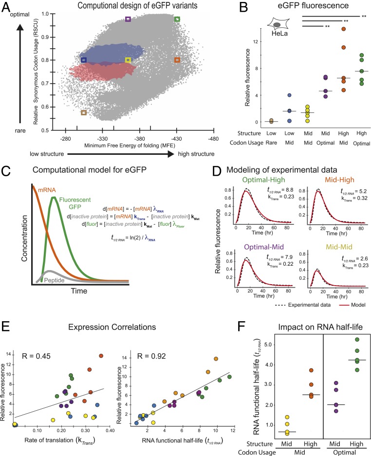
Messenger RNAs (mRNAs) encode information in both their primary sequence and their higher order structure. The independent contributions of factors like codon usage and secondary structure to regulating protein expression are difficult to establish as they are often highly correlated in endogenous sequences. Here, we used 2 approaches, global inclusion of modified nucleotides and rational sequence design of exogenously delivered constructs, to understand the role of mRNA secondary structure independent from codon usage. Unexpectedly, highly expressed mRNAs contained a highly structured coding sequence (CDS). Modified nucleotides that stabilize mRNA secondary structure enabled high expression across a wide variety of primary sequences. Using a set of eGFP mRNAs with independently altered codon usage and CDS structure, we find that the structure of the CDS regulates protein expression through changes in functional mRNA half-life (i.e., mRNA being actively translated). This work highlights an underappreciated role of mRNA secondary structure in the regulation of mRNA stability.
Overview
The claims and statistics of this paper haven’t been checked yet.
Free: it reads the open-access text and checks the first 5 claims that cite a source we can check. Create a free account to check every claim.
Integrity signals
Registry
Cites
Statistics
Figures
Verification
Claims the paper makes
The claims and statistics of this paper haven’t been checked yet.
Free: it reads the open-access text and checks the first 5 claims that cite a source we can check. Create a free account to check every claim.
Claims other papers make about this one
Each is checked against this paper’s own text: does it say what the citing paper says it does?
The source backs part of the claim, not all of it — or backs it for a different population, condition or comparison.
In the citing paper
“Increasing secondary structure lengthens mRNA half-life, which, together with optimal codons, improves protein expression5.”
From Algorithm for optimized mRNA design improves stability and immunogenicity (2023) · cites this paper as [5]
Experimental — accuracy not yet measuredThe source backs part of the claim, not all of it — or backs it for a different population, condition or comparison.
In the citing paper
“Previous work5 established two main objectives for mRNA design, stability and codon optimality, which synergize to increase protein expression.”
From Algorithm for optimized mRNA design improves stability and immunogenicity (2023) · cites this paper as [5]
Experimental — accuracy not yet measuredThe source backs part of the claim, not all of it — or backs it for a different population, condition or comparison.
In the citing paper
“However, mRNA molecules are chemically unstable and prone to degrade, which leads to insufficient protein expression5”
From Algorithm for optimized mRNA design improves stability and immunogenicity (2023) · cites this paper as [5]
Experimental — accuracy not yet measuredThe source backs part of the claim, not all of it — or backs it for a different population, condition or comparison.
In the citing paper
“Increasing secondary structure lengthens mRNA half-life, which, together with optimal codons, improves protein expression5.”
From Algorithm for optimized mRNA design improves stability and immunogenicity (2023) · cites this paper as [5]
Experimental — accuracy not yet measuredThe source backs part of the claim, not all of it — or backs it for a different population, condition or comparison.
In the citing paper
“However, mRNA molecules are chemically unstable and prone to degrade, which leads to insufficient protein expression5”
From Algorithm for optimized mRNA design improves stability and immunogenicity (2023) · cites this paper as [5]
Experimental — accuracy not yet measured
17 more claims about this paper
Create a free account to see every claim other papers make about this one.
Self-check
Percentages, sums and reported differences re-computed from the paper’s own numbers.
The self-check needs the paper’s full text.
Citations
Cites
Karikó, K., Buckstein, M., Ni, H., & Weissman, D. (2005) · Immunity · doi:10.1016/j.immuni.2005.06.008 · Cited by 2,728 · cited 1×
“To assess the degree to which the above conclusions from cell lines translated to animals, we examined protein expression in mice from formulated hEpo and Luc mRNA variants containing two nucleotide modifications shown to have reduced immunogenicity (m(1)Y and mo(5)U) (Kariko et al., 2005).”
A "Silent" Polymorphism in the MDR 1 Gene Changes Substrate Specificity
Kimchi-Sarfaty, C., Oh, J. M., Kim, I.-W., Sauna, Z. E., Calcagno, A. M., Ambudkar, S. V., & Gottesman, M. M. (2007) · Science · doi:10.1126/science.1135308 · Cited by 2,416 · cited 1×
“Another possibility is that structure leads to ribosome pausing at specific locations in the peptide, crucial for proper protein folding, and therefore activity, of certain multidomain proteins (32, 33).”
Karikó, K., Muramatsu, H., Welsh, F. A., Ludwig, J., Kato, H., Akira, S., & Weissman, D. (2008) · Molecular Therapy · doi:10.1038/mt.2008.200 · Cited by 1,930 · cited 2×
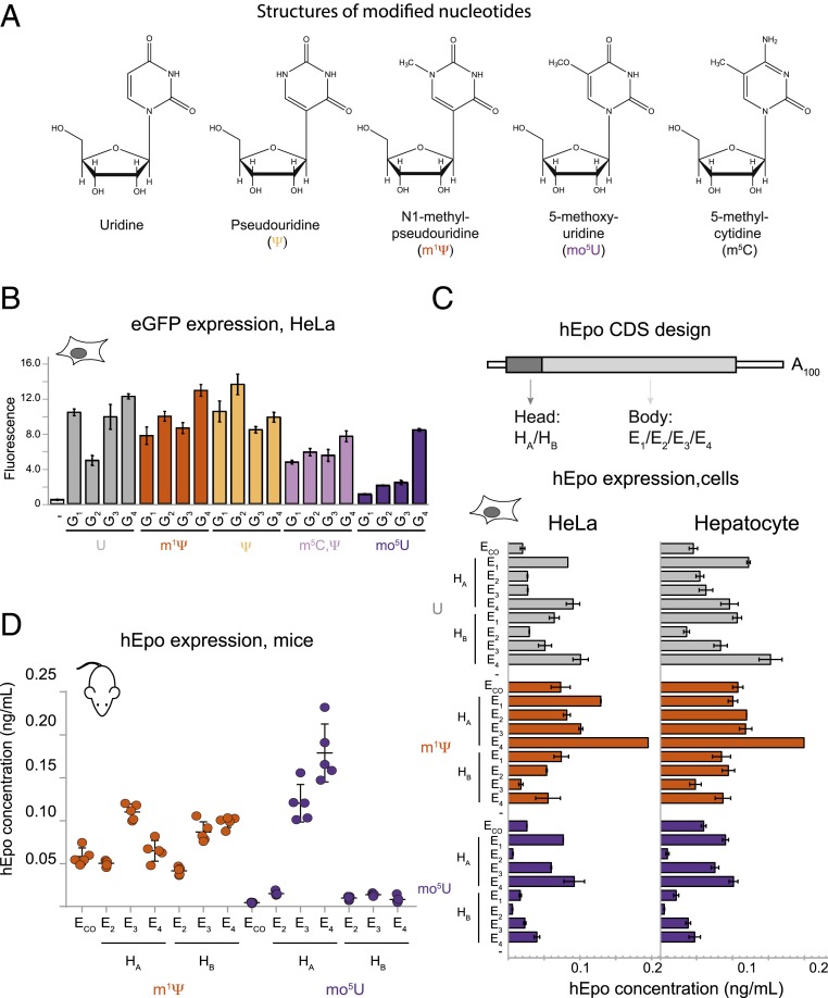
“For comparison with a previous study documenting the effects of modified nucleotides on RNA immunogenicity (16), we also made eGFP mRNA wherein both U and C were substituted with Ψ and 5-methyl-cytidine (m(5)C), respectively.”
Synonymous but not the same: the causes and consequences of codon bias
Plotkin, J. B., & Kudla, G. (2011) · Nature Reviews Genetics · doi:10.1038/nrg2899 · Cited by 1,685 · cited 1×
“Highly expressed mRNAs tended to have increased GC content, consistent with previous reports (17), but not all high GC sequences were high expressers (SI Appendix, Figs.”
ResultCodon bias and heterologous protein expression
Gustafsson, C., Govindarajan, S., & Minshull, J. (2004) · Trends in Biotechnology · doi:10.1016/j.tibtech.2004.04.006 · Cited by 1,330 · cited 1×
“Synonymous codon choice directly impacts translation, with highly expressed genes tending to include more "optimal" codons (Gustafsson et al., 2004; Hinnebusch et al., 2016; Horstick et al., 2015; Pop et al., 2014).”
Codon usage tabulated from international DNA sequence databases: status for the year 2000
Nakamura, Y. (2000) · Nucleic Acids Research · doi:10.1093/nar/28.1.292 · Cited by 1,254 · cited 2×
“Using probabilities weighted by frequency in the human transcriptome (Nakamura et al., 2000) generates a similar-shaped distribution, but shifted to both significantly higher RSCU (0.”
MethodInfluentialImproved Green Fluorescent Protein by Molecular Evolution Using DNA Shuffling
Crameri, A., Whitehorn, E. A., Tate, E., & Stemmer, W. P. C. (1996) · Nature Biotechnology · doi:10.1038/nbt0396-315 · Cited by 1,187 · cited 1×
“Realtime, continuous expression data were fit by a model including rate constants for mRNA translation, mRNA functional half-life, maturation of eGFP protein into its fluorescent form (28), and eGFP protein degradation (Fig.”
Xia, T., SantaLucia, J., Burkard, M. E., Kierzek, R., Schroeder, S. J., Jiao, X., Cox, C., & Turner, D. H. (1998) · Biochemistry · doi:10.1021/bi9809425 · Cited by 1,140 · cited 8×
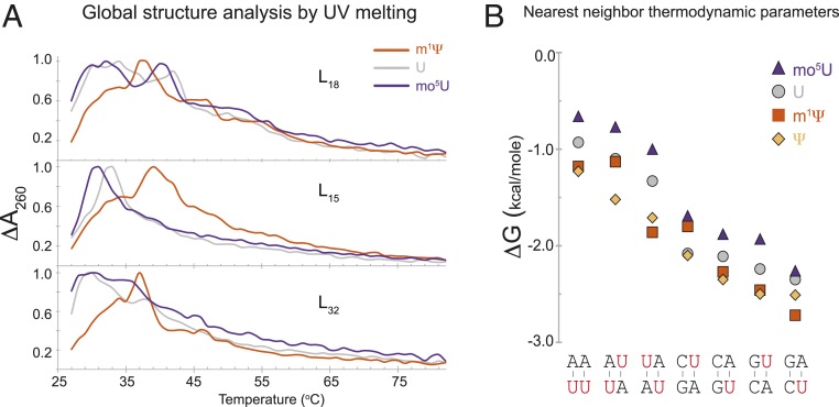
“Nearest-neighbor thermodynamic parameters along with the experimentally determined error for Watson-crick base pairs containing unmodified uridine (values from (Xia et al., 1998)), Y, m(1)Y, or mo(5)U.”
InfluentialTranslational control by 5′-untranslated regions of eukaryotic mRNAs
Hinnebusch, A. G., Ivanov, I. P., & Sonenberg, N. (2016) · Science · doi:10.1126/science.aad9868 · Cited by 1,079 · cited 1×
“Synonymous codon choice directly impacts translation, with highly expressed genes tending to include more "optimal" codons (Gustafsson et al., 2004; Hinnebusch et al., 2016; Horstick et al., 2015; Pop et al., 2014).”
Codon Optimality Is a Major Determinant of mRNA Stability
Presnyak, V., Alhusaini, N., Chen, Y.-H., Martin, S., Morris, N., Kline, N., Olson, S., Weinberg, D., Baker, K. E., Graveley, B. R., et al. (2015) · Cell · doi:10.1016/j.cell.2015.02.029 · Cited by 1,078 · cited 5×
“Regions of increased rare codon frequency have been shown to decrease protein expression and mRNA stability (Presnyak et al., 2015; Weinberg et al., 2016).”
InfluentialSelf-citationThe dynamic N1-methyladenosine methylome in eukaryotic messenger RNA
Dominissini, D., Nachtergaele, S., Moshitch-Moshkovitz, S., Peer, E., Kol, N., Ben-Haim, M. S., Dai, Q., Di Segni, A., Salmon-Divon, M., Clark, W. C., et al. (2016) · Nature · doi:10.1038/nature16998 · Cited by 1,021 · cited 1×
“By destabilizing local secondary structure in endogenous mRNAs, and thereby increasing the regulatory protein accessibility to specific sequence elements, N1-methyl and N6-methyl adenosine are emerging as key regulators of protein expression (Dominissini et al., 2016; Spitale et al., 2015).”
BackgroundAn evolutionarily conserved mechanism for controlling the efficiency of protein translation
Tuller, T., Carmi, A., Vestsigian, K., Navon, S., Dorfan, Y., Zaborske, J., Pan, T., Dahan, O., Furman, I., & Pilpel, Y. (2010) · Cell · doi:10.1016/j.cell.2010.03.031 · Cited by 874 · cited 2×
“Other mRNA sequence features reported to correlate with protein output are dinucleotide frequency in the CDS (Tulloch et al., 2014), and the effect of codon order on locally-accessible charged tRNA pools (Tuller et al., 2010).”
BackgroundGeneration of Destabilized Green Fluorescent Protein as a Transcription Reporter
Li, X., Zhao, X., Fang, Y., Jiang, X., Duong, T., Fan, C., Huang, C.-C., & Kain, S. R. (1998) · Journal of Biological Chemistry · doi:10.1074/jbc.273.52.34970 · Cited by 846 · cited 1×
“The eGFP-degron construct reduces protein half-life to under 1 hour, so that changes in eGFP fluorescence over time directly correlate to the kinetics of translation and mRNA decay (Li et al., 1998).”
Architecture and secondary structure of an entire HIV-1 RNA genome
Watts, J. M., Dang, K. K., Gorelick, R. J., Leonard, C. W., Bess Jr, J. W., Swanstrom, R., Burch, C. L., & Weeks, K. M. (2009) · Nature · doi:10.1038/nature08237 · Cited by 837 · cited 4×
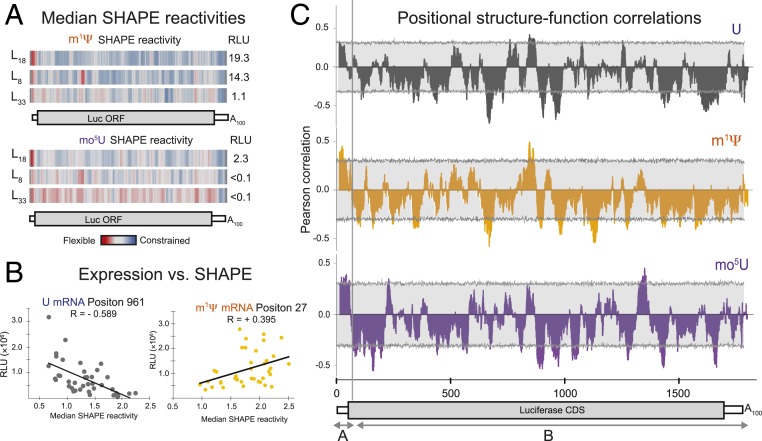
“Regions displaying structural differences were identified using 31-nucleotide sliding window median reactivities, as previously described (Watts et al., 2009).”
BackgroundMethodInfluentialSabnis, S., Kumarasinghe, E. S., Salerno, T., Mihai, C., Ketova, T., Senn, J. J., Lynn, A., Bulychev, A., McFadyen, I., Chan, J., et al. (2018) · Molecular Therapy · doi:10.1016/j.ymthe.2018.03.010 · Cited by 820 · cited 1×
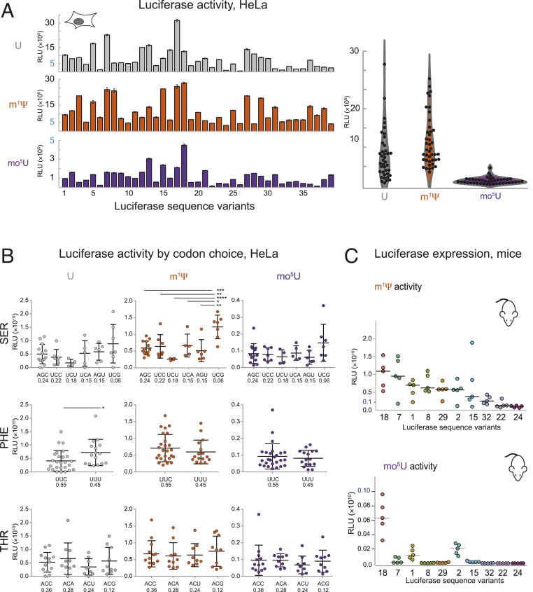
“As expected from the known biodistribution of mRNA-containing lipid nanoparticles (19), the liver was the main site of protein expression ( SI Appendix , Fig.”
Self-citationIn vivo genome-wide profiling of RNA secondary structure reveals novel regulatory features
Ding, Y., Tang, Y., Kwok, C. K., Zhang, Y., Bevilacqua, P. C., & Assmann, S. M. (2014) · Nature · doi:10.1038/nature12756 · Cited by 775 · cited 1×
“Transcriptome-wide RNA structure characterization is beginning to reveal global relationships between the structure content in different mRNA regions and protein expression (10, 11).”
Genome-wide measurement of RNA secondary structure in yeast
Kertesz, M., Wan, Y., Mazor, E., Rinn, J. L., Nutter, R. C., Chang, H. Y., & Segal, E. (2010) · Nature · doi:10.1038/nature09322 · Cited by 765 · cited 1×
“Transcriptomewide RNA structure characterization is beginning to reveal global relationships between the structure content in different mRNA regions and protein expression (Ding et al., 2014; Kertesz et al., 2010; Ramani et al.; Wan et al., 2014).”
Structural imprints in vivo decode RNA regulatory mechanisms
Spitale, R. C., Flynn, R. A., Zhang, Q. C., Crisalli, P., Lee, B., Jung, J.-W., Kuchelmeister, H. Y., Batista, P. J., Torre, E. A., Kool, E. T., et al. (2015) · Nature · doi:10.1038/nature14263 · Cited by 745 · cited 1×
“By destabilizing local secondary structure in endogenous mRNAs, and thereby increasing the regulatory protein accessibility to specific sequence elements, N1-methyl and N6-methyl adenosine are emerging as key regulators of protein expression (Dominissini et al., 2016; Spitale et al., 2015).”
Expression of therapeutic proteins after delivery of chemically modified mRNA in mice
Kormann, M. S. D., Hasenpusch, G., Aneja, M. K., Nica, G., Flemmer, A. W., Herber-Jonat, S., Huppmann, M., Mays, L. E., Illenyi, M., Schams, A., et al. (2011) · Nature Biotechnology · doi:10.1038/nbt.1733 · Cited by 733 · cited 1×
“Unmodified mRNAs were not included because in vivo protein expression can be obscured by strong activation of innate immunity (18).”
RNA motif discovery by SHAPE and mutational profiling (SHAPE-MaP)
Siegfried, N. A., Busan, S., Rice, G. M., Nelson, J. A. E., & Weeks, K. M. (2014) · Nature Methods · doi:10.1038/nmeth.3029 · Cited by 654 · cited 1×
“To investigate how modified nucleotides impact mRNA structure at single nucleotide resolution, we used selective 2’-hydroxyl acylation analyzed by primer-extension - mutational profiling (SHAPEMaP) to probe RNA structure (22).”
Landscape and variation of RNA secondary structure across the human transcriptome
Wan, Y., Qu, K., Zhang, Q. C., Flynn, R. A., Manor, O., Ouyang, Z., Zhang, J., Spitale, R. C., Snyder, M. P., Segal, E., et al. (2014) · Nature · doi:10.1038/nature12946 · Cited by 555 · cited 1×
“Transcriptome-wide RNA structure characterization is beginning to reveal global relationships between the structure content in different mRNA regions and protein expression (10, 11).”
Following translation by single ribosomes one codon at a time
Wen, J.-D., Lancaster, L., Hodges, C., Zeri, A.-C., Yoshimura, S. H., Noller, H. F., Bustamante, C., & Tinoco, I. (2008) · Nature · doi:10.1038/nature06716 · Cited by 492 · cited 1×
“Biophysical experiments have shown that secondary structure can slow ribosome processivity (34).”
Insights into RNA structure and function from genome-wide studies
Mortimer, S. A., Kidwell, M. A., & Doudna, J. A. (2014) · Nature Reviews Genetics · doi:10.1038/nrg3681 · Cited by 472 · cited 1×
“In addition to dictating encoded protein identity, its primary sequence also determines an mRNA ’ s propensity to form secondary and tertiary structure (9).”
Rate-Limiting Steps in Yeast Protein Translation
Shah, P., Ding, Y., Niemczyk, M., Kudla, G., & Plotkin, J. B. (2013) · Cell · doi:10.1016/j.cell.2013.05.049 · Cited by 463 · cited 4×
“…with previous reports that a relatively unstructured 5'-end is associated with higher protein expression (Ding et al., 2014; Gu et al., 2010; Shah et al., 2013; Tuller and Zur, 2015; Wan et al., 2014), and with our current understanding of translation initiation, which requires…”
BackgroundResultInfluentialZNF598 Is a Quality Control Sensor of Collided Ribosomes
Juszkiewicz, S., Chandrasekaran, V., Lin, Z., Kraatz, S., Ramakrishnan, V., & Hegde, R. S. (2018) · Molecular Cell · doi:10.1016/j.molcel.2018.08.037 · Cited by 424 · cited 1×
“One suggested mechanism for this regulation is the action of the ubiquitin ligase, ZNF598, on the small subunit proteins of collided diribosomes (42).”
BackgroundSynonymous Codons Direct Cotranslational Folding toward Different Protein Conformations
Buhr, F., Jha, S., Thommen, M., Mittelstaet, J., Kutz, F., Schwalbe, H., Rodnina, M. V., & Komar, A. A. (2016) · Molecular Cell · doi:10.1016/j.molcel.2016.01.008 · Cited by 395 · cited 1×
“Although most such pauses are driven by selective maintenance of rare codons (Buhr et al., 2016), regions of high secondary structure may play a similar role in HIV mRNAs encoding proteins with inter-domain junctions (Watts et al.”
Weinberg, D. E., Shah, P., Eichhorn, S. W., Hussmann, J. A., Plotkin, J. B., & Bartel, D. P. (2016) · Cell Reports · doi:10.1016/j.celrep.2016.01.043 · Cited by 392
Design Parameters to Control Synthetic Gene Expression in Escherichia coli
Welch, M., Govindarajan, S., Ness, J. E., Villalobos, A., Gurney, A., Minshull, J., & Gustafsson, C. (2009) · PLoS ONE · doi:10.1371/journal.pone.0007002 · Cited by 352 · cited 2×
“(D) Serum concentrations of hEpo protein measured by ELISA (ng/mL, y axis) in BALB-c mice (5 per group) following IV injection of LNP-formulated mRNA of 6 sequence variants (described in B above, x axis) plus 1 codon optimized variant (ECO) (44) containing m(1)Ψ (orange) or mo(5)U (dark purple).”
Ribosome Collision Is Critical for Quality Control during No-Go Decay
Simms, C. L., Yan, L. L., & Zaher, H. S. (2017) · Molecular Cell · doi:10.1016/j.molcel.2017.08.019 · Cited by 319 · cited 1×
“Notably, ribosome collisions were recently demonstrated to activate the No-Go mRNA decay (NGD) pathway and decrease mRNA half-life (Simms et al., 2017).”
BackgroundGu, W., Zhou, T., & Wilke, C. O. (2010) · PLoS Computational Biology · doi:10.1371/journal.pcbi.1000664 · Cited by 313 · cited 2×
“humans, have shown that secondary structure in the 5' untranslated region (5¢ UTR) generally reduces translation initiation efficiency and therefore overall protein output (Ding et al., 2014; Gu et al., 2010; Shah et al., 2013; Tuller and Zur, 2015; Wan et al., 2014).”
The DEAD-Box Protein Dhh1p Couples mRNA Decay and Translation by Monitoring Codon Optimality
Radhakrishnan, A., Chen, Y.-H., Martin, S., Alhusaini, N., Green, R., & Coller, J. (2016) · Cell · doi:10.1016/j.cell.2016.08.053 · Cited by 289 · cited 1×
“Further, ribosomal pauses induced by rare codons have recently been directly linked to ribosomal frame shifting (38) and mRNA degradation (4, 39).”
Svitkin, Y. V., Cheng, Y. M., Chakraborty, T., Presnyak, V., John, M., & Sonenberg, N. (2017) · Nucleic Acids Research · doi:10.1093/nar/gkx135 · Cited by 278 · cited 1×
“While counterintuitive, the increase in protein output is consistent with previous work in cell-free lysates, showing that mRNAs containing m(1)Ψ slow ribosome elongation, increase total protein output, and improve initiation through decreased phosphorylation of the initiation factor, eIF2α (35).”
Self-citationPervasive Regulatory Functions of mRNA Structure Revealed by High-Resolution SHAPE Probing
Mustoe, A. M., Busan, S., Rice, G. M., Hajdin, C. E., Peterson, B. K., Ruda, V. M., Kubica, N., Nutiu, R., Baryza, J. L., & Weeks, K. M. (2018) · Cell · doi:10.1016/j.cell.2018.02.034 · Cited by 258 · cited 1×
“Although secondary structure likely slows ribosome progression, the ribosome is inherently a very powerful helicase that necessarily unwinds mRNA structure during elongation (Mustoe et al., 2018).”
BackgroundCausal signals between codon bias, mRNA structure, and the efficiency of translation and elongation
Pop, C., Rouskin, S., Ingolia, N. T., Han, L., Phizicky, E. M., Weissman, J. S., & Koller, D. (2014) · Molecular Systems Biology · doi:10.15252/msb.20145524 · Cited by 237 · cited 2×
“Synonymous codon choice directly impacts translation, with highly expressed genes tending to include more "optimal" codons (Gustafsson et al., 2004; Hinnebusch et al., 2016; Horstick et al., 2015; Pop et al., 2014).”
BackgroundIshimura, R., Nagy, G., Dotu, I., Chuang, J. H., & Ackerman, S. L. (2016) · eLife · doi:10.7554/elife.14295 · Cited by 221
The endonuclease Cue2 cleaves mRNAs at stalled ribosomes during No Go Decay
D'Orazio, K. N., Wu, C. C.-C., Sinha, N., Loll-Krippleber, R., Brown, G. W., & Green, R. (2019) · eLife · doi:10.7554/elife.49117 · Cited by 207 · cited 1×
“Notably, ribosome collisions were demonstrated to activate the mRNA degradation through the no-go decay (NGD) pathway and decrease mRNA half-life (40, 41).”
Multiple roles of the coding sequence 5′ end in gene expression regulation
Tuller, T., & Zur, H. (2015) · Nucleic Acids Research · doi:10.1093/nar/gku1313 · Cited by 206 · cited 2×
“humans, have shown that secondary structure in the 5' untranslated region (5¢ UTR) generally reduces translation initiation efficiency and therefore overall protein output (Ding et al., 2014; Gu et al., 2010; Shah et al., 2013; Tuller and Zur, 2015; Wan et al., 2014).”
Chemical and structural effects of base modifications in messenger RNA
Harcourt, E. M., Kietrys, A. M., & Kool, E. T. (2017) · Nature · doi:10.1038/nature21351 · Cited by 204
Kierzek, E. (2003) · Nucleic Acids Research · doi:10.1093/nar/gkg633 · Cited by 197 · cited 1×
“Such modified nucleotides can either stabilize (14) or destabilize (15) base pairs and hence overall mRNA structure.”
Translation elongation can control translation initiation on eukaryotic mRNAs
Chu, D., Kazana, E., Bellanger, N., Singh, T., Tuite, M. F., & von der Haar, T. (2014) · The EMBO Journal · doi:10.1002/embj.201385651 · Cited by 188 · cited 1×
“Interestingly, a recent study in mammalian neurons links codon-specific ribosome stalling directly to eIF2α phosphorylation through the kinase GCN2 (36), suggesting that translation initiation and elongation may be highly interconnected (37).”
Tulloch, F., Atkinson, N. J., Evans, D. J., Ryan, M. D., & Simmonds, P. (2014) · eLife · doi:10.7554/elife.04531 · Cited by 178
Lü, Z., Turner, D. H., & Mathews, D. H. (2006) · Nucleic Acids Research · doi:10.1093/nar/gkl472 · Cited by 152
NEWBY, M. I., & GREENBAUM, N. L. (2001) · RNA · doi:10.1017/s1355838201002308 · Cited by 117
High-throughput determination of RNA structure by proximity ligation
Ramani, V., Qiu, R., & Shendure, J. (2015) · Nature Biotechnology · doi:10.1038/nbt.3289 · Cited by 108
Horstick, E. J., Jordan, D. C., Bergeron, S. A., Tabor, K. M., Serpe, M., Feldman, B., & Burgess, H. A. (2015) · Nucleic Acids Research · doi:10.1093/nar/gkv035 · Cited by 106 · cited 1×
“Synonymous codon choice directly impacts translation, with highly expressed genes tending to include more “optimal” codons (1, 2).”
Hudson, G. A., Bloomingdale, R. J., & Znosko, B. M. (2013) · RNA · doi:10.1261/rna.039610.113 · Cited by 106
Kearse, M. G., Goldman, D. H., Choi, J., Nwaezeapu, C., Liang, D., Green, K. M., Goldstrohm, A. C., Todd, P. K., Green, R., & Wilusz, J. E. (2019) · Genes & Development · doi:10.1101/gad.324715.119 · Cited by 77 · cited 1×
“Another possibility is that ribosome queuing near the start codon may facilitate translation initiation (31).”
Mechanisms of endonuclease‐mediated mRNA decay
Schoenberg, D. R. (2011) · WIREs RNA · doi:10.1002/wrna.78 · Cited by 75 · cited 1×
“It is also possible that more structured RNAs are less prone to cleavage by single strand-specific endonucleases, although endonucleolytic cleavage is thought to only contribute to the degradation in specific cases (Schoenberg, 2011; Wilamowski et al., 2018).”
BackgroundRibosome Collisions Result in +1 Frameshifting in the Absence of No-Go Decay
Simms, C. L., Yan, L. L., Qiu, J. K., & Zaher, H. S. (2019) · Cell Reports · doi:10.1016/j.celrep.2019.07.046 · Cited by 63 · cited 1×
“Further, ribosomal pauses induced by rare codons have recently been directly linked to ribosomal frame shifting (38) and mRNA degradation (4, 39).”
A novel translational control mechanism involving RNA structures within coding sequences
Jungfleisch, J., Nedialkova, D. D., Dotú, I., Sloan, K. E., Martínez‐Bosch, N., Brüning, L., Raineri, E., Navarro, P., Bohnsack, M. T., Leidel, S. A., et al. (2016) · Genome Research · doi:10.1101/gr.209015.116 · Cited by 59 · cited 1×
“It is possible that higher mRNA structure improves interaction with RNA binding proteins (RBPs) that positively impact translation, such as double-stranded RNA binding protein Staufen (29) or reduces accessibility to single strand-specific endonucleases, although endonucleolytic cleavage is thought to be limited to sp…”
Rauscher, R., & Ignatova, Z. (2018) · Biochemical Society Transactions · doi:10.1042/bst20170422 · Cited by 49 · cited 1×
“Another possibility is that structure leads to ribosome pausing at specific locations in the peptide, crucial for proper protein folding, and therefore activity, of certain multidomain proteins (32, 33).”
BackgroundOn the Ribosomal Density that Maximizes Protein Translation Rate
Zarai, Y., Margaliot, M., & Tuller, T. (2016) · PLOS ONE · doi:10.1371/journal.pone.0166481 · Cited by 46 · cited 1×
“So, the explanation for how more local secondary structure extends functional mRNA half-life may simply be a protective buffering effect that increases the spacing between ribosomes and thereby decreases collisions (Zarai et al., 2016).”
BackgroundSubstrate specificity of human MCPIP1 endoribonuclease
Wilamowski, M., Górecki, A., Dziedzicka‐Wasylewska, M., & Jura, J. (2018) · Scientific Reports · doi:10.1038/s41598-018-25765-2 · Cited by 35 · cited 1×
“It is also possible that more structured RNAs are less prone to cleavage by single strand-specific endonucleases, although endonucleolytic cleavage is thought to only contribute to the degradation in specific cases (Schoenberg, 2011; Wilamowski et al., 2018).”
Simmonds, P., Tulloch, F., Evans, D. J., & Ryan, M. D. (2015) · Proceedings of the National Academy of Sciences · doi:10.1073/pnas.1507339112 · Cited by 28 · cited 1×
“Because these effects are interdependent on mRNA sequence, teasing apart their individual contributions to protein output is difficult and often controversial (7, 8).”
Reply to Simmonds et al.: Codon pair and dinucleotide bias have not been functionally distinguished
Futcher, B., Gorbatsevych, O., Shen, S. H., Stauft, C. B., Song, Y., Wang, B., Leatherwood, J., Gardin, J., Yurovsky, A., Mueller, S., et al. (2015) · Proceedings of the National Academy of Sciences · doi:10.1073/pnas.1507710112 · Cited by 25 · cited 1×
“Because these effects are interdependent on mRNA sequence, teasing apart their individual contributions to protein output is difficult and often controversial (7, 8).”
BackgroundScherer, S. (2010) · Cold Spring Harbor Laboratory eBooks · Cited by 22
mRNA structure regulates protein expression through changes in functional half-lifePreprint
Mauger, D., Cabral, B. J., Presnyak, V., et al. (2019) · bioRxiv (Cold Spring Harbor Laboratory) · doi:10.1101/549022 · Cited by 16
Self-citation
Cited by
SARS-CoV-2 mRNA Vaccine Design Enabled by Prototype Pathogen Preparedness
Corbett, K. S., Edwards, D. K., Leist, S. R., Abiona, O. M., Boyoglu-Barnum, S., Gillespie, R. A., Himansu, S., Schäfer, A., Ziwawo, C. T., DiPiazza, A. T., et al. (2020) · Nature · doi:10.1038/s41586-020-2622-0 · Cited by 1,615
Self-citationmRNA-lipid nanoparticle COVID-19 vaccines: structure and stability
Schoenmaker, L., Witzigmann, D., Kulkarni, J. A., Verbeke, R., Kersten, G., Jiskoot, W., & Crommelin, D. J. A. (2021) · International Journal of Pharmaceutics · doi:10.1016/j.ijpharm.2021.120586 · Cited by 1,566
“Moreover, there are also indications that m1 Ψ increases base stacking and the melting point of mRNA, thereby making mRNA more stable (Mauger et al., 2019; Zhao and He, 2015).”
mRNA-based therapeutics: powerful and versatile tools to combat diseases
Qin, S., Tang, X., Chen, Y., Chen, K., Fan, N., Xiao, W., Zheng, Q., Li, G., Teng, Y., Wu, M., et al. (2022) · Signal Transduction and Targeted Therapy · doi:10.1038/s41392-022-01007-w · Cited by 771
“Increasing the GC content by replacing rare codons in ORFs protects mRNA from endoribonuclease degradation and enhances mRNA protein expression in vivo.(71,72) In addition, functional peptides are crucial for mRNA Fig.”
BackgroundAdvances in COVID-19 mRNA vaccine development
Fang, E., Liu, X., Li, M., Zhang, Z., Song, L., Zhu, B., Wu, X., Liu, J., Zhao, D., & Li, Y. (2022) · Signal Transduction and Targeted Therapy · doi:10.1038/s41392-022-00950-y · Cited by 601
“Currently, nucleoside-modified mRNA vaccines employ 100% chemically modified nucleosides replacing natural nucleosides, and m(1)ψ is often used to replace uridine during IVT to improve the safety and stability of mRNA vaccines.(52,134,172) At present, mRNA vaccines that have entered clinical trials or been approved fo…”
MethodRoles of mRNA poly(A) tails in regulation of eukaryotic gene expression
Passmore, L. A., & Coller, J. (2022) · Nature Reviews Molecular Cell Biology · doi:10.1038/s41580-021-00417-y · Cited by 569
Metadata
- Authors
- Mauger, D. M., Cabral, B. J., Presnyak, V., Su, S. V., Reid, D. W., Goodman, B., Link, K., Khatwani, N., Reynders, J., Moore, M. J., & McFadyen, I. J.
- Affiliations
- Platform Research, Moderna, Inc., Cambridge, MA 02139
- Corresponding author
- Moore, Melissa J.
- Venue
- Proceedings of the National Academy of Sciences
- Publisher
- National Academy of Sciences
- Published
- 2019-11-11
- Volume
- 116
- Issue
- 48
- Pages
- 24075-24083
- ISSN
- 0027-8424 (ISSN-L)
- 1091-6490
- PMC
- PMC6883848
- OpenAlex
- W2983889514
- Semantic Scholar
- 5893e4255a022371c1bbef16899698c3e704ff58
- Cited by
- 555
- References
- 56
- Citations by year
- 2026: 76 · 2025: 105 · 2024: 104 · 2023: 73 · 2022: 98 · 2021: 71 · 2020: 26 · 2019: 2
- FWCI
- 16.54
- Type
- journal article
- Language
- en
- Open access
- hybrid
- doi.org
- Licence
- Locations
- Proceedings of the National Academy of Sciences (publishedVersion, cc-by)
- bioRxiv (Cold Spring Harbor Laboratory) (acceptedVersion)
- PubMed Central (publishedVersion, cc-by)
- PubMed (publishedVersion)
- Funders
- Moderna
- Topics
- Life Sciences › Biochemistry, Genetics and Molecular Biology › Molecular Biology › RNA and protein synthesis mechanisms
- Life Sciences › Biochemistry, Genetics and Molecular Biology › Molecular Biology › RNA Research and Splicing
- Life Sciences › Biochemistry, Genetics and Molecular Biology › Molecular Biology › RNA Interference and Gene Delivery
- Keywords
- mRNA secondary structure, mRNA stability, mRNA half-life, codon usage, protein expression regulation, modified nucleotides, mRNA translation
- MeSH terms
- Half-Life, HeLa Cells, Humans, Nucleic Acid Conformation, Proteins, RNA, Messenger, Protein Biosynthesis, RNA Stability
About these checks
Registry, retraction, Cites and statistics results are computed, not judged, with one exception: a printed reference that no index lists is read once by a model before it counts as a citation error. Claims are extracted by a model and kept only when their quote appears verbatim in the text; each cited source is then checked, with a model’s help, for whether it says what the claim relies on. Those verdicts can be wrong — read the claim’s quote and the cited source before relying on one.
Statistics and arithmetic are recomputed, not judged.
Confidence bands are shown only for fields calibrated on our annotated test set. None is calibrated yet, so no band is shown.
What counts as a claim ›How we measure confidence ›How we check this ›
Are you an author, or do you see a problem with this page? Request a review
Data sources
Bibliographic data: Semantic Scholar, OpenAlex, Crossref.
Contains information from the Semantic Scholar Academic Graph Datasets, made available under the ODC Attribution License (ODC-BY 1.0).
Semantic Scholar’s data is described in: Kinney, R., et al. (2023). The Semantic Scholar Open Data Platform. arXiv:2301.10140.
