

The DOI matches its Crossref record; 35 of the 35 references it cites were found in a registry or an index.
Glyburide inhibits the Cryopyrin/Nalp3 inflammasome
Open accessAbstract
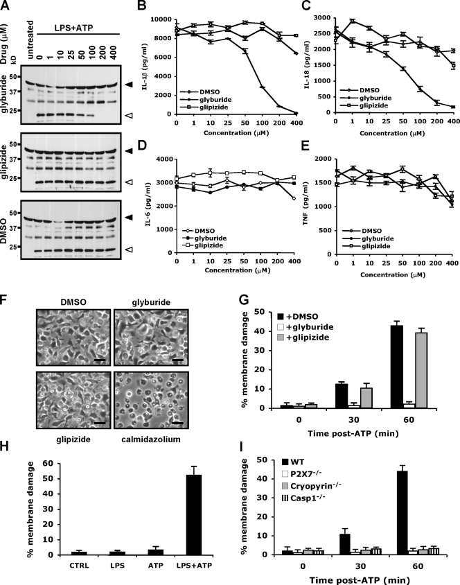
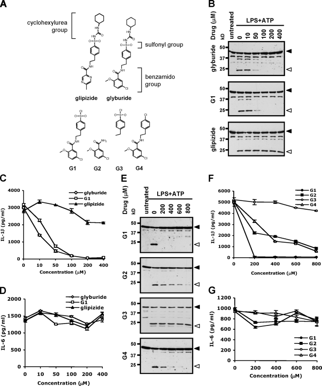
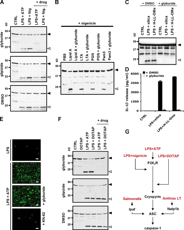
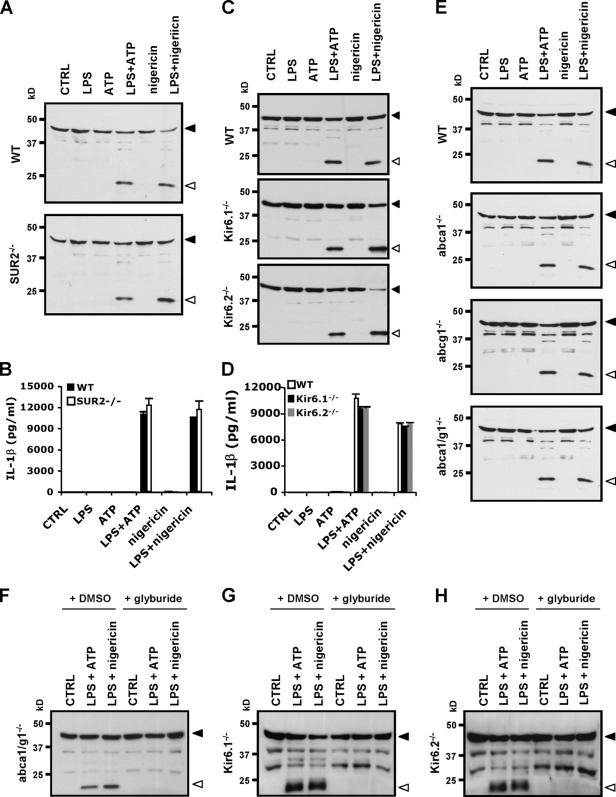
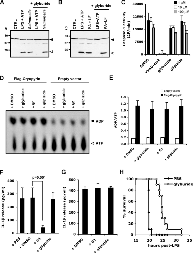
Inflammasomes activate caspase-1 for processing and secretion of the cytokines interleukin-1beta (IL-1beta) and IL-18. Cryopyrin/NALP3/NLRP3 is an essential component of inflammasomes triggered by microbial ligands, danger-associated molecular patterns (DAMPs), and crystals. Inappropriate Cryopyrin activity has been incriminated in the pathogenesis of gouty arthritis, Alzheimer's, and silicosis. Therefore, inhibitors of the Nalp3 inflammasome offer considerable therapeutic promise. In this study, we show that the type 2 diabetes drug glyburide prevented activation of the Cryopyrin inflammasome. Glyburide's cyclohexylurea group, which binds to adenosine triphosphatase (ATP)-sensitive K(+) (K(ATP)) channels for insulin secretion, is dispensable for inflammasome inhibition. Macrophages lacking K(ATP) subunits or ATP-binding cassette transporters also activate the Cryopyrin inflammasome normally. Glyburide analogues inhibit ATP- but not hypothermia-induced IL-1beta secretion from human monocytes expressing familial cold-associated autoinflammatory syndrome-associated Cryopyrin mutations, thus suggesting that inhibition occurs upstream of Cryopyrin. Concurrent with the role of Cryopyrin in endotoxemia, glyburide significantly delays lipopolysaccharide-induced lethality in mice. Therefore, glyburide is the first identified compound to prevent Cryopyrin activation and microbial ligand-, DAMP-, and crystal-induced IL-1beta secretion.
Overview
The claims and statistics of this paper haven’t been checked yet.
Run the check — free, no account needed. It reads the open-access text and checks the sources it cites.
Integrity signals
Registry
Cites
Statistics
Figures
Verification
Claims the paper makes
The claims and statistics of this paper haven’t been checked yet.
Run the check — free, no account needed. It reads the open-access text and checks the sources it cites.
Claims other papers make about this one
Each is checked against this paper’s own text: does it say what the citing paper says it does?
In the citing paper
“Glyburide is a commonly used sulfonylurea drug for the treatment of type 2 diabetes and was recently shown to inhibit the NLRP-3 inflammasome through a mechanism that appears to be different from that involved in its ability to promote insulin secretion from pancreatic β-cells (38).”
From Sustained Inflammasome Activity in Macrophages Impairs Wound Healing in Type 2 Diabetic Humans and Mice (2014) · cites this paper as [38]
Self-check
Percentages, sums and reported differences re-computed from the paper’s own numbers.
The self-check needs the paper’s full text.
Citations
Cites
Gout-associated uric acid crystals activate the NALP3 inflammasome
Martinon, F., Pétrilli, V., Mayor, A., Tardivel, A., & Tschopp, J. (2006) · Nature · doi:10.1038/nature04516 · Cited by 5,326
Cryopyrin activates the inflammasome in response to toxins and ATP
Mariathasan, S., Weiss, D. S., Newton, K., McBride, J., O'Rourke, K., Roose-Girma, M., Lee, W. P., Weinrauch, Y., Monack, D. M., & Dixit, V. M. (2006) · Nature · doi:10.1038/nature04515 · Cited by 3,080
Self-citationHornung, V., Bauernfeind, F., Halle, A., Samstad, E. O., Kono, H., Rock, K. L., Fitzgerald, K. A., & Latz, E. (2008) · Nature Immunology · doi:10.1038/ni.1631 · Cited by 3,065
Innate Immune Activation Through Nalp3 Inflammasome Sensing of Asbestos and Silica
Dostert, C., Pétrilli, V., Van Bruggen, R., Steele, C., Mossman, B. T., & Tschopp, J. (2008) · Science · doi:10.1126/science.1156995 · Cited by 2,658
The NALP3 inflammasome is involved in the innate immune response to amyloid-β
Halle, A., Hornung, V., Petzold, G. C., Stewart, C. R., Monks, B. G., Reinheckel, T., Fitzgerald, K. A., Latz, E., Moore, K. J., & Golenbock, D. T. (2008) · Nature Immunology · doi:10.1038/ni.1636 · Cited by 2,441
Differential activation of the inflammasome by caspase-1 adaptors ASC and Ipaf
Mariathasan, S., Newton, K., Monack, D. M., Vucic, D., French, D. M., Lee, W. P., Roose-Girma, M., Erickson, S. L., & Dixit, V. M. (2004) · Nature · doi:10.1038/nature02664 · Cited by 1,864
Self-citationHoffman, H. M., Mueller, J. L., Broide, D. H., Wanderer, A. A., & Kolodner, R. D. (2001) · Nature Genetics · doi:10.1038/ng756 · Cited by 1,679
Self-citationPannexin‐1 mediates large pore formation and interleukin‐1β release by the ATP‐gated P2X7 receptor
Pelegrı́n, P., & Surprenant, A. (2006) · The EMBO Journal · doi:10.1038/sj.emboj.7601378 · Cited by 1,490
Cloning of the β Cell High-Affinity Sulfonylurea Receptor: a Regulator of Insulin Secretion
Aguilar-Bryan, L., Nichols, C. G., Wechsler, S. W., Clement, J. P., Boyd, A. E., González, G., Herrera-Sosa, H., Nguy, K., Bryan, J., & Nelson, D. A. (1995) · Science · doi:10.1126/science.7716547 · Cited by 1,315
Bacterial RNA and small antiviral compounds activate caspase-1 through cryopyrin/Nalp3
Kanneganti, T., Özören, N., Body–Malapel, M., Amer, A. O., Park, J., Franchi, L., Whitfield, J. R., Barchet, W., Colonna, M., Vandenabeele, P., et al. (2006) · Nature · doi:10.1038/nature04517 · Cited by 1,149
Cited by
Sorted by citations
Inflammasomes: mechanism of action, role in disease, and therapeutics
Guo, H., Callaway, J. B., & Ting, J. P.-Y. (2015) · Nature Medicine · doi:10.1038/nm.3893 · Cited by 3,340
Nakahira, K., Haspel, J. A., Rathinam, V. A. K., Lee, S.-J., Dolinay, T., Lam, H. C., Englert, J. A., Rabinovitch, M., Cernadas, M., Kim, H. P., et al. (2011) · Nature Immunology · doi:10.1038/ni.1980 · Cited by 3,049
Activation and regulation of the inflammasomes
Latz, E., Xiao, T. S., & Stutz, A. (2013) · Nature Reviews Immunology · doi:10.1038/nri3452 · Cited by 2,884
A small-molecule inhibitor of the NLRP3 inflammasome for the treatment of inflammatory diseases
Coll, R. C., Robertson, A. A. B., Chae, J. J., Higgins, S. C., Muñoz-Planillo, R., Inserra, M. C., Vetter, I., Dungan, L. S., Monks, B. G., Stutz, A., et al. (2015) · Nature Medicine · doi:10.1038/nm.3806 · Cited by 2,849
Thioredoxin-interacting protein links oxidative stress to inflammasome activation
Zhou, R., Tardivel, A., Thorens, B., Choi, I., & Tschopp, J. (2010) · Nature Immunology · doi:10.1038/ni.1831 · Cited by 2,708
The NLRP3 inflammasome instigates obesity-induced inflammation and insulin resistance
Vandanmagsar, B., Youm, Y.-H., Ravussin, A., Galgani, J. E., Stadler, K., Mynatt, R. L., Ravussin, E., Stephens, J. M., & Dixit, V. D. (2011) · Nature Medicine · doi:10.1038/nm.2279 · Cited by 2,655
Self-citationOxidized mitochondrial DNA activates the NLRP3 inflammasome during apoptosis
Shimada, K., Crother, T. R., Karlin, J., Dagvadorj, J., Chiba, N., Chen, S., Ramanujan, V. K., Wolf, A. J., Vergnes, L., Ojcius, D. M., et al. (2012) · Immunity · doi:10.1016/j.immuni.2012.01.009 · Cited by 2,282
Inflammasomes in health and disease
Strowig, T., Henao-Mejia, J., Elinav, E., & Flavell, R. (2012) · Nature · doi:10.1038/nature10759 · Cited by 2,230
Molecular and cellular mechanisms of liver fibrosis and its regression
Kisseleva, T., & Brenner, D. (2021) · Nature Reviews Gastroenterology & Hepatology · doi:10.1038/s41575-020-00372-7 · Cited by 2,024
Targeting the NLRP3 inflammasome in inflammatory diseases
Mangan, M. S. J., Olhava, E. J., Roush, W. R., Seidel, H. M., Glick, G. D., & Latz, E. (2018) · Nature Reviews Drug Discovery · doi:10.1038/nrd.2018.97 · Cited by 1,885
The Inflammasome NLRs in Immunity, Inflammation, and Associated Diseases
Davis, B. K., Wen, H., & Ting, J. P.-Y. (2011) · Annual Review of Immunology · doi:10.1146/annurev-immunol-031210-101405 · Cited by 1,618
Understanding the mechanism of IL-1β secretion
Lopez-Castejon, G., & Brough, D. (2011) · Cytokine & Growth Factor Reviews · doi:10.1016/j.cytogfr.2011.10.001 · Cited by 1,519
Masters, S. L., Dunne, A., Subramanian, S. L., Hull, R. L., Tannahill, G. M., Sharp, F. A., Becker, C., Franchi, L., Yoshihara, E., Chen, Z., et al. (2010) · Nature Immunology · doi:10.1038/ni.1935 · Cited by 1,330
Cell death by pyroptosis drives CD4 T-cell depletion in HIV-1 infection
Doitsh, G., Galloway, N. L. K., Geng, X., Yang, Z., Monroe, K. M., Zepeda, O., Hunt, P. W., Hatano, H., Sowinski, S., Muñoz-Arias, I., et al. (2013) · Nature · doi:10.1038/nature12940 · Cited by 1,216
The NLRP3 Inflammasome: A Sensor for Metabolic Danger?
Schroder, K., Zhou, R., & Tschopp, J. (2010) · Science · doi:10.1126/science.1184003 · Cited by 1,153
Inflammasome activation and regulation: toward a better understanding of complex mechanisms
Zheng, D., Liwinski, T., & Elinav, E. (2020) · Cell Discovery · doi:10.1038/s41421-020-0167-x · Cited by 1,078
Caspases in Cell Death, Inflammation, and Pyroptosis
Kesavardhana, S., Malireddi, R. K. S., & Kanneganti, T. (2020) · Annual Review of Immunology · doi:10.1146/annurev-immunol-073119-095439 · Cited by 954
The inflammasome: an integrated view
Gross, O., Thomas, C. J., Guarda, G., & Tschopp, J. (2011) · Immunological Reviews · doi:10.1111/j.1600-065x.2011.01046.x · Cited by 778
NLRP3 and pyroptosis blockers for treating inflammatory diseases
Coll, R. C., Schroder, K., & Pelegrín, P. (2022) · Trends in Pharmacological Sciences · doi:10.1016/j.tips.2022.04.003 · Cited by 790
Inflammasomes in neuroinflammatory and neurodegenerative diseases
Voet, S., Srinivasan, S., Lamkanfi, M., & van Loo, G. (2019) · EMBO Molecular Medicine · doi:10.15252/emmm.201810248 · Cited by 761
Self-citationMechanism of NLRP3 inflammasome activation
Sutterwala, F. S., Haasken, S., & Cassel, S. L. (2014) · Annals of the New York Academy of Sciences · doi:10.1111/nyas.12458 · Cited by 736
Pharmacological Inhibitors of the NLRP3 Inflammasome
Zahid, A., Li, B., Kombe, A. J. K., Jin, T., & Tao, J. (2019) · Frontiers in Immunology · doi:10.3389/fimmu.2019.02538 · Cited by 671
The NLRP3 inflammasome in acute myocardial infarction
Toldo, S. A., & Abbate, A. (2017) · Nature Reviews Cardiology · doi:10.1038/nrcardio.2017.161 · Cited by 694
Diverging inflammasome signals in tumorigenesis and potential targeting
Karki, R., & Kanneganti, T. (2019) · Nature reviews. Cancer · doi:10.1038/s41568-019-0123-y · Cited by 648
Hyperferritinemia and inflammation
Kernan, K. F., & Carcillo, J. A. (2017) · International Immunology · doi:10.1093/intimm/dxx031 · Cited by 638
Walsh, J. G., Muruve, D. A., & Power, C. (2014) · Nature Reviews Neuroscience · doi:10.1038/nrn3638 · Cited by 637
Anti-inflammatory Compounds Parthenolide and Bay 11-7082 Are Direct Inhibitors of the Inflammasome
Juliana, C. A., Fernandes‐Alnemri, T., Wu, J., Datta, P., Solórzano, L., Yu, J., Meng, R., Quong, A. A., Latz, E., Scott, C. P., et al. (2010) · Journal of Biological Chemistry · doi:10.1074/jbc.m109.082305 · Cited by 630
Inflammasome activation and function in liver disease
Szabó, G., & Petrasek, J. (2015) · Nature Reviews Gastroenterology & Hepatology · doi:10.1038/nrgastro.2015.94 · Cited by 600
Blevins, H. M., Xu, Y., Biby, S., & Zhang, S. (2022) · Frontiers in Aging Neuroscience · doi:10.3389/fnagi.2022.879021 · Cited by 587
The inflammasome: Pathways linking psychological stress, depression, and systemic illnesses
Iwata, M., Ota, K. T., & Duman, R. S. (2013) · Brain, Behavior, and Immunity · doi:10.1016/j.bbi.2012.12.008 · Cited by 580
Tranilast directly targets NLRP3 to treat inflammasome‐driven diseases
Huang, Y., Jiang, H., Chen, Y., Wang, X., Yang, Y., Tao, J., Deng, X., Liang, G., Zhang, H., Jiang, W., et al. (2018) · EMBO Molecular Medicine · doi:10.15252/emmm.201708689 · Cited by 531
Inhibitor of Apoptosis Proteins Limit RIP3 Kinase-Dependent Interleukin-1 Activation
Vince, J. E., Wong, W. W.-L., Gentle, I., Lawlor, K. E., Allam, R., O'Reilly, L., Mason, K., Gross, O., Ma, S., Guarda, G., et al. (2012) · Immunity · doi:10.1016/j.immuni.2012.01.012 · Cited by 491
Reactive oxygen species at the crossroads of inflammasome and inflammation
Harijith, A. K., Ebenezer, D. L., & Natarajan, V. (2014) · Frontiers in Physiology · doi:10.3389/fphys.2014.00352 · Cited by 474
Cooke, K. R., Luznik, L., Sarantopoulos, S., Hakim, F. T., Jagasia, M., Fowler, D. H., van den Brink, M. R. M., Hansen, J. A., Parkman, R., Miklos, D. B., et al. (2016) · Transplantation and Cellular Therapy · doi:10.1016/j.bbmt.2016.09.023 · Cited by 483
Inflammasome-Dependent Release of the Alarmin HMGB1 in Endotoxemia
Lamkanfi, M., Sarkar, A., Walle, L. V., Vitari, A. C., Amer, A. O., Wewers, M. D., Tracey, K. J., Kanneganti, T., & Dixit, V. M. (2010) · The Journal of Immunology · doi:10.4049/jimmunol.1000803 · Cited by 482
Self-citationNLRP3 inflammasome in neurological diseases, from functions to therapies
Song, L., Pei, L., Yao, S., Wu, Y., & Shang, Y. (2017) · Frontiers in Cellular Neuroscience · doi:10.3389/fncel.2017.00063 · Cited by 467
A role for the NLRP3 inflammasome in metabolic diseases—did Warburg miss inflammation?
Wen, H., Ting, J. P. ‐., & O'Neill, L. A. J. (2012) · Nature Immunology · doi:10.1038/ni.2228 · Cited by 464
The mechanisms of NLRP3 inflammasome/pyroptosis activation and their role in Parkinson's disease
Wang, S., Yuan, Y., Chen, N., & Wang, H. (2018) · International Immunopharmacology · doi:10.1016/j.intimp.2018.12.019 · Cited by 452
Emerging inflammasome effector mechanisms
Lamkanfi, M. (2011) · Nature reviews. Immunology · doi:10.1038/nri2936 · Cited by 448
Self-citationNLRP3 inflammasome plays a key role in the regulation of intestinal homeostasis
Hirota, S. A., Ng, J., Lueng, A., Khajah, M. A., Parhar, K. K. S., Li, Y., Lam, V. L., Potentier, M. S., Ng, K., Bawa, M., et al. (2010) · Inflammatory Bowel Diseases · doi:10.1002/ibd.21478 · Cited by 440
Adipose tissue macrophages: going off track during obesity
Boutens, L., & Stienstra, R. (2016) · Diabetologia · doi:10.1007/s00125-016-3904-9 · Cited by 454
The NLRP3 inflammasome: Mechanism of action, role in disease and therapies
Wang, L., & Hauenstein, A. V. (2020) · Molecular Aspects of Medicine · doi:10.1016/j.mam.2020.100889 · Cited by 426
The role of the NLRP3 inflammasome and pyroptosis in cardiovascular diseases
Toldo, S. A., & Abbate, A. (2023) · Nature Reviews Cardiology · doi:10.1038/s41569-023-00946-3 · Cited by 461
Mitochondria: diversity in the regulation of the NLRP3 inflammasome
Gurung, P., Lukens, J. R., & Kanneganti, T.-D. (2015) · Trends in Molecular Medicine · doi:10.1016/j.molmed.2014.11.008 · Cited by 393
The Acute Respiratory Distress Syndrome: From Mechanism to Translation
Han, S., & Mallampalli, R. K. (2015) · The Journal of Immunology · doi:10.4049/jimmunol.1402513 · Cited by 404
Iwata, M., Ota, K. T., Li, X.-Y., Sakaue, F., Li, N., Dutheil, S., Banasr, M., Duric, V., Yamanashi, T., Kaneko, K., et al. (2016) · Biological Psychiatry · doi:10.1016/j.biopsych.2015.11.026 · Cited by 387
Lupfer, C., Thomas, P. G., Anand, P. K., Vogel, P., Milasta, S., Martinez, J., Huang, G., Green, M., Kundu, M., Chi, H., et al. (2013) · Nature Immunology · doi:10.1038/ni.2563 · Cited by 384
Self-citationTargeting the NLRP3 inflammasome in chronic inflammatory diseases: current perspectives
Doyle, S., Ozaki, E., & Campbell, M. (2015) · Journal of Inflammation Research · doi:10.2147/jir.s51250 · Cited by 389
Insulin and Insulin Resistance in Alzheimer’s Disease
Sędzikowska, A., & Szablewski, L. (2021) · International Journal of Molecular Sciences · doi:10.3390/ijms22189987 · Cited by 414
Karki, R., Man, S. M., & Kanneganti, T. (2017) · Cancer Immunology Research · doi:10.1158/2326-6066.cir-16-0269 · Cited by 387
Inflammasome, pyroptosis, and cytokines in myocardial ischemia-reperfusion injury
Toldo, S. A., Mauro, A. G., Cutter, Z., & Abbate, A. (2018) · American Journal of Physiology-Heart and Circulatory Physiology · doi:10.1152/ajpheart.00158.2018 · Cited by 377
Baker, P. J., Boucher, D., Bierschenk, D., Tebartz, C., Whitney, P. G., D’Silva, D. B., Tanzer, M. C., Monteleone, M. M., Robertson, A. A. B., Cooper, M. A., et al. (2015) · European Journal of Immunology · doi:10.1002/eji.201545655 · Cited by 370
Drugging the NLRP3 inflammasome: from signalling mechanisms to therapeutic targets
Walle, L. V., & Lamkanfi, M. (2023) · Nature Reviews Drug Discovery · doi:10.1038/s41573-023-00822-2 · Cited by 397
Self-citationInflammasome-derived IL-1β production induces nitric oxide–mediated resistance to Leishmania
Lima-Junior, D. S., Costa, D. L., Carregaro, V., Cunha, L. D., Silva, A. L. N., Mineo, T. W. P., Gutierrez, F. R. S., Bellio, M., Bortoluci, K. R., Flavell, R. A., et al. (2013) · Nature Medicine · doi:10.1038/nm.3221 · Cited by 358
Pathogenesis of acute stroke and the role of inflammasomes
Fann, D. Y., Lee, S.-Y., Manzanero, S., Chunduri, P., Sobey, C. G., & Arumugam, T. V. (2013) · Ageing Research Reviews · doi:10.1016/j.arr.2013.09.004 · Cited by 334
Targeting the NLRP3 inflammasome in cardiovascular diseases
Toldo, S. A., Mezzaroma, E., Buckley, L. F., Potere, N., Nisio, M. D., Biondi‐Zoccai, G., Tassell, B. W. V., & Abbate, A. (2021) · Pharmacology & Therapeutics · doi:10.1016/j.pharmthera.2021.108053 · Cited by 346
Cell death pathways: intricate connections and disease implications
Kist, M., & Vucic, D. (2021) · The EMBO Journal · doi:10.15252/embj.2020106700 · Cited by 337
Mirza, R. E., Fang, M. M., Weinheimer-Haus, E. M., Ennis, W. J., & Koh, T. J. (2014) · Diabetes · doi:10.2337/db13-0927 · Cited by 319
“Glyburide is a commonly used sulfonylurea drug for the treatment of type 2 diabetes and was recently shown to inhibit the NLRP-3 inflammasome through a mechanism that appears to be different from that involved in its ability to promote insulin secretion from pancreatic b-cells (38).”
BackgroundMacrophage functions in lean and obese adipose tissue
Thomas, D. D., & Apovian, C. M. (2017) · Metabolism · doi:10.1016/j.metabol.2017.04.005 · Cited by 333
Vaccari, J. P. D. R., Dietrich, W. D., & Keane, R. W. (2014) · Journal of Cerebral Blood Flow & Metabolism · doi:10.1038/jcbfm.2013.227 · Cited by 319
NLRP3 Inflammasome and Inflammatory Diseases
Wang, Z., Zhang, S., Xiao, Y., Zhang, W., Wu, S., Qin, T., Yue, Y., Qian, W., & Li, L. (2020) · Oxidative Medicine and Cellular Longevity · doi:10.1155/2020/4063562 · Cited by 321
Therapeutic potential of PANoptosis: innate sensors, inflammasomes, and RIPKs in PANoptosomes
Pandeya, A., & Kanneganti, T. (2023) · Trends in Molecular Medicine · doi:10.1016/j.molmed.2023.10.001 · Cited by 317
The role of inflammasomes in kidney disease
Komada, T., & Muruve, D. A. (2019) · Nature Reviews Nephrology · doi:10.1038/s41581-019-0158-z · Cited by 317
Diabetes is an inflammatory disease: evidence from traditional Chinese medicines
Xie, W., & Du, L. (2010) · Diabetes Obesity and Metabolism · doi:10.1111/j.1463-1326.2010.01336.x · Cited by 305
Perera, A. P., Fernando, R., Shinde, T., Gundamaraju, R., Southam, B., Sohal, S. S., Robertson, A. A. B., Schroder, K., Kunde, D., & Eri, R. D. (2018) · Scientific Reports · doi:10.1038/s41598-018-26775-w · Cited by 306
Mitochondria: Sovereign of inflammation?
Tschopp, J. (2011) · European Journal of Immunology · doi:10.1002/eji.201141436 · Cited by 303
Focus on the Role of NLRP3 Inflammasome in Diseases
Fusco, R., Siracusa, R., Genovese, T., Cuzzocrea, S., & Paola, R. D. (2020) · International Journal of Molecular Sciences · doi:10.3390/ijms21124223 · Cited by 301
Programming inflammatory cell death for therapy
Christgen, S. L., Tweedell, R. E., & Kanneganti, T. (2021) · Pharmacology & Therapeutics · doi:10.1016/j.pharmthera.2021.108010 · Cited by 298
p38 MAPK inhibits autophagy and promotes microglial inflammatory responses by phosphorylating ULK1
He, Y., She, H., Zhang, T., Xu, H., Cheng, L., Yepes, M., Zhao, Y., & Mao, Z. (2017) · The Journal of Cell Biology · doi:10.1083/jcb.201701049 · Cited by 293
Role of Pyroptosis in Cardiovascular Diseases and its Therapeutic Implications
Zeng, C., Wang, R., & Tan, H. (2019) · International Journal of Biological Sciences · doi:10.7150/ijbs.33568 · Cited by 288
The role of type 2 diabetes in neurodegeneration
Verdile, G., Fuller, S. J., & Martins, R. N. (2015) · Neurobiology of Disease · doi:10.1016/j.nbd.2015.04.008 · Cited by 290
Innate Immune Recognition in Infectious and Noninfectious Diseases of the Lung
Opitz, B., van Laak, V., Eitel, J., & Suttorp, N. (2010) · American Journal of Respiratory and Critical Care Medicine · doi:10.1164/rccm.200909-1427so · Cited by 287
He, Y., Varadarajan, S., Muñoz-Planillo, R., Burberry, A., Nakamura, Y., & Núñez, G. (2013) · Journal of Biological Chemistry · doi:10.1074/jbc.m113.515080 · Cited by 275
The Inflammasome Puts Obesity in the Danger Zone
Stienstra, R., Tack, C. J., Kanneganti, T., Joosten, L. A. B., & Netea, M. G. (2012) · Cell Metabolism · doi:10.1016/j.cmet.2011.10.011 · Cited by 275
Marchetti, C., Chojnacki, J. E., Toldo, S. A., Mezzaroma, E., Tranchida, N., Rose, S. W., Federici, M., Tassell, B. W. V., Zhang, S., & Abbate, A. (2013) · Journal of Cardiovascular Pharmacology · doi:10.1097/fjc.0000000000000053 · Cited by 268
Ng, J., Hirota, S. A., Groß, O., Li, Y., Ulke‐Lemée, A., Potentier, M. S., Schenck, L. P., Vilaysane, A., Seamone, M. E., Feng, H., et al. (2010) · Gastroenterology · doi:10.1053/j.gastro.2010.04.005 · Cited by 268
Anti-inflammatory Agents in the Treatment of Diabetes and Its Vascular Complications
Pollack, R. M., Donath, M. Y., LeRoith, D., & Leibowitz, G. (2016) · Diabetes Care · doi:10.2337/dcs15-3015 · Cited by 266
Kébaïer, C., Chamberland, R. R., Allen, I. C., Gao, X., Broglie, P. M., Hall, J. D., Jania, C. M., Doerschuk, C. M., Tilley, S. L., & Duncan, J. A. (2012) · The Journal of Infectious Diseases · doi:10.1093/infdis/jir846 · Cited by 267
The role of pattern recognition receptors in intestinal inflammation
Fukata, M., & Arditi, M. (2013) · Mucosal Immunology · doi:10.1038/mi.2013.13 · Cited by 266
Reperfusion Injury in Patients With Acute Myocardial Infarction
Welt, F. G. P., Batchelor, W., Spears, J. R., Penna, C. R. R., Pagliaro, P., Ibáñez, B., Drakos, S. G., Dangas, G., & Kapur, N. K. (2024) · Journal of the American College of Cardiology · doi:10.1016/j.jacc.2024.02.056 · Cited by 263
The NLRP3 Inflammasome in Alzheimer’s Disease
Tan, M., Yu, J., Jiang, T., Zhu, X., & Tan, L. (2013) · Molecular Neurobiology · doi:10.1007/s12035-013-8475-x · Cited by 249
Targeting the Microglial NLRP3 Inflammasome and Its Role in Parkinson's Disease
Haque, M. E., Akther, M., Jakaria, M., Kim, I., Azam, S., & Choi, D. (2019) · Movement Disorders · doi:10.1002/mds.27874 · Cited by 251
NLRP3 signaling drives macrophage-induced adaptive immune suppression in pancreatic carcinoma
Daley, D., Mani, V. R., Mohan, N., Akkad, N., Pandian, G. S. D. B., Savadkar, S., Lee, K. B., Torres-Hernandez, A., Aykut, B., Diskin, B., et al. (2017) · Journal of Experimental Medicine · doi:10.1084/jem.20161707 · Cited by 244
Cho, J. S., Guo, Y., Ramos, R. I., Hebroni, F., Plaisier, S. B., Xuan, C., Granick, J. L., Matsushima, H., Takashima, A., Iwakura, Y., et al. (2012) · PLoS Pathogens · doi:10.1371/journal.ppat.1003047 · Cited by 237
Diverse Activators of the NLRP3 Inflammasome Promote IL-1β Secretion by Triggering Necrosis
Cullen, S. P., Kearney, C. J., Clancy, D. M., & Martin, S. J. (2015) · Cell Reports · doi:10.1016/j.celrep.2015.05.003 · Cited by 234
The Role of Microglia and the Nlrp3 Inflammasome in Alzheimer's Disease
Hanslik, K. L., & Ulland, T. K. (2020) · Frontiers in Neurology · doi:10.3389/fneur.2020.570711 · Cited by 236
Necroinflammation in Kidney Disease
Mulay, S. R., Linkermann, A., & Anders, H. (2015) · Journal of the American Society of Nephrology · doi:10.1681/asn.2015040405 · Cited by 233
Rapid inflammasome activation in microglia contributes to brain disease in HIV/AIDS
Walsh, J. G., Reinke, S. N., Mamik, M. K., McKenzie, B., Maingat, F. G., Branton, W. G., Broadhurst, D., & Power, C. (2014) · Retrovirology · doi:10.1186/1742-4690-11-35 · Cited by 236
Modulatory mechanisms controlling the NLRP3 inflammasome in inflammation: recent developments
Haneklaus, M., O'Neill, L. A. J., & Coll, R. C. (2013) · Current Opinion in Immunology · doi:10.1016/j.coi.2012.12.004 · Cited by 236
Molecular pathways: targeting mechanisms of asbestos and erionite carcinogenesis in mesothelioma
Carbone, M., & Yang, H. (2012) · Clinical Cancer Research · doi:10.1158/1078-0432.ccr-11-2259 · Cited by 229
The impact of diabetes on the pathogenesis of sepsis
Koh, G. C. K. W., Peacock, S. J., van der Poll, T., & Wiersinga, W. J. (2011) · European Journal of Clinical Microbiology & Infectious Diseases · doi:10.1007/s10096-011-1337-4 · Cited by 234
Pourcet, B., Zecchin, M., Ferri, L., Beauchamp, J., Sitaula, S., Billon, C., Delhaye, S., Vanhoutte, J., Mayeuf-Louchart, A., Thorel, Q., et al. (2017) · Gastroenterology · doi:10.1053/j.gastro.2017.12.019 · Cited by 232
Therapeutic modulation of inflammasome pathways
Chauhan, D., Walle, L. V., & Lamkanfi, M. (2020) · Immunological Reviews · doi:10.1111/imr.12908 · Cited by 219
Self-citationThe Role of NLRP3 Inflammasome in Alzheimer’s Disease and Potential Therapeutic Targets
Liang, T., Zhang, Y., Wu, S., Chen, Q., & Wang, L. (2022) · Frontiers in Pharmacology · doi:10.3389/fphar.2022.845185 · Cited by 230
Sharma, A., Tate, M., Mathew, G., Vince, J. E., Ritchie, R. H., & de Haan, J. B. (2018) · Frontiers in Physiology · doi:10.3389/fphys.2018.00114 · Cited by 221
McElwain, C. J., Tuboly, E., McCarthy, F. P., & McCarthy, C. (2020) · Frontiers in Endocrinology · doi:10.3389/fendo.2020.00655 · Cited by 236
The NLR family of innate immune and cell death sensors
Sundaram, B., Tweedell, R. E., Kumar, S. P., & Kanneganti, T. (2024) · Immunity · doi:10.1016/j.immuni.2024.03.012 · Cited by 224
Critical role for NLRP3 in necrotic death triggered by Mycobacterium tuberculosis
Wong, K., & Jacobs Jr, W. R. (2011) · Cellular Microbiology · doi:10.1111/j.1462-5822.2011.01625.x · Cited by 209
Kuwar, R., Rolfe, A., Di, L., Xu, H., Liu, H., Jiang, Y., Zhang, S., & Sun, D. (2019) · Journal of Neuroinflammation · doi:10.1186/s12974-019-1471-y · Cited by 208
NLRP3 Inflammasome Activation Induced by Engineered Nanomaterials
Sun, B., Wang, X., Ji, Z., Li, R., & Xia, T. (2012) · Small · doi:10.1002/smll.201201962 · Cited by 213
Metadata
- Authors
- Lamkanfi, M., Mueller, J. L., Vitari, A. C., Misaghi, S., Fedorova, A., Deshayes, K., Lee, W. P., Hoffman, H. M., & Dixit, V. M.
- Affiliations
- Genentech (US)
- University of California San Diego (US)
- Corresponding author
- Dixit, Vishva M.
- Venue
- Journal of Cell Biology
- Publisher
- Rockefeller University Press
- Published
- 2009-10-05
- Volume
- 187
- Issue
- 1
- Pages
- 61-70
- ISSN
- 0021-9525 (ISSN-L)
- 1540-8140
- PMC
- PMC2762099
- OpenAlex
- W2134007048
- Cited by
- 820
- References
- 35
- Citations by year
- 2026: 25 · 2025: 44 · 2024: 63 · 2023: 50 · 2022: 67 · 2021: 52 · 2020: 66 · 2019: 64 · 2018: 62 · 2017: 47 · 2016: 42 · 2015: 68 · 2014: 47 · 2013: 44 · 2012: 31
- FWCI
- 10.48
- Type
- journal article
- Language
- en
- Open access
- hybrid
- doi.org
- Licence
- cc-by-nc-sa
- Locations
- The Journal of Cell Biology (publishedVersion, cc-by-nc-sa)
- PubMed Central (publishedVersion, cc-by-nc-sa)
- PubMed (publishedVersion)
- Ghent University Academic Bibliography (Ghent University) (submittedVersion, other-oa)
- Funders
- National Institute of Allergy and Infectious Diseases (R01AI052430)
- Topics
- Life Sciences › Biochemistry, Genetics and Molecular Biology › Molecular Biology › Inflammasome and immune disorders
- Life Sciences › Immunology and Microbiology › Immunology › Immune Response and Inflammation
- Life Sciences › Immunology and Microbiology › Immunology › IL-33, ST2, and ILC Pathways
- Keywords
- glibenclamide, NLRP3 inflammasome, NLRP3, caspase-1, IL-1β, IL-18, ATP-sensitive potassium channels, ABC transporters, monocytes, lipopolysaccharide, endotoxemia, gout, type 2 diabetes drugs, inflammasome inhibitors
- MeSH terms
- NLR Family, Pyrin Domain-Containing 3 Protein, Adenosine Triphosphatases, Adenosine Triphosphate, Animals, Carrier Proteins, Cells, Cultured, Enzyme Activation, Glyburide, Humans, Hypoglycemic Agents, Inflammation, Lipopolysaccharides, Macrophages, Male, Mice, Inbred BALB C, Mice, Inbred C57BL, Monocytes, Mutation, Random Allocation, Salmonella typhimurium, Mice, Knockout, Caspase 1, Interleukin-18, Mice, Interleukin-1beta
About these checks
Registry, retraction, Cites and statistics results are computed, not judged, with one exception: a printed reference that no index lists is read once by a model before it counts as a citation error. Claims are extracted by a model and kept only when their quote appears verbatim in the text; each cited source is then checked, with a model’s help, for whether it says what the claim relies on. Those verdicts can be wrong — read the claim’s quote and the cited source before relying on one.
Statistics and arithmetic are recomputed, not judged.
Confidence bands are shown only for fields calibrated on our annotated test set. None is calibrated yet, so no band is shown.
What counts as a claim ›How we measure confidence ›How we check this ›
Are you an author, or do you see a problem with this page? Request a review
