

The DOI matches its Crossref record; 75 of the 90 references it cites were found in a registry or an index; 4 of the 70 sources its claims cite were checked for support.
DeepIGeoS: A Deep Interactive Geodesic Framework for Medical Image Segmentation
Open accessAbstract
Accurate medical image segmentation is essential for diagnosis, surgical planning and many other applications. Convolutional Neural Networks (CNNs) have become the state-of-the-art automatic segmentation methods. However, fully automatic results may still need to be refined to become accurate and robust enough for clinical use. We propose a deep learning-based interactive segmentation method to improve the results obtained by an automatic CNN and to reduce user interactions during refinement for higher accuracy. We use one CNN to obtain an initial automatic segmentation, on which user interactions are added to indicate mis-segmentations. Another CNN takes as input the user interactions with the initial segmentation and gives a refined result. We propose to combine user interactions with CNNs through geodesic distance transforms, and propose a resolution-preserving network that gives a better dense prediction. In addition, we integrate user interactions as hard constraints into a back-propagatable Conditional Random Field. We validated the proposed framework in the context of 2D placenta segmentation from fetal MRI and 3D brain tumor segmentation from FLAIR images. Experimental results show our method achieves a large improvement from automatic CNNs, and obtains comparable and even higher accuracy with fewer user interventions and less time compared with traditional interactive methods.
Overview
AI snapshot
Written from the paper’s full text.
TL;DR
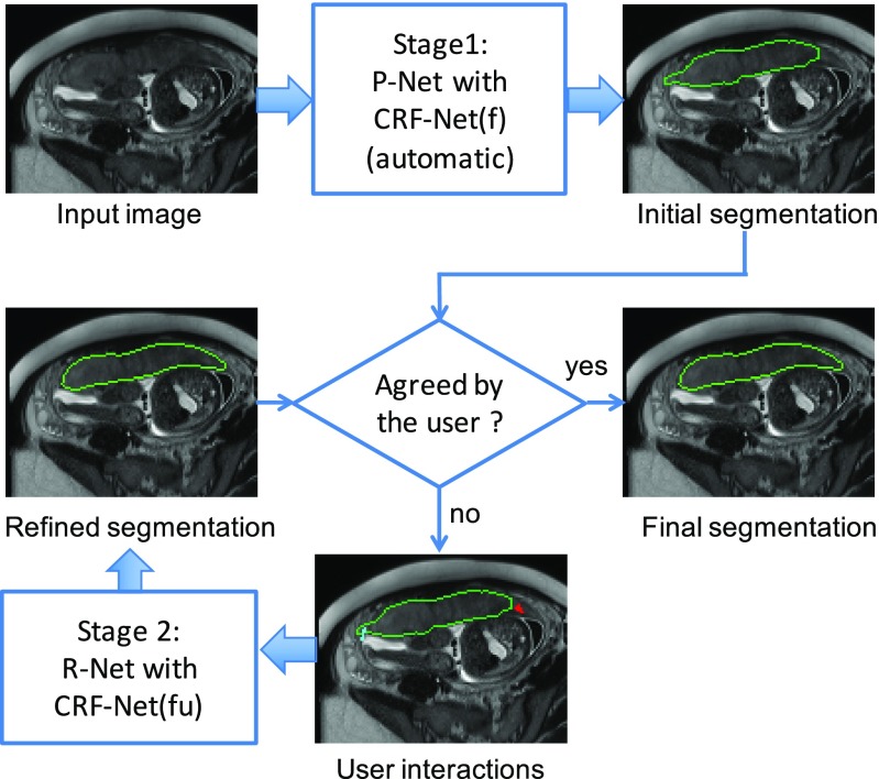
DeepIGeoS is a two-stage interactive segmentation framework in which a CNN proposes an initial segmentation and a second CNN refines it using user clicks/scribbles encoded as geodesic distance maps, with a trainable CRF that can enforce user interactions as hard constraints. It improved over automatic CNNs and achieved comparable or higher accuracy with fewer interactions and less user time than traditional interactive methods in 2D fetal MRI placenta and 3D brain tumor segmentation.
Key findings
- In 2D placenta segmentation, 2D P-Net + CRF-Net(f) reached Dice 85.86 ± 11.67% and ASSD 1.85 ± 1.30 pixels; after interactive refinement, 2D R-Net + CRF-Net(fu) reached Dice 89.31 ± 5.33% and ASSD 1.22 ± 0.55 pixels.
- In 3D brain tumor segmentation, 3D P-Net + CRF-Net(f) reached Dice 87.55 ± 6.72% and ASSD 2.04 ± 1.70 pixels; after refinement, 3D R-Net + CRF-Net(fu) reached Dice 89.93 ± 6.49% and ASSD 1.43 ± 1.16 pixels.
- Geodesic distance maps outperformed Euclidean distance maps for refinement, and CRF-Net(fu) further improved results; CRF-Net(f) with freeform pairwise potentials outperformed Dense CRF and CRF-Net(g) in the reported comparisons.
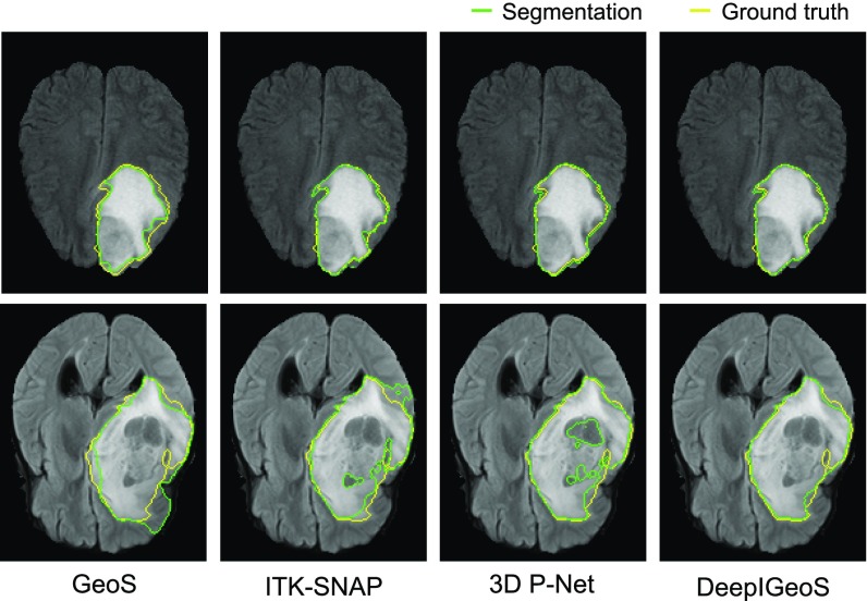
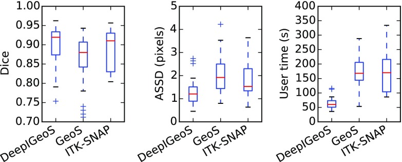
- Compared with traditional interactive methods, DeepIGeoS required far fewer user interactions and less user time; for 3D brain tumor segmentation, user time was about one third of that for GeoS and ITK-SNAP.
Methods
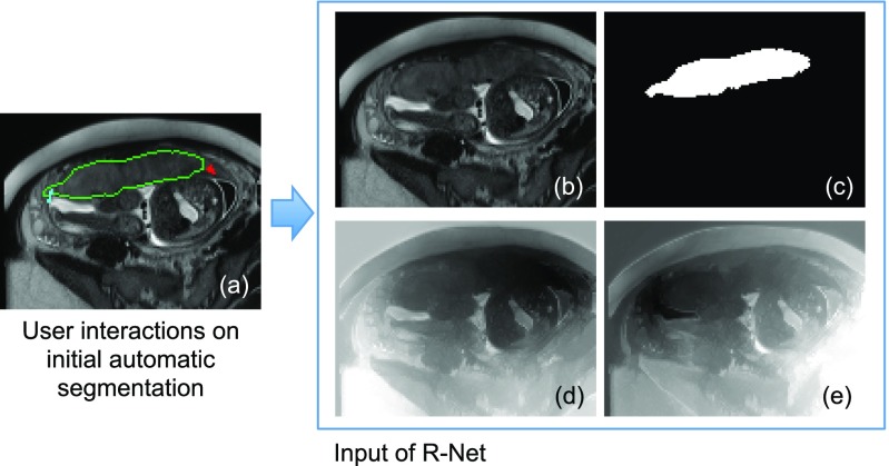
- Two-stage pipeline: P-Net produces an automatic initial segmentation; R-Net takes the original image, initial segmentation, and user interactions converted into geodesic distance maps to refine the result.
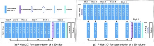
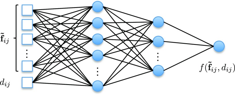
- Networks use a resolution-preserving dilated-convolution structure adapted from VGG-16, with multi-scale feature concatenation; a back-propagatable CRF-Net uses freeform pairwise potentials and, in R-Net, user scribbles as hard constraints.
- Data: 2D placenta from T2-weighted fetal MRI of 25 pregnant women, with 17 volumes/624 slices for training, 3 volumes/122 slices for validation, and 5 volumes/179 slices for testing; 3D brain tumor from the 2015 BraTS training set, using FLAIR images only, with 234 cases for training and 40 for testing. Evaluation used Dice and ASSD, with Student’s t-test for significance.
Limitations
- The paper does not include a dedicated limitations section; it states that the framework can be extended to deal with multiple organs in the future.
- Evident from the experiments: validation is limited to two binary segmentation tasks, and the 3D experiment uses only FLAIR images and segments only the whole tumor, not multi-label or multi-modality tumor subregions.
- Evident from the text: R-Net training uses simulated user interactions generated from mis-segmented regions, which may not fully match real user behavior.
Model-written summary. Check it against the paper before relying on it.
Integrity signals
Registry
Cites
Statistics
Figures
Verification
Claims the paper makes
39 Not checked yet · 34 Own result · 9 Own interpretation · 6 No basis given · 3 Own method · 2 Partly supported · 1 Supported · accuracy not yet measured
A result the paper reports from its own data — it cites no other source for it, so there is none to check. The quote shows it is in the paper.
In the paper
“Experimental results show our method achieves a large improvement from automatic CNNs”
§ DeepIGeoS: A Deep Interactive Geodesic Framework for Medical Image Segmentation > Abstract
plaindirect- Asserted: passes — The authors put it forward as true.
- Checkable: passes — It can be checked against evidence.
- Load-bearing: passes — The paper’s conclusions depend on it.
- Based: passes — The paper gives a reason why it holds.
A result the paper reports from its own data — it cites no other source for it, so there is none to check. The quote shows it is in the paper.
In the paper
“3D P-Net outperforms these three counterparts”
§ DeepIGeoS: A Deep Interactive Geodesic Framework for Medical Image Segmentation > Experiments > 3D Brain Tumor Segmentation from FLAIR Images
plaindirect- Asserted: passes — The authors put it forward as true.
- Checkable: passes — It can be checked against evidence.
- Load-bearing: unclear — It is unclear whether the paper’s conclusions depend on it.
- Based: passes — The paper gives a reason why it holds.
A result the paper reports from its own data — it cites no other source for it, so there is none to check. The quote shows it is in the paper.
In the paper
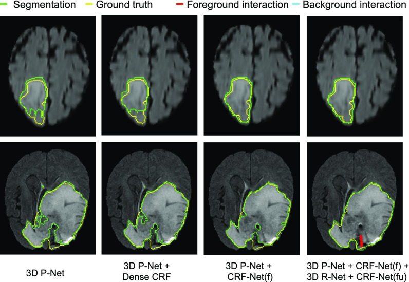
“only CRF-Net(f) obtains significantly better segmentation than 3D P-Net”
§ DeepIGeoS: A Deep Interactive Geodesic Framework for Medical Image Segmentation > Experiments > 3D Brain Tumor Segmentation from FLAIR Images
plaindirect- Asserted: passes — The authors put it forward as true.
- Checkable: passes — It can be checked against evidence.
- Load-bearing: passes — The paper’s conclusions depend on it.
- Based: passes — The paper gives a reason why it holds.
A result the paper reports from its own data — it cites no other source for it, so there is none to check. The quote shows it is in the paper.
In the paper
“CRF-Net(fu) can further improve the segmentation.”
§ DeepIGeoS: A Deep Interactive Geodesic Framework for Medical Image Segmentation > Experiments > 2D Placenta Segmentation from Fetal MRI
plaindirect- Asserted: passes — The authors put it forward as true.
- Checkable: passes — It can be checked against evidence.
- Load-bearing: passes — The paper’s conclusions depend on it.
- Based: passes — The paper gives a reason why it holds.
- Rests on an assumption: Simulated user interactions (random clicks in mis-segmented regions) are representative of real user interactions for training R-Net.
- Rests on an assumption: The manually delineated ground truth is accurate.
A result the paper reports from its own data — it cites no other source for it, so there is none to check. The quote shows it is in the paper.
In the paper
“Notice no initial scribbles are needed for DeepIGeoS.”
§ DeepIGeoS: A Deep Interactive Geodesic Framework for Medical Image Segmentation > Experiments > 2D Placenta Segmentation from Fetal MRI
plain- Asserted: passes — The authors put it forward as true.
- Checkable: passes — It can be checked against evidence.
- Load-bearing: passes — The paper’s conclusions depend on it.
- Based: passes — The paper gives a reason why it holds.
A result the paper reports from its own data — it cites no other source for it, so there is none to check. The quote shows it is in the paper.
In the paper
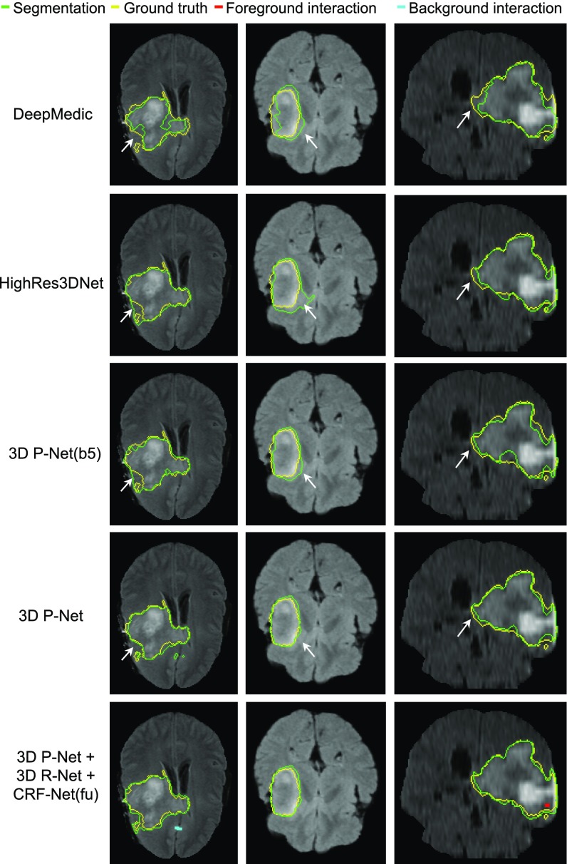
“DeepMedic achieves an average dice score of 83.87 percent.”
§ DeepIGeoS: A Deep Interactive Geodesic Framework for Medical Image Segmentation > Experiments > 3D Brain Tumor Segmentation from FLAIR Images
plain- Asserted: passes — The authors put it forward as true.
- Checkable: passes — It can be checked against evidence.
- Load-bearing: unclear — It is unclear whether the paper’s conclusions depend on it.
- Based: passes — The paper gives a reason why it holds.
A result the paper reports from its own data — it cites no other source for it, so there is none to check. The quote shows it is in the paper.
In the paper
“the combination of the proposed 2D R-Net using geodesic distance and CRF-Net(fu) leads to more accurate segmentations than the other refinement methods with the same set of user interactions.”
§ DeepIGeoS: A Deep Interactive Geodesic Framework for Medical Image Segmentation > Experiments > 2D Placenta Segmentation from Fetal MRI
plaindirectSingle case- Asserted: passes — The authors put it forward as true.
- Checkable: passes — It can be checked against evidence.
- Load-bearing: passes — The paper’s conclusions depend on it.
- Based: passes — The paper gives a reason why it holds.
- Rests on an assumption: Simulated user interactions (random clicks in mis-segmented regions) are representative of real user interactions for training R-Net.
- Rests on an assumption: The manually delineated ground truth is accurate.
A result the paper reports from its own data — it cites no other source for it, so there is none to check. The quote shows it is in the paper.
In the paper
“Fig. 7 and Table 1 indicate that large mis-segmentation exists in some images”
§ DeepIGeoS: A Deep Interactive Geodesic Framework for Medical Image Segmentation > Experiments > 2D Placenta Segmentation from Fetal MRI
hedged- Asserted: passes — The authors put it forward as true.
- Checkable: passes — It can be checked against evidence.
- Load-bearing: passes — The paper’s conclusions depend on it.
- Based: passes — The paper gives a reason why it holds.
- Rests on an assumption: The manually delineated ground truth is accurate.
A result the paper reports from its own data — it cites no other source for it, so there is none to check. The quote shows it is in the paper.
In the paper
“while the last two CRFs significantly improve the segmentation”
§ DeepIGeoS: A Deep Interactive Geodesic Framework for Medical Image Segmentation > Experiments > 2D Placenta Segmentation from Fetal MRI
plaindirect- Asserted: passes — The authors put it forward as true.
- Checkable: passes — It can be checked against evidence.
- Load-bearing: passes — The paper’s conclusions depend on it.
- Based: passes — The paper gives a reason why it holds.
- Rests on an assumption: The manually delineated ground truth is accurate.
A result the paper reports from its own data — it cites no other source for it, so there is none to check. The quote shows it is in the paper.
In the paper
“2.14”
§ DeepIGeoS: A Deep Interactive Geodesic Framework for Medical Image Segmentation > Experiments > 3D Brain Tumor Segmentation from FLAIR Images
plain- Asserted: passes — The authors put it forward as true.
- Checkable: passes — It can be checked against evidence.
- Load-bearing: unclear — It is unclear whether the paper’s conclusions depend on it.
- Based: passes — The paper gives a reason why it holds.
- It may state two separate claims as one. 31
- Probably not a substantial claim. 26
- The quote only partly states this claim. 6
- Borderline: one of the tests for a substantial claim is unclear. 1
The cited sources support only part of this claim — read each source’s verdict below.
In the paper
“Machine learning methods [6], [37], [38] have been proposed to learn from user interactions. They can achieve higher segmentation accuracy with fewer user interactions. However, they are limited by hand-crafted features that depend on the user's experience.”
§ DeepIGeoS: A Deep Interactive Geodesic Framework for Medical Image Segmentation > Related Works > Interactive Image Segmentation
What the sources say
- Not checked yetcites [6] Wang G., Zuluaga M. A., Pratt R., Aertsen M., Doel T., Klusmann M., David A. L., Deprest J., Vercauteren T., and Ourselin S., “ Slic-Seg: A minimally interactive segmentation of the placenta from sparse and motion-corrupted fetal MRI in multiple views,” Med. Image Anal., vol. 34, pp. 137– 147, 2016. 27179367 PMC5052128 source
- Not checked yetcites [37] Barinova O., Shapovalov R., Sudakov S., and Velizhev A., “ Online random forest for interactive image segmentation,” in Proc. Int. Workshop Exp. Economics Mach. Learn., 2012, pp. 1– 8.
- Partly supportedcites [38] Luengo I., Darrow M. C., Spink M. C., Sun Y., Dai W., He C. Y., Chiu W., Pridmore T., Ashton A. W., Duke E. M., Basham M., and French A. P., “ SuRVoS: Super-region volume segmentation workbench,” J. Structural Biol., vol. 198, no. 1, pp. 43– 53, 2017. PMC5405849 28246039 sourceExperimental — accuracy not yet measured
The Introduction describes machine-learning segmentation that learns from user annotations and requires discriminative features. Model training reports segmenting a biological volume with just two brief annotations, while Data Pre-processing and the Discussion say users must choose appropriate features and parameters. The paper does not establish that these methods achieve higher segmentation accuracy with fewer interactions, or explicitly identify dependence on user experience as a general limitation. (LLM, conf=0.82)
plaindirect- Asserted: passes — The authors put it forward as true.
- Checkable: passes — It can be checked against evidence.
- Load-bearing: passes — The paper’s conclusions depend on it.
- Based: passes — The paper gives a reason why it holds.
It may state two separate claims as one.
Every source the paper cites for this claim was checked and supports it.
In the paper
“To overcome the drawbacks of successive max-pooling and downsampling that lead to a loss of feature map resolution, dilated convolution [22], [23] was proposed to preserve the resolution of feature maps and enlarge the receptive field to incorporate larger contextual information.”
§ DeepIGeoS: A Deep Interactive Geodesic Framework for Medical Image Segmentation > Related Works > Image Segmentation Based on CNNs
What the sources say
- Supportedcites [22] Chen L.-C., Papandreou G., Kokkinos I., Murphy K., and Yuille A. L., “ Semantic image segmentation with deep convolutional nets and fully connected CRFs,” arXiv preprint arXiv:1412.7062. 28463186 source
Section 3.1 (pp. 4–5) says atrous convolution enables denser, higher-resolution feature maps despite downsampling and enlarges filters’ field of view to assimilate more context. The Introduction (p. 1) also states both points together. (LLM, conf=0.94)
- Supportedcites [23] Yu F. and Koltun V., “ Multi-scale context aggregation by dilated convolutions,” arXiv preprint arXiv:1511.07122. source
The Introduction (p. 2) says the dilated-convolution module aggregates multi-scale contextual information without losing resolution. Section 2 (p. 2) states that dilated convolutions expand the receptive field without losing resolution or coverage. (LLM, conf=0.94)
plaindirect- Asserted: passes — The authors put it forward as true.
- Checkable: passes — It can be checked against evidence.
- Load-bearing: fails — The paper’s conclusions may not depend on it.
- Based: passes — The paper gives a reason why it holds.
Probably not a substantial claim. The paper’s conclusions may not depend on it. It may state two separate claims as one.
- Supported
The cited sources support only part of this claim — read each source’s verdict below.
In the paper
“However, most of these methods rely on low-level features and require a relatively large amount of user interactions to deal with images with low contrast and ambiguous boundaries.”
§ DeepIGeoS: A Deep Interactive Geodesic Framework for Medical Image Segmentation > Related Works > Interactive Image Segmentation
What the sources say
- Not checked yetcites [8] Boykov Y. Y. and Jolly M. P., “ Interactive graph cuts for optimal boundary & region segmentation of objects in N-D images,” in Proc. 8th IEEE Int. Conf. Comput. Vis., 2001, pp. 105– 112. source
- Partly supportedcites [35] Bai X. and Sapiro G., “ A geodesic framework for fast interactive image and video segmentation and matting,” in Proc. IEEE 11th Int. Conf. Comput. Vis., 2007, pp. 1– 8. sourceExperimental — accuracy not yet measured
The Conclusions (page not provided) say the paper’s own method relies mainly on pixel-value distributions and that overlapping distributions may take enough user interactions to become tedious. The Introduction also describes ambiguous images as a reason for user intervention. These passages do not establish that most interactive segmentation methods require relatively many interactions specifically for low-contrast images or ambiguous boundaries. (LLM, conf=0.83)
- Not checked yetcites [7] Grady L., “ Random walks for image segmentation,” IEEE Trans. Pattern Anal. Mach. Intell., vol. 28, no. 11, pp. 1768– 1783, Nov. 2006. 17063682 source
plaindirect- Asserted: passes — The authors put it forward as true.
- Checkable: passes — It can be checked against evidence.
- Load-bearing: passes — The paper’s conclusions depend on it.
- Based: passes — The paper gives a reason why it holds.
It may state two separate claims as one.
A result the paper reports from its own data — it cites no other source for it, so there is none to check. The quote shows it is in the paper.
In the paper
“obtains comparable and even higher accuracy with fewer user interventions and less time compared with traditional interactive methods.”
§ DeepIGeoS: A Deep Interactive Geodesic Framework for Medical Image Segmentation > Abstract
plaindirect- Asserted: passes — The authors put it forward as true.
- Checkable: passes — It can be checked against evidence.
- Load-bearing: passes — The paper’s conclusions depend on it.
- Based: passes — The paper gives a reason why it holds.
It may state two separate claims as one.
A result the paper reports from its own data — it cites no other source for it, so there is none to check. The quote shows it is in the paper.
In the paper
“HighRes3DNet and 3D P-Net(b5) achieve similar performance, and they are better than DeepMedic.”
§ DeepIGeoS: A Deep Interactive Geodesic Framework for Medical Image Segmentation > Experiments > 3D Brain Tumor Segmentation from FLAIR Images
plain- Asserted: passes — The authors put it forward as true.
- Checkable: passes — It can be checked against evidence.
- Load-bearing: unclear — It is unclear whether the paper’s conclusions depend on it.
- Based: passes — The paper gives a reason why it holds.
It may state two separate claims as one.
A result the paper reports from its own data — it cites no other source for it, so there is none to check. The quote shows it is in the paper.
In the paper
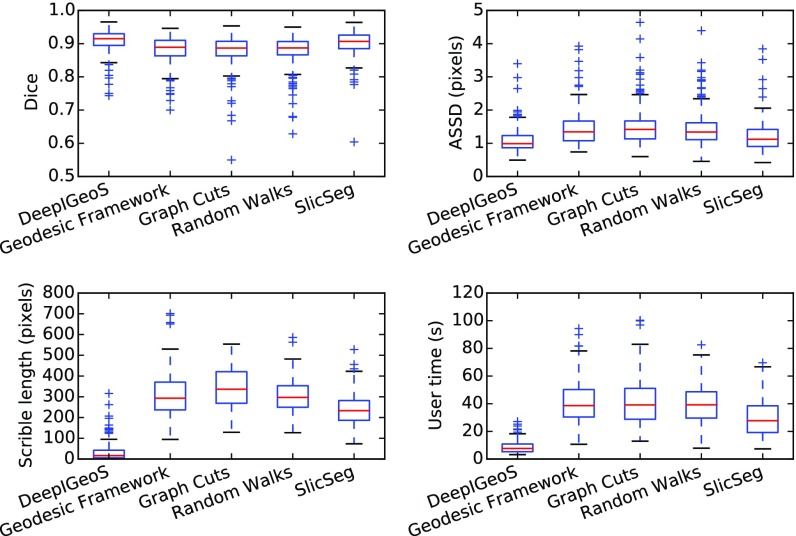
“DeepIGeoS requires far fewer user interactions and less user time.”
§ DeepIGeoS: A Deep Interactive Geodesic Framework for Medical Image Segmentation > Experiments > 2D Placenta Segmentation from Fetal MRI
plaindirect- Asserted: passes — The authors put it forward as true.
- Checkable: passes — It can be checked against evidence.
- Load-bearing: passes — The paper’s conclusions depend on it.
- Based: passes — The paper gives a reason why it holds.
It may state two separate claims as one.
The paper states this without a source or data behind it here, so there is nothing to check it against. The quote shows it is in the paper.
In the paper
“In addition, CRF-Net(f) is better than CRF-Net(g).”
§ DeepIGeoS: A Deep Interactive Geodesic Framework for Medical Image Segmentation > Experiments > 2D Placenta Segmentation from Fetal MRI
plaindirect- Asserted: passes — The authors put it forward as true.
- Checkable: passes — It can be checked against evidence.
- Load-bearing: unclear — It is unclear whether the paper’s conclusions depend on it.
- Based: passes — The paper gives a reason why it holds.
The quote only partly states this claim.
- Rests on an assumption: The manually delineated ground truth is accurate.
The paper states this without a source or data behind it here, so there is nothing to check it against. The quote shows it is in the paper.
In the paper
“the proposed 3D P-Net has far fewer parameters compared with HighRes3DNet.”
§ DeepIGeoS: A Deep Interactive Geodesic Framework for Medical Image Segmentation > Experiments > 3D Brain Tumor Segmentation from FLAIR Images
plain- Asserted: passes — The authors put it forward as true.
- Checkable: passes — It can be checked against evidence.
- Load-bearing: passes — The paper’s conclusions depend on it.
- Based: fails — The paper may give no reason why it holds.
Probably not a substantial claim. The paper may give no reason why it holds.
A result the paper reports from its own data — it cites no other source for it, so there is none to check. The quote shows it is in the paper.
In the paper
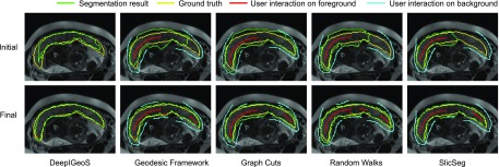
“DeepIGeoS only needs two short strokes to get an accurate segmentation, while the other methods require far more scribbles to get similar results.”
§ DeepIGeoS: A Deep Interactive Geodesic Framework for Medical Image Segmentation > Experiments > 2D Placenta Segmentation from Fetal MRI
plaindirect- Asserted: passes — The authors put it forward as true.
- Checkable: passes — It can be checked against evidence.
- Load-bearing: passes — The paper’s conclusions depend on it.
- Based: passes — The paper gives a reason why it holds.
It may state two separate claims as one.
A result the paper reports from its own data — it cites no other source for it, so there is none to check. The quote shows it is in the paper.
In the paper
“Significant improvement from 2D R-Net”
§ DeepIGeoS: A Deep Interactive Geodesic Framework for Medical Image Segmentation > Experiments > 2D Placenta Segmentation from Fetal MRI
plaindirect- Asserted: passes — The authors put it forward as true.
- Checkable: passes — It can be checked against evidence.
- Load-bearing: passes — The paper’s conclusions depend on it.
- Based: passes — The paper gives a reason why it holds.
The quote only partly states this claim.
A result the paper reports from its own data — it cites no other source for it, so there is none to check. The quote shows it is in the paper.
In the paper
“3D P-Net outperforms these three counterparts with 86.68”
§ DeepIGeoS: A Deep Interactive Geodesic Framework for Medical Image Segmentation > Experiments > 3D Brain Tumor Segmentation from FLAIR Images
plain- Asserted: passes — The authors put it forward as true.
- Checkable: passes — It can be checked against evidence.
- Load-bearing: unclear — It is unclear whether the paper’s conclusions depend on it.
- Based: passes — The paper gives a reason why it holds.
The quote only partly states this claim.
A result the paper reports from its own data — it cites no other source for it, so there is none to check. The quote shows it is in the paper.
In the paper
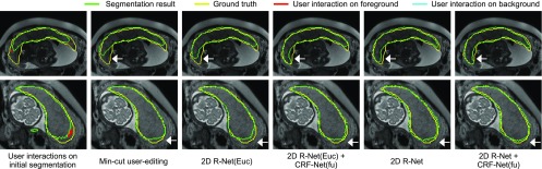
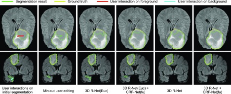
“Fig. 8 shows 2D R-Net with geodesic distance performs better than min-cut user-editing and 2D R-Net(Euc) that uses euclidean distance.”
§ DeepIGeoS: A Deep Interactive Geodesic Framework for Medical Image Segmentation > Experiments > 2D Placenta Segmentation from Fetal MRI
plaindirect- Asserted: passes — The authors put it forward as true.
- Checkable: passes — It can be checked against evidence.
- Load-bearing: passes — The paper’s conclusions depend on it.
- Based: passes — The paper gives a reason why it holds.
It may state two separate claims as one.
- Rests on an assumption: Simulated user interactions (random clicks in mis-segmented regions) are representative of real user interactions for training R-Net.
- Rests on an assumption: The manually delineated ground truth is accurate.
A result the paper reports from its own data — it cites no other source for it, so there is none to check. The quote shows it is in the paper.
In the paper
“our proposed method achieves better results than automatic CNNs.”
§ DeepIGeoS: A Deep Interactive Geodesic Framework for Medical Image Segmentation > Conclusion
plaindirect- Asserted: passes — The authors put it forward as true.
- Checkable: passes — It can be checked against evidence.
- Load-bearing: passes — The paper’s conclusions depend on it.
- Based: passes — The paper gives a reason why it holds.
It may state two separate claims as one.
A result the paper reports from its own data — it cites no other source for it, so there is none to check. The quote shows it is in the paper.
In the paper
“Significant improvement from 3D R-Net”
§ DeepIGeoS: A Deep Interactive Geodesic Framework for Medical Image Segmentation > Experiments > 3D Brain Tumor Segmentation from FLAIR Images
plaindirect- Asserted: passes — The authors put it forward as true.
- Checkable: passes — It can be checked against evidence.
- Load-bearing: passes — The paper’s conclusions depend on it.
- Based: passes — The paper gives a reason why it holds.
The quote only partly states this claim.
A result the paper reports from its own data — it cites no other source for it, so there is none to check. The quote shows it is in the paper.
In the paper
“ASSD of 1.43”
§ DeepIGeoS: A Deep Interactive Geodesic Framework for Medical Image Segmentation > Experiments > 3D Brain Tumor Segmentation from FLAIR Images
plain- Asserted: passes — The authors put it forward as true.
- Checkable: passes — It can be checked against evidence.
- Load-bearing: passes — The paper’s conclusions depend on it.
- Based: passes — The paper gives a reason why it holds.
The quote only partly states this claim.
The paper states this without a source or data behind it here, so there is nothing to check it against. The quote shows it is in the paper.
In the paper
“It requires far less user time compared with traditional interactive methods”
§ DeepIGeoS: A Deep Interactive Geodesic Framework for Medical Image Segmentation > Conclusion
plaindirect- Asserted: passes — The authors put it forward as true.
- Checkable: passes — It can be checked against evidence.
- Load-bearing: passes — The paper’s conclusions depend on it.
- Based: unclear — It is unclear why the paper says it holds.
Borderline substantial claim. It is unclear why the paper says it holds.
A result the paper reports from its own data — it cites no other source for it, so there is none to check. The quote shows it is in the paper.
In the paper
“Dice score of 89.93”
§ DeepIGeoS: A Deep Interactive Geodesic Framework for Medical Image Segmentation > Experiments > 3D Brain Tumor Segmentation from FLAIR Images
plain- Asserted: passes — The authors put it forward as true.
- Checkable: passes — It can be checked against evidence.
- Load-bearing: passes — The paper’s conclusions depend on it.
- Based: passes — The paper gives a reason why it holds.
The quote only partly states this claim.
The paper states this without a source or data behind it here, so there is nothing to check it against. The quote shows it is in the paper.
In the paper
“However, the euclidean distance does not take advantage of image context information.”
- Also stated in: § User Interaction-Based Geodesic Distance Maps · plain
“However, the euclidean distance treats each direction equally and does not take the image context into account.”
§ DeepIGeoS: A Deep Interactive Geodesic Framework for Medical Image Segmentation > Related Works > Interactive Image Segmentation
plain- Asserted: passes — The authors put it forward as true.
- Checkable: passes — It can be checked against evidence.
- Load-bearing: passes — The paper’s conclusions depend on it.
- Based: passes — The paper gives a reason why it holds.
It may state two separate claims as one.
- Also stated in: § User Interaction-Based Geodesic Distance Maps · plain
In the paper
“In [40], a deep interactive object selection method was proposed where user-provided clicks are transformed into euclidean distance maps and then concatenated with the input of FCNs.”
§ DeepIGeoS: A Deep Interactive Geodesic Framework for Medical Image Segmentation > Related Works > Interactive Image Segmentation
What the sources say
- Not checked yetcites [40] Xu N., Price B., Cohen S., Yang J., and Huang T., “ Deep interactive object selection,” in Proc. IEEE Conf. Comput. Vis. Pattern Recognit., 2016, pp. 373– 381. source
plaindirect- Asserted: passes — The authors put it forward as true.
- Checkable: passes — It can be checked against evidence.
- Load-bearing: passes — The paper’s conclusions depend on it.
- Based: passes — The paper gives a reason why it holds.
Probably not a substantial claim.
A conclusion the paper draws from its own results — it cites no other source for it, so there is none to check. The quote shows it is in the paper.
In the paper
“Third, it is efficient and can respond to user interactions in real time, which leads to very short user time.”
§ DeepIGeoS: A Deep Interactive Geodesic Framework for Medical Image Segmentation > Introduction
plaindirect- Asserted: passes — The authors put it forward as true.
- Checkable: passes — It can be checked against evidence.
- Load-bearing: passes — The paper’s conclusions depend on it.
- Based: unclear — It is unclear why the paper says it holds.
It may state two separate claims as one.
In the paper
“DeepCut [32] employs user-provided bounding boxes as annotations to train CNNs for the segmentation of fetal MRI.”
§ DeepIGeoS: A Deep Interactive Geodesic Framework for Medical Image Segmentation > Related Works > Interactive Image Segmentation
What the sources say
- Not checked yetcites [32] Rajchl M., Lee M., Oktay O., Kamnitsas K., Passerat-Palmbach J., Bai W., Rutherford M., Hajnal J., Kainz B., and Rueckert D., “ DeepCut: Object segmentation from bounding box annotations using convolutional neural networks,” IEEE Trans. Medical Imaging, vol. 36, no. 2, pp. 674– 683, Feb. 2017. 27845654 PMC7115996 source
plaindirect- Asserted: passes — The authors put it forward as true.
- Checkable: passes — It can be checked against evidence.
- Load-bearing: fails — The paper’s conclusions may not depend on it.
- Based: passes — The paper gives a reason why it holds.
Probably not a substantial claim. The paper’s conclusions may not depend on it.
In the paper
“Recently, deep learning with convolutional neural networks (CNNs) has achieved the state-of-the-art performance in many image analysis applications [10].”
§ DeepIGeoS: A Deep Interactive Geodesic Framework for Medical Image Segmentation > Introduction
What the sources say
- Not checked yetcites [10] Litjens G., Kooi T., Bejnordi B. E., Setio A. A. A., Ciompi F., Ghafoorian M., van der Laak J. A., Ginneken B. V., and Sánchez C. I., “ A survey on deep learning in medical image analysis,” Med. Image Anal., vol. 42, pp. 60– 88, 2017. 28778026 source
plain- Asserted: passes — The authors put it forward as true.
- Checkable: passes — It can be checked against evidence.
- Load-bearing: fails — The paper’s conclusions may not depend on it.
- Based: passes — The paper gives a reason why it holds.
Probably not a substantial claim. The paper’s conclusions may not depend on it.
In the paper
“SlicSeg [6] accepts user-provided scribbles in a single start slice to train an Online Random Forest for 3D segmentation, but lacks in flexibility to allow further user-editing.”
§ DeepIGeoS: A Deep Interactive Geodesic Framework for Medical Image Segmentation > Introduction
What the sources say
- Not checked yetcites [6] Wang G., Zuluaga M. A., Pratt R., Aertsen M., Doel T., Klusmann M., David A. L., Deprest J., Vercauteren T., and Ourselin S., “ Slic-Seg: A minimally interactive segmentation of the placenta from sparse and motion-corrupted fetal MRI in multiple views,” Med. Image Anal., vol. 34, pp. 137– 147, 2016. 27179367 PMC5052128 source
plain- Asserted: passes — The authors put it forward as true.
- Checkable: passes — It can be checked against evidence.
- Load-bearing: passes — The paper’s conclusions depend on it.
- Based: passes — The paper gives a reason why it holds.
It may state two separate claims as one.
In the paper
“For example, the widely used ITK-SNAP [5] takes user-provided seed pixels or blobs as a starting point and employs an active contour model for segmentation. It requires most of the user interactions to be given at the beginning and the underpinning model can hardly be refined with additional user interactions once an initial segmentation is obtained.”
§ DeepIGeoS: A Deep Interactive Geodesic Framework for Medical Image Segmentation > Introduction
What the sources say
- Not checked yetcites [5] Yushkevich P. A., Piven J., Hazlett H. C., Smith R. G., Ho S., Gee J. C., and Gerig G., “ User-guided 3D active contour segmentation of anatomical structures: Significantly improved efficiency and reliability,” NeuroImage, vol. 31, no. 3, pp. 1116– 1128, 2006. 16545965 source
plaindirect- Asserted: passes — The authors put it forward as true.
- Checkable: passes — It can be checked against evidence.
- Load-bearing: passes — The paper’s conclusions depend on it.
- Based: passes — The paper gives a reason why it holds.
It may state two separate claims as one.
None of the sources cited for this claim have been checked yet.
In the paper
“Typical CNNs [16], [17], [18] were originally designed for image classification tasks.”
§ DeepIGeoS: A Deep Interactive Geodesic Framework for Medical Image Segmentation > Related Works > Image Segmentation Based on CNNs
What the sources say
- Not checked yetcites [16] Krizhevsky A., Sutskever I., and Hinton G. E., “ ImageNet classification with deep convolutional neural networks,” in Proc. 25th Int. Conf. Neural Inf. Process. Syst., 2012, pp. 1097– 1105.
- Not checked yetcites [17] Simonyan K. and Zisserman A., “ Very deep convolutional networks for large-scale image recognition,” arXiv preprint arXiv:1409.1556. source
- Not checked yetcites [18] He K., Zhang X., Ren S., and Sun J., “ Deep residual learning for image recognition,” in Proc. IEEE Conf. Comput. Vis. Pattern Recognit., 2016, pp. 770– 778. source
plain- Asserted: passes — The authors put it forward as true.
- Checkable: passes — It can be checked against evidence.
- Load-bearing: fails — The paper’s conclusions may not depend on it.
- Based: passes — The paper gives a reason why it holds.
Probably not a substantial claim. The paper’s conclusions may not depend on it.
None of the sources cited for this claim have been checked yet.
In the paper
“Some early works adapted such networks for pixel labeling with patch or region-based methods [19], [20]. Such methods achieved higher accuracy than traditional methods that relied on hand-crafted features. However, they suffered from inefficiency for testing.”
§ DeepIGeoS: A Deep Interactive Geodesic Framework for Medical Image Segmentation > Related Works > Image Segmentation Based on CNNs
What the sources say
- Not checked yetcites [19] Havaei M., Davy A., Warde-Farley D., Biard A., Courville A., Bengio Y., Pal C., Jodoin P.-M., and Larochelle H., “ Brain tumor segmentation with deep neural networks,” Med. Image Anal., vol. 35, pp. 18– 31, 2016. 27310171 source
- Not checked yetcites [20] Girshick R., Donahue J., Darrell T., and Malik J., “ Rich feature hierarchies for accurate object detection and semantic segmentation,” in Proc. IEEE Conf. Comput. Vis. Pattern Recognit., 2014, pp. 580– 587. source
plaindirect- Asserted: passes — The authors put it forward as true.
- Checkable: passes — It can be checked against evidence.
- Load-bearing: fails — The paper’s conclusions may not depend on it.
- Based: passes — The paper gives a reason why it holds.
Probably not a substantial claim. The paper’s conclusions may not depend on it. It may state two separate claims as one.
None of the sources cited for this claim have been checked yet.
In the paper
“GrabCut [9] works with a user-provided bounding box to start the segmentation and requires fewer interactions compared with Graph Cuts [8], but the performance is still limited by the representativity of the underpinning GMM.”
§ DeepIGeoS: A Deep Interactive Geodesic Framework for Medical Image Segmentation > Introduction
What the sources say
- Not checked yetcites [9] Rother C., Kolmogorov V., and Blake A., “ GrabCut: Interactive foreground extraction using iterated graph cuts,” ACM Trans. Graph., vol. 23, no. 3, pp. 309– 314, 2004. source
- Not checked yetcites [8] Boykov Y. Y. and Jolly M. P., “ Interactive graph cuts for optimal boundary & region segmentation of objects in N-D images,” in Proc. 8th IEEE Int. Conf. Comput. Vis., 2001, pp. 105– 112. source
plaindirect- Asserted: passes — The authors put it forward as true.
- Checkable: passes — It can be checked against evidence.
- Load-bearing: passes — The paper’s conclusions depend on it.
- Based: passes — The paper gives a reason why it holds.
It may state two separate claims as one.
In the paper
“ScribbleSup [39] also trains CNNs for semantic segmentation supervised by scribbles.”
§ DeepIGeoS: A Deep Interactive Geodesic Framework for Medical Image Segmentation > Related Works > Interactive Image Segmentation
What the sources say
- Not checked yetcites [39] Lin D., Dai J., Jia J., He K., and Sun J., “ ScribbleSup: Scribble-supervised convolutional networks for semantic segmentation,” in Proc. IEEE Conf. Comput. Vis. Pattern Recognit., 2016, pp. 3159– 3167. source
plaindirect- Asserted: passes — The authors put it forward as true.
- Checkable: passes — It can be checked against evidence.
- Load-bearing: fails — The paper’s conclusions may not depend on it.
- Based: passes — The paper gives a reason why it holds.
Probably not a substantial claim. The paper’s conclusions may not depend on it.
In the paper
“FCNs [11] take an entire image as input and give a dense segmentation. In order to overcome the problem of loss of spatial resolution due to multi-stage max-pooling and downsampling, it uses a stack of deconvolution (a.k.a. upsampling) layers and activation functions to upsample the feature maps.”
§ DeepIGeoS: A Deep Interactive Geodesic Framework for Medical Image Segmentation > Related Works > Image Segmentation Based on CNNs
What the sources say
- Not checked yetcites [11] Long J., Shelhamer E., and Darrell T., “ Fully convolutional networks for semantic segmentation,” in Proc. IEEE Conf. Comput. Vis. Pattern Recognit., 2015, pp. 3431– 3440. 27244717
plaindirect- Asserted: passes — The authors put it forward as true.
- Checkable: passes — It can be checked against evidence.
- Load-bearing: fails — The paper’s conclusions may not depend on it.
- Based: passes — The paper gives a reason why it holds.
Probably not a substantial claim. The paper’s conclusions may not depend on it. It may state two separate claims as one.
The paper states this without a source or data behind it here, so there is nothing to check it against. The quote shows it is in the paper.
In the paper
“However, they have not yet been adapted to interactive medical image segmentation.”
§ DeepIGeoS: A Deep Interactive Geodesic Framework for Medical Image Segmentation > Introduction
plain- Asserted: passes — The authors put it forward as true.
- Checkable: passes — It can be checked against evidence.
- Load-bearing: passes — The paper’s conclusions depend on it.
- Based: fails — The paper may give no reason why it holds.
Probably not a substantial claim. The paper may give no reason why it holds.
None of the sources cited for this claim have been checked yet.
In the paper
“Multi-scale features extracted from CNNs have been shown to be effective for improving segmentation accuracy [11], [22], [23].”
§ DeepIGeoS: A Deep Interactive Geodesic Framework for Medical Image Segmentation > Related Works > Image Segmentation Based on CNNs
What the sources say
- Not checked yetcites [11] Long J., Shelhamer E., and Darrell T., “ Fully convolutional networks for semantic segmentation,” in Proc. IEEE Conf. Comput. Vis. Pattern Recognit., 2015, pp. 3431– 3440. 27244717 source
- Not checked yetcites [22] Chen L.-C., Papandreou G., Kokkinos I., Murphy K., and Yuille A. L., “ Semantic image segmentation with deep convolutional nets and fully connected CRFs,” arXiv preprint arXiv:1412.7062. 28463186 source
- Not checked yetcites [23] Yu F. and Koltun V., “ Multi-scale context aggregation by dilated convolutions,” arXiv preprint arXiv:1511.07122. source
boosteddirect- Asserted: passes — The authors put it forward as true.
- Checkable: passes — It can be checked against evidence.
- Load-bearing: fails — The paper’s conclusions may not depend on it.
- Based: passes — The paper gives a reason why it holds.
Probably not a substantial claim. The paper’s conclusions may not depend on it.
None of the sources cited for this claim have been checked yet.
In the paper
“the geodesic distance transform [35], [36], [41] encodes spatial regularization and contrast-sensitivity”
§ DeepIGeoS: A Deep Interactive Geodesic Framework for Medical Image Segmentation > Related Works > Interactive Image Segmentation
What the sources say
- Not checked yetcites [35] Bai X. and Sapiro G., “ A geodesic framework for fast interactive image and video segmentation and matting,” in Proc. IEEE 11th Int. Conf. Comput. Vis., 2007, pp. 1– 8. source
- Not checked yetcites [36] Criminisi A., Sharp T., and Blake A., “ GeoS: Geodesic image segmentation,” in Proc. Eur. Conf. Comput. Vis., 2008, pp. 99– 112. source
- Not checked yetcites [41] Kontschieder P., Kohli P., Shotton J., and Criminisi A., “ GeoF geodesic forests for learning coupled predictors,” in Proc. IEEE Conf. Comput. Vis. Pattern Recognit., 2013, pp. 65– 72. source
plaindirect- Asserted: passes — The authors put it forward as true.
- Checkable: passes — It can be checked against evidence.
- Load-bearing: passes — The paper’s conclusions depend on it.
- Based: passes — The paper gives a reason why it holds.
It may state two separate claims as one.
None of the sources cited for this claim have been checked yet.
In the paper
“Random Walks [7] and Graph Cuts [8] learn from scribbles and allow the user to provide additional scribbles for refinement. They use Random Walker and Gaussian Mixture Model (GMM) as the underpinning model. However, they need a large number of scribbles to get satisfactory segmentation.”
§ DeepIGeoS: A Deep Interactive Geodesic Framework for Medical Image Segmentation > Introduction
What the sources say
- Not checked yetcites [7] Grady L., “ Random walks for image segmentation,” IEEE Trans. Pattern Anal. Mach. Intell., vol. 28, no. 11, pp. 1768– 1783, Nov. 2006. 17063682 source
- Not checked yetcites [8] Boykov Y. Y. and Jolly M. P., “ Interactive graph cuts for optimal boundary & region segmentation of objects in N-D images,” in Proc. 8th IEEE Int. Conf. Comput. Vis., 2001, pp. 105– 112. source
plaindirect- Asserted: passes — The authors put it forward as true.
- Checkable: passes — It can be checked against evidence.
- Load-bearing: passes — The paper’s conclusions depend on it.
- Based: passes — The paper gives a reason why it holds.
It may state two separate claims as one.
None of the sources cited for this claim have been checked yet.
In the paper
“Graphical models such as CRFs [22], [42], [43] have been widely used to enhance segmentation accuracy by introducing spatial consistency.”
§ DeepIGeoS: A Deep Interactive Geodesic Framework for Medical Image Segmentation > Related Works > CRFs for Spatial Regularization
What the sources say
- Not checked yetcites [22] Chen L.-C., Papandreou G., Kokkinos I., Murphy K., and Yuille A. L., “ Semantic image segmentation with deep convolutional nets and fully connected CRFs,” arXiv preprint arXiv:1412.7062. 28463186 source
- Not checked yetcites [42] Boykov Y. and Kolmogorov V., “ An experimental comparison of min-cut/max-flow algorithms for energy minimization in vision,” IEEE Trans. Pattern Anal. Mach. Intell., vol. 26, no. 9, pp. 1124– 1137, Sep. 2004. 15742889 source
- Not checked yetcites [43] Yuan J., Bae E., and Tai X. C., “ A study on continuous max-flow and min-cut approaches,” in Proc. IEEE Comput. Society Conf. Comput. Vis. Pattern Recognit., 2010, pp. 2217– 2224. source
boosteddirect- Asserted: passes — The authors put it forward as true.
- Checkable: passes — It can be checked against evidence.
- Load-bearing: fails — The paper’s conclusions may not depend on it.
- Based: passes — The paper gives a reason why it holds.
Probably not a substantial claim. The paper’s conclusions may not depend on it.
In the paper
“Despite the large number of existing interactive segmentation methods [3], most of them are confronted by requiring a large amount of user interactions and long user time, or limited learning ability with their underpinning models.”
§ DeepIGeoS: A Deep Interactive Geodesic Framework for Medical Image Segmentation > Introduction
What the sources say
- Not checked yetcites [3] Zhao F. and Xie X., “ An overview of interactive medical image segmentation,” Ann. BMVA, vol. 2013, no. 7, pp. 1– 22, 2013.
plain- Asserted: passes — The authors put it forward as true.
- Checkable: passes — It can be checked against evidence.
- Load-bearing: passes — The paper’s conclusions depend on it.
- Based: passes — The paper gives a reason why it holds.
It may state two separate claims as one.
In the paper
“For instance, 3D U-Net [21] learns from sparsely annotated images and can be used for semi-automatic segmentation.”
§ DeepIGeoS: A Deep Interactive Geodesic Framework for Medical Image Segmentation > Related Works > Interactive Image Segmentation
What the sources say
- Not checked yetcites [21] Abdulkadir A., Lienkamp S. S., Brox T., and Ronneberger O., “ 3D U-Net: Learning dense volumetric segmentation from sparse annotation,” in Proc. Medical Image Comput. Comput.-Assisted Intervention, 2016, pp. 424– 432. source
plaindirect- Asserted: passes — The authors put it forward as true.
- Checkable: passes — It can be checked against evidence.
- Load-bearing: fails — The paper’s conclusions may not depend on it.
- Based: passes — The paper gives a reason why it holds.
Probably not a substantial claim. The paper’s conclusions may not depend on it. It may state two separate claims as one.
None of the sources cited for this claim have been checked yet.
In the paper
“Alternatively, mean-field approximation [45], [50], [52] is often used for efficient inference of the CRF while allowing learning parameters by back-propagation.”
§ DeepIGeoS: A Deep Interactive Geodesic Framework for Medical Image Segmentation > Method > Back-Propagatable CRF-Net with Freeform Pairwise Potentials and User Constraints
What the sources say
- Not checked yetcites [45] Krähenbühl P. and Koltun V., “ Efficient inference in fully connected CRFs with gaussian edge potentials,” in Proc. Neural Inf. Process. Syst. Conf., 2011, pp. 109– 117. source
- Not checked yetcites [50] Zheng S., Jayasumana S., Romera-Paredes B., Vineet V., Su Z., Du D., Huang C., and Torr P. H. S., “ Conditional random fields as recurrent neural networks,” in Proc. IEEE Int. Conf. Comput. Vis., 2015, pp. 1529– 1537. source
- Not checked yetcites [52] Vemulapalli R., Tuzel O., Liu M.-y., and Chellappa R., “ Gaussian conditional random field network for semantic segmentation,” in Proc. IEEE Conf. Comput. Vis. Pattern Recognit., 2016, pp. 3224– 3233. source
plaindirect- Asserted: passes — The authors put it forward as true.
- Checkable: passes — It can be checked against evidence.
- Load-bearing: fails — The paper’s conclusions may not depend on it.
- Based: passes — The paper gives a reason why it holds.
Probably not a substantial claim. The paper’s conclusions may not depend on it.
What the paper did — it cites no source for this, so there is none to check. The quote shows it is in the paper.
In the paper
“in which the same initial segmentation and the same set of user interactions were used by the five refinement methods.”
§ DeepIGeoS: A Deep Interactive Geodesic Framework for Medical Image Segmentation > Experiments > 2D Placenta Segmentation from Fetal MRI
plain- Asserted: passes — The authors put it forward as true.
- Checkable: passes — It can be checked against evidence.
- Load-bearing: passes — The paper’s conclusions depend on it.
- Based: passes — The paper gives a reason why it holds.
Probably not a substantial claim.
In the paper
“In [52], a Gaussian Mean Field (GMF) network was proposed and combined with CNNs where all the parameters are trainable.”
§ DeepIGeoS: A Deep Interactive Geodesic Framework for Medical Image Segmentation > Related Works > CRFs for Spatial Regularization
What the sources say
- Not checked yetcites [52] Vemulapalli R., Tuzel O., Liu M.-y., and Chellappa R., “ Gaussian conditional random field network for semantic segmentation,” in Proc. IEEE Conf. Comput. Vis. Pattern Recognit., 2016, pp. 3224– 3233. source
plain- Asserted: passes — The authors put it forward as true.
- Checkable: passes — It can be checked against evidence.
- Load-bearing: fails — The paper’s conclusions may not depend on it.
- Based: passes — The paper gives a reason why it holds.
Probably not a substantial claim. The paper’s conclusions may not depend on it. It may state two separate claims as one.
In the paper
“In [54], a generic CNN-CRF model was proposed to handle arbitrary potentials for labeling body parts in depth images. However, it has not yet been validated with other segmentation applications.”
§ DeepIGeoS: A Deep Interactive Geodesic Framework for Medical Image Segmentation > Related Works > CRFs for Spatial Regularization
What the sources say
- Not checked yetcites [54] Kirillov A., Zheng S., Schlesinger D., Forkel W., Zelenin A., Torr P., and Rother C., “ Efficient likelihood learning of a generic CNN-CRF model for semantic segmentation,” arXiv preprint arXiv:1511.05067. source
plain- Asserted: passes — The authors put it forward as true.
- Checkable: passes — It can be checked against evidence.
- Load-bearing: fails — The paper’s conclusions may not depend on it.
- Based: passes — The paper gives a reason why it holds.
Probably not a substantial claim. The paper’s conclusions may not depend on it. It may state two separate claims as one.
In the paper
“GeoF [41] uses the geodesic distance to encode variable dependencies in the feature space and it is combined with Random Forests for semantic segmentation. However, it is not designed to deal with user interactions.”
§ DeepIGeoS: A Deep Interactive Geodesic Framework for Medical Image Segmentation > Method > User Interaction-Based Geodesic Distance Maps
What the sources say
- Not checked yetcites [41] Kontschieder P., Kohli P., Shotton J., and Criminisi A., “ GeoF geodesic forests for learning coupled predictors,” in Proc. IEEE Conf. Comput. Vis. Pattern Recognit., 2013, pp. 65– 72. source
plain- Asserted: passes — The authors put it forward as true.
- Checkable: passes — It can be checked against evidence.
- Load-bearing: passes — The paper’s conclusions depend on it.
- Based: passes — The paper gives a reason why it holds.
It may state two separate claims as one.
None of the sources cited for this claim have been checked yet.
In the paper
“More freeform pairwise potentials for a pair of super-pixels or image patches were proposed in [27], [53], but such CRFs have a low resolution.”
§ DeepIGeoS: A Deep Interactive Geodesic Framework for Medical Image Segmentation > Related Works > CRFs for Spatial Regularization
What the sources say
- Not checked yetcites [27] Lin G., Shen C., Reid I., and van dan Hengel A., “ Efficient piecewise training of deep structured models for semantic segmentation,” in Proc. IEEE Conf. Comput. Vis. Pattern Recognit., 2016, pp. 3194– 3203. source
- Not checked yetcites [53] Liu F., Shen C., and Lin G., “ Deep convolutional neural fields for depth estimation from a single image,” in Proc. IEEE Conf. Comput. Vis. Pattern Recognit., 2014, pp. 5162– 5170. source
plaincorrelational- Asserted: passes — The authors put it forward as true.
- Checkable: passes — It can be checked against evidence.
- Load-bearing: fails — The paper’s conclusions may not depend on it.
- Based: passes — The paper gives a reason why it holds.
Probably not a substantial claim. The paper’s conclusions may not depend on it. It may state two separate claims as one.
None of the sources cited for this claim have been checked yet.
In the paper
“However, the pair-wise potentials in [50] are limited to weighted Gaussians and not all the parameters are trainable due to the Permutohedral lattice implementation [51].”
§ DeepIGeoS: A Deep Interactive Geodesic Framework for Medical Image Segmentation > Related Works > CRFs for Spatial Regularization
What the sources say
- Not checked yetcites [50] Zheng S., Jayasumana S., Romera-Paredes B., Vineet V., Su Z., Du D., Huang C., and Torr P. H. S., “ Conditional random fields as recurrent neural networks,” in Proc. IEEE Int. Conf. Comput. Vis., 2015, pp. 1529– 1537. source
- Not checked yetcites [51] Adams A., Baek J., and Davis M. A., “ Fast high-dimensional filtering using the permutohedral lattice,” Comput. Graph. Forum, vol. 29, no. 2, pp. 753– 762, 2010. source
plaindirect- Asserted: passes — The authors put it forward as true.
- Checkable: passes — It can be checked against evidence.
- Load-bearing: fails — The paper’s conclusions may not depend on it.
- Based: passes — The paper gives a reason why it holds.
Probably not a substantial claim. The paper’s conclusions may not depend on it. It may state two separate claims as one.
What the paper did — it cites no source for this, so there is none to check. The quote shows it is in the paper.
In the paper
“The ground truth was manually delineated by an experienced Radiologist.”
§ DeepIGeoS: A Deep Interactive Geodesic Framework for Medical Image Segmentation > Experiments > 2D Placenta Segmentation from Fetal MRI
plain- Asserted: passes — The authors put it forward as true.
- Checkable: passes — It can be checked against evidence.
- Load-bearing: passes — The paper’s conclusions depend on it.
- Based: passes — The paper gives a reason why it holds.
Probably not a substantial claim.
In the paper
“The 2D segmentation results can also be used for motion correction and high-resolution volume reconstruction [63].”
§ DeepIGeoS: A Deep Interactive Geodesic Framework for Medical Image Segmentation > Experiments > 2D Placenta Segmentation from Fetal MRI
What the sources say
- Not checked yetcites [63] Keraudren K., Kuklisova-Murgasova M., Kyriakopoulou V., Malamateniou C., Rutherford M. A., Kainz B., Hajnal J. V., and Rueckert D., “ Automated fetal brain segmentation from 2D MRI slices for motion correction,” NeuroImage, vol. 101, pp. 633– 643, 2014. 25058899 source
plaindirect- Asserted: passes — The authors put it forward as true.
- Checkable: passes — It can be checked against evidence.
- Load-bearing: fails — The paper’s conclusions may not depend on it.
- Based: passes — The paper gives a reason why it holds.
Probably not a substantial claim. The paper’s conclusions may not depend on it. It may state two separate claims as one.
A conclusion the paper draws from its own results — it cites no other source for it, so there is none to check. The quote shows it is in the paper.
In the paper
“It is more memory efficient and therefore can perform inference on a 3D volume in interactive time.”
§ DeepIGeoS: A Deep Interactive Geodesic Framework for Medical Image Segmentation > Experiments > 3D Brain Tumor Segmentation from FLAIR Images
plaindirect- Asserted: passes — The authors put it forward as true.
- Checkable: passes — It can be checked against evidence.
- Load-bearing: passes — The paper’s conclusions depend on it.
- Based: passes — The paper gives a reason why it holds.
It may state two separate claims as one.
In the paper
“Differently from previous works [55] that re-train the learning model each time when new user interactions are given, the proposed R-Net is only trained with user interactions once since it takes a considerable time to re-train a CNN model with a large training set.”
§ DeepIGeoS: A Deep Interactive Geodesic Framework for Medical Image Segmentation > Method
What the sources say
- Not checked yetcites [55] Wang G., Zuluaga M. A., Pratt R., Aertsen M., Doel T., Klusmann M., David A. L., Deprest J., Vercauteren T., and Ourselin S., “ Dynamically balanced online random forests for interactive scribble-based segmentation,” in Proc. Medical Image Comput. Comput.-Assisted Intervention, 2016, pp. 352– 360. source
plaindirect- Asserted: passes — The authors put it forward as true.
- Checkable: passes — It can be checked against evidence.
- Load-bearing: passes — The paper’s conclusions depend on it.
- Based: passes — The paper gives a reason why it holds.
It may state two separate claims as one.
In the paper
“Continuous max-flow [43] can also be used for the minimization, but its parameters are manually designed.”
§ DeepIGeoS: A Deep Interactive Geodesic Framework for Medical Image Segmentation > Method > Back-Propagatable CRF-Net with Freeform Pairwise Potentials and User Constraints
What the sources say
- Not checked yetcites [43] Yuan J., Bae E., and Tai X. C., “ A study on continuous max-flow and min-cut approaches,” in Proc. IEEE Comput. Society Conf. Comput. Vis. Pattern Recognit., 2010, pp. 2217– 2224. source
plain- Asserted: passes — The authors put it forward as true.
- Checkable: passes — It can be checked against evidence.
- Load-bearing: unclear — It is unclear whether the paper’s conclusions depend on it.
- Based: passes — The paper gives a reason why it holds.
It may state two separate claims as one.
What the paper did — it cites no source for this, so there is none to check. The quote shows it is in the paper.
In the paper
“Two users (an Obstetrician and a Radiologist) respectively used these interactive methods to segment every test image until the result was visually acceptable.”
§ DeepIGeoS: A Deep Interactive Geodesic Framework for Medical Image Segmentation > Experiments > Comparison Methods and Evaluation Metrics
plain- Asserted: passes — The authors put it forward as true.
- Checkable: passes — It can be checked against evidence.
- Load-bearing: passes — The paper’s conclusions depend on it.
- Based: passes — The paper gives a reason why it holds.
Probably not a substantial claim.
In the paper
“In [50], a CRF based on RNN was proposed and it can be trained by back-propagation.”
§ DeepIGeoS: A Deep Interactive Geodesic Framework for Medical Image Segmentation > Method > Back-Propagatable CRF-Net with Freeform Pairwise Potentials and User Constraints
What the sources say
- Not checked yetcites [50] Zheng S., Jayasumana S., Romera-Paredes B., Vineet V., Su Z., Du D., Huang C., and Torr P. H. S., “ Conditional random fields as recurrent neural networks,” in Proc. IEEE Int. Conf. Comput. Vis., 2015, pp. 1529– 1537. source
plain- Asserted: passes — The authors put it forward as true.
- Checkable: passes — It can be checked against evidence.
- Load-bearing: passes — The paper’s conclusions depend on it.
- Based: passes — The paper gives a reason why it holds.
Probably not a substantial claim. It may state two separate claims as one.
In the paper
“The CRF-RNN network [50] formulated dense CRFs as RNNs so that the CNNs and CRFs can be jointly trained in an end-to-end system for segmentation.”
§ DeepIGeoS: A Deep Interactive Geodesic Framework for Medical Image Segmentation > Related Works > CRFs for Spatial Regularization
What the sources say
- Not checked yetcites [50] Zheng S., Jayasumana S., Romera-Paredes B., Vineet V., Su Z., Du D., Huang C., and Torr P. H. S., “ Conditional random fields as recurrent neural networks,” in Proc. IEEE Int. Conf. Comput. Vis., 2015, pp. 1529– 1537. source
plaindirect- Asserted: passes — The authors put it forward as true.
- Checkable: passes — It can be checked against evidence.
- Load-bearing: fails — The paper’s conclusions may not depend on it.
- Based: passes — The paper gives a reason why it holds.
Probably not a substantial claim. The paper’s conclusions may not depend on it.
A conclusion the paper draws from its own results — it cites no other source for it, so there is none to check. The quote shows it is in the paper.
In the paper
“P-Net and R-Net use a resolution-preserving structure that captures high-level features from a large receptive field without loss of resolution.”
§ DeepIGeoS: A Deep Interactive Geodesic Framework for Medical Image Segmentation > Method
plaindirect- Asserted: passes — The authors put it forward as true.
- Checkable: passes — It can be checked against evidence.
- Load-bearing: passes — The paper’s conclusions depend on it.
- Based: unclear — It is unclear why the paper says it holds.
It may state two separate claims as one.
None of the sources cited for this claim have been checked yet.
In the paper
“Second, though long-distance dependency helps to improve segmentation in most RGB images [22], [45], [50]”
§ DeepIGeoS: A Deep Interactive Geodesic Framework for Medical Image Segmentation > Method > Implementation Details
What the sources say
- Not checked yetcites [22] Chen L.-C., Papandreou G., Kokkinos I., Murphy K., and Yuille A. L., “ Semantic image segmentation with deep convolutional nets and fully connected CRFs,” arXiv preprint arXiv:1412.7062. 28463186 source
- Not checked yetcites [45] Krähenbühl P. and Koltun V., “ Efficient inference in fully connected CRFs with gaussian edge potentials,” in Proc. Neural Inf. Process. Syst. Conf., 2011, pp. 109– 117. source
- Not checked yetcites [50] Zheng S., Jayasumana S., Romera-Paredes B., Vineet V., Su Z., Du D., Huang C., and Torr P. H. S., “ Conditional random fields as recurrent neural networks,” in Proc. IEEE Int. Conf. Comput. Vis., 2015, pp. 1529– 1537. source
plaindirect- Asserted: passes — The authors put it forward as true.
- Checkable: passes — It can be checked against evidence.
- Load-bearing: passes — The paper’s conclusions depend on it.
- Based: passes — The paper gives a reason why it holds.
Probably not a substantial claim.
In the paper
“It is fast due to accessing the image memory in contiguous blocks.”
§ DeepIGeoS: A Deep Interactive Geodesic Framework for Medical Image Segmentation > Method > Implementation Details
What the sources say
- Not checked yetcites [36] Criminisi A., Sharp T., and Blake A., “ GeoS: Geodesic image segmentation,” in Proc. Eur. Conf. Comput. Vis., 2008, pp. 99– 112.
plaindirect- Asserted: passes — The authors put it forward as true.
- Checkable: passes — It can be checked against evidence.
- Load-bearing: fails — The paper’s conclusions may not depend on it.
- Based: passes — The paper gives a reason why it holds.
Probably not a substantial claim. The paper’s conclusions may not depend on it.
The paper relies on this without support.
Poor image quality, large variations among patients, inhomogeneous appearances from pathology, and variability of protocols among clinicians lead to limitations of automatic segmentation.
“This is typically due to poor image quality (with noise, partial volume effect, artifacts and low contrast), large variations among patients, inhomogeneous appearances brought by pathology, and variability of protocols among clinicians leading to different definitions of a given structure boundary.”
The paper relies on this without support.
Providing manual annotations for segmentation is time-consuming and labor-intensive.
“As providing manual annotations for segmentation is time-consuming and labor-intensive, an efficient interactive segmentation tool is of great importance for practical use.”
The paper relies on this without support.
The initial automatic segmentation from P-Net is high-quality.
“the method starts with a high-quality automatic segmentation”
The paper relies on this without support.
Simulated user interactions (random clicks in mis-segmented regions) are representative of real user interactions for training R-Net.
“we automatically simulate user interactions to train R-Net with CRF-Net(fu)”
The paper relies on this without support.
The manually delineated ground truth is accurate.
“The ground truth was manually delineated by an experienced Radiologist.”
The paper relies on this without support.
Automatic brain tumor segmentation remains technically challenging because tumor size, shape, and localization vary among patients and boundaries between adjacent structures are often ambiguous.
“automatic brain tumor segmentation remains technically challenging because 1) the size, shape, and localization of brain tumors have considerable variations among patients; 2) the boundaries between adjacent structures are often ambiguous.”
The paper relies on this without support.
Segmentation of brain tumors can provide better volumetric measurements and therefore has enormous potential value for improved diagnosis, treatment planning, and follow-up of individual patients.
“Segmentation of brain tumors can provide better volumetric measurements and therefore has enormous potential value for improved diagnosis, treatment planning, and follow-up of individual patients.”
Claims other papers make about this one
Each is checked against this paper’s own text: does it say what the citing paper says it does?
The source backs part of the claim, not all of it — or backs it for a different population, condition or comparison.
In the citing paper
“Semi- or fully automatic segmentation methods can significantly reduce the time and labor required, increase consistency, and enable the analysis of large-scale datasets4.”
From Segment anything in medical images (2024) · cites this paper as [4]
Experimental — accuracy not yet measuredThe source backs part of the claim, not all of it — or backs it for a different population, condition or comparison.
In the citing paper
“while existing deep learning-based interactive segmentation methods focus mainly on limited tasks and image modalities.”
From Segment anything in medical images (2024) · cites this paper as [4]
Experimental — accuracy not yet measuredThe source backs part of the claim, not all of it — or backs it for a different population, condition or comparison.
In the citing paper
“In annotation workflows, an interactive model can reduce the amount of slice-by-slice contouring needed to create training labels [28,31].”
From Correction-aware interactive 3D tumor segmentation with sparse and revisable prompts (2026) · cites this paper as [28]
Experimental — accuracy not yet measured
Citations
Cites
Deep Residual Learning for Image Recognition
He, K., Zhang, X., Ren, S., & Sun, J. (2016) · 2016 IEEE Conference on Computer Vision and Pattern Recognition (CVPR) · doi:10.1109/cvpr.2016.90 · Cited by 235,474 · cited 2×
“Typical CNNs such as AlexNet [22], GoogleNet [23], VGG [24] and ResNet [25] were originally designed for image classification tasks.”
MethodVery Deep Convolutional Networks for Large-Scale Image Recognition
Simonyan, K., & Zisserman, A. (2014) · arXiv · doi:10.48550/arxiv.1409.1556 · Cited by 114,148 · cited 4×
“To obtain an exponential increase of the receptive field, VGG-16 uses a max-pooling and downsampling layer after each block.”
BackgroundMethodInfluentialImageNet classification with deep convolutional neural networks
Krizhevsky, A., Sutskever, I., & Hinton, G. E. (2017) · Communications of the ACM · doi:10.1145/3065386 · Cited by 112,155 · cited 1×
“Typical CNNs [16], [17], [18] were originally designed for image classification tasks.”
BackgroundU-Net: Convolutional Networks for Biomedical Image Segmentation
Ronneberger, O., Fischer, P., & Brox, T. (2015) · Lecture notes in computer science · doi:10.1007/978-3-319-24574-4_28 · Cited by 99,404 · cited 1×
“Inspired by the convolution and deconvolution framework of FCN, a U-shape network (U-Net) [28] and its 3D version [18] were proposed for biomedical image segmentation.”
MethodGoing deeper with convolutions
Szegedy, C., Liu, W., Jia, Y., Sermanet, P., Reed, S., Anguelov, D., Erhan, D., Vanhoucke, V., & Rabinovich, A. (2015) · 2015 IEEE Conference on Computer Vision and Pattern Recognition (CVPR) · doi:10.1109/cvpr.2015.7298594 · Cited by 48,562 · cited 2×
“Typical CNNs such as AlexNet [22], GoogleNet [23], VGG [24] and ResNet [25] were originally designed for image classification tasks.”
MethodFully convolutional networks for semantic segmentation
Long, J., Shelhamer, E., & Darrell, T. (2015) · 2015 IEEE Conference on Computer Vision and Pattern Recognition (CVPR) · doi:10.1109/cvpr.2015.7298965 · Cited by 38,834
Rich Feature Hierarchies for Accurate Object Detection and Semantic Segmentation
Girshick, R., Donahue, J., Darrell, T., & Malik, J. (2014) · 2014 IEEE Conference on Computer Vision and Pattern Recognition · doi:10.1109/cvpr.2014.81 · Cited by 32,684
Chen, L.-C., Papandreou, G., Kokkinos, I., Murphy, K., & Yuille, A. L. (2018) · IEEE Transactions on Pattern Analysis and Machine Intelligence · doi:10.1109/tpami.2017.2699184 · Cited by 22,392
Chen, L.-C., Papandreou, G., Kokkinos, I., Murphy, K., & Yuille, A. L. (2016) · arXiv · doi:10.48550/arxiv.1606.00915 · Cited by 22,392
TensorFlow: A system for large-scale machine learningPreprint
Abadi, M., Barham, P., Chen, J., Chen, Z., Davis, A., Dean, J., Devin, M., Ghemawat, S., Irving, G., Isard, M., et al. (2016) · arXiv (Cornell University) · doi:10.5555/3026877.3026899 · Cited by 16,548
Research works printed but not linked: 1
- payet, 2010, (RF)^2 – random forest random field, Proc Neural Inf Process Syst Conf, 1885
Cited by
Sorted by citations
Segment anything in medical images
Ma, J., He, Y., Li, F., Han, L., You, C., & Wang, B. (2024) · Nature Communications · doi:10.1038/s41467-024-44824-z · Cited by 2,913
“However, their application to medical image segmentation has been challenging due to the substantial domain gap [6] (Supplementary Related work).”
Embracing imperfect datasets: A review of deep learning solutions for medical image segmentation
Tajbakhsh, N., Jeyaseelan, L., Li, Q., Chiang, J. N., Wu, Z., & Ding, X. (2020) · Medical Image Analysis · doi:10.1016/j.media.2020.101693 · Cited by 1,095
Interactive medical image segmentation using deep learning with image-specific fine tuning
Wang, G., Li, W., Zuluaga, M. A., Pratt, R., Patel, P. A., Aertsen, M., Doel, T., David, A. L., Deprest, J., Ourselin, S., et al. (2018) · IEEE Transactions on Medical Imaging · doi:10.1109/tmi.2018.2791721 · Cited by 843
Self-citationMedical image segmentation using deep learning: A survey
Wang, R., Lei, T., Cui, R., Zhang, B., Meng, H., & Nandi, A. K. (2022) · IET Image Processing · doi:10.1049/ipr2.12419 · Cited by 1,012
“[121] proposed the DeepIGeoS using the cascade of two CNNs for interactive segmentation of 2D and 3D medical images.”
MethodGu, R., Wang, G., Song, T., Huang, R., Aertsen, M., Deprest, J. A., Sebastien Ourselin, A., Vercauteren, T. K., & Zhang, S. (2020) · IEEE Transactions on Medical Imaging · doi:10.1109/tmi.2020.3035253 · Cited by 780
Self-citationSemi-supervised Medical Image Segmentation through Dual-task Consistency
Luo, X., Chen, J., Song, T., & Wang, G. (2021) · Proceedings of the AAAI Conference on Artificial Intelligence · doi:10.1609/aaai.v35i10.17066 · Cited by 744
Self-citationA survey on active learning and human-in-the-loop deep learning for medical image analysis
Budd, S., Robinson, E. C., & Kainz, B. (2021) · Medical Image Analysis · doi:10.1016/j.media.2021.102062 · Cited by 606
NiftyNet: a deep-learning platform for medical imaging
Gibson, E., Li, W., Sudre, C., Fidon, L., Shakir, D. I., Wang, G., Eaton-Rosen, Z., Gray, R., Doel, T., Hu, Y., et al. (2018) · Computer Methods and Programs in Biomedicine · doi:10.1016/j.cmpb.2018.01.025 · Cited by 552
Self-citationAutomatic Brain Tumor Segmentation Using Cascaded Anisotropic Convolutional Neural Networks
Wang, G., Li, W., Sebastien Ourselin, A., & Vercauteren, T. K. (2018) · Lecture notes in computer science · doi:10.1007/978-3-319-75238-9_16 · Cited by 579
“HighRes3DNet [20] proposes a compact end-to-end 3D CNN structure that maintains high-resolution multi-scale features with dilated convolution and residual connection [29,6].”
BackgroundSelf-citationCombo loss: Handling input and output imbalance in multi-organ segmentation
Taghanaki, S. A., Zheng, Y., Zhou, S. K., Georgescu, B., Sharma, P., Xu, D., Comaniciu, D., & Hamarneh, G. (2019) · Computerized Medical Imaging and Graphics · doi:10.1016/j.compmedimag.2019.04.005 · Cited by 500
Semi-supervised medical image segmentation via uncertainty rectified pyramid consistency
Luo, X., Wang, G., Liao, W., Chen, J., Song, T., Chen, Y., Zhang, S., Metaxas, D., & Zhang, S. (2022) · Medical Image Analysis · doi:10.1016/j.media.2022.102517 · Cited by 419
Self-citationEmbracing Imperfect Datasets: A Review of Deep Learning Solutions for Medical Image SegmentationPreprint
Tajbakhsh, N., Jeyaseelan, L., Li, Q., et al. (2019) · arXiv (Cornell University) · doi:10.48550/arxiv.1908.10454 · Cited by 324
Boundary-aware context neural network for medical image segmentation
Wang, R., Chen, S., Ji, C., Fan, J., & Li, Y. (2022) · Medical Image Analysis · doi:10.1016/j.media.2022.102395 · Cited by 267
“In recent years, deep convolutional neural networks have been successfully applied to a wide variety of problems in computer vision, which shows striking profit using encoder-decoder framework for image segmentation tasks [31]–[33].”
KiU-Net: Overcomplete Convolutional Architectures for Biomedical Image and Volumetric Segmentation
Valanarasu, J. M. J., Sindagi, V. A., Hacihaliloglu, I., & Patel, V. M. (2021) · IEEE Transactions on Medical Imaging · doi:10.1109/tmi.2021.3130469 · Cited by 295
“In [14], Wang et al. proposes an interactive geodesic framework where the resolution-preserving network is proposed by combining user interactions and geodesic distance transforms.”
BackgroundInfluentialWang, G., Li, W., Sebastien Ourselin, A., & Vercauteren, T. K. (2019) · Frontiers in Computational Neuroscience · doi:10.3389/fncom.2019.00056 · Cited by 250
“Some of the automatically segmented results can also be interactively refined to improve the robustness of brain tumor segmentation for clinic use (Wang et al., 2019b).”
BackgroundSelf-citationDeep reinforcement learning in computer vision: a comprehensive survey
Le, N., Rathour, V. S., Yamazaki, K., Luu, K., & Savvides, M. (2021) · Artificial Intelligence Review · doi:10.1007/s10462-021-10061-9 · Cited by 271
Introducing Biomedisa as an open-source online platform for biomedical image segmentation
Lösel, P. D., van de Kamp, T., Jayme, A., Ershov, A., Faragó, T., Pichler, O., Jerome, N. T., Aadepu, N., Bremer, S., Chilingaryan, S. A., et al. (2020) · Nature Communications · doi:10.1038/s41467-020-19303-w · Cited by 240
“Convolutional neural networks (CNNs) have also been used for semi-automatic segmentation 32 – 34 .”
MethodLarge-Scale Interactive Object Segmentation With Human Annotators
Benenson, R., Popov, S., & Ferrari, V. (2019) · IEEE Computer Society Conference on Computer Vision and Pattern Recognition/Proceedings - IEEE Computer Society Conference on Computer Vision and Pattern Recognition/Proceedings · doi:10.1109/cvpr.2019.01197 · Cited by 255
“It has been revisited in the recent years under the deep learning umbrella [36, 20, 10, 34, 22, 16, 15, 1], focusing on how to best use clicks and scribbles corrections.”
BackgroundHu, K., Gan, Q., Zhang, Y., Deng, S., Xiao, F., Huang, W., Cao, C., & Gao, X. (2019) · IEEE Access · doi:10.1109/access.2019.2927433 · Cited by 188
Paluru, N., Dayal, A., Jenssen, H. B., Sakinis, T., Cenkeramaddi, L. R., Prakash, J., & Yalavarthy, P. K. (2021) · IEEE Transactions on Neural Networks and Learning Systems · doi:10.1109/tnnls.2021.3054746 · Cited by 183
“Wang et al. [39] proposed a user in the loop strategy for 2-”
BackgroundLuo, X., Liao, W., Xiao, J., Chen, J., Song, T., Zhang, X., Li, K., Metaxas, D., Wang, G., & Zhang, S. (2022) · Medical Image Analysis · doi:10.1016/j.media.2022.102642 · Cited by 237
Self-citationStructure Boundary Preserving Segmentation for Medical Image With Ambiguous Boundary
Lee, H. J., Kim, J. U., Lee, S., Kim, H. G., & Ro, Y. M. (2020) · 2020 IEEE/CVF Conference on Computer Vision and Pattern Recognition (CVPR) · doi:10.1109/cvpr42600.2020.00487 · Cited by 180
“In medical image segmentation, it shows great performance [29, 19, 27, 20] since it encodes the experts knowledge to segmentation network with several interactions.”
BackgroundInfluentialInteractive Image Segmentation With First Click Attention
Zheng, L. L., Zhang, Z., Chen, L.-Z., Cheng, M., & Lu, S.-P. (2020) · IEEE Computer Society Conference on Computer Vision and Pattern Recognition/Proceedings - IEEE Computer Society Conference on Computer Vision and Pattern Recognition/Proceedings · doi:10.1109/cvpr42600.2020.01335 · Cited by 196
“It directly ben-efits many applications, e.g . image editing [ 8, 11, 29] and medical imaging analysis [46].”
BackgroundSMU-Net: Saliency-Guided Morphology-Aware U-Net for Breast Lesion Segmentation in Ultrasound Image
Ning, Z., Zhong, S., Feng, Q., Chen, W., & Zhang, Y. (2021) · IEEE Transactions on Medical Imaging · doi:10.1109/tmi.2021.3116087 · Cited by 176
“In view of the requirement of simple user interaction for the proposed method, we compared two semi-automatic interactive meth-ods, i.e., iFCN [56] and DeepIGeoS [55], which both provide seed points to facilitate segmentation.”
MethodFew-shot medical image segmentation using a global correlation network with discriminative embedding
Sun, L., Li, C., Ding, X., Huang, Y., Chen, Z., Wang, G., Yu, Y., & Paisley, J. W. (2021) · Computers in Biology and Medicine · doi:10.1016/j.compbiomed.2021.105067 · Cited by 172
“Precise segmentation of tissues, organs or lesions on imaging scans provide essential information in diagnosis [14], treatment [15] and prognosis [16].”
Self-citationMONAI Label: A framework for AI-assisted interactive labeling of 3D medical images
Diaz‐Pinto, A., Alle, S., Nath, V., Tang, Y., Ihsani, A., Asad, M., Pérez‐García, F., Mehta, P., Li, W., Flores, M. G., et al. (2024) · Medical Image Analysis · doi:10.1016/j.media.2024.103207 · Cited by 171
“An alternate method that first performs an automatic CNN segmentation step, followed by an optional refinement through user clicks or scribbles, was proposed by [15].”
MethodSelf-citationMedical Image Segmentation: A Comprehensive Review of Deep Learning-Based Methods
Gao, Y., Jiang, Y., Peng, Y., Yuan, F., Zhang, X., & Wang, J. (2025) · Tomography · doi:10.3390/tomography11050052 · Cited by 192
Deep learning–based detection and segmentation-assisted management of brain metastases
Xue, J., Wang, B., Yang, M., Liu, X., Jiang, Z., Wang, C., Liu, X., Chen, L., Qu, J., Xu, S., et al. (2019) · Neuro-Oncology · doi:10.1093/neuonc/noz234 · Cited by 133
Interactive Few-Shot Learning: Limited Supervision, Better Medical Image Segmentation
Feng, R., Zheng, X., Gao, T., Chen, J., Wang, W., Chen, D. Z., & Wu, J. (2021) · IEEE Transactions on Medical Imaging · doi:10.1109/tmi.2021.3060551 · Cited by 141
Semi-supervised semantic segmentation network for surface crack detection
Wang, W., & Su, C. (2021) · Automation in Construction · doi:10.1016/j.autcon.2021.103786 · Cited by 136
Luo, X., Wang, G., Song, T., Zhang, J., Aertsen, M., Deprest, J., Ourselin, S., Vercauteren, T., & Zhang, S. (2021) · Medical Image Analysis · doi:10.1016/j.media.2021.102102 · Cited by 126
“Following DeepIGeoS (Wang et al., 2018b), we used open source code to compute geodesic distance 1 and solve Eq.”
BackgroundMethodInfluentialSelf-citationCAI4CAI: The Rise of Contextual Artificial Intelligence in Computer Assisted Interventions
Vercauteren, T., Unberath, M., Padoy, N., & Navab, N. (2020) · Proceedings of the IEEE · doi:10.1109/jproc.2019.2946993 · Cited by 125
“Interactive deep learning methodologies are emerging to combine rich prior knowledge embedded in retrospective data from previous patients with as-sparseas-possible annotations provided by clinicians [8], [18].”
BackgroundMethodSelf-citationShapey, J., Wang, G., Dorent, R., Dimitriadis, A., Li, W., Paddick, I., Kitchen, N. D., Bisdas, S., Saeed, S. R., Sebastien Ourselin, A., et al. (2019) · Journal of neurosurgery · doi:10.3171/2019.9.jns191949 · Cited by 119
Self-citationVolumetric memory network for interactive medical image segmentation
Zhou, T., Li, L., Bredell, G., Li, J., Unkelbach, J., & Konukoglu, E. (2023) · Medical Image Analysis · doi:10.1016/j.media.2022.102599 · Cited by 109
Liao, X., Li, W., Xu, Q., Wang, X., Jin, B., Zhang, X., Wang, Y., & Zhang, Y. (2020) · 2020 IEEE/CVF Conference on Computer Vision and Pattern Recognition (CVPR) · doi:10.1109/cvpr42600.2020.00941 · Cited by 113
“While previous works [5, 20] output directly the segmentation probability from the network, we here predict the adjustment amount based on the previous probability as actions to make the result more stable without abrupt changes.”
BackgroundMethodInfluentialSelf-citationHu, M., Zhang, J., Matkovic, L. A., Liu, T., & Yang, X. (2023) · Journal of Applied Clinical Medical Physics · doi:10.1002/acm2.13898 · Cited by 125
“The refined final segmentation result outperformed Min-Cut (Boykov & Kolmogorov, 2004), DeepGeoS(R-Net) (Wang et al., 2018), and InterCNN (Bredell et al., 2018) on all the BRATS20015, MM-WHS, NCI-ICBI2013 datasets.”
SESV: Accurate Medical Image Segmentation by Predicting and Correcting Errors
Xie, Y., Zhang, J., Lü, H., Shen, C., & Xia, Y. (2020) · IEEE Transactions on Medical Imaging · doi:10.1109/tmi.2020.3025308 · Cited by 104
“[31] employed user interactions to generate the error map of each segmentation result, and then fed the error map and other image information to a deep model to improve segmentation accuracy.”
MethodInfluentialAn MRI-based deep learning approach for efficient classification of brain tumors
Haq, E. U., Jianjun, H., Li, K., Haq, H. U., & Zhang, T. (2021) · Journal of Ambient Intelligence and Humanized Computing · doi:10.1007/s12652-021-03535-9 · Cited by 112
Segmentation and classification in MRI and US fetal imaging: Recent trends and future prospects
Torrents-Barrena, J., Piella, G., Masoller, N., Gratacós, E., Eixarch, E., Ceresa, M., & Ballester, M. Á. G. (2019) · Medical Image Analysis · doi:10.1016/j.media.2018.10.003 · Cited by 97
Medical Image Segmentation With Limited Supervision: A Review of Deep Network Models
Peng, J., & Wang, Y. (2021) · IEEE Access · doi:10.1109/access.2021.3062380 · Cited by 115
Dhombres, F., Bonnard, J., Bailly, K., Maurice, P., Papageorghiou, A. T., & Jouannic, J.-M. (2022) · Journal of Medical Internet Research · doi:10.2196/35465 · Cited by 93
“When novel algorithms are developed, computer science journals are naturally preferred [80-84]; for example, one of the first convolutional neural networks able to perform automated plane recognition during a fetal ultrasound scan was reported in a computer science journal [85].”
BackgroundContent-Aware Multi-Level Guidance for Interactive Instance Segmentation
Majumder, S., & Yao, A. (2019) · IEEE Computer Society Conference on Computer Vision and Pattern Recognition/Proceedings - IEEE Computer Society Conference on Computer Vision and Pattern Recognition/Proceedings · doi:10.1109/cvpr.2019.01187 · Cited by 93
“The segmented results are useful for downstream applications such as image/video editing [6, 30], image-based medical diagnosis [50, 51], human-machine collaborative annotation [2], etc .”
Data Anonymization for Pervasive Health Care: Systematic Literature Mapping Study
Zuo, Z., Watson, M., Budgen, D., Hall, R., Kennelly, C., & Moubayed, N. A. (2021) · JMIR Medical Informatics · doi:10.2196/29871 · Cited by 101
“…health [1] encompasses several distinct domains, including but not limited to automatic visual diagnostic systems [2], medical image segmentation [3], continuous patient monitoring [4], clinical data–driven decision support systems [5-7], connected biometric sensors [8,9], and…”
VoCo: A Simple-Yet-Effective Volume Contrastive Learning Framework for 3D Medical Image Analysis
Wu, L., Zhuang, J., & Chen, H. (2024) · IEEE Computer Society Conference on Computer Vision and Pattern Recognition/Proceedings - IEEE Computer Society Conference on Computer Vision and Pattern Recognition/Proceedings · doi:10.1109/cvpr52733.2024.02158 · Cited by 88
“Deep learning has demonstrated outstanding achievements in 3D medical image analysis [54, 21, 40, 33, 39], yet is heavily hampered by the expensive cost of the required expert annotations [51, 23].”
BackgroundInput Augmentation with SAM: Boosting Medical Image Segmentation with Segmentation Foundation Model
Zhang, Y., Zhou, T., Wang, S., Liang, P., Zhang, Y., & Chen, D. Z. (2023) · Lecture notes in computer science · doi:10.1007/978-3-031-47401-9_13 · Cited by 112
MRI brain tumor medical images analysis using deep learning techniques: a systematic review
Al-Galal, S. A. Y., Alshaikhli, I. F. T., & Abdulrazzaq, M. M. (2021) · Health and Technology · doi:10.1007/s12553-020-00514-6 · Cited by 79
Interactive medical image annotation using improved Attention U-net with compound geodesic distance
Zhang, Y., Chen, J., Ma, X., Wang, G., Bhatti, U. A., & Huang, M. (2023) · Expert Systems with Applications · doi:10.1016/j.eswa.2023.121282 · Cited by 81
Self-citationAn end-to-end approach to segmentation in medical images with CNN and posterior-CRF
Chen, S., Gamechi, Z. S., Dubost, F., van Tulder, G., & de Bruijne, M. (2021) · Medical Image Analysis · doi:10.1016/j.media.2021.102311 · Cited by 71
MedLSAM: Localize and segment anything model for 3D CT images
Lei, W., Xu, W., Li, K., Zhang, X., & Zhang, S. (2024) · Medical Image Analysis · doi:10.1016/j.media.2024.103370 · Cited by 135
Automatic Pancreas Segmentation in CT Images With Distance-Based Saliency-Aware DenseASPP Network
Hu, P., Li, X., Tian, Y., Tang, T., Zhou, T., Bai, X., Zhu, S., Liang, T., & Li, J. (2020) · IEEE Journal of Biomedical and Health Informatics · doi:10.1109/jbhi.2020.3023462 · Cited by 71
“Wang et al. [35] performed user interactive refinement with geodesic distance transforms for fetal MRI and 3D brain tumor segmentation.”
BackgroundToward human intervention-free clinical diagnosis of intracranial aneurysm via deep neural network
Bo, Z.-H., Qiao, H., Tian, C., Guo, Y., Li, W., Liang, T., Li, D., Liao, D., Zeng, X., Mei, L., et al. (2021) · Patterns · doi:10.1016/j.patter.2020.100197 · Cited by 71
“Inspired by the development of deep learning on segmentation for natural image interpretation, several deep neural networks, such as U-Net, 18 V-net, 19 3D U-Net, 20 and P-Net, 21 have been validated successfully on biomedical datasets recently.”
BackgroundSelf-citationSegment Anything in Medical Images
Ma, J., He, Y., Li, F., Han, L., You, C., & Wang, B. (2023) · arXiv · doi:10.48550/arxiv.2304.12306 · Cited by 67
“However, their application to medical image segmentation has been challenging due to the substantial domain gap [6] (Supplementary Related work).”
FocusCut: Diving into a Focus View in Interactive Segmentation
Zheng, L. L., Duan, Z.-P., Zhang, Z., Guo, C., & Cheng, M. (2022) · IEEE Computer Society Conference on Computer Vision and Pattern Recognition/Proceedings - IEEE Computer Society Conference on Computer Vision and Pattern Recognition/Proceedings · doi:10.1109/cvpr52688.2022.00266 · Cited by 71
“The Average Symmetric Surface Distance (ASSD), which is used to evaluate the similarity of the boundaries of the prediction and ground truth, has also been used in interactive medical image segmentation [45].”
MethodGoing to Extremes: Weakly Supervised Medical Image Segmentation
Roth, H. R., Yang, D., Xu, Z., Wang, X., & Xu, D. (2021) · Machine Learning and Knowledge Extraction · doi:10.3390/make3020026 · Cited by 67
McGrath, H., Li, P., Dorent, R., Bradford, R., Saeed, S. R., Bisdas, S., Sebastien Ourselin, A., Shapey, J., & Vercauteren, T. K. (2020) · International Journal of Computer Assisted Radiology and Surgery · doi:10.1007/s11548-020-02222-y · Cited by 76
Self-citationAhmadi, M., Sharifi, A., Hassantabar, S., & Enayati, S. (2021) · BioMed Research International · doi:10.1155/2021/6653879 · Cited by 63
“The second case is the transfer of versions on a di ff erent scale from the input image using the same network [10].”
BackgroundMethodGao, F., Hu, M., Zhong, M., Feng, S., Tian, X., Meng, X., Nijiati, M., Huang, Z.-P., Lv, M., Song, T., et al. (2022) · Medical Image Analysis · doi:10.1016/j.media.2022.102515 · Cited by 62
LW-CTrans: A lightweight hybrid network of CNN and Transformer for 3D medical image segmentation
Kuang, H., Wang, Y., Tan, X., Yang, J., Sun, J., Liu, J., Qiu, W., Zhang, J., Wang, J., Yang, C., et al. (2025) · Medical Image Analysis · doi:10.1016/j.media.2025.103545 · Cited by 64
Lai, J. W., Ang, C. K. E., Acharya, U. R., & Cheong, K. H. (2021) · International Journal of Environmental Research and Public Health · doi:10.3390/ijerph18116099 · Cited by 64
“been well explored for creating predictive models and have been used extensively in a variety of medical and healthcare purposes [3,4].”
BackgroundAn empirical study on the robustness of the segment anything model (SAM)
Wang, Y., Zhao, Y. C., & Petzold, L. R. (2024) · Pattern Recognition · doi:10.1016/j.patcog.2024.110685 · Cited by 61
A physics-guided modular deep-learning based automated framework for tumor segmentation in PET
Leung, K. H., Marashdeh, W., Wray, R., et al. (2020) · Physics in Medicine and Biology · doi:10.1088/1361-6560/ab8535 · Cited by 58
Graph‐convolutional‐network‐based interactive prostate segmentation in MR images
Tian, Z., Li, X., Zheng, Y., Chen, Z., Zhong, S. S., Liu, L., & Fei, B. (2020) · Medical Physics · doi:10.1002/mp.14327 · Cited by 57
“Wang 12 presented an interactive medical image segmentation method based on a bounding box that adds an image-specific adaptation model for CNNs to obtain accurate segmentation.”
MethodInteractive segmentation of medical images through fully convolutional neural networksPreprint
Sakinis, T., Milletarì, F., Roth, H. R., Korfiatis, P. D., Kostandy, P., Philbrick, K. A., Akkus, Z., Xu, Z., Xu, D., & Erickson, B. J. (2019) · arXiv (Cornell University) · doi:10.48550/arxiv.1903.08205 · Cited by 59
“DeepIGeoS [34] uses DL and feeds geodesic distance transforms of user scribbles as additional CNN channels to allow interactive segmentation.”
MethodVinayaki, V. D., & Kalaiselvi, R. (2022) · Neural Processing Letters · doi:10.1007/s11063-021-10734-0 · Cited by 58
Nakagami-Fuzzy imaging framework for precise lesion segmentation in MRI
Alpar, O., Doležal, R., Ryška, P., & Krejcar, O. (2022) · Pattern Recognition · doi:10.1016/j.patcog.2022.108675 · Cited by 43
Tang, Z., Chen, K., Pan, M., Wang, M., & Song, Z. (2019) · IEEE Access · doi:10.1109/access.2019.2941154 · Cited by 58
“This method can segment the image automatically by 3D P-Net with CRF-Net and then interactively refine the results by 3D R-Net with CRF-Net [11].”
MethodAnderson, B., Wahid, K. A., & Brock, K. K. (2021) · Practical Radiation Oncology · doi:10.1016/j.prro.2021.02.003 · Cited by 49
Reviewing 3D convolutional neural network approaches for medical image segmentation
Ilesanmi, A. E., Ilesanmi, T. O., & Ajayi, B. O. (2024) · Heliyon · doi:10.1016/j.heliyon.2024.e27398 · Cited by 67
“Zhang et al. [69] used the 3D P-Net [70] for the segmentation of multiple organs in MRI.”
MethodMulti-level semantic adaptation for few-shot segmentation on cardiac image sequences
Guo, S., Xu, L., Feng, C., Xiong, H., Gao, Z., & Zhang, H. (2021) · Medical Image Analysis · doi:10.1016/j.media.2021.102170 · Cited by 53
Bagheri, F., Tarokh, M. J., & Ziaratban, M. (2021) · Biomedical Signal Processing and Control · doi:10.1016/j.bspc.2021.102533 · Cited by 52
Qian, L., Huang, H., Xia, X., Li, Y., & Zhou, X. Y. (2022) · The Visual Computer · doi:10.1007/s00371-022-02705-w · Cited by 52
ScribblePrompt: Fast and Flexible Interactive Segmentation for Any Biomedical Image
Wong, H. E., Rakic, M., Guttag, J. V., & Dalca, A. V. (2024) · Lecture notes in computer science · doi:10.1007/978-3-031-73661-2_12 · Cited by 85
“Previous deep-learning models trained for scribble-based segmentation either relied on collecting large datasets of manually drawn scribbles [70, 73, 152] or simplistic approaches such as sampling random points [6, 9, 134] or Bezier curves [3].”
BackgroundMethodInfluentialIntelligent labeling based on fisher information for medical image segmentation using deep learning
Sourati, J., Gholipour, A., Dy, J. G., Tomas-Fernandez, X., Kurugol, S., & Warfield, S. K. (2019) · IEEE Transactions on Medical Imaging · doi:10.1109/tmi.2019.2907805 · Cited by 50
“they either lack any AL component [37], [38], or simply use uncertainty sampling [39], [40].”
BackgroundNeural Volumetric Object Selection
Ren, Z., Agarwala, A., Russell, B. C., Schwing, A. G., & Wang, O. (2022) · IEEE Computer Society Conference on Computer Vision and Pattern Recognition/Proceedings - IEEE Computer Society Conference on Computer Vision and Pattern Recognition/Proceedings · doi:10.1109/cvpr52688.2022.00604 · Cited by 52
Multi‐needle Localization with Attention U‐Net in US‐guided HDR Prostate Brachytherapy
Zhang, Y., Lei, Y., Qiu, R. L. J., Wang, T., Wang, H., Jani, A. B., Curran, W. J., Patel, P. R., Liu, T., & Yang, X. (2020) · Medical Physics · doi:10.1002/mp.14128 · Cited by 50
Self-citationDeep learning for screening of interstitial lung disease patterns in high-resolution CT images
Agarwala, S., Kale, M., Kumar, D. K. S., Swaroop, R., Kumar, A., Dhara, A. K., Thakur, S. B., Sadhu, A. K., & Nandi, D. (2020) · Clinical Radiology · doi:10.1016/j.crad.2020.01.010 · Cited by 48
Vessel-CAPTCHA: An efficient learning framework for vessel annotation and segmentation
Dang, V. N., Galati, F., Cortese, R., Giacomo, G. D., Marconetto, V., Mathur, P., Lekadir, K., Lorenzi, M., Prados, F., & Zuluaga, M. A. (2021) · Medical Image Analysis · doi:10.1016/j.media.2021.102263 · Cited by 50
Self-citationDeep Interactive Thin Object Selection
Liew, J. H., Cohen, S., Price, B., Mai, L., & Feng, J. (2021) · 2021 IEEE Winter Conference on Applications of Computer Vision (WACV) · doi:10.1109/wacv48630.2021.00035 · Cited by 48
“…in which user clicks first go through Euclidean distance transformation before concatenated with the image for training an FCN. Recent methods focus on better exploiting useful context from user-provided inputs [23, 15, 27] or deriving better representation for user clicks [32, 45, 19, 31].”
MethodZhai, S., Wang, G., Luo, X., Yue, Q., Li, K., & Zhang, S. (2023) · IEEE Transactions on Medical Imaging · doi:10.1109/tmi.2023.3245068 · Cited by 49
“To deal with this problem, we utilize geodesic distance transform [17], [18] to expand the raw annotation seeds to derive the annotations of more pixels, as shown in the third column of Fig.”
Self-citationReview on deep learning fetal brain segmentation from Magnetic Resonance images
Ciceri, T., Squarcina, L., Giubergia, A., Bertoldo, A., Brambilla, P., & Peruzzo, D. (2023) · Artificial Intelligence in Medicine · doi:10.1016/j.artmed.2023.102608 · Cited by 50
Interactive image segmentation using label propagation through complex networks
Breve, F. (2019) · Expert Systems with Applications · doi:10.1016/j.eswa.2019.01.031 · Cited by 47
“Price et al., 2010). Other approaches rely on graph-based machine learning (Grady, 2006; Wang et al., 2007a; Duchenne et al., 2008; Ducournau and Bretto, 2014; Breve et al., 2015b,a; Oh et al., 2017; Wang et al., 2018b; Dong et al., 2016; Wang et al., 2016b,a), where the image is modeled as an anity graph, where edges…”
BackgroundMethodInfluentialTorrents‐Barrena, J., Piella, G., Masoller, N., Gratacós, E., Eixarch, E., Ceresa, M., & Ballester, M. Á. G. (2019) · Medical Image Analysis · doi:10.1016/j.media.2019.03.008 · Cited by 48
Deep Interactive Segmentation of Medical Images: A Systematic Review and Taxonomy
Marinov, Z., Jäger, P. F., Egger, J., Kleesiek, J., & Stiefelhagen, R. (2024) · IEEE Transactions on Pattern Analysis and Machine Intelligence · doi:10.1109/tpami.2024.3452629 · Cited by 50
“However,traditional methodsoftenrequirehandcraftedfeaturestoincorporatehigh-levelsemanticsrelatedtotheobject-of-interest[5],[65],[140].”
InfluentialM-Net: An encoder-decoder architecture for medical image analysis using ensemble learning
Sreelakshmi, S., Malu, G., Sherly, E., et al. (2023) · Results in Engineering · doi:10.1016/j.rineng.2023.100927 · Cited by 44
Dissecting multi drug resistance in head and neck cancer cells using multicellular tumor spheroids
Azharuddin, M., Roberg, K. B., Dhara, A. K., Jain, M. V., D’Arcy, P., Hinkula, J., Slater, N. K. H., & Patra, H. K. (2019) · Scientific Reports · doi:10.1038/s41598-019-56273-6 · Cited by 47
From IC Layout to Die Photograph: A CNN-Based Data-Driven Approach
Shao, H.-C., Peng, C.-Y., Wu, J.-R., Lin, C., Fang, S., Tsai, P.-Y., & Liu, Y.-H. (2020) · IEEE Transactions on Computer-Aided Design of Integrated Circuits and Systems · doi:10.1109/tcad.2020.3015469 · Cited by 45
“The second set of images can be derived by applying either manual labeling, advanced thresholding techniques [28], [29], interactive segmentation [30], [31], or pseudo-background subtraction [32] on the source SEM images.”
MethodMachine learning and disease prediction in obstetrics
Arain, Z., Iliodromiti, S., Slabaugh, G., David, A. L., & Chowdhury, T. T. (2023) · Current Research in Physiology · doi:10.1016/j.crphys.2023.100099 · Cited by 40
“Accurate reconstructions of placental images are important when planning fetal surgery in conditions such as twin to twin transfusion syndrome (Wang et al., 2018).”
BackgroundSelf-citationDiscriminative error prediction network for semi-supervised colon gland segmentation
Zhang, Z., Tian, C., Bai, H. X., Jiao, Z., & Tian, X. (2022) · Medical Image Analysis · doi:10.1016/j.media.2022.102458 · Cited by 42
Torrents-Barrena, J., Monill, N., Piella, G., Gratacós, E., Eixarch, E., Ceresa, M., & González Ballester, M. A. (2021) · Academic Radiology · doi:10.1016/j.acra.2019.11.006 · Cited by 42
Data-Driven Deep Supervision for Medical Image Segmentation
Mishra, S., Zhang, Y., Chen, D. Z., & Hu, X. S. (2022) · IEEE Transactions on Medical Imaging · doi:10.1109/tmi.2022.3143371 · Cited by 42
Zhang, F., Liu, H., Cao, C., et al. (2022) · Information Sciences · doi:10.1016/j.ins.2022.03.035 · Cited by 40
Upadhyay, A. K., & Bhandari, A. K. (2023) · Archives of Computational Methods in Engineering · doi:10.1007/s11831-023-10028-9 · Cited by 45
One-Shot Weakly-Supervised Segmentation in 3D Medical Images
Lei, W., Su, Q., Jiang, T., Gu, R., Wang, N., Liu, X., Wang, G., Zhang, X., & Zhang, S. (2023) · IEEE Transactions on Medical Imaging · doi:10.1109/tmi.2023.3294975 · Cited by 42
“3) Self-Support Boundary Refinement: Based on the pseudo scribbles, we apply geodesic image segmentation [39], [41] and refine the boundary area based on their embedding similarity with the self-support prototypes.”
MethodSelf-citationMultiorgan segmentation using distance-aware adversarial networks
Trullo, R., Petitjean, C., Dubray, B. M., & Ruan, S. (2019) · Journal of medical imaging · doi:10.1117/1.jmi.6.1.014001 · Cited by 35
Q-Net: Query-Informed Few-Shot Medical Image Segmentation
Shen, Q., Li, Y., Jin, J., & Liu, B. (2024) · Lecture notes in networks and systems · doi:10.1007/978-3-031-47724-9_40 · Cited by 64
Automatic Segmentation of Human Placenta Images With U-Net
Han, M., Bao, Y., Sun, Z., Wen, S., Xia, L., Zhao, J., Du, J., & Yan, Z. (2019) · IEEE Access · doi:10.1109/access.2019.2958133 · Cited by 37
“In 2018, Wang et al. [64] proposed a deep learning-based interactive segmentation method to improve the segmentation results of convolutional neural network.”
MethodParallelNet: multiple backbone network for detection tasks on thigh bone fracture
Wang, M., Yao, J., Zhang, G., Guan, B., Wang, X., & Zhang, Y. (2021) · Multimedia Systems · doi:10.1007/s00530-021-00783-9 · Cited by 36
Nguyen, A., Kundrat, D., Dagnino, G., Chi, W., Abdelaziz, M. E. M. K., Guo, Y., Ma, Y., Kwok, T. M. Y., Riga, C., & Yang, G. (2020) · Proceedings - IEEE International Conference on Robotics and Automation/Proceedings · doi:10.1109/icra40945.2020.9197307 · Cited by 39
“Recently, machine learning, especially deep learning has been widely adopted as a novel approach for medical image segmentation [17]–[19].”
BackgroundWang, L., Guo, D., Wang, G., & Zhang, S. (2020) · IEEE Transactions on Medical Imaging · doi:10.1109/tmi.2020.3047807 · Cited by 46
“In addition, some intelligent interactive segmentation/annotation tools [12], [13] have also been developed to reduce the human efforts for pixel-level annotation.”
Self-citationDeep reinforcement learning and its applications in medical imaging and radiation therapy: a survey
Xu, L., Zhu, S., & Wen, N. (2022) · Physics in Medicine & Biology · doi:10.1088/1361-6560/ac9cb3 · Cited by 35
Metadata
- Authors
- Wang, G., Zuluaga, M. A., Li, W., Pratt, R., Patel, P. A., Aertsen, M., Doel, T., David, A. L., Deprest, J., Ourselin, S., & Vercauteren, T.
- Affiliations
- Wellcome / EPSRC Centre for Interventional and Surgical Sciences (GB)
- University College London (GB)
- KU Leuven (BE)
- Corresponding author
- Wang, Guotai
- ORCID
- Venue
- IEEE Transactions on Pattern Analysis and Machine Intelligence
- Published
- 2019-07-01
- Volume
- 41
- Issue
- 7
- Pages
- 1559-1572
- ISSN
- 0162-8828 (ISSN-L)
- 2160-9292
- 1939-3539
- PMC
- PMC6594450
- OpenAlex
- W2732931556
- Semantic Scholar
- d66c118307ca332e24b1d53bcf152563e3c48b6b
- Cited by
- 502
- References
- 90
- Citations by year
- 2026: 47 · 2025: 74 · 2024: 64 · 2023: 75 · 2022: 76 · 2021: 82 · 2020: 42 · 2019: 31
- FWCI
- 20.76
- Type
- journal article
- Language
- en
- Open access
- hybrid
- doi.org
- Locations
- IEEE Transactions on Pattern Analysis and Machine Intelligence (publishedVersion, cc-by)
- Europe PMC (PubMed Central) (publishedVersion, cc-by)
- PubMed Central (publishedVersion, cc-by)
- PubMed (publishedVersion)
- arXiv (Cornell University) (submittedVersion)
- Lirias (submittedVersion, other-oa)
- UCL Discovery (University College London) (submittedVersion)
- Research Portal (King's College London) (submittedVersion, cc-by)
- arXiv (Cornell University)
- Funders
- Wellcome Trust (WT101957)
- Engineering and Physical Sciences Research Council (NS/A000027/1, EP/H046410/1, EP/J020990/1, EP/K005278)
- Wellcome/EPSRC (203145Z/16/Z)
- National Institute for Health Research (NIHR BRC UCLH/UCL)
- Royal Society (RG160569)
- Topics
- Physical Sciences › Computer Science › Computer Vision and Pattern Recognition › Medical Image Segmentation Techniques
- Physical Sciences › Computer Science › Computer Vision and Pattern Recognition › Advanced Neural Network Applications
- Physical Sciences › Computer Science › Computer Vision and Pattern Recognition › Brain Tumor Detection and Classification
- Keywords
- interactive segmentation, medical image segmentation, user interaction, conditional random fields, placenta segmentation, fetal MRI, brain tumor segmentation, FLAIR MRI, dense prediction, segmentation refinement
- MeSH terms
- Brain Neoplasms, Female, Humans, Image Interpretation, Computer-Assisted, Magnetic Resonance Imaging, Placenta, Pregnancy, Neural Networks, Computer, Imaging, Three-Dimensional
About these checks
Registry, retraction, Cites and statistics results are computed, not judged, with one exception: a printed reference that no index lists is read once by a model before it counts as a citation error. Claims are extracted by a model and kept only when their quote appears verbatim in the text; each cited source is then checked, with a model’s help, for whether it says what the claim relies on. Those verdicts can be wrong — read the claim’s quote and the cited source before relying on one.
A language model drew the claims from the paper on 2026-10-08, under version 3 of the claim definition (model deepseek-v4.1-flash, prompt extract-v3).
A second model labelled each claim on 2026-10-08 (model jev-1.13-20260917, definition revision gold-guideline-v0.2).
Cited sources are checked with a language model’s help (gpt-6-sol).
Statistics and arithmetic are recomputed, not judged.
Confidence bands are shown only for fields calibrated on our annotated test set. None is calibrated yet, so no band is shown.
What counts as a claim ›How we measure confidence ›How we check this ›
Are you an author, or do you see a problem with this page? Request a review
