

The DOI matches its Crossref record; 85 of the 88 references it cites were found in a registry or an index.
On the Analysis of Fingertip Photoplethysmogram Signals
Open accessAbstract
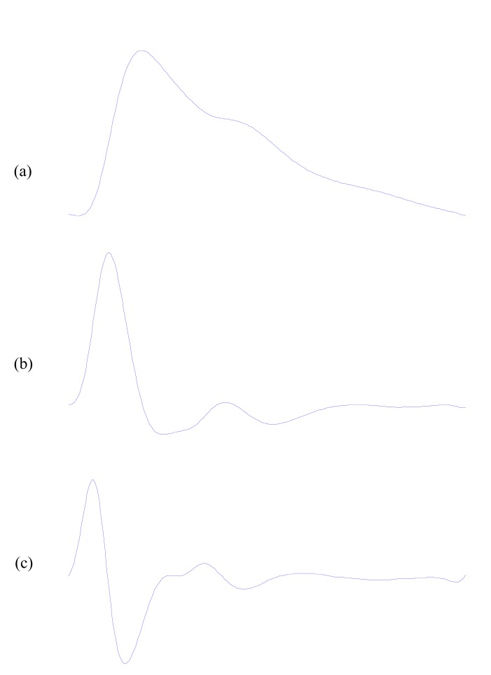
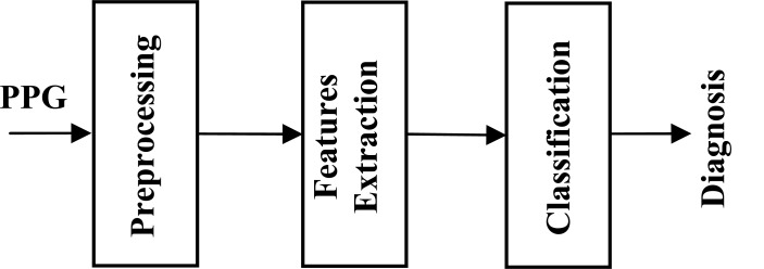
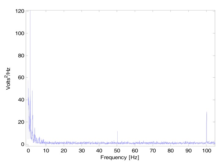
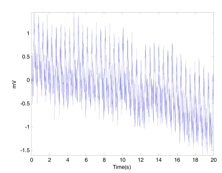
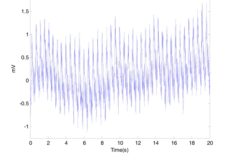
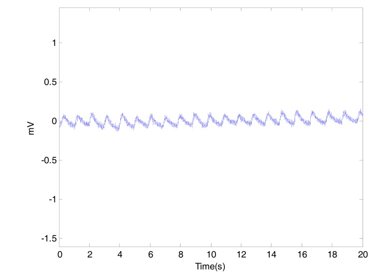
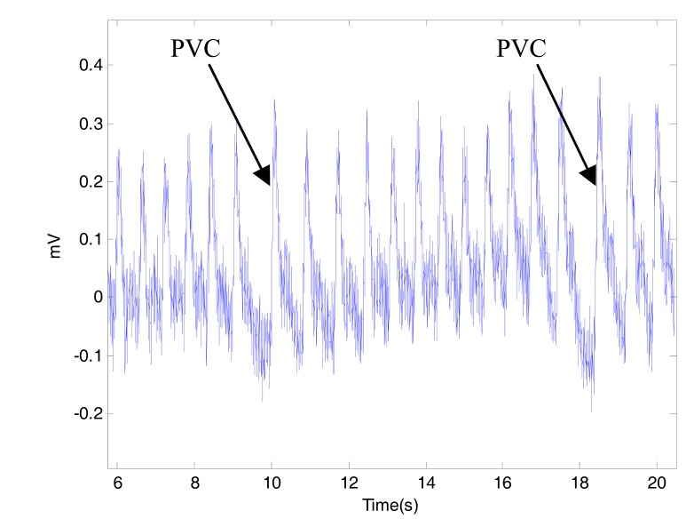
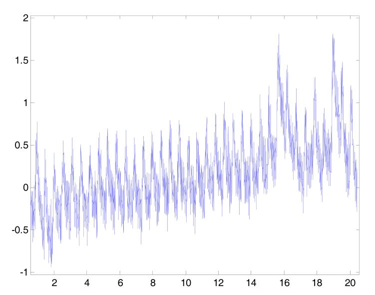
Photoplethysmography (PPG) is used to estimate the skin blood flow using infrared light. Researchers from different domains of science have become increasingly interested in PPG because of its advantages as non-invasive, inexpensive, and convenient diagnostic tool. Traditionally, it measures the oxygen saturation, blood pressure, cardiac output, and for assessing autonomic functions. Moreover, PPG is a promising technique for early screening of various atherosclerotic pathologies and could be helpful for regular GP-assessment but a full understanding of the diagnostic value of the different features is still lacking. Recent studies emphasise the potential information embedded in the PPG waveform signal and it deserves further attention for its possible applications beyond pulse oximetry and heart-rate calculation. Therefore, this overview discusses different types of artifact added to PPG signal, characteristic features of PPG waveform, and existing indexes to evaluate for diagnoses. Keywords: Photoplethysmography, acceleration plethysmogram, second derivative plethysmogram, digital volume pulse, ageing, artery, autonomic function, blood pressure, cardiovascular, heart rate, pulse wave analysis, vascular disease
Overview
The claims and statistics of this paper haven’t been checked yet.
Run the check — free, no account needed. It reads the open-access text and checks the sources it cites.
Integrity signals
Registry
Cites
Statistics
Figures
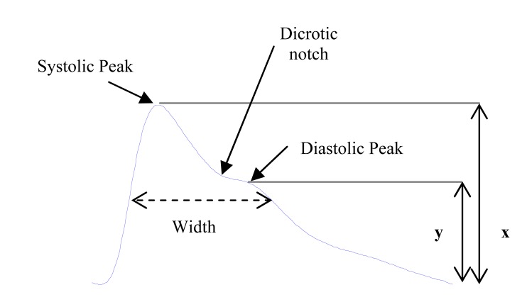
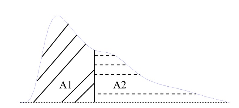
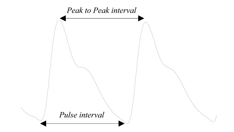
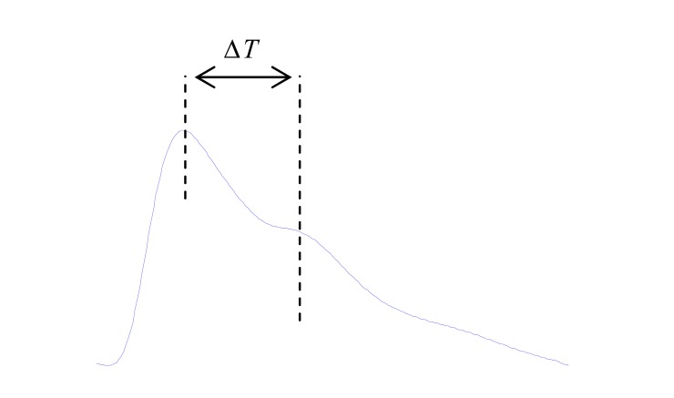
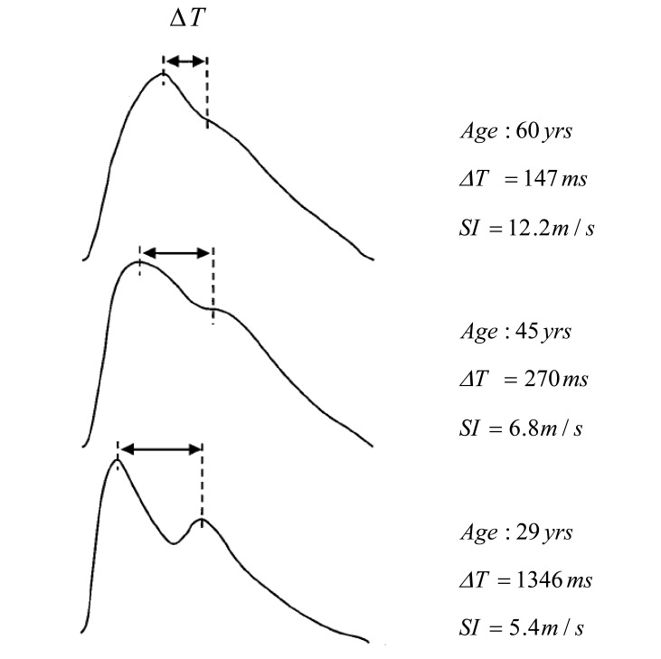
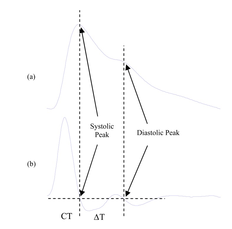
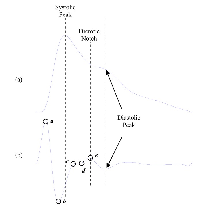
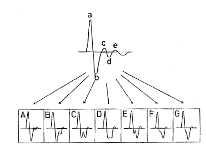
Verification
Claims the paper makes
The claims and statistics of this paper haven’t been checked yet.
Run the check — free, no account needed. It reads the open-access text and checks the sources it cites.
Claims other papers make about this one
Each is checked against this paper’s own text: does it say what the citing paper says it does?
In the citing paper
“The shape of a clean PPG signal is closely related to the blood pulse wave of the human body25”
From Data quality evaluation in wearable monitoring (2022) · cites this paper as [25]
In the citing paper
“In this case, the pulsatile component of the PPG can be of the order of 1 mV in amplitude [55] although lower amplitudes can be caused by technical factors, such as inappropriate gain control, or physiological factors, such as vasoconstriction [9].”
From Wearable Photoplethysmography for Cardiovascular Monitoring (2022) · cites this paper as [9]
In the citing paper
“In particular, obesity can lead to changes in skin thickness, blood flow, and oxygen saturation. This affects the optical properties of skin in addition to the distance light has to travel to reach a target vessel or vessels [48,59,60].”
From Sources of inaccuracy in photoplethysmography for continuous cardiovascular monitoring (2021) · cites this paper as [59]
In the citing paper
“While, to the authors’ knowledge, publications that experimentally and explicitly demonstrate the effect of obesity or BMI on the PPG waveform are limited, we will explore works which suggest that there would be a substantial effect [56,59,60,61,62,63,64,65,66,67,68,69,70].”
From Sources of inaccuracy in photoplethysmography for continuous cardiovascular monitoring (2021) · cites this paper as [59]
In the citing paper
“The ratio b/a has been found to correlate positively to age, while c/a, d/a and e/a have all been correlated negatively to age [29,59].”
From Sources of inaccuracy in photoplethysmography for continuous cardiovascular monitoring (2021) · cites this paper as [59]
In the citing paper
“| e/a | Decrease | Takazawa et al., 1998 Elgendi et al., 2012 | [29,59] |”
From Sources of inaccuracy in photoplethysmography for continuous cardiovascular monitoring (2021) · cites this paper as [59]
In the citing paper
“| d/a | Decrease | Takazawa et al., 1998 Elgendi et al., 2012 | [29,59] |”
From Sources of inaccuracy in photoplethysmography for continuous cardiovascular monitoring (2021) · cites this paper as [59]
In the citing paper
“| c/a | Decrease | Takazawa et al., 1998 Elgendi et al., 2012 | [29,59] |”
From Sources of inaccuracy in photoplethysmography for continuous cardiovascular monitoring (2021) · cites this paper as [59]
In the citing paper
“Furthermore, PPG can be used to index heart rate variability (HRV), blood pressure, oxygen saturation, and cardiac output24, but as of yet have not be widely rolled out in consumer-use devices and therefore data collection other than HR will not be discussed further.”
From Guidelines for wrist-worn consumer wearable assessment of heart rate in biobehavioral research (2020) · cites this paper as [24]
Self-check
Percentages, sums and reported differences re-computed from the paper’s own numbers.
The self-check needs the paper’s full text.
Citations
Cites
Photoplethysmography and its application in clinical physiological measurement
Allen, J. (2007) · Physiological Measurement · doi:10.1088/0967-3334/28/3/r01 · Cited by 3,967
Assessment of fat-free mass using bioelectrical impedance measurements of the human body
Lukaski, H., Johnson, P., Bolonchuk, W., & Lykken, G. (1985) · The American Journal of Clinical Nutrition · doi:10.1093/ajcn/41.4.810 · Cited by 1,896
Investigation of Chronic Venous Insufficiency: A Consensus Statement
Nicolaides, A. N. (2000) · Circulation · doi:10.1161/01.cir.102.20.e126 · Cited by 945
Fifteen years experience with finger arterial pressure monitoring: assessment of the technology
Imholz, B. P. M. (1998) · Cardiovascular Research · doi:10.1016/s0008-6363(98)00067-4 · Cited by 841
Determination of age-related increases in large artery stiffness by digital pulse contour analysis
Millasseau, S., Kelly, R. P., Ritter, J., & Chowienczyk, P. (2002) · Clinical Science · doi:10.1042/cs1030371 · Cited by 624
Takazawa, K., Tanaka, N., Fujita, M., Matsuoka, O., Saiki, T., Aikawa, M., Tamura, S., & Ibukiyama, C. (1998) · Hypertension · doi:10.1161/01.hyp.32.2.365 · Cited by 589
Photoelectric measurement of blood pressure, volume and flow in the finger
Peňáz, J. (1973) · Medical Entomology and Zoology · Cited by 528
Mobile monitoring with wearable photoplethysmographic biosensors
Asada, H. H., Shaltis, P., Reisner, A., Rhee, S., & Hutchinson, R. C. (2003) · IEEE Engineering in Medicine and Biology Magazine · doi:10.1109/memb.2003.1213624 · Cited by 466
Gil, E., Orini, M., Bailón, R., Vergara, J. M., Mainardi, L., & Laguna, P. (2010) · Physiological Measurement · doi:10.1088/0967-3334/31/9/015 · Cited by 461
THE BLOOD SUPPLY OF VARIOUS SKIN AREAS AS ESTIMATED BY THE PHOTOELECTRIC PLETHYSMOGRAPH
Hertzman, A. B. (1938) · American Journal of Physiology-Legacy Content · doi:10.1152/ajplegacy.1938.124.2.328 · Cited by 455
Research works printed but not linked: 3
- [59]Takazawa KTN Fujita M Matsuoka O Saiki T Aikawa M Tamura S Ibukiyama C Assessment of vasocative agents and vascular aging by the second derivative of photoplethysmogram waveform Hypertension 1998 32 365 70 9719069
- [80]Elgendi M Jonkman M De Boer F Fred A Filipe J Gamboa h Heart Rate Variability and Acceleration Plethysmogram measured at rest Biomedical Engineering Systems and Technologies: 2011 Springer 266 77
- [81]Tokutaka H MY Gonda E Yamamoto M Construction of a General Physical Condition Judgment System Using Acceleration Plethysmogram Pulse-Wave Analysis 2009 Berlin/Heidelberg Springer 5629 307 15
Cited by
Sorted by citations
Items 801–900 of 1,001
Diabetes‐Associated Changes in the Toe Photoplethysmogram During Local Heating Test
Lapitan, D. G., Xing, X., Glazkov, A. A., Krasulina, K. A., Kovaleva, Y. A., Glazkova, P. A., Альбертович, Б. Т., Загаров, С. С., Николаевич, Л. Р., & Рогаткин, Д. А. (2025) · Journal of Biophotonics · doi:10.1002/jbio.202500190 · Cited by 2
Monitoring of hemorrhage and whole blood resuscitation in non-human primates
Arshad, S. H., Hinojosa‐Laborde, C., Snider, E. J., Telfer, B. A., & Convertino, V. A. (2025) · npj Biosensing · doi:10.1038/s44328-025-00049-8 · Cited by 2
Georgieva-Tsaneva, G. N. (2025) · Computation · doi:10.3390/computation13120273 · Cited by 2
Tayade, A., Kumar, S., Shrivastava, A., & Ravi, B. (2025) · Scientific Reports · doi:10.1038/s41598-025-28223-y · Cited by 2
An AI-Driven Wearable Framework for Remote Health Monitoring and AR-Based Notifications
Jacob, S. S., Hariprasad, S., Varghese, L. J., Pavithra, M., & Jejo, J. (2025) · International Conference on Intelligent Technologies for Sustainable Electric and Communications Systems (iTech SECOM) · doi:10.1109/itechsecom64750.2025.11307504 · Cited by 2
Yi, M.-K., Lee, J., Lee, J., & Kim, I. Y. (2026) · IEEE Access · doi:10.1109/access.2026.3658724 · Cited by 2
A multi-domain feature-based ensemble learning approach for cuffless blood pressure estimation
Kumar, V., & Muduli, P. R. (2026) · Artificial Intelligence in Emergency Medicine · doi:10.1016/j.aiemed.2026.100004 · Cited by 2
Mustafa, S., Li, G., Wang, K., Fu, G., Anwar, F., Arshad, G., & Lin, L. (2026) · Applied Soft Computing · doi:10.1016/j.asoc.2026.114909 · Cited by 2
Woelk, S. P., Garfinkel, S. N., Mayiwar, L., & Knöferle, K. (2026) · Behavior Research Methods · doi:10.3758/s13428-026-02953-x · Cited by 2
Signal Quality of Reflective-Mode Photoplethysmograms Across Anatomical Sites
Ricci, F., Vivarelli, C., Mattei, E., & Calcagnini, G. (2026) · Sensors · doi:10.3390/s26102986 · Cited by 2
Harmonic-Selective Gaussian Filtering for Morphology and Timing Preservation in PPG Signals
Dominguez-Hernandez, S., Паез, Г., & Padilla, M. (2026) · Sensors · doi:10.3390/s26123710 · Cited by 2
Cuff-less Blood Pressure measurement from Wireless ECG and PPG signals
Dave, T., Pandya, U. T., & Joshi, M. M. (2021) · IEEE International Symposium on Smart Electronic Systems (iSES) · doi:10.1109/ises52644.2021.00020 · Cited by 2
Nimi, W. S., Jose, P. S. H., Jose, P. S. H., Jegan, R., & El‐Shahat, A. (2022) · International Journal of Reliable and Quality E-Healthcare · doi:10.4018/ijrqeh.298632 · Cited by 2
Sviridova, N., Zhao, T., Nakano, A., & Ikeguchi, T. (2022) · Sensors · doi:10.3390/s22145154 · Cited by 2
A photoplethysmography-based system for talking detection in bedridden patients
Prada, E. J. A., Cantín, M. A. D., & Victoria, J. C. (2022) · Biomedical Signal Processing and Control · doi:10.1016/j.bspc.2022.104477 · Cited by 2
Study on the tolerance of motion artifacts in estimating variance of blood pressure by single PPG
Chang, C.-L., & Wang, L. (2023) · AIP conference proceedings · doi:10.1063/5.0133887 · Cited by 2
Cuffless BP Measurement Using Single Site Photoplethysmography
Maharajan, R. (2023) · International Conference on Bio Signals, Images, and Instrumentation (ICBSII) · doi:10.1109/icbsii58188.2023.10181082 · Cited by 2
Phonocardiography and Photoplethysmography Circuit for Integrated Physiological Signal Acquisition
Giri, A., & Chaskar, U. M. (2023) · World Conference on Communication & Computing (WCONF) · doi:10.1109/wconf58270.2023.10235138 · Cited by 2
Agitated Behaviors Detection in Children with ASD Using Wearable Data
Montassar, I., Chikhaoui, B., & Wang, S. (2023) · Lecture notes in computer science · doi:10.1007/978-3-031-43950-6_8 · Cited by 2
A new e-health cloud-based system for cardiovascular risk assessment
Tatsis, G., Baldoumas, G., Christofilakis, V., Kostarakis, P., Varotsos, P. A., Sarlis, N. V., Skordas, E. S., Bechlioulis, A., Michalis, L. K., & Naka, K. Κ. (2023) · Frontiers in Electronics · doi:10.3389/felec.2023.1315132 · Cited by 2
Analysis of Age-Related Variations in Photoplethysmography: A Machine Learning Approach
Lombardi, S., Tavernise, F., Francia, P., & Bocchi, L. (2024) · World Congress on Medical Physics and Biomedical Engineering, September 7 - 12, 2009, Munich, Germany · doi:10.1007/978-3-031-61625-9_11 · Cited by 2
Noninvasive ML-Based BUN Prediction from Ten-Second Fingertip Video Using Generative AI
Sridevi, P., Arefin, K. Z., Das, D., Khan, R. A., & Ahamed, S. I. (2024) · IEEE Annual International Computer Software and Applications Conference (COMPSAC) · doi:10.1109/compsac61105.2024.00256 · Cited by 2
Tang, Q., Tao, C., Li, X., Hu, H., Chu, X., Liu, S., Zhang, L., Su, B., Xu, J., & An, H. (2024) · Computers in Biology and Medicine · doi:10.1016/j.compbiomed.2024.109076 · Cited by 2
Noninvasive Vital Signs Monitoring in the Neonatal Intensive Care Unit
Qian, C., Li, J., & Khine, M. (2024) · Journal of Biomedical Research & Environmental Sciences · doi:10.37871/jbres1958 · Cited by 2
Yang, T., Okada, E., Todoroki, M., Liu, S., Athurupana, R., Kataoka, K., Kashino, C., Mitsui, T., Hasegawa, T., Kamada, Y., et al. (2024) · International Journal of Gynecology & Obstetrics · doi:10.1002/ijgo.16051 · Cited by 2
Tayade, A., Kumar, S., Kumar, P., & Ravi, B. (2024) · Proceedings of IEEE Sensors/Proceedings of IEEE Sensors ... · doi:10.1109/sensors60989.2024.10785088 · Cited by 2
Neural network models for predicting vascular age from PPG signals: A comparative study
Abrisham, K. P., Alipour, K., Tarvirdizadeh, B., & Ghamari, M. (2024) · IET Wireless Sensor Systems · doi:10.1049/wss2.12103 · Cited by 2
Ipar, E., Cymberknop, L. J., & Armentano, R. L. (2025) · Physiological Measurement · doi:10.1088/1361-6579/adc366 · Cited by 2
Dehghanojamahalleh, S., Gamage, P. T., Ahmed, M., Petersen, C., Matthew, B. P., Hyacinth, K., Weerasinghe, Y., Subasi, E., Subasi, M. M., & Kaya, M. (2025) · BioMedInformatics · doi:10.3390/biomedinformatics5020024 · Cited by 2
Strada, M., Galli, M., & Cattaneo, R. (2025) · Journal of Bodywork and Movement Therapies · doi:10.1016/j.jbmt.2025.05.050 · Cited by 3
Smart Wristband Based on a Magnetic Soft Sensor for Pulse Wave Measurement
Romano, C., Silvestri, S., Schena, E., & Massaroni, C. (2025) · IEEE Sensors Journal · doi:10.1109/jsen.2025.3594238 · Cited by 2
Continuous Cardiac Arrest Prediction in ICU using PPG Foundation Model
Kataria, S., Xiao, R., Ruchti, T. L., Clark, M. P. A., Lu, J., Lee, R. J., Grunwell, J. R., & Hu, X. (2025) · Annual International Conference of the IEEE Engineering in Medicine and Biology Society (EMBC) · doi:10.1109/embc58623.2025.11254649 · Cited by 4
Sparse characterization of PPG based on K- SVD for beat-to-beat blood pressure prediction
Bose, S. S. N., & Kandaswamy, A. (2018) · Biomedical Research - India · doi:10.4066/biomedicalresearch.29-17-1438 · Cited by 2
Analysis of Vital Signals Acquired from Wearable Device
Kumar, D., Adarsh, A., Chandrika, S., Kishor, N., & Mala, R. (2019) · Lecture notes in electrical engineering · doi:10.1007/978-981-13-5802-9_39 · Cited by 2
A Photoplethysmographic Monitor for Local Pulse Wave Velocity Measurement
Matheus, P., Antonio, W., Pedro, H., & Talles, M. (2020) · International Journal of Computer Applications · doi:10.5120/ijca2020919811 · Cited by 2
Algoritmo para el análisis en conjunto de las señales del ECG y PPG
Martínez-Reyes, D. C. (2021) · Revista UIS Ingenierías · doi:10.18273/revuin.v20n4-2021004 · Cited by 2
Early detection of mental disorder signs using photoplethysmogram : A review
Awang, A., & Nayan, N. A. (2021) · Journal of Discrete Mathematical Sciences and Cryptography · doi:10.1080/09720529.2021.2009191 · Cited by 2
Zerwas, F. K., Ebo, R., Allison, G. O., Karnilowicz, H. R., Carrillo, B., Wilhelm, F. H., & Mauss, I. B. (2022) · International Journal of Psychophysiology · doi:10.1016/j.ijpsycho.2022.06.001 · Cited by 2
Williamson, S., Daniel‐Watanabe, L., Finnemann, J., Powell, C., Teed, A. R., Allen, M., Paulus, M. P., Khalsa, S. S., & Fletcher, P. C. (2022) · Wellcome Open Research · doi:10.12688/wellcomeopenres.17855.1 · Cited by 2
Rahman, N. A., Chellapan, K., Ong, P. Y., Adnan, A., & Din, N. M. (2024) · Retina · doi:10.1097/iae.0000000000004297 · Cited by 2
Investigating the correlation between smoking and blood pressure via photoplethysmography
Qananwah, Q. M., Quran, H. A., Dagamseh, A., Blažek, V., & Leonhardt, S. K. (2025) · BioMedical Engineering OnLine · doi:10.1186/s12938-025-01373-w · Cited by 2
Zhao, Y., Lahdenoja, O., Elnaggar, I., Vasankari, T., Jaakkola, S., Kiviniemi, T., Airaksinen, J. K. E., Kaisti, M., & Koivisto, T. (2025) · IEEE Sensors Journal · doi:10.1109/jsen.2025.3574232 · Cited by 2
Rahman, M. M., Hasan, M., & Hao, Z. (2025) · Biosensors · doi:10.3390/bios15090578 · Cited by 2
Kuribayashi, R., & Furukawa, H. (2015) · Proceedings of SPIE, the International Society for Optical Engineering/Proceedings of SPIE · doi:10.1117/12.2078769 · Cited by 2
Preliminary study for localizing c, d and e waves in photoplethysmogram signals
Elgendi, M. (2014) · Annual International Conference of the IEEE Engineering in Medicine and Biology Society (EMBC) · doi:10.1109/embc.2014.6943529 · Cited by 2
Self-citationHubená, G., Osina, O., Prindesova, B., & Vasicko, T. (2015) · Acta Medica Martiniana · doi:10.1515/acm-2015-0008 · Cited by 2
Estimation Model for Blood Pressure Based on Clustering and Gradient Boosting
Zhang, Z., Yu, M., Meng, L., Xie, X., Pan, H., & Feng, X. (2017) · Chinese Conference on Computer Supported Cooperative Work and Social Computing (ChineseCSCW) · doi:10.1145/3127404.3127413 · Cited by 2
An accurate method for fourier synthesis of photoplethysmographic signals
Abeysekera, S. S. (2017) · IEEE International Symposium on Circuits and Systems proceedings · doi:10.1109/iscas.2017.8050419 · Cited by 2
Tremoleda, J. L., Alvarez, K., Aden, A., Donnan, R. S., Michael-Titus, A. T., & Tomlins, P. (2017) · Journal of Biomedical Optics · doi:10.1117/1.jbo.22.12.121710 · Cited by 2
An image-based pulse rate detection system using an FPGA platform
Lin, Y.-C., Lin, G., Lin, J.-W., Tsai, Y.-L., & Lin, Y. (2017) · IEEE/SICE International Symposium on System Integration (SII) · doi:10.1109/sii.2017.8279267 · Cited by 2
System for ECG, PPG Signal Acquisition and Peak Detection
Menon, R. B., & Mohan, N. M. (2017) · IEEE India Conference · doi:10.1109/indicon.2017.8487860 · Cited by 2
A Model for Waveform Dissimilarities in Dual-Depth Reflectance-PPG
Moço, A., Stuijk, S., de Haan, G., Wang, R. K., & Verkruysse, W. (2018) · Annual International Conference of the IEEE Engineering in Medicine and Biology Society (EMBC) · doi:10.1109/embc.2018.8513484 · Cited by 2
Human Activity and Heart Rate Monitoring System in a Mobile Platform
Manjarrés, J., Russo, V., Peñaranda, J., & Pardo, M. (2018) · Congreso Internacional de Innovación y Tendencias en Ingeniería (CONIITI) · doi:10.1109/coniiti.2018.8587094 · Cited by 2
Motion Artefact Removal using Single Beat Classification of Photoplethysmographic Signals
Li, L., & Abeysekera, S. S. (2019) · IEEE International Symposium on Circuits and Systems proceedings · doi:10.1109/iscas.2019.8702180 · Cited by 2
Implementation and Validation of a Non-Contact Measurement of Cardiac Activity
Kausalya, R., & Priya, E. (2019) · International Conference on Computing and Communications Technologies (ICCCT) · doi:10.1109/iccct2.2019.8824864 · Cited by 2
Sensors based Biomedical Framework to Monitor Patient’s Vital Parameters
Jegan, R., Nimi, W. S., & Ramson, S. R. J. (2020) · International Conference on Devices, Circuits and Systems (ICDCS) · doi:10.1109/icdcs48716.2020.243557 · Cited by 2
The influence of physiological characteristics on blood pressure estimation using only PPG signals
Sen, Y., Morgan, S. P., Cho, S., & Yaping, Z. (2019) · IEEE International Conference on Electronic Measurement & Instruments · doi:10.1109/icemi46757.2019.9101868 · Cited by 2
Raj, R., Selvakumar, J., & Maik, V. (2021) · Journal of Physics Conference Series · doi:10.1088/1742-6596/2007/1/012056 · Cited by 2
Assessment of Blood Viscosity from Photoplethysmogram
Sulochana, H., & Safwaana, A. (2022) · International Conference on Electrical, Electronics, Information and Communication Technologies (ICEEICT) · doi:10.1109/iceeict53079.2022.9768640 · Cited by 2
Reddy, G., Bhat, K. K., Lunia, U., & Krupa, N. (2022) · Lecture notes in electrical engineering · doi:10.1007/978-981-19-2004-2_34 · Cited by 2
Cuffless Hypertension Risk Assessment and the Significance of Calibration
Cano, J., Fácila, L., Hornero, F., Langley, P., Alcaraz, R., & Rieta, J. J. (2022) · Computing in cardiology · doi:10.22489/cinc.2022.092 · Cited by 2
ECG and PPG-Based Hypertension Screening Under Non-Hypertensive Blood Pressure Recordings
Cano, J., Bertomeu‐González, V., Fácila, L., Moreno-Arribas, J., Alcaraz, R., & Rieta, J. J. (2022) · Computing in cardiology · doi:10.22489/cinc.2022.093 · Cited by 2
Nonlinear Pulse Wave Dynamics in Prediction of Coronary Heart Disease and Myocardial Infarction
Kumar, R., Aggarwal, Y., Nigam, V. K., & Sinha, R. K. (2023) · IETE Journal of Research · doi:10.1080/03772063.2023.2245353 · Cited by 2
Machikhin, A. S., Guryleva, A. V., Chakraborty, A., Хохлов, Д. Д., Selyukov, A., Shuman, L. A., Bukova, V. I., Efremova, E. V., Rudenko, E., & Burlakov, A. B. (2024) · Journal of Biophotonics · doi:10.1002/jbio.202400111 · Cited by 2
Kim, Y., Kim, Y. H., & Lee, J. S. (2016) · Journal of Mechanical Science and Technology · doi:10.1007/s12206-016-0734-0 · Cited by 2
Real-Time Pulse Waveform Profiling Algorithm for Wearable Applications
Liu, H., Fujita, D., Zhang, L., & Suzuki, A. (2018) · IEEE Access · doi:10.1109/access.2018.2875548 · Cited by 2
Appending Photoplethysmograph as a Security Key for Encryption of Medical Images Using Watermarking
Vidya, M. J., & Padmaja, K. (2018) · Advances in intelligent systems and computing · doi:10.1007/978-981-13-1708-8_33 · Cited by 2
Aluminium Nitride based bio-MEMS for vascular graft monitoring
Natta, L., Mastronardi, V., Guido, F., Algieri, L., Puce, S., Pisano, F., Rizzi, F., Pulli, R., Qualtieri, A., & Vittorio, M. D. (2019) · European Microelectronics and Packaging Conference · doi:10.23919/empc44848.2019.8951796 · Cited by 2
SMARTPHONE-BASED DIAGNOSTIC MODEL FOR HYPERTENSION USING FEATURES FROM PHOTOPLETHYSMOGRAM
Devaki, V., & Jayanthi, T. (2020) · Biomedical Engineering Applications Basis and Communications · doi:10.4015/s1016237220500271 · Cited by 2
Sustained Attention Detection System in Learning Environments
da Costa, B. N., Postolache, O. A., & Araujo, J. (2020) · International Conference and Exposition on Electrical and Power Engineering (EPE) · doi:10.1109/epe50722.2020.9305027 · Cited by 2
Mouney, F., Tiplica, T., Fasquel, J.-B., Hallab, M., & Dinomais, M. (2021) · Lecture notes of the Institute for Computer Sciences, Social Informatics and Telecommunications Engineering · doi:10.1007/978-3-030-76063-2_4 · Cited by 2
Cano, J., Quesada, A., Ravelli, F., Zangróniz, R., Alcaraz, R., & Rieta, J. J. (2021) · E-Health and Bioengineering Conference (EHB) · doi:10.1109/ehb52898.2021.9657546 · Cited by 2
Cano, J., Hornero, F., Quesada, A., Martínez‐Rodrigo, A., Alcaraz, R., & Rieta, J. J. (2021) · Computing in cardiology · doi:10.23919/cinc53138.2021.9662894 · Cited by 2
Hemodynamic Monitoring with the Clinic: Back to Basics
Nieto, P., Guerrero Gutiérrez, M. A., La, M., Jl, F., Zamarrón López, E. I., Soriano Orozco, R., Sánchez Díaz, J. S., Uribe Moya, S. E., Díaz-Martínez, M. A., Escarraman, et al. (2020) · Journal of Emergency Medicine & Critical Care · doi:10.13188/2469-4045.1000021 · Cited by 2
Continuous monitoring of blood pressure from photophlethysmograph signal
Ackshaiyaa, A. G., Jothi, E. S. J., & Rajasekaran, K. (2017) · International Conference on Innovations in Electrical, Electronics, Instrumentation and Media Technology (ICEEIMT) · doi:10.1109/icieeimt.2017.8116833 · Cited by 1
AIM and Transdermal Optical Imaging
Barszczyk, A., Zhou, W., & Lee, K. (2022) · Springer eBooks · doi:10.1007/978-3-030-64573-1_250 · Cited by 1
Distributed Model Architecture for Cuff-Less Blood Pressure Measurement System
Dave, T., Joshi, M. M., & Pandya, U. T. (2022) · SN Computer Science · doi:10.1007/s42979-022-01391-8 · Cited by 1
Ghosal, P., Himavathi, S., & Srinivasan, E. (2023) · International Conference on Intelligent Systems for Communication, IoT and Security (ICISCoIS) · doi:10.1109/iciscois56541.2023.10100487 · Cited by 1
The Physiology of Musical Preference
Mehdizadeh, S. K., & Leslie, G. A. (2023) · Music Perception An Interdisciplinary Journal · doi:10.1525/mp.2023.40.5.395 · Cited by 1
Physiological Measures in VR Experiments: Some Aspects of Plethysmogram and Heart Rate
Miyake, S., Kurosaka, C., & Kuraoka, H. (2023) · Lecture notes in computer science · doi:10.1007/978-3-031-35132-7_32 · Cited by 1
The effect of contact pressure on photoplethysmography
Gong, X., Yu, C., & Xuan, Y. (2024) · Indonesian Journal of Electrical Engineering and Computer Science · doi:10.11591/ijeecs.v33.i3.pp1589-1603 · Cited by 1
Open-source synthetic photoplethysmographic signal generator with analog output
Gentilin, A. (2024) · Proceedings of the Institution of Mechanical Engineers Part H Journal of Engineering in Medicine · doi:10.1177/09544119241272833 · Cited by 1
Effect of acupuncture at Shaofu acupoint on the cardiovascular system of healthy individuals
Saleem, A. F., Al-Wahab, Z. N., & Al‐Gailani, B. T. (2024) · Polish Journal of Medical Physics And Engineering · doi:10.2478/pjmpe-2024-0015 · Cited by 1
Causal Inference Based Hierarchical Regression Model for Cuffless Blood Pressure Estimation
Song, X., Zan, X., Fu, Z., & Ding, X. (2024) · Annual International Conference of the IEEE Engineering in Medicine and Biology Society (EMBC) · doi:10.1109/embc53108.2024.10782557 · Cited by 1
Spontaneous motor tempo modulates the effect of music tempo on arousal levels
Hine, K., Abe, K., & Nakauchi, S. (2025) · Psychology of Music · doi:10.1177/03057356241311288 · Cited by 1
Swin Transformer-based Cuffless Blood Pressure Estimation using Cardiovascular Signals
Kumar, V., Mishra, M., Gadani, M., Jayarajan, J., Sharma, P., & Muduli, P. R. (2024) · IEEE India Conference · doi:10.1109/indicon63790.2024.10958421 · Cited by 1
Feasibility of Photoplethysmography in Detecting Arterial Stiffness in Hypertension
Karimpour, P., May, J. M., & Kyriacou, P. A. (2025) · Photonics · doi:10.3390/photonics12050430 · Cited by 1
Method for the Measurement of the Dicrotic Notch in the Photoplethysmography Signal
Forster, P., Laska, B., Goubran, R. A., Wallace, B., Liu, P. X., & Sveistrup, H. (2025) · IEEE International Instrumentation and Measurement Technology Conference · doi:10.1109/i2mtc62753.2025.11079209 · Cited by 1
A 10.42μW/Ch. PPG Sensor with a Zoomed Sampling Based on Velocity of Blood Flow
Choi, K., Kim, B.-J., & Sim, J. (2025) · IEEE Symposium on VLSI Technology and Circuits (VLSI Technology and Circuits) · doi:10.23919/vlsitechnologyandcir65189.2025.11074909 · Cited by 1
Contactless Hemodynamic Monitoring Based on Multi-Scale Gaussian Filtering via Imaging PPG
Tong, Y., Huang, Z., Qiu, F., Wu, C., Tao, X., Huang, W. F., & Qin, F. (2025) · IEEE Journal of Biomedical and Health Informatics · doi:10.1109/jbhi.2025.3605591 · Cited by 1
Park, H., Kang, S., Kim, J., Lee, J., Park, S., & Park, S. M. (2025) · Micromachines · doi:10.3390/mi16101086 · Cited by 1
Ali, N. F., Al-Nashash, H. A., Elfadel, I. M., & Atef, M. (2025) · IEEE Access · doi:10.1109/access.2025.3639253 · Cited by 1
Rahbar, S., Ronaghi, S., & Varsi, A. (2025) · Annual International Conference of the IEEE Engineering in Medicine and Biology Society (EMBC) · doi:10.1109/embc58623.2025.11253113 · Cited by 1
Non-contact blood pressure estimation by imaging photoplethysmography
Sahl, F., Woyczyk, A., Fleischhauer, V., & Zaunseder, S. (2025) · Annual International Conference of the IEEE Engineering in Medicine and Biology Society (EMBC) · doi:10.1109/embc58623.2025.11252987 · Cited by 1
Tate, A. J., Mouradian, G. C., & Lipinski, D. M. (2025) · Biomedical Optics Express · doi:10.1364/boe.575278 · Cited by 1
Machine Learning for Classifying Vascular Age Using the Second Derivative of the PPG Signal
Otesteanu, C. F., Diana, G., Scardulla, F., Puleo, S., Pasta, S., D’Acquisto, L., & Menon, C. (2025) · International Conference on Communications, Information, Electronic and Energy Systems (CIEES) · doi:10.1109/ciees66347.2025.11300216 · Cited by 1
Li, F., Li, L., Wang, L., & Xu, H. (2026) · IEEE Access · doi:10.1109/access.2026.3657471 · Cited by 1
Seo, W.-D., Choi, I., Kim, G. C., Song, H. S., & Kim, Y. K. (2026) · Biomedical Signal Processing and Control · doi:10.1016/j.bspc.2026.109852 · Cited by 1
Naskinova, I., Kolev, M., Milev, M., & Mitev, P. (2026) · AI · doi:10.3390/ai7030098 · Cited by 1
Decoding brain-heart dynamics: Effective connectivity predictors of heart rate variability
Bello, M. D., & McIntosh, R. C. (2026) · NeuroImage · doi:10.1016/j.neuroimage.2026.121887 · Cited by 1
Metadata
- Authors
- Elgendi, M.
- Affiliations
- Charles Darwin University (AU)
- Corresponding author
- Elgendi, Mohamed
- ORCID
- Elgendi, Mohamed
- Venue
- Current Cardiology Reviews
- Publisher
- Bentham Science Publishers Ltd.
- Published
- 2012-06-01
- Volume
- 8
- Issue
- 1
- Pages
- 14-25
- ISSN
- 1573-403X (ISSN-L)
- PMC
- PMC3394104
- OpenAlex
- W1981918735
- Cited by
- 1,206
- References
- 88
- Citations by year
- 2026: 120 · 2025: 106 · 2024: 116 · 2023: 109 · 2022: 130 · 2021: 114 · 2020: 118 · 2019: 118 · 2018: 86 · 2017: 65 · 2016: 46 · 2015: 42 · 2014: 24 · 2013: 11 · 2012: 1
- FWCI
- 2.17
- Type
- journal article
- Language
- en
- Open access
- bronze
- doi.org
- Locations
- Current Cardiology Reviews (publishedVersion)
- Europe PMC (PubMed Central) (publishedVersion, cc-by)
- PubMed Central (publishedVersion, cc-by)
- PubMed (publishedVersion)
- DR-NTU (Nanyang Technological University) (submittedVersion)
- Funders
- Univerzita Karlova v Praze
- Australian Government
- Charles Darwin University
- Topics
- Physical Sciences › Engineering › Biomedical Engineering › Non-Invasive Vital Sign Monitoring
- Health Sciences › Medicine › Cardiology and Cardiovascular Medicine › Cardiovascular Health and Disease Prevention
- Health Sciences › Medicine › Cardiology and Cardiovascular Medicine › Heart Rate Variability and Autonomic Control
- Keywords
- photoplethysmography, digital volume pulse, aging, autonomic function, blood pressure, heart rate, pulse wave analysis, cardiovascular disease, vascular disease, atherosclerosis screening
- MeSH terms
- Fingers, Hemodynamics, Humans, Skin, Artifacts, Photoplethysmography, Pulse Wave Analysis
About these checks
Registry, retraction, Cites and statistics results are computed, not judged, with one exception: a printed reference that no index lists is read once by a model before it counts as a citation error. Claims are extracted by a model and kept only when their quote appears verbatim in the text; each cited source is then checked, with a model’s help, for whether it says what the claim relies on. Those verdicts can be wrong — read the claim’s quote and the cited source before relying on one.
Statistics and arithmetic are recomputed, not judged.
Confidence bands are shown only for fields calibrated on our annotated test set. None is calibrated yet, so no band is shown.
What counts as a claim ›How we measure confidence ›How we check this ›
Are you an author, or do you see a problem with this page? Request a review
