

The DOI matches its Crossref record; 194 of the 198 references it cites were found in a registry or an index; 6 of the 222 sources its claims cite were checked for support.
ROS Generation in Microglia: Understanding Oxidative Stress and Inflammation in Neurodegenerative Disease
Open accessAbstract
Neurodegenerative disorders, such as Alzheimer’s disease, are a global public health burden with poorly understood aetiology. Neuroinflammation and oxidative stress (OS) are undoubtedly hallmarks of neurodegeneration, contributing to disease progression. Protein aggregation and neuronal damage result in the activation of disease-associated microglia (DAM) via damage-associated molecular patterns (DAMPs). DAM facilitate persistent inflammation and reactive oxygen species (ROS) generation. However, the molecular mechanisms linking DAM activation and OS have not been well-defined; thus targeting these cells for clinical benefit has not been possible. In microglia, ROS are generated primarily by NADPH oxidase 2 (NOX2) and activation of NOX2 in DAM is associated with DAMP signalling, inflammation and amyloid plaque deposition, especially in the cerebrovasculature. Additionally, ROS originating from both NOX and the mitochondria may act as second messengers to propagate immune activation; thus intracellular ROS signalling may underlie excessive inflammation and OS. Targeting key kinases in the inflammatory response could cease inflammation and promote tissue repair. Expression of antioxidant proteins in microglia, such as NADPH dehydrogenase 1 (NQO1), is promoted by transcription factor Nrf2, which functions to control inflammation and limit OS. Lipid droplet accumulating microglia (LDAM) may also represent a double-edged sword in neurodegenerative disease by sequestering peroxidised lipids in non-pathological ageing but becoming dysregulated and pro-inflammatory in disease. We suggest that future studies should focus on targeted manipulation of NOX in the microglia to understand the molecular mechanisms driving inflammatory-related NOX activation. Finally, we discuss recent evidence that therapeutic target identification should be unbiased and founded on relevant pathophysiological assays to facilitate the discovery of translatable antioxidant and anti-inflammatory therapeutics.
Overview
AI snapshot
Written from the paper’s full text.
TL;DR
This review argues that microglial reactive oxygen species (ROS), generated mainly by NOX2 and NOX4, link neuroinflammation and oxidative stress in neurodegenerative disease, with ROS also acting as second messengers and lipid droplet-accumulating microglia contributing to pathology. It proposes targeting microglial NOX, Nrf2-driven antioxidant pathways, and unbiased disease-relevant drug discovery.
Key findings
- NOX2 (CYBB) is the most highly expressed NOX isotype transcript in human and mouse brain microglia, with NOX4 second-highest at a much lower level; NOX2 activation in disease-associated microglia is associated with DAMP signalling, inflammation, and amyloid plaque deposition, especially in the cerebrovasculature.
- ROS from both NOX and mitochondria may act as second messengers to propagate immune activation: H₂O₂ can activate NFκB, and suppressing mitochondrial ROS with mitoTEMPO reduced MAPK/NFκB activation and pro-inflammatory factors (TNFα, IL1β, IL6, iNOS, Cox-2) in LPS-stimulated microglia.
- Nrf2 controls antioxidant genes such as NQO1 and GSTs in microglia; Nrf2 deficiency promotes inflammatory markers (IL6, Cox2, iNOS) and inhibits anti-inflammatory markers, while sulforaphane enhances Nrf2, limits pro-inflammatory microglial markers, and confers neuroprotection.
- Lipid droplet-accumulating microglia (LDAM) may be a double-edged sword: ROS/JNK drive lipid droplet formation in flies, antioxidant treatment or SOD1 expression reduces it, and GRN−/− mice show ROS-generating, pro-inflammatory LDAM defective in phagocytosis; ToxSeq identified a ROS-positive microglial signature rescued by acivin in EAE.
Methods
- Narrative review; no primary data, experimental sample, or statistical analysis are reported.
- The paper synthesizes literature on microglial NOX/ROS biology, oxidative stress, neuroinflammation, lipid droplet-accumulating microglia, antioxidant regulation, and therapeutic strategies.
- It discusses evidence from human post-mortem tissue, rodent models, cell lines, single-cell RNA-seq, and drug-screening studies, and proposes future directions including cell-specific NOX manipulation.
Limitations
- The paper does not state limitations of its own review; it repeatedly notes limitations in the underlying literature, including lack of stringent controls and specific antibodies for NOX expression studies, possible artefactual NOX1 expression in Cx3cr1-EGFP reporter mice, and a human microglial cell line lacking NOX2.
- It also notes that apocynin specificity for NOX4 has been questioned, mitoTEMPO selectivity is questioned especially at higher concentrations, and a causal relationship between ROS and NFκB activation in microglia remains undetermined.
- Evident from the text: as a narrative review, no systematic search or formal quality assessment is described, and many conclusions rely on preclinical models or immortalized cells, with calls for validation in primary and iPSC-derived microglia.
Model-written summary. Check it against the paper before relying on it.
Integrity signals
Registry
Cites
Statistics
Figures
Verification
Claims the paper makes
130 Not checked yet · 37 Own interpretation · 8 No basis given · 3 Partly supported · 1 Mixed support · accuracy not yet measured
Some cited sources support this claim and some don’t — read each source’s verdict below.
In the paper
“Multiple studies have shown that H2O2-mediated NFκB activation results from phosphorylation of IκB tyrosine residues [143,144].”
§ ROS Generation in Microglia: Understanding Oxidative Stress and Inflammation in Neurodegenerative Disease > 5. ROS Are Secondary Messengers Activating Pro-inflammatory Pathways in Microglia > 5.1. NFκB, a Master Regulator of Inflammation in Microglia, Is Associated with NOX Expression
What the sources say
- Disputedcites [143] Schoonbroodt, 2000, Crucial Role of the Amino-Terminal Tyrosine Residue 42 and the Carboxyl-Terminal PEST Domain of IκBα in NF-κB Activation by an Oxidative Stress, J. Immunol. sourceExperimental — accuracy not yet measured
The Discussion (page unavailable) says H₂O₂-induced NF-κB activation probably relies on an unmodified IκBα tyrosine 42 and phosphorylation of the carboxyl-terminal PEST sequence. The paper finds tyrosine 42 necessary for activation, but does not identify phosphorylation of tyrosine residues as its cause; its proposed mechanism points elsewhere. (LLM, conf=0.88)
Checked by a reasoning model — may be wrong.
- Supportedcites [144] Takada, 2003, Hydrogen Peroxide Activates NF-κB through Tyrosine Phosphorylation of IκBα and Serine Phosphorylation of p65 evidence for the involvement of IκBα kinase and Syk protein-tyrosine kinase, J. Biol. Chem. source
The Discussion states that H₂O₂ induces tyrosine phosphorylation of IκBα and that Syk-dependent phosphorylation is critical for NF-κB activation. The paper’s summary likewise identifies Syk-mediated tyrosine phosphorylation of IκBα as the activation mechanism. (LLM, conf=0.94)
boosteddirect- Asserted: passes — The authors put it forward as true.
- Checkable: passes — It can be checked against evidence.
- Load-bearing: unclear — It is unclear whether the paper’s conclusions depend on it.
- Based: passes — The paper gives a reason why it holds.
- Disputed
The cited sources support only part of this claim — read each source’s verdict below.
In the paper
“As phagocytes, microglia are capable of mediating an “oxidative/respiratory burst” in response to pathogen- and damage-AMPs, including aggregated protein and cellular debris [40,41].”
§ ROS Generation in Microglia: Understanding Oxidative Stress and Inflammation in Neurodegenerative Disease > 1. Introduction > 1.3. The Brain Is Especially Susceptible to Oxidative Stress
What the sources say
- Partly supportedcites [40] Claude, 2013, Microglial CD33-related Siglec-E inhibits neurotoxicity by preventing the phagocytosis-associated oxidative burst, J. Neurosci. Off. J. Soc. Neurosci. sourceExperimental — accuracy not yet measured
The Introduction describes ROS production during microglial phagocytosis as an oxidative burst, and the section “Siglec-E prevents superoxide release triggered by neural debris” reports increased superoxide production after exposure to neural debris. The cited paper does not establish an oxidative-burst response to aggregated proteins or pathogen-associated molecular patterns. Page numbers are unavailable in our copy. (LLM, conf=0.86)
- Partly supportedcites [41] Herzog, C., Pons Garcia, L., Keatinge, M., Greenald, D., Moritz, C., Peri, F., and Herrgen, L. (2019). Rapid clearance of cellular debris by microglia limits secondary neuronal cell death after brain injury in vivo. Dev. Camb. Engl., 146. sourceExperimental — accuracy not yet measured
The Introduction says phagocytosis can trigger a respiratory burst and reports ROS production after microglia take up neural debris. It does not address pathogen- or damage-associated molecular patterns as triggers, or aggregated protein specifically. No page number is available in our copy. (LLM, conf=0.86)
plaindirect- Asserted: passes — The authors put it forward as true.
- Checkable: passes — It can be checked against evidence.
- Load-bearing: passes — The paper’s conclusions depend on it.
- Based: passes — The paper gives a reason why it holds.
- Partly supported
The cited sources support only part of this claim — read each source’s verdict below.
In the paper
“Inhibiting NOX with the naturally occurring compound, apocynin, or genetic deletion of gp91phox in mice (Tg2576) overexpressing mutant amyloid precursor protein (APP), attenuates cerebral amyloid angiopathy (CAA) [115,116].”
§ ROS Generation in Microglia: Understanding Oxidative Stress and Inflammation in Neurodegenerative Disease > 4. Microglial NOX Is Activated in Inflammation and Neurodegeneration > 4.3. NOX Is Activated in Chronic Disease-Associated Microglia (DAM)—A Focus on Alzheimer’s Disease
What the sources say
- Partly supportedcites [115] Han, 2015, Contribution of reactive oxygen species to cerebral amyloid angiopathy, vasomotor dysfunction, and microhemorrhage in aged Tg2576 mice, Proc. Natl. Acad. Sci. USA sourceExperimental — accuracy not yet measured
The ‘Apocynin Selectively Decreases CAA Formation’ section reports an 80% reduction in CAA deposits after apocynin treatment in aged Tg2576 mice. The Significance section says genetic depletion of Nox2 improved cerebrovascular function, but those studies did not examine CAA. This paper therefore does not establish that gp91phox deletion attenuates CAA. (LLM, conf=0.90)
- Not checked yetcites [116] Park, 2005, NADPH-oxidase-derived reactive oxygen species mediate the cerebrovascular dysfunction induced by the amyloid beta peptide, J. Neurosci. Off. J. Soc. Neurosci. source
plaindirect- Asserted: passes — The authors put it forward as true.
- Checkable: passes — It can be checked against evidence.
- Load-bearing: passes — The paper’s conclusions depend on it.
- Based: passes — The paper gives a reason why it holds.
- Partly supported
A conclusion the paper draws from its own results — it cites no other source for it, so there is none to check. The quote shows it is in the paper.
In the paper
“Critically, this identifies a mechanistic link between microglial activation in AD and the development of tau pathology.”
§ ROS Generation in Microglia: Understanding Oxidative Stress and Inflammation in Neurodegenerative Disease > 1. Introduction > 1.2. Microglial Activation and Oxidative Stress Are Hallmarks of Neurodegenerative Disease
boostedcorrelational- Asserted: passes — The authors put it forward as true.
- Checkable: passes — It can be checked against evidence.
- Load-bearing: passes — The paper’s conclusions depend on it.
- Based: passes — The paper gives a reason why it holds.
A conclusion the paper draws from its own results — it cites no other source for it, so there is none to check. The quote shows it is in the paper.
In the paper
“activation of NOX2 in DAM is associated with DAMP signalling, inflammation and amyloid plaque deposition, especially in the cerebrovasculature.”
§ ROS Generation in Microglia: Understanding Oxidative Stress and Inflammation in Neurodegenerative Disease > Abstract
plaincorrelational- Asserted: passes — The authors put it forward as true.
- Checkable: passes — It can be checked against evidence.
- Load-bearing: passes — The paper’s conclusions depend on it.
- Based: unclear — It is unclear why the paper says it holds.
In the paper
“NOX is best characterised in peripheral phagocytes (macrophages and neutrophils), where NOX2 or Phox (phagocyte NOX, gp91phox) mediates the oxidative burst during phagocytosis or in response to pro-inflammatory signals, such as IFNγ [45].”
§ ROS Generation in Microglia: Understanding Oxidative Stress and Inflammation in Neurodegenerative Disease > 2. Inflammatory-Related ROS Production—NOX Mediates Production of ROS in Microglia > 2.1. NOX Are a Family of Enzyme Subunits that Mediate Deliberate Production of ROS in Inflammation
What the sources say
- Not checked yetcites [45] Nathan, 1986, Secretion of toxic oxygen products by macrophages: Regulatory cytokines and their effects on the oxidase, Ciba Found. Symp.
plaindirect- Asserted: passes — The authors put it forward as true.
- Checkable: passes — It can be checked against evidence.
- Load-bearing: unclear — It is unclear whether the paper’s conclusions depend on it.
- Based: passes — The paper gives a reason why it holds.
A conclusion the paper draws from its own results — it cites no other source for it, so there is none to check. The quote shows it is in the paper.
In the paper
“thus intracellular ROS signalling may underlie excessive inflammation and OS.”
§ ROS Generation in Microglia: Understanding Oxidative Stress and Inflammation in Neurodegenerative Disease > Abstract
hedgedconditional- Asserted: passes — The authors put it forward as true.
- Checkable: passes — It can be checked against evidence.
- Load-bearing: passes — The paper’s conclusions depend on it.
- Based: passes — The paper gives a reason why it holds.
None of the sources cited for this claim have been checked yet.
In the paper
“since there is a strong correlation between immune activation and oxidative damage in neurodegenerative disease [42,43,44]”
§ ROS Generation in Microglia: Understanding Oxidative Stress and Inflammation in Neurodegenerative Disease > 1. Introduction > 1.3. The Brain Is Especially Susceptible to Oxidative Stress
What the sources say
- Not checked yetcites [42] Heneka, 2015, Neuroinflammation in Alzheimer’s disease, Lancet Neurol. source
- Not checked yetcites [43] Trushina, 2017, Oxidative Stress, Synaptic Dysfunction, and Alzheimer’s Disease, J. Alzheimers Dis. JAD source
- Not checked yetcites [44] Reiss, 2018, Amyloid toxicity in Alzheimer’s disease, Rev. Neurosci. source
boostedcorrelational- Asserted: passes — The authors put it forward as true.
- Checkable: passes — It can be checked against evidence.
- Load-bearing: passes — The paper’s conclusions depend on it.
- Based: passes — The paper gives a reason why it holds.
A conclusion the paper draws from its own results — it cites no other source for it, so there is none to check. The quote shows it is in the paper.
In the paper
“microglial ROS likely contribute to oxidative stress associated with neurodegeneration.”
§ ROS Generation in Microglia: Understanding Oxidative Stress and Inflammation in Neurodegenerative Disease > 1. Introduction > 1.2. Microglial Activation and Oxidative Stress Are Hallmarks of Neurodegenerative Disease
hedgedconditional- Asserted: passes — The authors put it forward as true.
- Checkable: passes — It can be checked against evidence.
- Load-bearing: passes — The paper’s conclusions depend on it.
- Based: passes — The paper gives a reason why it holds.
None of the sources cited for this claim have been checked yet.
In the paper
“Protein aggregation and neuronal damage result in the activation of disease-associated microglia (DAM) via damage-associated molecular patterns (DAMPs).”
§ ROS Generation in Microglia: Understanding Oxidative Stress and Inflammation in Neurodegenerative Disease > Abstract
What the sources say
- Not checked yetcites [27] Veerhuis, 1996, Amyloid beta protein primes cultured rat microglial cells for an enhanced phorbol 12-myristate 13-acetate-induced respiratory burst activity, J. Neurochem. source
- Not checked yetcites [28] Klegeris, 1997, beta-amyloid protein enhances macrophage production of oxygen free radicals and glutamate, J. Neurosci. Res. source
- Not checked yetcites [29] Schilling, 2011, Amyloid-β-induced reactive oxygen species production and priming are differentially regulated by ion channels in microglia, J. Cell. Physiol. source
plaindirect- Asserted: passes — The authors put it forward as true.
- Checkable: passes — It can be checked against evidence.
- Load-bearing: passes — The paper’s conclusions depend on it.
- Based: passes — The paper gives a reason why it holds.
- It may state two separate claims as one. 61
- Probably not a substantial claim. 24
The cited sources support only part of this claim — read each source’s verdict below.
In the paper
“The authors were able to characterise the molecular mechanism of acivin, as well as demonstrate improved functional outcome’s in a mammalian model of neurodegeneration.”
§ ROS Generation in Microglia: Understanding Oxidative Stress and Inflammation in Neurodegenerative Disease > 8. Conclusions and Future Directions > 8.2. At the Forefront of Drug Discovery: ToxSeq Analysis of ROS-Generating Microglia
What the sources say
- Partly supportedcites [181] Mendiola, 2020, Transcriptional profiling and therapeutic targeting of oxidative stress in neuroinflammation, Nat. Immunol. sourceExperimental — accuracy not yet measured
The Results section, “Acivicin suppresses oxidative stress in innate immune cells” (page unavailable), examines how acivicin inhibits GGT-associated oxidative stress. “Therapeutic effects of acivicin in neuroinflammation” reports reduced clinical signs in mouse EAE models, but the separate LPS-induced mouse model of neurodegeneration reports improved neuronal survival, not a measured functional outcome. The claim conflates outcomes from those models. (LLM, conf=0.84)
plaindirect- Asserted: passes — The authors put it forward as true.
- Checkable: passes — It can be checked against evidence.
- Load-bearing: passes — The paper’s conclusions depend on it.
- Based: passes — The paper gives a reason why it holds.
It may state two separate claims as one.
- Partly supported
A conclusion the paper draws from its own results — it cites no other source for it, so there is none to check. The quote shows it is in the paper.
In the paper
“ROS originating from both NOX and the mitochondria may act as second messengers to propagate immune activation”
§ ROS Generation in Microglia: Understanding Oxidative Stress and Inflammation in Neurodegenerative Disease > Abstract
hedgedconditional- Asserted: passes — The authors put it forward as true.
- Checkable: passes — It can be checked against evidence.
- Load-bearing: passes — The paper’s conclusions depend on it.
- Based: fails — The paper may give no reason why it holds.
Probably not a substantial claim. The paper may give no reason why it holds.
None of the sources cited for this claim have been checked yet.
In the paper
“Furthermore, microglia respond to damage-associated molecular patterns (DAMPs)—molecules released from damaged cells—by the production of ROS, which becomes elevated and sustained in disease [27,28,29].”
§ ROS Generation in Microglia: Understanding Oxidative Stress and Inflammation in Neurodegenerative Disease > 1. Introduction > 1.2. Microglial Activation and Oxidative Stress Are Hallmarks of Neurodegenerative Disease
What the sources say
- Not checked yetcites [27] Veerhuis, 1996, Amyloid beta protein primes cultured rat microglial cells for an enhanced phorbol 12-myristate 13-acetate-induced respiratory burst activity, J. Neurochem. source
- Not checked yetcites [28] Klegeris, 1997, beta-amyloid protein enhances macrophage production of oxygen free radicals and glutamate, J. Neurosci. Res. source
- Not checked yetcites [29] Schilling, 2011, Amyloid-β-induced reactive oxygen species production and priming are differentially regulated by ion channels in microglia, J. Cell. Physiol. source
plaindirect- Asserted: passes — The authors put it forward as true.
- Checkable: passes — It can be checked against evidence.
- Load-bearing: passes — The paper’s conclusions depend on it.
- Based: passes — The paper gives a reason why it holds.
It may state two separate claims as one.
None of the sources cited for this claim have been checked yet.
In the paper
“In Alzheimer’s disease (AD), the most common form of dementia, microglia surround amyloid plaques and respond to amyloid-β with a pro-inflammatory phenotype characterised by cytokine expression (e.g., interleukins (IL) 1β and 6 and tumour necrosis factor α (TNFα)) that may initially limit amyloidosis but likely becomes overzealous and neurotoxic [12,13,14].”
§ ROS Generation in Microglia: Understanding Oxidative Stress and Inflammation in Neurodegenerative Disease > 1. Introduction > 1.2. Microglial Activation and Oxidative Stress Are Hallmarks of Neurodegenerative Disease
What the sources say
- Not checked yetcites [12] Mielke, 2011, Reactive glia not only associates with plaques but also parallels tangles in Alzheimer’s disease, Am. J. Pathol. source
- Not checked yetcites [13] Hickman, 2008, Microglial Dysfunction and Defective β-Amyloid Clearance Pathways in Aging Alzheimer’s Disease Mice, J. Neurosci. source
- Not checked yetcites [14] Zhao, 2017, Microglia limit the expansion of β-amyloid plaques in a mouse model of Alzheimer’s disease, Mol. Neurodegener. source
hedgedconditional- Asserted: passes — The authors put it forward as true.
- Checkable: passes — It can be checked against evidence.
- Load-bearing: passes — The paper’s conclusions depend on it.
- Based: passes — The paper gives a reason why it holds.
It may state two separate claims as one.
A conclusion the paper draws from its own results — it cites no other source for it, so there is none to check. The quote shows it is in the paper.
In the paper
“DAM facilitate persistent inflammation and reactive oxygen species (ROS) generation.”
§ ROS Generation in Microglia: Understanding Oxidative Stress and Inflammation in Neurodegenerative Disease > Abstract
plaindirect- Asserted: passes — The authors put it forward as true.
- Checkable: passes — It can be checked against evidence.
- Load-bearing: passes — The paper’s conclusions depend on it.
- Based: unclear — It is unclear why the paper says it holds.
It may state two separate claims as one.
None of the sources cited for this claim have been checked yet.
In the paper
“Microglia also phagocytose α-synuclein, the constituent protein of Lewy bodies in Parkinson’s and Lewy Body dementias, and become pro-inflammatory in the process, expressing cytokines (IL1β, IL6, TNFα), reactive oxygen species (ROS) and prostaglandin synthesising cyclooxygenase 2 (COX2) [24,25,26].”
§ ROS Generation in Microglia: Understanding Oxidative Stress and Inflammation in Neurodegenerative Disease > 1. Introduction > 1.2. Microglial Activation and Oxidative Stress Are Hallmarks of Neurodegenerative Disease
What the sources say
- Not checked yetcites [24] Croisier, 2005, Microglial inflammation in the parkinsonian substantia nigra: Relationship to alpha-synuclein deposition, J. Neuroinflamm. source
- Not checked yetcites [25] Su, 2008, Synuclein activates microglia in a model of Parkinson’s disease, Neurobiol. Aging source
- Not checked yetcites [26] Kim, 2013, Neuron-released oligomeric α-synuclein is an endogenous agonist of TLR2 for paracrine activation of microglia, Nat. Commun. source
plaindirect- Asserted: passes — The authors put it forward as true.
- Checkable: passes — It can be checked against evidence.
- Load-bearing: passes — The paper’s conclusions depend on it.
- Based: passes — The paper gives a reason why it holds.
It may state two separate claims as one.
None of the sources cited for this claim have been checked yet.
In the paper
“Since oxidative stress is a hallmark of AD, indicated by lipid peroxidation [30,31,32,33], protein oxidation [34,35] and mitochondrial DNA damage [36]”
§ ROS Generation in Microglia: Understanding Oxidative Stress and Inflammation in Neurodegenerative Disease > 1. Introduction > 1.2. Microglial Activation and Oxidative Stress Are Hallmarks of Neurodegenerative Disease
What the sources say
- Not checked yetcites [30] Sung, 2004, Lipid peroxidation and oxidative imbalance: Early functional events in Alzheimer’s disease, J. Alzheimers Dis. JAD source
- Not checked yetcites [31] Clark, 2000, Increased 8,12-iso-iPF2α-VI in Alzheimer’s disease: Correlation of a noninvasive index of lipid peroxidation with disease severity, Ann. Neurol. source
- Not checked yetcites [32] Lovell, 1995, Elevated thiobarbituric acid-reactive substances and antioxidant enzyme activity in the brain in Alzheimer’s disease, Neurology source
- Not checked yetcites [33] Subbarao, 1990, Autopsy samples of Alzheimer’s cortex show increased peroxidation in vitro, J. Neurochem. source
- Not checked yetcites [34] Hensley, 1995, Brain regional correspondence between Alzheimer’s disease histopathology and biomarkers of protein oxidation, J. Neurochem. source
- Not checked yetcites [35] Smith, 1991, Excess brain protein oxidation and enzyme dysfunction in normal aging and in Alzheimer disease, Proc. Natl. Acad. Sci. USA source
- Not checked yetcites [36] Mecocci, 1994, Oxidative damage to mitochondrial DNA is increased in Alzheimer’s disease, Ann. Neurol. source
plaincorrelational- Asserted: passes — The authors put it forward as true.
- Checkable: passes — It can be checked against evidence.
- Load-bearing: passes — The paper’s conclusions depend on it.
- Based: passes — The paper gives a reason why it holds.
It may state two separate claims as one.
A conclusion the paper draws from its own results — it cites no other source for it, so there is none to check. The quote shows it is in the paper.
In the paper
“it is hypothesised that elevated and dysregulated ROS production from DAM contributes to oxidative stress, leading to neuronal death.”
§ ROS Generation in Microglia: Understanding Oxidative Stress and Inflammation in Neurodegenerative Disease > 1. Introduction > 1.3. The Brain Is Especially Susceptible to Oxidative Stress
hedgeddirect- Asserted: fails — The authors may not put it forward as true.
- Checkable: passes — It can be checked against evidence.
- Load-bearing: passes — The paper’s conclusions depend on it.
- Based: passes — The paper gives a reason why it holds.
Probably not a substantial claim. The authors may not put it forward as true.
None of the sources cited for this claim have been checked yet.
In the paper
“Neuroinflammation and oxidative stress (OS) are undoubtedly hallmarks of neurodegeneration, contributing to disease progression.”
§ ROS Generation in Microglia: Understanding Oxidative Stress and Inflammation in Neurodegenerative Disease > Abstract
What the sources say
- Not checked yetcites [42] Heneka, 2015, Neuroinflammation in Alzheimer’s disease, Lancet Neurol. source
- Not checked yetcites [43] Trushina, 2017, Oxidative Stress, Synaptic Dysfunction, and Alzheimer’s Disease, J. Alzheimers Dis. JAD source
- Not checked yetcites [44] Reiss, 2018, Amyloid toxicity in Alzheimer’s disease, Rev. Neurosci. source
boosteddirect- Asserted: passes — The authors put it forward as true.
- Checkable: passes — It can be checked against evidence.
- Load-bearing: passes — The paper’s conclusions depend on it.
- Based: passes — The paper gives a reason why it holds.
It may state two separate claims as one.
A conclusion the paper draws from its own results — it cites no other source for it, so there is none to check. The quote shows it is in the paper.
In the paper
“Nevertheless, data suggest NOX2, along with mediating sustained ROS production, is a driver of excessive inflammation in response to neuronal injury and may impede inflammatory resolution in vivo.”
§ ROS Generation in Microglia: Understanding Oxidative Stress and Inflammation in Neurodegenerative Disease > 4. Microglial NOX Is Activated in Inflammation and Neurodegeneration > 4.1. NOX2 and NOX4 Are Activated by Acute, Pro-Inflammatory Stimulation of Microglia
hedgedconditional- Asserted: passes — The authors put it forward as true.
- Checkable: passes — It can be checked against evidence.
- Load-bearing: passes — The paper’s conclusions depend on it.
- Based: passes — The paper gives a reason why it holds.
It may state two separate claims as one.
In the paper
“NOX activation is evident in the AD brain. The NOX2 regulatory subunits, p47phox and p67phox, are translocated from the cytosolic to the membranous fractions of AD brains, suggesting NOX2 activation [113].”
§ ROS Generation in Microglia: Understanding Oxidative Stress and Inflammation in Neurodegenerative Disease > 4. Microglial NOX Is Activated in Inflammation and Neurodegeneration > 4.3. NOX Is Activated in Chronic Disease-Associated Microglia (DAM)—A Focus on Alzheimer’s Disease
What the sources say
- Not checked yetcites [113] Shimohama, 2000, Activation of NADPH oxidase in Alzheimer’s disease brains, Biochem. Biophys. Res. Commun. source
hedgedcorrelational- Asserted: passes — The authors put it forward as true.
- Checkable: passes — It can be checked against evidence.
- Load-bearing: passes — The paper’s conclusions depend on it.
- Based: passes — The paper gives a reason why it holds.
It may state two separate claims as one.
The paper states this without a source or data behind it here, so there is nothing to check it against. The quote shows it is in the paper.
In the paper
“NOX4 is expressed in the mammalian brain, however specific proteomic expression in microglia is unknown.”
§ ROS Generation in Microglia: Understanding Oxidative Stress and Inflammation in Neurodegenerative Disease > 3. Expression and Regulation of NOX in Microglia > Microglia Express NOX2 and NOX4
plain- Asserted: passes — The authors put it forward as true.
- Checkable: passes — It can be checked against evidence.
- Load-bearing: unclear — It is unclear whether the paper’s conclusions depend on it.
- Based: passes — The paper gives a reason why it holds.
It may state two separate claims as one.
None of the sources cited for this claim have been checked yet.
In the paper
“NOX1 transcript and protein expression have been described in microglia isolated from Cx3cr1-enhanced green fluorescent protein (EGFP) reporter mice [77], although expression has not been demonstrated in the mouse brain elsewhere, to our knowledge [65].”
§ ROS Generation in Microglia: Understanding Oxidative Stress and Inflammation in Neurodegenerative Disease > 3. Expression and Regulation of NOX in Microglia > Microglia Express NOX2 and NOX4
What the sources say
hedged- Asserted: passes — The authors put it forward as true.
- Checkable: passes — It can be checked against evidence.
- Load-bearing: unclear — It is unclear whether the paper’s conclusions depend on it.
- Based: passes — The paper gives a reason why it holds.
It may state two separate claims as one.
In the paper
“Unlike mitochondrial ROS, which are by-products of oxidative phosphorylation, ROS generated from NOX are deliberate and part of the antimicrobial armoury of phagocytes [58].”
§ ROS Generation in Microglia: Understanding Oxidative Stress and Inflammation in Neurodegenerative Disease > 2. Inflammatory-Related ROS Production—NOX Mediates Production of ROS in Microglia > 2.1. NOX Are a Family of Enzyme Subunits that Mediate Deliberate Production of ROS in Inflammation
What the sources say
- Not checked yetcites [58] Darrah, 2000, Cooperation between reactive oxygen and nitrogen intermediates in killing of Rhodococcus equi by activated macrophages, Infect. Immun. source
plaindirect- Asserted: passes — The authors put it forward as true.
- Checkable: passes — It can be checked against evidence.
- Load-bearing: unclear — It is unclear whether the paper’s conclusions depend on it.
- Based: passes — The paper gives a reason why it holds.
It may state two separate claims as one.
In the paper
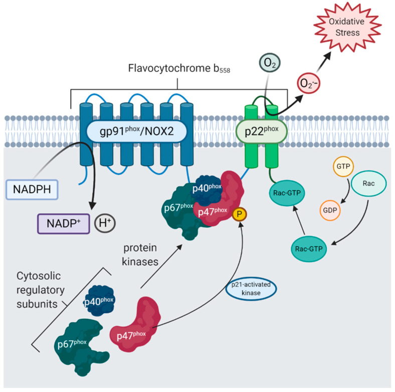
“Indeed, the expression of cytosolic regulatory subunits of NOX2 (p67phox, p47phox and p40phox) is positively correlated with disease progression and oxidative stress in AD, whilst expression of membrane-bound flavocytochrome b558 subunits (gp91phox and p22phox) is stable [68].”
§ ROS Generation in Microglia: Understanding Oxidative Stress and Inflammation in Neurodegenerative Disease > 3. Expression and Regulation of NOX in Microglia
What the sources say
- Not checked yetcites [68] Ansari, 2011, NADPH-oxidase activation and cognition in Alzheimer disease progression, Free Radic. Biol. Med. source
plaincorrelational- Asserted: passes — The authors put it forward as true.
- Checkable: passes — It can be checked against evidence.
- Load-bearing: passes — The paper’s conclusions depend on it.
- Based: passes — The paper gives a reason why it holds.
It may state two separate claims as one.
None of the sources cited for this claim have been checked yet.
In the paper
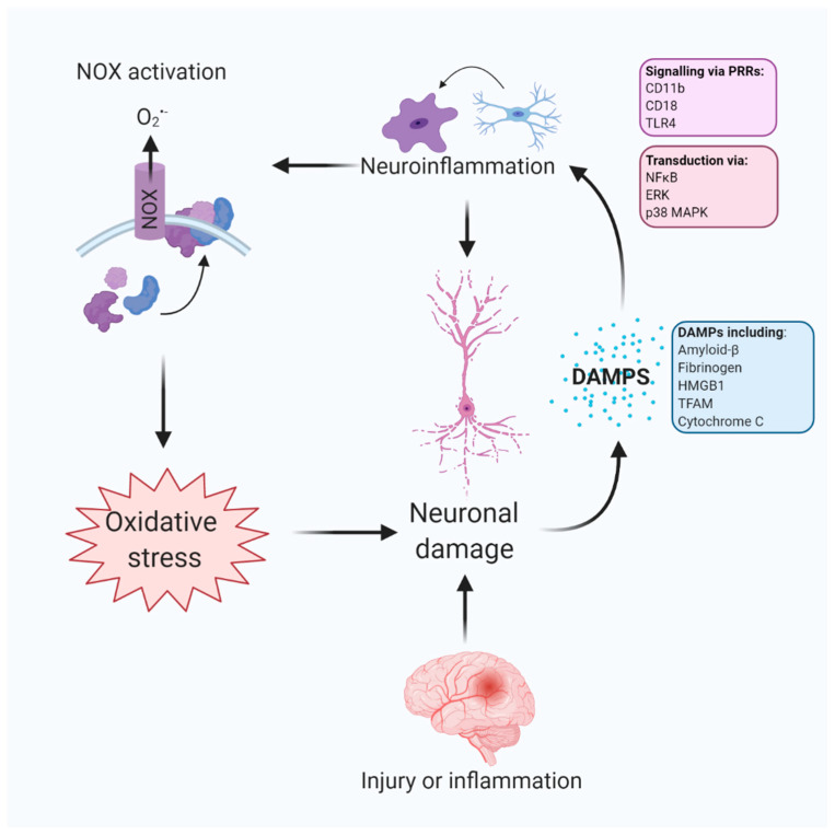
“Hence, DAMP signalling on these receptors may underpin excessive ROS production by inflammatory-related phagocytic activation and prime a vicious cycle of DAMP-induced neuronal damage by oxidative stress [86,96] (Figure 2).”
§ ROS Generation in Microglia: Understanding Oxidative Stress and Inflammation in Neurodegenerative Disease > 4. Microglial NOX Is Activated in Inflammation and Neurodegeneration > 4.2. Damage-Associated Molecular Patterns (DAMPs) Originating from Neurons Mediate NOX Activation via Pattern Recognition Receptors CR3 and TLR4
What the sources say
- Not checked yetcites [86] Hou, 2018, Complement receptor 3 mediates NADPH oxidase activation and dopaminergic neurodegeneration through a Src-Erk-dependent pathway, Redox Biol. source
- Not checked yetcites [96] Bell, 2013, Toll-like receptor 4-dependent microglial activation mediates spinal cord ischemia-reperfusion injury, Circulation source
hedgedconditional- Asserted: passes — The authors put it forward as true.
- Checkable: passes — It can be checked against evidence.
- Load-bearing: passes — The paper’s conclusions depend on it.
- Based: passes — The paper gives a reason why it holds.
It may state two separate claims as one.
None of the sources cited for this claim have been checked yet.
In the paper
“Upon activation by the assembly of multiple subunits, NOX enzymes generate the superoxide radical (O2•–). The exception is NOX4, which is constitutively active, independent of co-expression with regulatory subunits, and mostly produces H2O2 [56,57].”
§ ROS Generation in Microglia: Understanding Oxidative Stress and Inflammation in Neurodegenerative Disease > 2. Inflammatory-Related ROS Production—NOX Mediates Production of ROS in Microglia > 2.1. NOX Are a Family of Enzyme Subunits that Mediate Deliberate Production of ROS in Inflammation
What the sources say
plaindirect- Asserted: passes — The authors put it forward as true.
- Checkable: passes — It can be checked against evidence.
- Load-bearing: unclear — It is unclear whether the paper’s conclusions depend on it.
- Based: passes — The paper gives a reason why it holds.
It may state two separate claims as one.
In the paper
“Knockdown of NOX4 in these cells suppresses both ROS and interleukin (IL)6 production, confirming a link between NOX4 activation and pro-inflammatory cytokine expression.”
§ ROS Generation in Microglia: Understanding Oxidative Stress and Inflammation in Neurodegenerative Disease > 3. Expression and Regulation of NOX in Microglia > Microglia Express NOX2 and NOX4
What the sources say
- Not checked yetcites [76] Li, 2009, NOX4 expression in human microglia leads to constitutive generation of reactive oxygen species and to constitutive IL-6 expression, J. Innate Immun. source
boosteddirect- Asserted: passes — The authors put it forward as true.
- Checkable: passes — It can be checked against evidence.
- Load-bearing: passes — The paper’s conclusions depend on it.
- Based: passes — The paper gives a reason why it holds.
It may state two separate claims as one.
A conclusion the paper draws from its own results — it cites no other source for it, so there is none to check. The quote shows it is in the paper.
In the paper
“NOX2 is expressed robustly in both human and murine microglia and the adult CNS and throughout development, indicating a strong evolutionary requirement for its expression in the CNS.”
§ ROS Generation in Microglia: Understanding Oxidative Stress and Inflammation in Neurodegenerative Disease > 3. Expression and Regulation of NOX in Microglia > Microglia Express NOX2 and NOX4
boostedcorrelational- Asserted: passes — The authors put it forward as true.
- Checkable: passes — It can be checked against evidence.
- Load-bearing: unclear — It is unclear whether the paper’s conclusions depend on it.
- Based: unclear — It is unclear why the paper says it holds.
It may state two separate claims as one.
None of the sources cited for this claim have been checked yet.
In the paper
“Loss-of-function mutations in NOX2 result in chronic granulomatous disease, a rare syndrome described in 1957 and characterised by recurrent infections caused by impaired phagocytosis [46,47,48].”
§ ROS Generation in Microglia: Understanding Oxidative Stress and Inflammation in Neurodegenerative Disease > 2. Inflammatory-Related ROS Production—NOX Mediates Production of ROS in Microglia > 2.1. NOX Are a Family of Enzyme Subunits that Mediate Deliberate Production of ROS in Inflammation
What the sources say
- Not checked yetcites [46] Berendes, 1957, A fatal granulomatosus of childhood: The clinical study of a new syndrome, Minn. Med.
- Not checked yetcites [47] Quie, 1967, In vitro bactericidal capacity of human polymorphonuclear leukocytes: Diminished activity in chronic granulomatous disease of childhood, J. Clin. Investig. source
- Not checked yetcites [48] Baehner, 1967, Leukocyte oxidase: Defective activity in chronic granulomatous disease, Science source
plaindirect- Asserted: passes — The authors put it forward as true.
- Checkable: passes — It can be checked against evidence.
- Load-bearing: fails — The paper’s conclusions may not depend on it.
- Based: passes — The paper gives a reason why it holds.
Probably not a substantial claim. The paper’s conclusions may not depend on it.
None of the sources cited for this claim have been checked yet.
In the paper
“Interestingly, however, this particular cell line does not express NOX2; this calls into question the validity of the model since NOX2 is the most highly expressed isotype in both human and murine microglia [72,73].”
§ ROS Generation in Microglia: Understanding Oxidative Stress and Inflammation in Neurodegenerative Disease > 3. Expression and Regulation of NOX in Microglia > Microglia Express NOX2 and NOX4
What the sources say
- Not checked yetcites [72] Zhang, 2014, An RNA-Sequencing Transcriptome and Splicing Database of Glia, Neurons, and Vascular Cells of the Cerebral Cortex, J. Neurosci. source
- Not checked yetcites [73] Zhang, 2016, Purification and Characterization of Progenitor and Mature Human Astrocytes Reveals Transcriptional and Functional Differences with Mouse, Neuron source
plain- Asserted: passes — The authors put it forward as true.
- Checkable: passes — It can be checked against evidence.
- Load-bearing: unclear — It is unclear whether the paper’s conclusions depend on it.
- Based: passes — The paper gives a reason why it holds.
It may state two separate claims as one.
None of the sources cited for this claim have been checked yet.
In the paper
“CR3, which is upregulated in human AD-associated microglia, contributes to persistent inflammation, NOX2 activation and O2•- release in response to DAMPs, such as amyloid-β [85,98,99,100].”
§ ROS Generation in Microglia: Understanding Oxidative Stress and Inflammation in Neurodegenerative Disease > 4. Microglial NOX Is Activated in Inflammation and Neurodegeneration > 4.2. Damage-Associated Molecular Patterns (DAMPs) Originating from Neurons Mediate NOX Activation via Pattern Recognition Receptors CR3 and TLR4
What the sources say
- Not checked yetcites [85] Fu, 2012, Complement component C3 and complement receptor type 3 contribute to the phagocytosis and clearance of fibrillar Aβ by microglia, Glia source
- Not checked yetcites [98] Zhang, 2011, Microglial MAC1 receptor and PI3K are essential in mediating β-amyloid peptide-induced microglial activation and subsequent neurotoxicity, J. Neuroinflamm. source
- Not checked yetcites [99] Hu, 2008, Macrophage antigen complex-1 mediates reactive microgliosis and progressive dopaminergic neurodegeneration in the MPTP model of Parkinson’s disease, J. Immunol. source
- Not checked yetcites [100] Akiyama, 1990, Brain microglia constitutively express beta-2 integrins, J. Neuroimmunol. source
plaindirect- Asserted: passes — The authors put it forward as true.
- Checkable: passes — It can be checked against evidence.
- Load-bearing: passes — The paper’s conclusions depend on it.
- Based: passes — The paper gives a reason why it holds.
It may state two separate claims as one.
None of the sources cited for this claim have been checked yet.
In the paper
“Pharmacological blockade or microglial-specific knockout of TLR4 or CR3 severely attenuates ROS production by NOX2, indicating NOX2 activation is of microglial origin, despite reported expression of NOX2 in neurons [64,102,103].”
§ ROS Generation in Microglia: Understanding Oxidative Stress and Inflammation in Neurodegenerative Disease > 4. Microglial NOX Is Activated in Inflammation and Neurodegeneration > 4.2. Damage-Associated Molecular Patterns (DAMPs) Originating from Neurons Mediate NOX Activation via Pattern Recognition Receptors CR3 and TLR4
What the sources say
- Not checked yetcites [64] Cooney, 2013, Cellular and temporal expression of NADPH oxidase (NOX) isotypes after brain injury, J. Neuroinflamm. source
- Not checked yetcites [102] Levesque, 2013, The role of MAC1 in diesel exhaust particle-induced microglial activation and loss of dopaminergic neuron function, J. Neurochem. source
- Not checked yetcites [103] Pei, 2007, MAC1 mediates LPS-induced production of superoxide by microglia: The role of pattern recognition receptors in dopaminergic neurotoxicity, Glia source
hedgeddirect- Asserted: passes — The authors put it forward as true.
- Checkable: passes — It can be checked against evidence.
- Load-bearing: passes — The paper’s conclusions depend on it.
- Based: passes — The paper gives a reason why it holds.
It may state two separate claims as one.
In the paper
“The rate-limiting step of NOX2 activation is hypothesised to be serine-threonine phosphorylation of p47phox by p21 (cdc42/Rac1)-activated kinase-1 [62].”
§ ROS Generation in Microglia: Understanding Oxidative Stress and Inflammation in Neurodegenerative Disease > 2. Inflammatory-Related ROS Production—NOX Mediates Production of ROS in Microglia > 2.3. Phagocytic NOX (NOX1-3) Are Regulated by the Assembly of Cytosolic Subunits
What the sources say
- Not checked yetcites [62] Roepstorff, 2008, Stimulus-dependent Regulation of the Phagocyte NADPH Oxidase by a VAV1, Rac1, and PAK1 Signaling Axis, J. Biol. Chem. source
hedgedconditional- Asserted: passes — The authors put it forward as true.
- Checkable: passes — It can be checked against evidence.
- Load-bearing: fails — The paper’s conclusions may not depend on it.
- Based: passes — The paper gives a reason why it holds.
Probably not a substantial claim. The paper’s conclusions may not depend on it.
A conclusion the paper draws from its own results — it cites no other source for it, so there is none to check. The quote shows it is in the paper.
In the paper
“Nonetheless, NOX is activated in in vivo models of AD and offsetting this upregulation may limit neurodegeneration and cognitive impairment.”
§ ROS Generation in Microglia: Understanding Oxidative Stress and Inflammation in Neurodegenerative Disease > 4. Microglial NOX Is Activated in Inflammation and Neurodegeneration > 4.3. NOX Is Activated in Chronic Disease-Associated Microglia (DAM)—A Focus on Alzheimer’s Disease
hedgedconditional- Asserted: passes — The authors put it forward as true.
- Checkable: passes — It can be checked against evidence.
- Load-bearing: passes — The paper’s conclusions depend on it.
- Based: passes — The paper gives a reason why it holds.
It may state two separate claims as one.
A conclusion the paper draws from its own results — it cites no other source for it, so there is none to check. The quote shows it is in the paper.
In the paper
“Evidence from mammalian studies indicates that NOX activation in response to amyloid-β contributes to neurodegeneration and is associated with cerebrovascular pathology.”
§ ROS Generation in Microglia: Understanding Oxidative Stress and Inflammation in Neurodegenerative Disease > 4. Microglial NOX Is Activated in Inflammation and Neurodegeneration > 4.3. NOX Is Activated in Chronic Disease-Associated Microglia (DAM)—A Focus on Alzheimer’s Disease
hedgeddirect- Asserted: passes — The authors put it forward as true.
- Checkable: passes — It can be checked against evidence.
- Load-bearing: passes — The paper’s conclusions depend on it.
- Based: passes — The paper gives a reason why it holds.
It may state two separate claims as one.
In the paper
“Furthermore, age-related amyloid deposition, microgliosis and ROS production were dependent on NOX2 expression and findings correlated with post-mortem tissue from young and aged human samples [125].”
§ ROS Generation in Microglia: Understanding Oxidative Stress and Inflammation in Neurodegenerative Disease > 4. Microglial NOX Is Activated in Inflammation and Neurodegeneration > 4.3. NOX Is Activated in Chronic Disease-Associated Microglia (DAM)—A Focus on Alzheimer’s Disease
What the sources say
- Not checked yetcites [125] Geng, L., Fan, L.M., Liu, F., Smith, C., and Li, J.-M. (2020). Nox2 dependent redox-regulation of microglial response to amyloid-β stimulation and microgliosis in aging. Sci. Rep., 10. source
plaindirect- Asserted: passes — The authors put it forward as true.
- Checkable: passes — It can be checked against evidence.
- Load-bearing: passes — The paper’s conclusions depend on it.
- Based: passes — The paper gives a reason why it holds.
It may state two separate claims as one.
In the paper
“Further, microglia in AD brains strongly express gp91phox, implying NOX2 activation is localised to microglia in AD [119].”
§ ROS Generation in Microglia: Understanding Oxidative Stress and Inflammation in Neurodegenerative Disease > 4. Microglial NOX Is Activated in Inflammation and Neurodegeneration > 4.3. NOX Is Activated in Chronic Disease-Associated Microglia (DAM)—A Focus on Alzheimer’s Disease
What the sources say
- Not checked yetcites [119] Gupta, 2010, NOX activity is increased in mild cognitive impairment, Antioxid. Redox Signal. source
hedgedcorrelational- Asserted: passes — The authors put it forward as true.
- Checkable: passes — It can be checked against evidence.
- Load-bearing: passes — The paper’s conclusions depend on it.
- Based: passes — The paper gives a reason why it holds.
It may state two separate claims as one.
In the paper
“Indeed, Aβ-42 induces NOX2 expression in microglia and this response is accentuated in aged mice [125].”
§ ROS Generation in Microglia: Understanding Oxidative Stress and Inflammation in Neurodegenerative Disease > 4. Microglial NOX Is Activated in Inflammation and Neurodegeneration > 4.3. NOX Is Activated in Chronic Disease-Associated Microglia (DAM)—A Focus on Alzheimer’s Disease
What the sources say
- Not checked yetcites [125] Geng, L., Fan, L.M., Liu, F., Smith, C., and Li, J.-M. (2020). Nox2 dependent redox-regulation of microglial response to amyloid-β stimulation and microgliosis in aging. Sci. Rep., 10. source
plaindirect- Asserted: passes — The authors put it forward as true.
- Checkable: passes — It can be checked against evidence.
- Load-bearing: passes — The paper’s conclusions depend on it.
- Based: passes — The paper gives a reason why it holds.
It may state two separate claims as one.
In the paper
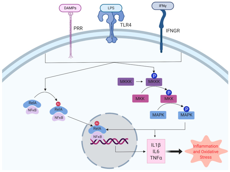
“For example, microglial activation by the TLR4 agonist, LPS, results in degradation of IκB, translocation of NFκB to the nucleus and activation of κB-containing DNA response elements, including TNFα, IL1β and cyclooxygenase-2 (COX2).”
§ ROS Generation in Microglia: Understanding Oxidative Stress and Inflammation in Neurodegenerative Disease > 5. ROS Are Secondary Messengers Activating Pro-inflammatory Pathways in Microglia > 5.1. NFκB, a Master Regulator of Inflammation in Microglia, Is Associated with NOX Expression
What the sources say
- Not checked yetcites [140] Dresselhaus, E.C., and Meffert, M.K. (2019). Cellular Specificity of NF-κB Function in the Nervous System. Front. Immunol., 10. source
plaindirect- Asserted: passes — The authors put it forward as true.
- Checkable: passes — It can be checked against evidence.
- Load-bearing: unclear — It is unclear whether the paper’s conclusions depend on it.
- Based: passes — The paper gives a reason why it holds.
It may state two separate claims as one.
In the paper
“whilst neurovascular changes were not reported, increased NOX activity and NOX4 expression were age-dependent and NOX activity was, again, strongly correlated with cognitive impairment [117].”
§ ROS Generation in Microglia: Understanding Oxidative Stress and Inflammation in Neurodegenerative Disease > 4. Microglial NOX Is Activated in Inflammation and Neurodegeneration > 4.3. NOX Is Activated in Chronic Disease-Associated Microglia (DAM)—A Focus on Alzheimer’s Disease
What the sources say
- Not checked yetcites [117] Gupta, 2011, Cognitive impairment in humanized APP×PS1 mice is linked to Aβ(1-42) and NOX activation, Neurobiol. Dis. source
boostedcorrelational- Asserted: passes — The authors put it forward as true.
- Checkable: passes — It can be checked against evidence.
- Load-bearing: passes — The paper’s conclusions depend on it.
- Based: passes — The paper gives a reason why it holds.
It may state two separate claims as one.
In the paper
“Furthermore, amyloid-β stimulates NOX2 in rat microglia and peripheral phagocytes, whilst phagocytes isolated from patients with chronic granulomatosis disease (caused by a genetic deficiency of NOX2) do not respond to stimulation with amyloid-β [121].”
§ ROS Generation in Microglia: Understanding Oxidative Stress and Inflammation in Neurodegenerative Disease > 4. Microglial NOX Is Activated in Inflammation and Neurodegeneration > 4.3. NOX Is Activated in Chronic Disease-Associated Microglia (DAM)—A Focus on Alzheimer’s Disease
What the sources say
- Not checked yetcites [121] Bianca, 1999, β-Amyloid Activates the O⨪2 Forming NADPH Oxidase in Microglia, Monocytes, and Neutrophils A POSSIBLE INFLAMMATORY MECHANISM OF NEURONAL DAMAGE IN ALZHEIMER’S DISEASE, J. Biol. Chem. source
plaindirect- Asserted: passes — The authors put it forward as true.
- Checkable: passes — It can be checked against evidence.
- Load-bearing: passes — The paper’s conclusions depend on it.
- Based: passes — The paper gives a reason why it holds.
It may state two separate claims as one.
In the paper
“Also, NOX1 and 3 expression is increased in early AD, followed by upregulation of markers of mitochondrial dysfunction [114].”
§ ROS Generation in Microglia: Understanding Oxidative Stress and Inflammation in Neurodegenerative Disease > 4. Microglial NOX Is Activated in Inflammation and Neurodegeneration > 4.3. NOX Is Activated in Chronic Disease-Associated Microglia (DAM)—A Focus on Alzheimer’s Disease
What the sources say
- Not checked yetcites [114] Wands, 2006, Molecular indices of oxidative stress and mitochondrial dysfunction occur early and often progress with severity of Alzheimer’s disease, J. Alzheimers Dis. JAD source
plaincorrelational- Asserted: passes — The authors put it forward as true.
- Checkable: passes — It can be checked against evidence.
- Load-bearing: passes — The paper’s conclusions depend on it.
- Based: passes — The paper gives a reason why it holds.
It may state two separate claims as one.
In the paper
“In LPS-stimulated macrophages, the peripheral cousins of microglia, the antioxidant lutein scavenges intracellular O2•– and H2O2 and reduces NFκB activation, whilst TLR4 signalling and MAPK activation remained [146].”
§ ROS Generation in Microglia: Understanding Oxidative Stress and Inflammation in Neurodegenerative Disease > 5. ROS Are Secondary Messengers Activating Pro-inflammatory Pathways in Microglia > 5.1. NFκB, a Master Regulator of Inflammation in Microglia, Is Associated with NOX Expression
What the sources say
- Not checked yetcites [146] Kim, 2008, The non-provitamin A carotenoid, lutein, inhibits NF-κB-dependent gene expression through redox-based regulation of the phosphatidylinositol 3-kinase/PTEN/Akt and NF-κB-inducing kinase pathways: Role of H2O2 in NF-κB activation, Free Radic. Biol. Med. source
plaindirect- Asserted: passes — The authors put it forward as true.
- Checkable: passes — It can be checked against evidence.
- Load-bearing: passes — The paper’s conclusions depend on it.
- Based: passes — The paper gives a reason why it holds.
It may state two separate claims as one.
None of the sources cited for this claim have been checked yet.
In the paper
“NFκB activation by TLR4 signalling results in neuroinflammation and cognitive impairment [141,142].”
§ ROS Generation in Microglia: Understanding Oxidative Stress and Inflammation in Neurodegenerative Disease > 5. ROS Are Secondary Messengers Activating Pro-inflammatory Pathways in Microglia > 5.1. NFκB, a Master Regulator of Inflammation in Microglia, Is Associated with NOX Expression
What the sources say
plaindirect- Asserted: passes — The authors put it forward as true.
- Checkable: passes — It can be checked against evidence.
- Load-bearing: passes — The paper’s conclusions depend on it.
- Based: passes — The paper gives a reason why it holds.
It may state two separate claims as one.
In the paper
“Knockdown of gp91phox or application of N-acetyl cysteine (the antioxidant, NAC) in infected macrophages reduced NFκB activation levels.”
§ ROS Generation in Microglia: Understanding Oxidative Stress and Inflammation in Neurodegenerative Disease > 5. ROS Are Secondary Messengers Activating Pro-inflammatory Pathways in Microglia > 5.1. NFκB, a Master Regulator of Inflammation in Microglia, Is Associated with NOX Expression
What the sources say
- Not checked yetcites [147] Li, H., Luo, Y.-F., Wang, Y.-S., Yang, Q., Xiao, Y.-L., Cai, H.-R., and Xie, C.-M. (2020, June 16). Using ROS as a Second Messenger, NADPH Oxidase 2 Mediates Macrophage Senescence via Interaction with NF-κB during Pseudomonas aeruginosa Infection. Available online: https://www.hindawi.com/journals/omcl/2018/9741838/. source
plaindirect- Asserted: passes — The authors put it forward as true.
- Checkable: passes — It can be checked against evidence.
- Load-bearing: passes — The paper’s conclusions depend on it.
- Based: passes — The paper gives a reason why it holds.
It may state two separate claims as one.
In the paper
“However, p47phox and p67phox are not expressed in neurons or astrocytes isolated from rats, but cytosolic translocation of both subunits does occur in microglia [113].”
§ ROS Generation in Microglia: Understanding Oxidative Stress and Inflammation in Neurodegenerative Disease > 4. Microglial NOX Is Activated in Inflammation and Neurodegeneration > 4.3. NOX Is Activated in Chronic Disease-Associated Microglia (DAM)—A Focus on Alzheimer’s Disease
What the sources say
- Not checked yetcites [113] Shimohama, 2000, Activation of NADPH oxidase in Alzheimer’s disease brains, Biochem. Biophys. Res. Commun. source
plain- Asserted: passes — The authors put it forward as true.
- Checkable: passes — It can be checked against evidence.
- Load-bearing: passes — The paper’s conclusions depend on it.
- Based: passes — The paper gives a reason why it holds.
It may state two separate claims as one.
None of the sources cited for this claim have been checked yet.
In the paper
“Microglial NFκB is a dimer of p50 (NFκB1) and p65 (Rel-A) [138,139] and is sequestered in the cytoplasm by inhibitor of κB (IκB).”
§ ROS Generation in Microglia: Understanding Oxidative Stress and Inflammation in Neurodegenerative Disease > 5. ROS Are Secondary Messengers Activating Pro-inflammatory Pathways in Microglia > 5.1. NFκB, a Master Regulator of Inflammation in Microglia, Is Associated with NOX Expression
What the sources say
- Not checked yetcites [138] Wang, W.-Y., Tan, M.-S., Yu, J.-T., and Tan, L. (2015). Role of pro-inflammatory cytokines released from microglia in Alzheimer’s disease. Ann. Transl. Med., 3.
- Not checked yetcites [139] Zheng, 2011, Structural studies of NF-κB signaling, Cell Res. source
plain- Asserted: passes — The authors put it forward as true.
- Checkable: passes — It can be checked against evidence.
- Load-bearing: fails — The paper’s conclusions may not depend on it.
- Based: passes — The paper gives a reason why it holds.
Probably not a substantial claim. The paper’s conclusions may not depend on it.
In the paper
“Interestingly, GRN−/− mice display high numbers of LDAM which are ROS-generating and pro-inflammatory, yet defective in phagocytosis [162].”
§ ROS Generation in Microglia: Understanding Oxidative Stress and Inflammation in Neurodegenerative Disease > 6. Lipid Droplet-Accumulating Microglia as a Source of OS in Neurodegenerative Disease > 6.1. Lipid Droplet Accumulation Is Stimulated by ROS and Correlated with Neuronal Death
What the sources say
- Not checked yetcites [162] Marschallinger, 2020, Lipid-droplet-accumulating microglia represent a dysfunctional and proinflammatory state in the aging brain, Nat. Neurosci. source
plaincorrelational- Asserted: passes — The authors put it forward as true.
- Checkable: passes — It can be checked against evidence.
- Load-bearing: passes — The paper’s conclusions depend on it.
- Based: passes — The paper gives a reason why it holds.
It may state two separate claims as one.
The paper states this without a source or data behind it here, so there is nothing to check it against. The quote shows it is in the paper.
In the paper
“The family includes extracellular signal-regulated kinases (ERK1/2), c-Jun N-terminal kinase (JNK) and p38 MAPKs.”
§ ROS Generation in Microglia: Understanding Oxidative Stress and Inflammation in Neurodegenerative Disease > 5. ROS Are Secondary Messengers Activating Pro-inflammatory Pathways in Microglia > 5.2. ROS Serves to Activate the MAPK Family Resulting in Microglial Activation
plain- Asserted: passes — The authors put it forward as true.
- Checkable: passes — It can be checked against evidence.
- Load-bearing: fails — The paper’s conclusions may not depend on it.
- Based: passes — The paper gives a reason why it holds.
Probably not a substantial claim. The paper’s conclusions may not depend on it.
In the paper
“This identifies endogenous ROS derived from mitochondria as critical mediators of the neuroimmune response to LPS, as well as exogenous ROS, i.e., from neuronal damage.”
§ ROS Generation in Microglia: Understanding Oxidative Stress and Inflammation in Neurodegenerative Disease > 5. ROS Are Secondary Messengers Activating Pro-inflammatory Pathways in Microglia > 5.2. ROS Serves to Activate the MAPK Family Resulting in Microglial Activation
What the sources say
- Not checked yetcites [158] Park, 2015, Mitochondrial ROS govern the LPS-induced pro-inflammatory response in microglia cells by regulating MAPK and NF-κB pathways, Neurosci. Lett. source
plaindirect- Asserted: passes — The authors put it forward as true.
- Checkable: passes — It can be checked against evidence.
- Load-bearing: passes — The paper’s conclusions depend on it.
- Based: passes — The paper gives a reason why it holds.
It may state two separate claims as one.
In the paper
“For example, IL1 signalling results in activation of the small G-protein, Ras, which in turn activates MKK6, resulting in activation of p38 MAPK [153].”
§ ROS Generation in Microglia: Understanding Oxidative Stress and Inflammation in Neurodegenerative Disease > 5. ROS Are Secondary Messengers Activating Pro-inflammatory Pathways in Microglia > 5.2. ROS Serves to Activate the MAPK Family Resulting in Microglial Activation
What the sources say
- Not checked yetcites [153] McDermott, 2002, Ras Participates in the Activation of p38 MAPK by Interleukin-1 by Associating with IRAK, IRAK2, TRAF6, and TAK-1, J. Biol. Chem. source
plaindirect- Asserted: passes — The authors put it forward as true.
- Checkable: passes — It can be checked against evidence.
- Load-bearing: unclear — It is unclear whether the paper’s conclusions depend on it.
- Based: passes — The paper gives a reason why it holds.
It may state two separate claims as one.
In the paper
“Moreover, BV-2 microglia treated with advanced oxidation protein products, a hallmark of oxidative stress, displayed increased NOX4 expression and elevated ROS, along with p38 MAPK and JNK activation [155].”
§ ROS Generation in Microglia: Understanding Oxidative Stress and Inflammation in Neurodegenerative Disease > 5. ROS Are Secondary Messengers Activating Pro-inflammatory Pathways in Microglia > 5.2. ROS Serves to Activate the MAPK Family Resulting in Microglial Activation
What the sources say
- Not checked yetcites [155] Liu, 2020, Advanced oxidation protein products induce microglia-mediated neuroinflammation via MAPKs-NF-κB signaling pathway and pyroptosis after secondary spinal cord injury, J. Neuroinflamm. source
plaindirect- Asserted: passes — The authors put it forward as true.
- Checkable: passes — It can be checked against evidence.
- Load-bearing: passes — The paper’s conclusions depend on it.
- Based: passes — The paper gives a reason why it holds.
It may state two separate claims as one.
None of the sources cited for this claim have been checked yet.
In the paper
“This evidence shows that lipid droplets are an important element of the redox response in microglia and these organelles are associated with AD-risk genes and neuronal death.”
§ ROS Generation in Microglia: Understanding Oxidative Stress and Inflammation in Neurodegenerative Disease > 6. Lipid Droplet-Accumulating Microglia as a Source of OS in Neurodegenerative Disease > 6.1. Lipid Droplet Accumulation Is Stimulated by ROS and Correlated with Neuronal Death
What the sources say
- Not checked yetcites [162] Marschallinger, 2020, Lipid-droplet-accumulating microglia represent a dysfunctional and proinflammatory state in the aging brain, Nat. Neurosci. source
- Not checked yetcites [163] Liu, 2015, Glial Lipid Droplets and ROS Induced by Mitochondrial Defects Promote Neurodegeneration, Cell source
- Not checked yetcites [164] Liu, 2017, The Glia-Neuron Lactate Shuttle and Elevated ROS Promote Lipid Synthesis in Neurons and Lipid Droplet Accumulation in Glia via APOE/D, Cell Metab. source
boostedcorrelational- Asserted: passes — The authors put it forward as true.
- Checkable: passes — It can be checked against evidence.
- Load-bearing: passes — The paper’s conclusions depend on it.
- Based: passes — The paper gives a reason why it holds.
It may state two separate claims as one.
In the paper
“Specific pharmacological suppression of mROS by mitoTEMPO in LPS-stimulated primary murine microglia resulted in reduced activation of all three MAPKs and limited NFκB nuclear translocation, along with the reduced expression of a battery of pro-inflammatory factors (TNFα, IL1β, IL6, iNOS and Cox-2) [158].”
§ ROS Generation in Microglia: Understanding Oxidative Stress and Inflammation in Neurodegenerative Disease > 5. ROS Are Secondary Messengers Activating Pro-inflammatory Pathways in Microglia > 5.2. ROS Serves to Activate the MAPK Family Resulting in Microglial Activation
What the sources say
- Not checked yetcites [158] Park, 2015, Mitochondrial ROS govern the LPS-induced pro-inflammatory response in microglia cells by regulating MAPK and NF-κB pathways, Neurosci. Lett. source
plaindirect- Asserted: passes — The authors put it forward as true.
- Checkable: passes — It can be checked against evidence.
- Load-bearing: passes — The paper’s conclusions depend on it.
- Based: passes — The paper gives a reason why it holds.
It may state two separate claims as one.
In the paper
“Ethanol-related neurodegeneration is associated with increased microglial NFκB activation and gp91phox expression [150].”
§ ROS Generation in Microglia: Understanding Oxidative Stress and Inflammation in Neurodegenerative Disease > 5. ROS Are Secondary Messengers Activating Pro-inflammatory Pathways in Microglia > 5.1. NFκB, a Master Regulator of Inflammation in Microglia, Is Associated with NOX Expression
What the sources say
- Not checked yetcites [150] Qin, 2012, NADPH oxidase and reactive oxygen species contribute to alcohol-induced microglial activation and neurodegeneration, J. Neuroinflamm. source
plaincorrelational- Asserted: passes — The authors put it forward as true.
- Checkable: passes — It can be checked against evidence.
- Load-bearing: unclear — It is unclear whether the paper’s conclusions depend on it.
- Based: passes — The paper gives a reason why it holds.
It may state two separate claims as one.
The paper states this without a source or data behind it here, so there is nothing to check it against. The quote shows it is in the paper.
In the paper
“The mitogen-activated protein kinase (MAPK) family comprises three subgroups of serine/threonine kinases activated in response to a range of cellular signals, including proinflammatory cytokines, environmental stress and growth factors.”
§ ROS Generation in Microglia: Understanding Oxidative Stress and Inflammation in Neurodegenerative Disease > 5. ROS Are Secondary Messengers Activating Pro-inflammatory Pathways in Microglia > 5.2. ROS Serves to Activate the MAPK Family Resulting in Microglial Activation
plaindirect- Asserted: passes — The authors put it forward as true.
- Checkable: passes — It can be checked against evidence.
- Load-bearing: fails — The paper’s conclusions may not depend on it.
- Based: passes — The paper gives a reason why it holds.
Probably not a substantial claim. The paper’s conclusions may not depend on it.
In the paper
“Lipid droplets (LD) are organelles formed in the endoplasmic reticulum, comprising of a neutral lipid (triacylglycerol and cholesteryl ester) core encapsulated by a phospholipid monolayer, thought to function as a store of energy [160].”
§ ROS Generation in Microglia: Understanding Oxidative Stress and Inflammation in Neurodegenerative Disease > 6. Lipid Droplet-Accumulating Microglia as a Source of OS in Neurodegenerative Disease
What the sources say
- Not checked yetcites [160] Jackson, 2019, Lipid droplet biogenesis, Curr. Opin. Cell Biol. source
plain- Asserted: passes — The authors put it forward as true.
- Checkable: passes — It can be checked against evidence.
- Load-bearing: fails — The paper’s conclusions may not depend on it.
- Based: passes — The paper gives a reason why it holds.
Probably not a substantial claim. The paper’s conclusions may not depend on it. It may state two separate claims as one.
In the paper
“Critically, these findings were correlated with human post-mortem brain samples from alcoholic patients, yet the effect of NOX inhibition on NFκB activation in humans was not noted [150].”
§ ROS Generation in Microglia: Understanding Oxidative Stress and Inflammation in Neurodegenerative Disease > 5. ROS Are Secondary Messengers Activating Pro-inflammatory Pathways in Microglia > 5.1. NFκB, a Master Regulator of Inflammation in Microglia, Is Associated with NOX Expression
What the sources say
- Not checked yetcites [150] Qin, 2012, NADPH oxidase and reactive oxygen species contribute to alcohol-induced microglial activation and neurodegeneration, J. Neuroinflamm. source
plaincorrelational- Asserted: passes — The authors put it forward as true.
- Checkable: passes — It can be checked against evidence.
- Load-bearing: unclear — It is unclear whether the paper’s conclusions depend on it.
- Based: passes — The paper gives a reason why it holds.
It may state two separate claims as one.
In the paper
“Antioxidant treatment or human superoxide dismutase 1 (SOD1) expression in flies reduces LD formation and attenuates neurodegeneration [163].”
§ ROS Generation in Microglia: Understanding Oxidative Stress and Inflammation in Neurodegenerative Disease > 6. Lipid Droplet-Accumulating Microglia as a Source of OS in Neurodegenerative Disease > 6.1. Lipid Droplet Accumulation Is Stimulated by ROS and Correlated with Neuronal Death
What the sources say
- Not checked yetcites [163] Liu, 2015, Glial Lipid Droplets and ROS Induced by Mitochondrial Defects Promote Neurodegeneration, Cell source
plaindirect- Asserted: passes — The authors put it forward as true.
- Checkable: passes — It can be checked against evidence.
- Load-bearing: passes — The paper’s conclusions depend on it.
- Based: passes — The paper gives a reason why it holds.
It may state two separate claims as one.
In the paper
“MAPKs themselves exert their effects on cellular physiology via myriad effectors, including transcription factors and further kinases [152].”
§ ROS Generation in Microglia: Understanding Oxidative Stress and Inflammation in Neurodegenerative Disease > 5. ROS Are Secondary Messengers Activating Pro-inflammatory Pathways in Microglia > 5.2. ROS Serves to Activate the MAPK Family Resulting in Microglial Activation
What the sources say
- Not checked yetcites [152] Kaminska, 2009, MAPK signal transduction underlying brain inflammation and gliosis as therapeutic target, Anat. Rec. source
plaindirect- Asserted: passes — The authors put it forward as true.
- Checkable: passes — It can be checked against evidence.
- Load-bearing: fails — The paper’s conclusions may not depend on it.
- Based: passes — The paper gives a reason why it holds.
Probably not a substantial claim. The paper’s conclusions may not depend on it.
In the paper
“MAPKs share a common chain of activation mediated by sequential, three-tiered phosphorylation of upstream kinases, i.e., signal transduction is mediated by phosphorylation of a MAPK kinase kinase (MKKK), which in turn activates a MAPK kinase (MKK) which activates the MAPK.”
§ ROS Generation in Microglia: Understanding Oxidative Stress and Inflammation in Neurodegenerative Disease > 5. ROS Are Secondary Messengers Activating Pro-inflammatory Pathways in Microglia > 5.2. ROS Serves to Activate the MAPK Family Resulting in Microglial Activation
What the sources say
- Not checked yetcites [152] Kaminska, 2009, MAPK signal transduction underlying brain inflammation and gliosis as therapeutic target, Anat. Rec. source
plaindirect- Asserted: passes — The authors put it forward as true.
- Checkable: passes — It can be checked against evidence.
- Load-bearing: fails — The paper’s conclusions may not depend on it.
- Based: passes — The paper gives a reason why it holds.
Probably not a substantial claim. The paper’s conclusions may not depend on it.
None of the sources cited for this claim have been checked yet.
In the paper
“This was mediated by both a reduction in heme availability and biliverdin and carbon monoxide, by-products of heme metabolism by HO-1 which may facilitate attenuation of TLR4 signalling by NOX inhibition [173,174].”
§ ROS Generation in Microglia: Understanding Oxidative Stress and Inflammation in Neurodegenerative Disease > 7. Balancing the Scales—Antioxidant Enzymes Limit Microglial Activation > 7.1. Expression of Classical Antioxidant Proteins Are Controlled by Nrf2 in Microglia
What the sources say
- Not checked yetcites [173] Nakahira, 2006, Carbon monoxide differentially inhibits TLR signaling pathways by regulating ROS-induced trafficking of TLRs to lipid rafts, J. Exp. Med. source
- Not checked yetcites [174] Lanone, 2004, Induction of heme oxygenase-1 inhibits NAD(P)H oxidase activity by down-regulating cytochrome b558 expression via the reduction of heme availability, J. Biol. Chem. source
hedgedconditional- Asserted: passes — The authors put it forward as true.
- Checkable: passes — It can be checked against evidence.
- Load-bearing: unclear — It is unclear whether the paper’s conclusions depend on it.
- Based: passes — The paper gives a reason why it holds.
It may state two separate claims as one.
None of the sources cited for this claim have been checked yet.
In the paper
“Furthermore, NCOA7 is induced in epithelial cells in response to rhinovirus infection and LPS-stimulated macrophages, the peripheral myeloid cousins of microglia [186,187].”
§ ROS Generation in Microglia: Understanding Oxidative Stress and Inflammation in Neurodegenerative Disease > 7. Balancing the Scales—Antioxidant Enzymes Limit Microglial Activation > 7.2. A Role for TLDc Proteins in Neuroinflammation
What the sources say
- Not checked yetcites [186] Baines, 2013, Novel immune genes associated with excessive inflammatory and antiviral responses to rhinovirus in COPD, Respir. Res. source
- Not checked yetcites [187] Alasoo, 2015, Transcriptional profiling of macrophages derived from monocytes and iPS cells identifies a conserved response to LPS and novel alternative transcription, Sci. Rep. source
plaindirect- Asserted: passes — The authors put it forward as true.
- Checkable: passes — It can be checked against evidence.
- Load-bearing: fails — The paper’s conclusions may not depend on it.
- Based: passes — The paper gives a reason why it holds.
Probably not a substantial claim. The paper’s conclusions may not depend on it. It may state two separate claims as one.
In the paper
“Loss of OXR1 results in pronounced microgliosis whilst neuronal OXR1 overexpression delays inflammation, indicating OXR1 regulation of microglial activation in a non-cell-autonomous fashion [184].”
§ ROS Generation in Microglia: Understanding Oxidative Stress and Inflammation in Neurodegenerative Disease > 7. Balancing the Scales—Antioxidant Enzymes Limit Microglial Activation > 7.2. A Role for TLDc Proteins in Neuroinflammation
What the sources say
- Not checked yetcites [184] Liu, 2015, Neuron-specific antioxidant OXR1 extends survival of a mouse model of amyotrophic lateral sclerosis, Brain source
hedgeddirect- Asserted: passes — The authors put it forward as true.
- Checkable: passes — It can be checked against evidence.
- Load-bearing: passes — The paper’s conclusions depend on it.
- Based: passes — The paper gives a reason why it holds.
It may state two separate claims as one.
A conclusion the paper draws from its own results — it cites no other source for it, so there is none to check. The quote shows it is in the paper.
In the paper
“Expression of these proteins by Nrf2 in microglia may represent a pathway that can be modulated to enhance innate defences and combat oxidative stress.”
§ ROS Generation in Microglia: Understanding Oxidative Stress and Inflammation in Neurodegenerative Disease > 8. Conclusions and Future Directions > 8.1. Understanding the Molecular Mechanisms of Neuroinflammation Is Key to Developing Effective Antioxidant Therapies
hedgedconditional- Asserted: passes — The authors put it forward as true.
- Checkable: passes — It can be checked against evidence.
- Load-bearing: passes — The paper’s conclusions depend on it.
- Based: passes — The paper gives a reason why it holds.
Probably not a substantial claim.
The paper states this without a source or data behind it here, so there is nothing to check it against. The quote shows it is in the paper.
In the paper
“The function of these redox regulators in microglia is unclear as of yet, however, many of these proteins are linked to inflammation, suggesting functionality in microglia.”
§ ROS Generation in Microglia: Understanding Oxidative Stress and Inflammation in Neurodegenerative Disease > 7. Balancing the Scales—Antioxidant Enzymes Limit Microglial Activation
plain- Asserted: passes — The authors put it forward as true.
- Checkable: passes — It can be checked against evidence.
- Load-bearing: fails — The paper’s conclusions may not depend on it.
- Based: passes — The paper gives a reason why it holds.
Probably not a substantial claim. The paper’s conclusions may not depend on it.
None of the sources cited for this claim have been checked yet.
In the paper
“Many antioxidant proteins are understood to function in the CNS, including classical antioxidants (superoxide dismutase, glutathione reductase, catalase, etc. reviewed in [38]) and more recently TBC, LysM, domain catalytic (TLDc) proteins (oxidation resistance 1, nuclear receptor coactivator 7, Tbc1d24, etc. reviewed in [166]).”
§ ROS Generation in Microglia: Understanding Oxidative Stress and Inflammation in Neurodegenerative Disease > 7. Balancing the Scales—Antioxidant Enzymes Limit Microglial Activation
What the sources say
plain- Asserted: passes — The authors put it forward as true.
- Checkable: passes — It can be checked against evidence.
- Load-bearing: fails — The paper’s conclusions may not depend on it.
- Based: passes — The paper gives a reason why it holds.
Probably not a substantial claim. The paper’s conclusions may not depend on it.
In the paper
“The two are undoubtedly linked; however, it is not clear how ROS interact with glia to elicit an immune response, although this may involve reciprocal regulation between NOX2 and NFκB [147].”
§ ROS Generation in Microglia: Understanding Oxidative Stress and Inflammation in Neurodegenerative Disease > 8. Conclusions and Future Directions > 8.1. Understanding the Molecular Mechanisms of Neuroinflammation Is Key to Developing Effective Antioxidant Therapies
What the sources say
- Not checked yetcites [147] Li, H., Luo, Y.-F., Wang, Y.-S., Yang, Q., Xiao, Y.-L., Cai, H.-R., and Xie, C.-M. (2020, June 16). Using ROS as a Second Messenger, NADPH Oxidase 2 Mediates Macrophage Senescence via Interaction with NF-κB during Pseudomonas aeruginosa Infection. Available online: https://www.hindawi.com/journals/omcl/2018/9741838/. source
hedgedconditional- Asserted: passes — The authors put it forward as true.
- Checkable: passes — It can be checked against evidence.
- Load-bearing: fails — The paper’s conclusions may not depend on it.
- Based: passes — The paper gives a reason why it holds.
Probably not a substantial claim. The paper’s conclusions may not depend on it.
None of the sources cited for this claim have been checked yet.
In the paper
“Intracranial injection of the isothiocyanate, sulforaphane, enhanced Nrf2 levels which limited expression of pro-inflammatory microglial markers and conferred neuroprotection [168,169,170].”
§ ROS Generation in Microglia: Understanding Oxidative Stress and Inflammation in Neurodegenerative Disease > 7. Balancing the Scales—Antioxidant Enzymes Limit Microglial Activation > 7.1. Expression of Classical Antioxidant Proteins Are Controlled by Nrf2 in Microglia
What the sources say
- Not checked yetcites [168] Innamorato, 2008, The transcription factor Nrf2 is a therapeutic target against brain inflammation, J. Immunol. Baltim. source
- Not checked yetcites [169] Rojo, 2010, Nrf2 regulates microglial dynamics and neuroinflammation in experimental Parkinson’s disease, Glia source
- Not checked yetcites [170] Jazwa, 2011, Pharmacological targeting of the transcription factor Nrf2 at the basal ganglia provides disease modifying therapy for experimental parkinsonism, Antioxid. Redox Signal. source
plaindirect- Asserted: passes — The authors put it forward as true.
- Checkable: passes — It can be checked against evidence.
- Load-bearing: passes — The paper’s conclusions depend on it.
- Based: passes — The paper gives a reason why it holds.
It may state two separate claims as one.
A conclusion the paper draws from its own results — it cites no other source for it, so there is none to check. The quote shows it is in the paper.
In the paper
“Finally, LDAM are important regulators of oxidative stress and a mechanism by which protective inflammation may become dysregulated and damaging in ageing and neurodegeneration.”
§ ROS Generation in Microglia: Understanding Oxidative Stress and Inflammation in Neurodegenerative Disease > 8. Conclusions and Future Directions > 8.1. Understanding the Molecular Mechanisms of Neuroinflammation Is Key to Developing Effective Antioxidant Therapies
plaindirect- Asserted: passes — The authors put it forward as true.
- Checkable: passes — It can be checked against evidence.
- Load-bearing: passes — The paper’s conclusions depend on it.
- Based: fails — The paper may give no reason why it holds.
Probably not a substantial claim. The paper may give no reason why it holds. It may state two separate claims as one.
In the paper
“This signature involved the downregulation of microglial homeostatic genes (Tmem119, Cx3cr1) and overexpression of ROS-related genes Cybb (p91phox), Cyba (p22phox) and Nos2 (nitric oxide synthase 2); and pro-inflammatory genes Il1b, Cd47 and Prkcd (protein kinase C-δ).”
§ ROS Generation in Microglia: Understanding Oxidative Stress and Inflammation in Neurodegenerative Disease > 8. Conclusions and Future Directions > 8.2. At the Forefront of Drug Discovery: ToxSeq Analysis of ROS-Generating Microglia
What the sources say
- Not checked yetcites [181] Mendiola, 2020, Transcriptional profiling and therapeutic targeting of oxidative stress in neuroinflammation, Nat. Immunol. source
plaincorrelational- Asserted: passes — The authors put it forward as true.
- Checkable: passes — It can be checked against evidence.
- Load-bearing: passes — The paper’s conclusions depend on it.
- Based: passes — The paper gives a reason why it holds.
It may state two separate claims as one.
None of the sources cited for this claim have been checked yet.
In the paper
“The mechanism of LD accumulation in response to elevated ROS in flies and the perturbation of this mechanism by ApoeE-ε4 indicates LDAM are protective, potentially by sequestering peroxidised lipids [163,164].”
§ ROS Generation in Microglia: Understanding Oxidative Stress and Inflammation in Neurodegenerative Disease > 6. Lipid Droplet-Accumulating Microglia as a Source of OS in Neurodegenerative Disease > 6.2. Lipid-Droplet Accumulating Microglia May Represent a Double-Edged Sword in Neurodegeneration
What the sources say
- Not checked yetcites [163] Liu, 2015, Glial Lipid Droplets and ROS Induced by Mitochondrial Defects Promote Neurodegeneration, Cell source
- Not checked yetcites [164] Liu, 2017, The Glia-Neuron Lactate Shuttle and Elevated ROS Promote Lipid Synthesis in Neurons and Lipid Droplet Accumulation in Glia via APOE/D, Cell Metab. source
hedgedconditional- Asserted: passes — The authors put it forward as true.
- Checkable: passes — It can be checked against evidence.
- Load-bearing: passes — The paper’s conclusions depend on it.
- Based: passes — The paper gives a reason why it holds.
It may state two separate claims as one.
A conclusion the paper draws from its own results — it cites no other source for it, so there is none to check. The quote shows it is in the paper.
In the paper
“The function of these redox regulators in microglia is unclear as of yet, however, many of these proteins are linked to inflammation, suggesting functionality in microglia.”
§ ROS Generation in Microglia: Understanding Oxidative Stress and Inflammation in Neurodegenerative Disease > 7. Balancing the Scales—Antioxidant Enzymes Limit Microglial Activation
hedgedcorrelational- Asserted: passes — The authors put it forward as true.
- Checkable: passes — It can be checked against evidence.
- Load-bearing: fails — The paper’s conclusions may not depend on it.
- Based: passes — The paper gives a reason why it holds.
Probably not a substantial claim. The paper’s conclusions may not depend on it.
A conclusion the paper draws from its own results — it cites no other source for it, so there is none to check. The quote shows it is in the paper.
In the paper
“However, it is feasible that ROS act as secondary messengers in microglia to propagate inflammatory states and may contribute to immune dysregulation, leading to persistent and inappropriate inflammation.”
§ ROS Generation in Microglia: Understanding Oxidative Stress and Inflammation in Neurodegenerative Disease > 8. Conclusions and Future Directions > 8.1. Understanding the Molecular Mechanisms of Neuroinflammation Is Key to Developing Effective Antioxidant Therapies
hedgedconditional- Asserted: passes — The authors put it forward as true.
- Checkable: passes — It can be checked against evidence.
- Load-bearing: passes — The paper’s conclusions depend on it.
- Based: fails — The paper may give no reason why it holds.
Probably not a substantial claim. The paper may give no reason why it holds. It may state two separate claims as one.
In the paper
“This implies LDAM are dysfunctional in neurodegeneration, where they may be unable to clear aggregated protein but would induce oxidative stress and inflammatory cascades.”
§ ROS Generation in Microglia: Understanding Oxidative Stress and Inflammation in Neurodegenerative Disease > 6. Lipid Droplet-Accumulating Microglia as a Source of OS in Neurodegenerative Disease > 6.2. Lipid-Droplet Accumulating Microglia May Represent a Double-Edged Sword in Neurodegeneration
What the sources say
- Not checked yetcites [162] Marschallinger, 2020, Lipid-droplet-accumulating microglia represent a dysfunctional and proinflammatory state in the aging brain, Nat. Neurosci. source
hedgedconditional- Asserted: passes — The authors put it forward as true.
- Checkable: passes — It can be checked against evidence.
- Load-bearing: passes — The paper’s conclusions depend on it.
- Based: passes — The paper gives a reason why it holds.
It may state two separate claims as one.
A conclusion the paper draws from its own results — it cites no other source for it, so there is none to check. The quote shows it is in the paper.
In the paper
“Molecular pathways in microglia contributing to neurodegeneration undoubtedly involve inflammatory-mediated ROS production by activation of NOX.”
§ ROS Generation in Microglia: Understanding Oxidative Stress and Inflammation in Neurodegenerative Disease > 8. Conclusions and Future Directions > 8.1. Understanding the Molecular Mechanisms of Neuroinflammation Is Key to Developing Effective Antioxidant Therapies
boosteddirect- Asserted: passes — The authors put it forward as true.
- Checkable: passes — It can be checked against evidence.
- Load-bearing: passes — The paper’s conclusions depend on it.
- Based: fails — The paper may give no reason why it holds.
Probably not a substantial claim. The paper may give no reason why it holds.
None of the sources cited for this claim have been checked yet.
In the paper
“In microglia, Nrf2-deficiency promotes inflammatory marker expression (IL6, Cox2, iNOS) whilst inhibiting anti-inflammatory marker expression (arginase 1, IL4) in response to LPS and MPTP [168,169].”
§ ROS Generation in Microglia: Understanding Oxidative Stress and Inflammation in Neurodegenerative Disease > 7. Balancing the Scales—Antioxidant Enzymes Limit Microglial Activation > 7.1. Expression of Classical Antioxidant Proteins Are Controlled by Nrf2 in Microglia
What the sources say
plaindirect- Asserted: passes — The authors put it forward as true.
- Checkable: passes — It can be checked against evidence.
- Load-bearing: passes — The paper’s conclusions depend on it.
- Based: passes — The paper gives a reason why it holds.
It may state two separate claims as one.
None of the sources cited for this claim have been checked yet.
In the paper
“The mechanism by which TLDc proteins confer stress resistance is unclear, however, specific disruption of the OXR1 TLDc domain in mice is sufficient to cause neurodegeneration and loss-of-function polymorphisms in human TLDc proteins result in a neurodegenerative phenotype [182,183].”
§ ROS Generation in Microglia: Understanding Oxidative Stress and Inflammation in Neurodegenerative Disease > 7. Balancing the Scales—Antioxidant Enzymes Limit Microglial Activation > 7.2. A Role for TLDc Proteins in Neuroinflammation
What the sources say
- Not checked yetcites [182] Finelli, 2016, The Evolutionarily Conserved Tre2/Bub2/Cdc16 (TBC), Lysin Motif (LysM), Domain Catalytic (TLDc) Domain Is Neuroprotective against Oxidative Stress, J. Biol. Chem. source
- Not checked yetcites [183] Wang, 2019, Loss of Oxidation Resistance 1, OXR1, Is Associated with an Autosomal-Recessive Neurological Disease with Cerebellar Atrophy and Lysosomal Dysfunction, Am. J. Hum. Genet. source
plaindirect- Asserted: passes — The authors put it forward as true.
- Checkable: passes — It can be checked against evidence.
- Load-bearing: unclear — It is unclear whether the paper’s conclusions depend on it.
- Based: passes — The paper gives a reason why it holds.
It may state two separate claims as one.
In the paper
“Finally, a unique, short-isoform of NCOA7, NCOA7-B or NCOA7-AS, is strongly linked to inflammatory activation, especially by viral-related stimuli such as interferons (IFNs), and functional inhibition of endosome-mediated viral-entry by NCOA7-B has been demonstrated [185].”
§ ROS Generation in Microglia: Understanding Oxidative Stress and Inflammation in Neurodegenerative Disease > 7. Balancing the Scales—Antioxidant Enzymes Limit Microglial Activation > 7.2. A Role for TLDc Proteins in Neuroinflammation
What the sources say
- Not checked yetcites [185] Doyle, 2018, The interferon-inducible isoform of NCOA7 inhibits endosome-mediated viral entry, Nat. Microbiol. source
boostedcorrelational- Asserted: passes — The authors put it forward as true.
- Checkable: passes — It can be checked against evidence.
- Load-bearing: unclear — It is unclear whether the paper’s conclusions depend on it.
- Based: passes — The paper gives a reason why it holds.
It may state two separate claims as one.
A conclusion the paper draws from its own results — it cites no other source for it, so there is none to check. The quote shows it is in the paper.
In the paper
“It is unlikely ROS are the primary trigger of inflammation in Alzheimer’s disease; this is likely mediated by DAMPs associated with toxic protein aggregation.”
§ ROS Generation in Microglia: Understanding Oxidative Stress and Inflammation in Neurodegenerative Disease > 8. Conclusions and Future Directions > 8.1. Understanding the Molecular Mechanisms of Neuroinflammation Is Key to Developing Effective Antioxidant Therapies
hedgeddirect- Asserted: passes — The authors put it forward as true.
- Checkable: passes — It can be checked against evidence.
- Load-bearing: passes — The paper’s conclusions depend on it.
- Based: fails — The paper may give no reason why it holds.
Probably not a substantial claim. The paper may give no reason why it holds. It may state two separate claims as one.
In the paper
“However, LDAM are also ROS-generating and pro-inflammatory yet defective in phagocytosis in murine models [162].”
§ ROS Generation in Microglia: Understanding Oxidative Stress and Inflammation in Neurodegenerative Disease > 6. Lipid Droplet-Accumulating Microglia as a Source of OS in Neurodegenerative Disease > 6.2. Lipid-Droplet Accumulating Microglia May Represent a Double-Edged Sword in Neurodegeneration
What the sources say
- Not checked yetcites [162] Marschallinger, 2020, Lipid-droplet-accumulating microglia represent a dysfunctional and proinflammatory state in the aging brain, Nat. Neurosci. source
plaindirect- Asserted: passes — The authors put it forward as true.
- Checkable: passes — It can be checked against evidence.
- Load-bearing: passes — The paper’s conclusions depend on it.
- Based: passes — The paper gives a reason why it holds.
It may state two separate claims as one.
None of the sources cited for this claim have been checked yet.
In the paper
“Microgliosis and sulforaphane neuroprotection were attenuated in Nrf2-deficient mice which confirms a mechanistic link between Nrf2 and immune regulation.”
§ ROS Generation in Microglia: Understanding Oxidative Stress and Inflammation in Neurodegenerative Disease > 7. Balancing the Scales—Antioxidant Enzymes Limit Microglial Activation > 7.1. Expression of Classical Antioxidant Proteins Are Controlled by Nrf2 in Microglia
What the sources say
- Not checked yetcites [168] Innamorato, 2008, The transcription factor Nrf2 is a therapeutic target against brain inflammation, J. Immunol. Baltim. source
- Not checked yetcites [169] Rojo, 2010, Nrf2 regulates microglial dynamics and neuroinflammation in experimental Parkinson’s disease, Glia source
- Not checked yetcites [170] Jazwa, 2011, Pharmacological targeting of the transcription factor Nrf2 at the basal ganglia provides disease modifying therapy for experimental parkinsonism, Antioxid. Redox Signal. source
boosteddirect- Asserted: passes — The authors put it forward as true.
- Checkable: passes — It can be checked against evidence.
- Load-bearing: passes — The paper’s conclusions depend on it.
- Based: passes — The paper gives a reason why it holds.
It may state two separate claims as one.
In the paper
“Furthermore, the expression of this short-isoform in ovarian follicles is regulated intracellularly by MAPKs, p38 and ERK, which are recognised mediators of the pro-inflammatory response in microglia [191].”
§ ROS Generation in Microglia: Understanding Oxidative Stress and Inflammation in Neurodegenerative Disease > 7. Balancing the Scales—Antioxidant Enzymes Limit Microglial Activation > 7.2. A Role for TLDc Proteins in Neuroinflammation
What the sources say
- Not checked yetcites [191] Shkolnik, 2008, Molecular characterization and bioinformatics analysis of Ncoa7B, a novel ovulation-associated and reproduction system-specific Ncoa7 isoform, Reproduction source
plaindirect- Asserted: passes — The authors put it forward as true.
- Checkable: passes — It can be checked against evidence.
- Load-bearing: fails — The paper’s conclusions may not depend on it.
- Based: passes — The paper gives a reason why it holds.
Probably not a substantial claim. The paper’s conclusions may not depend on it. It may state two separate claims as one.
A conclusion the paper draws from its own results — it cites no other source for it, so there is none to check. The quote shows it is in the paper.
In the paper
“For example, the concerted kinase activity of MAPKs and PKC may create a strong pro-oxidant signal by vigorous and persistent phosphorylation of both each other’s activation pathways and NOX2 regulatory subunits, resulting in enhanced and sustained ROS production.”
§ ROS Generation in Microglia: Understanding Oxidative Stress and Inflammation in Neurodegenerative Disease > 8. Conclusions and Future Directions > 8.1. Understanding the Molecular Mechanisms of Neuroinflammation Is Key to Developing Effective Antioxidant Therapies
hedgedconditional- Asserted: passes — The authors put it forward as true.
- Checkable: passes — It can be checked against evidence.
- Load-bearing: passes — The paper’s conclusions depend on it.
- Based: fails — The paper may give no reason why it holds.
Probably not a substantial claim. The paper may give no reason why it holds.
The paper states this without a source or data behind it here, so there is nothing to check it against. The quote shows it is in the paper.
In the paper
“Notably, many experiments examining the role have been conducted in immortalised microglia, e.g., BV2 cells.”
§ ROS Generation in Microglia: Understanding Oxidative Stress and Inflammation in Neurodegenerative Disease > 7. Balancing the Scales—Antioxidant Enzymes Limit Microglial Activation > 7.1. Expression of Classical Antioxidant Proteins Are Controlled by Nrf2 in Microglia
plain- Asserted: passes — The authors put it forward as true.
- Checkable: passes — It can be checked against evidence.
- Load-bearing: fails — The paper’s conclusions may not depend on it.
- Based: passes — The paper gives a reason why it holds.
Probably not a substantial claim. The paper’s conclusions may not depend on it.
A conclusion the paper draws from its own results — it cites no other source for it, so there is none to check. The quote shows it is in the paper.
In the paper
“This likely involves a vicious cycle in which neuroinflammatory signals induce ROS release, which then promotes further deleterious inflammation.”
§ ROS Generation in Microglia: Understanding Oxidative Stress and Inflammation in Neurodegenerative Disease > 8. Conclusions and Future Directions > 8.1. Understanding the Molecular Mechanisms of Neuroinflammation Is Key to Developing Effective Antioxidant Therapies
hedgeddirect- Asserted: passes — The authors put it forward as true.
- Checkable: passes — It can be checked against evidence.
- Load-bearing: passes — The paper’s conclusions depend on it.
- Based: fails — The paper may give no reason why it holds.
Probably not a substantial claim. The paper may give no reason why it holds.
In the paper
“Gene network analysis identified upregulation of glutathione transferase activity in ROS-generating microglia, which was rescued by acivin, a compound which inhibits glutathione degradation [181].”
§ ROS Generation in Microglia: Understanding Oxidative Stress and Inflammation in Neurodegenerative Disease > 8. Conclusions and Future Directions > 8.2. At the Forefront of Drug Discovery: ToxSeq Analysis of ROS-Generating Microglia
What the sources say
- Not checked yetcites [181] Mendiola, 2020, Transcriptional profiling and therapeutic targeting of oxidative stress in neuroinflammation, Nat. Immunol. source
plaindirect- Asserted: passes — The authors put it forward as true.
- Checkable: passes — It can be checked against evidence.
- Load-bearing: passes — The paper’s conclusions depend on it.
- Based: passes — The paper gives a reason why it holds.
It may state two separate claims as one.
In the paper
“Acivin effectively reduced oxidative stress and neuronal death in the EAE model without toxicity to other CNS cell types.”
§ ROS Generation in Microglia: Understanding Oxidative Stress and Inflammation in Neurodegenerative Disease > 8. Conclusions and Future Directions > 8.2. At the Forefront of Drug Discovery: ToxSeq Analysis of ROS-Generating Microglia
What the sources say
- Not checked yetcites [181] Mendiola, 2020, Transcriptional profiling and therapeutic targeting of oxidative stress in neuroinflammation, Nat. Immunol. source
plaindirect- Asserted: passes — The authors put it forward as true.
- Checkable: passes — It can be checked against evidence.
- Load-bearing: passes — The paper’s conclusions depend on it.
- Based: passes — The paper gives a reason why it holds.
It may state two separate claims as one.
Claims other papers make about this one
Each is checked against this paper’s own text: does it say what the citing paper says it does?
In the citing paper
“Treatment options for neuroinflammation associated with AD have been investigated, including IL-1β antagonists such as canakinumab [206,207,208].”
From Recent Advances in Therapeutics for the Treatment of Alzheimer’s Disease (2024) · cites this paper as [207]
In the citing paper
“Research on cytokine inhibition has focused on anti-inflammatory medications that interact with pro-inflammatory cytokines such as TNF-a and IL-1 beta.”
From Recent Advances in Therapeutics for the Treatment of Alzheimer’s Disease (2024) · cites this paper as [207]
Citations
Cites
Amyloid oligomers exacerbate tau pathology in a mouse model of tauopathy
Selenica, M.-L. B., Brownlow, M., Jimenez, J. P., Lee, D. C., Pena, G., Dickey, C. A., Gordon, M. N., & Morgan, D. (2013) · Neurodegenerative Diseases · doi:10.1159/000337230 · Cited by 10,359
Neuroinflammation in Alzheimer's disease
Heneka, M. T., Carson, M. J., Khoury, J. E., Landreth, G. E., Brosseron, F., Feinstein, D. L., Jacobs, A. H., Wyss-Coray, T., Vitorica, J., Ransohoff, R. M., et al. (2015) · The Lancet Neurology · doi:10.1016/s1474-4422(15)70016-5 · Cited by 6,226
Fate Mapping Analysis Reveals That Adult Microglia Derive from Primitive Macrophages
Ginhoux, F., Greter, M., Leboeuf, M., Nandi, S., See, P., Gokhan, S., Mehler, M. F., Conway, S. J., Ng, L. G., Stanley, E. R., et al. (2010) · Science · doi:10.1126/science.1194637 · Cited by 5,175 · cited 1×
“Whilst peripheral macrophages and microglia share origin in the yolk sac, microglia are an ontogenetically distinct, locally maintained cell population [69,70] and so NOX expression in microglia may not exactly mirror that of their macrophage relatives.”
BackgroundZhang, Y., Chen, K., Sloan, S. A., Bennett, M. L., Scholze, A. R., O'Keeffe, S., Phatnani, H. P., Guarnieri, P., Caneda, C., Ruderisch, N., et al. (2014) · The Journal of Neuroscience · doi:10.1523/jneurosci.1860-14.2014 · Cited by 4,875 · cited 5×
“There remains a small increase in ROS production in response to LPS and α-synuclein in these NOX2 knockout cultures, likely a result of NOX4, since this isoform is expressed at the next highest level in microglia [72].”
BackgroundInfluentialMicroglia Sculpt Postnatal Neural Circuits in an Activity and Complement-Dependent Manner
Schafer, D. P., Lehrman, E. K., Kautzman, A. G., Koyama, R., Mardinly, A. R., Yamasaki, R., Ransohoff, R. M., Greenberg, M. E., Barres, B. A., & Stevens, B. (2012) · Neuron · doi:10.1016/j.neuron.2012.03.026 · Cited by 4,089 · cited 1×
“Considering CR3 signalling is an essential functional component in synaptic pruning by microglia [93], NOX activation by PRRs is likely a functional inflammatory-related phagocytic response to eliminate dead and dying neurons and aggregated protein, which has been elucidated by 2-photon live imaging [94,95].”
BackgroundItoh, K., Chiba, T., Takahashi, S., Ishii, T., Igarashi, K., Katoh, Y., Oyake, T., Hayashi, N., Satoh, K., Hatayama, I., et al. (1997) · Biochemical and Biophysical Research Communications · doi:10.1006/bbrc.1997.6943 · Cited by 3,999 · cited 1×
“, genes encoding NADH-quinone oxidoreductase 1 (NQO1) and glutathione-S-transferases (GSTs) [167].”
Toll-like receptors and their crosstalk with other innate receptors in infection and immunity
Kawai, T., & Akira, S. (2011) · Immunity · doi:10.1016/j.immuni.2011.05.006 · Cited by 3,658 · cited 6×
“These signals are transduced in microglia by nuclear factor κB (NFκB) and mitogen-activated protein kinases (MAPKs), such as p38 and extracellular signal-regulated kinase (ERK).”
BackgroundInfluentialOncogenic Signaling Pathways in The Cancer Genome Atlas
Sanchez-Vega, F., Mina, M., Armenia, J., Chatila, W. K., Luna, A., La, K. C., Dimitriadoy, S., Liu, D. L., Kantheti, H. S., Saghafinia, S., et al. (2018) · Cell · doi:10.1016/j.cell.2018.03.035 · Cited by 3,201 · cited 1×
“Investigating this dynamic interaction between convergent pro-inflammatory pathways, similar to what has been divulged in oncogenic pathways in cancer [193], will be key to understanding inappropriate inflammation in neurodegenerative disease.”
BackgroundNOX enzymes and the biology of reactive oxygen
Lambeth, J. D. (2004) · Nature Reviews Immunology · doi:10.1038/nri1312 · Cited by 3,125
Variant of TREM2 Associated with the Risk of Alzheimer's Disease
Jonsson, T., Stefansson, H., Steinberg, S., Jonsdottir, I., Jonsson, P. V., Snaedal, J., Bjornsson, S., Huttenlocher, J., Levey, A. I., Lah, J. J., et al. (2013) · New England Journal of Medicine · doi:10.1056/nejmoa1211103 · Cited by 2,558 · cited 1×
“Characterisation of gene ontology networks in post-mortem AD brains has identified dysregulation of microglia-specific networks as the most strongly correlated to neuropathology [15] and many microglial AD risk-factor genes have been identified [16,17].”
Background
Research works printed but not linked: 1
- 2018, Epidemiology of Alzheimer’s disease and other dementias, Rev. Neurol., 66, 377
Cited by
Sorted by citations
Items 501–600 of 1,001
Yang, Y., Deng, C., Aldali, F., Huang, Y., Luo, H., Liu, Y., Huang, D., Cao, X., Zhou, Q., Xu, J. M., et al. (2025) · Cells · doi:10.3390/cells14060409 · Cited by 11
“Notably, oxidative stress can activate the neuroinflammatory response, and neuroinflammation in turn can exacerbate oxidative stress by generating more reactive oxygen species (ROS) [24].”
Antioxidant Combo Therapy Protects White Matter After Traumatic Brain Injury
Chandran, R., Mehta, S. L., & Vemuganti, R. L. (2021) · NeuroMolecular Medicine · doi:10.1007/s12017-021-08645-3 · Cited by 10
Dey, R., & Bishayi, B. (2021) · International Immunopharmacology · doi:10.1016/j.intimp.2021.107695 · Cited by 10
Risperidone Toxicity on Human Blood Lymphocytes in Nano molar Concentrations
Yousefsani, B. S., Salimi, A., Imani, F., Ramezani, M., Shirani, K., Seydi, E., & Pourahmad, J. (2022) · Drug Research · doi:10.1055/a-1830-8701 · Cited by 10
Microglial reprogramming by Hv1 antagonism protects neurons from inflammatory and glutamate toxicity
Hernández-Espinosa, D. R., Gale, J. R., Scrabis, M. G., & Aizenman, E. (2023) · Journal of Neurochemistry · doi:10.1111/jnc.15760 · Cited by 11
“Significance is shown as **p 0.01, ***p 0.005 and ****p 0.001 (a) (b) (c) (d) (e) (f) (g) inflammatory neurotoxicity (Simpson & Oliver, 2020), we first assessed whether antioxidant exposure could increase neuronal viability using the MTS assay.”
BackgroundInfluentialPark, J. H., Hwang, Y., Nguyen, Y. N. D., Kim, H., & Shin, E. (2023) · Journal of Pineal Research · doi:10.1111/jpi.12921 · Cited by 11
“111,112 Thus, oxidative stress and neuroinflammation form a positive feedback loop, and the antioxidant effects of ramelteon and melatonin may break this vicious amplification cycle, as shown in this study.”
BackgroundLeng, T., Zhang, L., Ma, J., Qu, X., & Lei, B. (2024) · Bioactive Materials · doi:10.1016/j.bioactmat.2024.07.002 · Cited by 10
“Compared to the conventional treatment protocols, the bioactive polymers-based nano system possesses the special advantages including the large-scale fabrication and tailored structure-functions, which has become the promising strategy in regulating the inflammation environment [17 – 20].”
BackgroundImpact of Fetal Growth Restriction on the Neonatal Microglial Proteome in the Rat
Zinni, M., Pansiot, J., Colella, M., Faivre, V., Delahaye‐Duriez, A., Guillonneau, F., Bruce, J., Salnot, V., Mairesse, J., Knoop, M. S., et al. (2021) · Nutrients · doi:10.3390/nu13113719 · Cited by 10
Kim, J., Kim, Y.-R., Jang, S., Wang, S. G., Cho, E., Mun, S., Jeon, H.-I., Kim, H.-K., Min, S., & Yang, C. (2021) · Antioxidants · doi:10.3390/antiox10121954 · Cited by 9
Ding, X.-M., Wang, Y.-F., Lyu, Y., Zou, Y., Wang, X., Ruan, S., Wu, W., Liu, H., Sun, Y., Zhang, R., et al. (2022) · Virus Research · doi:10.1016/j.virusres.2022.198716 · Cited by 10
Integrated evaluation the antioxidant activity of epicatechin from cell dynamics
Qing, Y., Xiang, X., Li, S., et al. (2023) · Biotechnology Progress · doi:10.1002/btpr.3328 · Cited by 10
“Furthermore, numerous age-related and neurodegenerative diseases, such as Alzheimer's disease (AD), Parkinson's disease (PD), and inherited mitochondrial disorders, are also directly associated with oxidative damage.(3) Cell morphology is an emergent outcome of many cellular processes, potentially carrying information…”
BackgroundNeuroprotective Actions of Different Exogenous Nucleotides in H2O2-Induced Cell Death in PC-12 Cells
Zhu, N., Liu, R., Xu, M., & Li, Y. (2023) · Molecules · doi:10.3390/molecules28031226 · Cited by 11
“The hallmarks of the neural dysfunction or degeneration include mitochondrial dysfunction [2], accumulation of oxidatively damaged molecules [3], dysregulated energy metabolism [4],”
BackgroundBöröczky, C., Paszternák, A., Laufer, R., Tarnóczi, K., Sikur, N., Bagaméry, F., Szökő, É., Varga, K., & Tábi, T. (2025) · International Journal of Molecular Sciences · doi:10.3390/ijms26146581 · Cited by 11
“Prolonged activation of microglia and astrocytes leads to an inflammatory environment, which exacerbates oxidative stress by increasing the production of ROS [6,12].”
From vulnerability to resilience: unraveling the role of oxidative stress in preterm brain injury
Jiang, Q., Wen, J., Ding, Y., & Cui, H. (2025) · The Italian Journal of Pediatrics/Italian journal of pediatrics · doi:10.1186/s13052-025-02079-4 · Cited by 17
“Changes in mitochondrial function in these cells further amplify ROS production [67, 68].”
Shifting Microglial Phenotypes: Targeting Disease-Associated Microglia in Neurodegeneration
Sasidharan, A., Somayaji, Y., & Fernandes, R. (2025) · ACS Chemical Neuroscience · doi:10.1021/acschemneuro.5c00159 · Cited by 11
Neural stem cells fate under neuroinflammatory conditions and oxidative stress response
Torrisi, F., Denaro, S., Ragonese, J., D’Aprile, S., Zappalà, A., & Parenti, R. (2025) · Frontiers in Cellular Neuroscience · doi:10.3389/fncel.2025.1736865 · Cited by 12
Kuwar, O. K., Dhingra, M. S., Maloo, A., Singh, T., Singh, I., Kumar, L., & Attri, M. (2025) · Molecular Biology Reports · doi:10.1007/s11033-025-11393-3 · Cited by 11
“Chronic inflammation induces the overproduction of ROS and RNS through the activation of immune cells such as microglia and peripheral macrophages [90].”
Serum NOX1 and Raftlin as New Potential Biomarkers of Interest in Schizophrenia: A Preliminary Study
Hurşıtoğlu, O., Kurutaş, E. B., Strawbridge, R., Uygur, Ö. F., Yildiz, E., & Reilly, T. J. (2022) · Neuropsychiatric Disease and Treatment · doi:10.2147/ndt.s385631 · Cited by 9
“Recently, Gunes et al reported that increased prolidase in patients with chronic SCZ had a very good diagnostic performance with an AUC value of 1.0.44 In this context, in one of our previous publications, we showed that increased malondialdehyde levels, a lipid peroxidation marker that plays an important role in OS,…”
BackgroundInfluentialCHIP ameliorates neuronal damage in H2O2-induced oxidative stress in HT22 cells and gerbil ischemia
Hahn, K. R., Kwon, H. J., Yoon, Y. S., Kim, D. W., & Hwang, I. K. (2022) · Scientific Reports · doi:10.1038/s41598-022-22766-0 · Cited by 9
Vinci, G., Prencipe, S. A., Armeli, F., & Businaro, R. (2023) · International Journal of Environmental Research and Public Health · doi:10.3390/ijerph20065057 · Cited by 9
Ipconazole Induces Oxidative Stress, Cell Death, and Proinflammation in SH-SY5Y Cells
Villaorduña, C., Mendoza-Carlos, M. M., Tomaylla, M. M. C., Avilés, J., Díaz, A. G. Á., Reátegui, R. M. L., Garcı́a-Ruiz, J. M., Panduro-Tenazoa, N. M., Flores, J. I. V., Moran-Quintanilla, Y., et al. (2023) · Toxics · doi:10.3390/toxics11070566 · Cited by 9
“Neuroinflammation and oxidative stress are processes that in a prolonged form lead to neurodegeneration [49].”
BackgroundGiménez, S., Millan, A., Mora-Morell, A., Ayuso, N., Gastaldo-Jordán, I., & Pardo, M. (2024) · Molecules · doi:10.3390/molecules29153580 · Cited by 14
“Notably, the impact of environmental circumstances, such as those inducing oxidative stress, have proved to alter the underlying mechanisms common to both conditions, inducing neurodegeneration (for example, [97]) as well as addiction [98].”
BackgroundUltrastructural features of psychological stress resilience in the brain: a microglial perspective
Ibáñez, F. G., VanderZwaag, J., Deslauriers, J., & Tremblay, M. (2024) · Open Biology · doi:10.1098/rsob.240079 · Cited by 11
Germelli, L., Angeloni, E., Pozzo, E. D., Tremolanti, C., Felice, M. D., Giacomelli, C., Marchetti, L., Muscatello, B., Barresi, E., Taliani, S., et al. (2025) · Cellular and Molecular Life Sciences · doi:10.1007/s00018-024-05544-1 · Cited by 10
Goleij, P., Amini, A., Tabari, M. A. K., Hadipour, M., Rezaee, A., Daglia, M., Aschner, M., Sanaye, P. M., Kumar, A. P., & Khan, H. Z. (2025) · International Immunopharmacology · doi:10.1016/j.intimp.2025.114399 · Cited by 9
Mohammed, V., Arasu, M. V., Muthuramamoorthy, M., Namasivayam, S. K. R., & Arockiaraj, J. (2025) · Toxicology Letters · doi:10.1016/j.toxlet.2025.06.022 · Cited by 9
Zamanian, M. Y., Khachatryan, L., Heidari, M., Darabi, R., Golmohammadi, M., Al-Aouadi, R. F. A., & Akkol, E. K. (2025) · BioFactors · doi:10.1002/biof.70047 · Cited by 14
“Activated microglia and astrocytes surrounding Aβ plaques release pro-inflammatory mediators and ROS, further contributing to oxidative stress and neuronal damage [80, 81].”
Walnut Oligopeptide Delayed Improved Aging-Related Learning and Memory Impairment in SAMP8 Mice
Du, Q., Xu, M., Wu, L., Fan, R., Hao, Y., Liu, X., Mao, R., Liu, R., & Li, Y. (2022) · Nutrients · doi:10.3390/nu14235059 · Cited by 9
“Neural dysfunction or degeneration manifests as mitochondrial dysfunction [2], accumulation of oxidatively damaged molecules [3], dysregulated energy metabolism [4], autophagy dysfunction [5], impaired DNA repair [6], disruption of neuronal networks [7], altered calcium homeostasis [8], and inflammation [9].”
BackgroundFasolino, I., Carvalho, E. D., Raucci, M. G., Bonadies, I., Soriente, A., Pezzella, A., Pêgo, A. P., & Ambrosio, L. (2023) · Biomaterials Advances · doi:10.1016/j.bioadv.2023.213312 · Cited by 9
The Fate of Oxidative Strand Breaks in Mitochondrial DNA
Trombly, G., Said, A. M., Kudin, A. P., Peeva, V., Altmüller, J., Becker, K., Köhrer, K., Zsurka, G., & Kunz, W. S. (2023) · Antioxidants · doi:10.3390/antiox12051087 · Cited by 10
The Effects of a High Concentration of Dissolved Oxygen on Actinobacteria from Lake Baikal
Dmitrieva, M. E., Владимировна, М. Е., Belyshenko, A. Y., Shelkovnikova, V. N., Imidoeva, N. A., Morgunova, M. M., Telnova, T., Vlasova, A. A., & Axenov‐Gribanov, D. V. (2023) · Metabolites · doi:10.3390/metabo13070830 · Cited by 9
ALS iPSC-derived microglia and motor neurons respond to astrocyte-targeted IL-10 and CCL2 modulation
Allison, R. L., & Ebert, A. D. (2023) · Human Molecular Genetics · doi:10.1093/hmg/ddad209 · Cited by 9
Rungruang, P., Rodthayoy, D., Hawangjoo, M., Klaypradit, W., Chonpathompikunlert, P., Sansri, V., Uthayopas, C., & Sroyraya, M. (2024) · Journal of Functional Foods · doi:10.1016/j.jff.2024.106134 · Cited by 9
Lauro, M. R., Marzocco, S., Rapa, S. F., Musumeci, T., Giannone, V., Picerno, P., Aquino, R. P., & Puglisi, G. (2020) · Pharmaceutics · doi:10.3390/pharmaceutics12090884 · Cited by 9
Dong, X., Li, L., Zhang, D., Su, Y., Yang, L., Li, X., Han, Y., Li, W., & Li, W. (2021) · Journal of Functional Foods · doi:10.1016/j.jff.2021.104791 · Cited by 9
Apweiler, M., Streyczek, J., Saliba, S. W., Ditrich, J., Muñóz, E., & Fiebich, B. L. (2021) · Frontiers in Pharmacology · doi:10.3389/fphar.2021.789074 · Cited by 10
“, 2020), and the modulation of oxidative stress by anti-oxidative compounds has been reported to decrease neuroinflammation (Simpson and Oliver, 2020).”
BackgroundBae, S.-J., Bak, S. B., & Kim, Y. W. (2022) · International Journal of Molecular Sciences · doi:10.3390/ijms232213730 · Cited by 9
“Oxidative stress contributes to the progression of various diseases in humans [1,2].”
BackgroundNeuroprotective compounds from marine invertebrates
Rivai, B., & Umar, A. K. (2023) · Beni-Suef University Journal of Basic and Applied Sciences · doi:10.1186/s43088-023-00407-3 · Cited by 12
Multi-omic Characterization of HIV Effects at Single Cell Level across Human Brain RegionsPreprint
Yang, J., Agrawal, K., Stanley, J. S., Li, R., Jacobs, N., Wang, H., Lu, C. F., Qu, R., Clarke, D., Chen, Y., et al. (2025) · bioRxiv (Cold Spring Harbor Laboratory) · doi:10.1101/2025.02.05.636707 · Cited by 10
Bariatric Surgery and Gut-Brain-Axis Driven Alterations in Cognition and Inflammation
Custers, E., Franco, A., & Kiliaan, A. J. (2023) · Journal of Inflammation Research · doi:10.2147/jir.s437156 · Cited by 9
“125 Finally, ROS is a modulator of NF-kB and mitogen-activated protein kinase pathways, 126 and can thereby activate the brain’s resident immune cells, the microglia, and increase levels of pro-inflammatory cytokines such as IL-1β, IL-6 and TNF-α.”
BackgroundLi, Z., Wu, J., Zhao, T., Wei, Y., Xu, Y., Liu, Z., Li, X., & Chen, X. (2024) · Frontiers in Cellular Neuroscience · doi:10.3389/fncel.2024.1296205 · Cited by 9
“Radiation-induced oxidative stress also emerges as a significant contributor to neurological harm and cognitive impairment, warranting careful consideration (Simpson and Oliver, 2020).”
BackgroundLi, W., Wan, P., Qiao, J., Liu, Y., Peng, Q., Zhang, Z., Shu, X., Xia, Y., & Sun, B. (2024) · Frontiers in Pharmacology · doi:10.3389/fphar.2024.1372110 · Cited by 8
Ram, K. R., Kumar, K., Singh, D., Chopra, D., Mani, V., Jaggi, A. S., & Singh, N. (2024) · Metabolic Brain Disease · doi:10.1007/s11011-024-01364-1 · Cited by 9
“Indeed, it has been depicted that neuroin - flammation and oxidative stress are key contributors to the pathophysiology of AD (Simpson and Oliver 2020 ; Teleanu et al. 2022 ).”
Mosalam, E. M., Elberri, A. I., Abdallah, M. S., Abdel‐Bar, H. M., Zidan, A. A., Batakoushy, H. A., & Mansour, H. E. A. (2024) · International Journal of Molecular Sciences · doi:10.3390/ijms25147790 · Cited by 11
“Indeed, activated microglia produce proinflammatory cytokines, oxidative enzymes, and ROS as a defense in the beginning, but later this become overzealous and neurotoxic [44].”
BackgroundIshikawa, M., Uchiyama, A., Kosaka, K., Nishio, M., Ogino, S., Yokoyama, Y., Torii, R., Akai, R., Iwawaki, T., Torii, S., et al. (2024) · Journal of Dermatological Science · doi:10.1016/j.jdermsci.2024.07.005 · Cited by 8
Glial peroxisome dysfunction induces axonal swelling and neuroinflammation in Drosophila
Sodders, M., Das, A., & Bai, H. (2024) · G3 Genes Genomes Genetics · doi:10.1093/g3journal/jkae243 · Cited by 9
Hu, C., Cui, T., Xu, Z., Yang, K., Wu, Y., Cai, W., Yu, J., & Qiu, Y. (2025) · International Immunopharmacology · doi:10.1016/j.intimp.2025.114192 · Cited by 9
Potential Antioxidant and Anti-inflammatory Effects of Astaxanthin Ionic Liquid Liposomes
Lü, B., Ruan, B., Zhang, J., Zhang, J., & Liang, Y. (2025) · ACS Applied Bio Materials · doi:10.1021/acsabm.4c01889 · Cited by 8
Zhang, Y., Xin, J., Zhao, D., Chen, G., Ji, P., Liu, P., Wei, H., Wang, H., Xia, Y., Wang, Y., et al. (2025) · Biomaterials · doi:10.1016/j.biomaterials.2025.123279 · Cited by 9
Zhao, J., Lu, J., Chen, J., Zhijie, Z., Liwen, D., He, Z., Zhang, J., Zeng, L., & Jianping, J. (2025) · International Immunopharmacology · doi:10.1016/j.intimp.2025.114954 · Cited by 9
Mondal, V., Ross-Munro, E., Balasuriya, G. K., Kumari, R., Hossen, M. M., Ageeli, M., Firipis, K., Nisbet, D. R., King, G. F., Williams, R. J., et al. (2025) · Cells · doi:10.3390/cells14120910 · Cited by 11
“Microglia and astrocytes also express ROS-producing NADPH oxidases (NOX) located in the cytosol, nucleus, endosome, endoplasmic reticulum, and lysosomal membranes [96,97].”
Uruaka, C. I., Ben‐Azu, B., Omeiza, N. A., Chidebe, E. O., Ajayi, A. M., Lemii, B. C., Nonju, T. I., Georgewill, U. O., & Georgewill, O. A. (2025) · Journal of Neuroimmune Pharmacology · doi:10.1007/s11481-025-10226-1 · Cited by 8
“This rapid-acting ancient transcription protein regulates inflammation and innate immunity, causing the retrograde production of more free radicals and inducing inflammatory protein expression (Simpson and Oliver 2020).”
Gonzalez-Ricon, R., Otero, A. M., Chalen, I., Savas, J. N., Adetunji, S. A., & Antonson, A. M. (2025) · Brain Behavior and Immunity · doi:10.1016/j.bbi.2025.106069 · Cited by 8
Repopulation of microglia and its implications for CNS disorders: Insights from the single-cell era
Chen, C., Qiao, G., Tang, D., Xu, F., Dong, J., Zhang, J., & Jin, B. (2025) · Neurobiology of Disease · doi:10.1016/j.nbd.2025.107066 · Cited by 8
Gautam, P., Singh, V., Kanwar, N., Shri, R., Singh, T., Singh, M., Singh, T. G., Grewal, A., Kumar, A., Singh, R., et al. (2025) · Neurotoxicology · doi:10.1016/j.neuro.2025.103310 · Cited by 10
Beyond amyloid: nanobody-mediated neuroinflammatory therapy for Alzheimer’s disease
Amniouel, S., Suh, J., Zheng, W. H., & Zhang, Q. (2025) · Translational Neurodegeneration · doi:10.1186/s40035-025-00513-5 · Cited by 11
The homeostatic function of Regnase‐2 restricts neuroinflammation
Sowińska, W., Wawro, M., Biswas, D. D., Kochan, J. A., Pustelny, K., Solecka, A., Gupta, A. S., Mockenhaupt, K., Polak, J., Kwinta, B. M., et al. (2023) · The FASEB Journal · doi:10.1096/fj.202201978r · Cited by 7
Molecular Targets Underlying the Neuroprotective Effects of Boswellic Acid: A Systematic Review
Khaafi, F., & Javadi, B. (2023) · Mini-Reviews in Medicinal Chemistry · doi:10.2174/1389557523666230330113611 · Cited by 11
Potential drugs for the treatment of Alzheimer’s disease
Montero-Cosme, T. G., Pascual-Mathey, L. I., Hernández‐Aguilar, M. E., Herrera‐Covarrubias, D., Rojas-Durán, F., & Aranda-Abreu, G. E. (2023) · Pharmacological Reports · doi:10.1007/s43440-023-00481-5 · Cited by 8
“Released species include cytokines and chemokines, ROS, and PGs, which aggravate neuroinflammatory conditions [66, 67].”
BackgroundGu, Y., Hu, J., Wang, C., et al. (2023) · Mediators of Inflammation · doi:10.1155/2023/1164147 · Cited by 8
Glänzel, N. M., Parmeggiani, B., Grings, M., et al. (2023) · Cells · doi:10.3390/cells12121557 · Cited by 8
“Since neuroinflammation is often correlated with elevated ROS [45,46], we characterized the expression of genes related to antioxidant defenses and signaling following sulfite exposure.”
BackgroundVolk, K. M., Schleh, M. W., Harrison, F. E., & Hasty, A. H. (2023) · Brain Behavior and Immunity · doi:10.1016/j.bbi.2023.12.020 · Cited by 8
Loffredo, L., Alfano, A. R., Ettorre, E., Desideri, G., Carnevale, R., Forte, M., Maglione, V., Bartimoccia, S., Castellani, V., Totè, C. M., et al. (2025) · Journal of Alzheimer s Disease · doi:10.1177/13872877251345157 · Cited by 12
“7,8 9 Furthermore, our research group demonstrated that LPS and NOX-2 activity is increased in AD.”
Investigations into the Role of Metabolism in the Inflammatory Response of BV2 Microglial Cells
Maher, P. A. (2021) · Antioxidants · doi:10.3390/antiox10010109 · Cited by 10
“Both NADPH oxidases (NOXs) and lipoxygenases (LOXs) promote ROS or lipid peroxide production, respectively, from various sites within cells and have been implicated in inflammation [5,32,33] so inhibitors of members of these families of enzymes were tested.”
BackgroundMethodJeong, J. Y., Wi, R., Chung, Y. C., & Jin, B. K. (2021) · International Journal of Molecular Sciences · doi:10.3390/ijms22073486 · Cited by 8
“Furthermore, microglia are known to participate in ROS/RNS production during neurodegenerative process in AD and dementia [8,15].”
BackgroundJeong, J. H., Kim, S. H., Park, M. N., Park, J. Y., Park, H. Y., Song, C. E., Moon, J. H., Choi, A. L., Kim, K. D., Lee, N., et al. (2021) · Current Issues in Molecular Biology · doi:10.3390/cimb43010030 · Cited by 10
“In addition to a series of events related to ROS accumulation-induced oxidative stress, neuro-inflammation, which is aggravated by adjacent microglia and astrocytes activated with neuronal apoptotic debris, is also known to be involved in fCI pathogenesis [11,12].”
BackgroundImmune-microbiome interplay and its implications in neurodegenerative disorders
Uniyal, A., Tiwari, V., Rani, M., & Tiwari, V. (2021) · Metabolic Brain Disease · doi:10.1007/s11011-021-00807-3 · Cited by 9
Persistent Acidic Environment Induces Impaired Phagocytosis via ERK in Microglia
Jang, K.-B., You, M., Yang, B., Rim, C., Kim, H.-J., & Kwon, M. (2022) · Neurochemical Research · doi:10.1007/s11064-022-03533-z · Cited by 7
“In addition, ROS contribute to inflammatory responses, including NF-κB and MAPK activation [34].”
BackgroundSubermaniam, K., Lew, S. Y., Yow, Y., Lim, S., Yu, W. S., Lim, L. W., & Wong, K.-H. (2023) · PubMed Central · doi:10.22038/ijbms.2023.67835.14842 · Cited by 8
“In CNS diseases, reactive microgliosis and ROS have been found to exacerbate the chronic deleterious activation of microglial (4).”
BackgroundMethamphetamine and MDMA Neurotoxicity: Biochemical and Molecular Mechanisms
Bisagno, V., & Cadet, J. L. (2021) · Springer eBooks · doi:10.1007/978-3-030-71519-9_80-1 · Cited by 8
Anti‐inflammatory Agents from Medicinal Plants
Tùng, B. T., Linh, T. V., Thao, T. P., & Thuan, N. D. (2023) · Wiley eBooks · doi:10.1002/9781119794127.ch9 · Cited by 8
CD200-CD200R Pathway: A Regulator of Microglial Polarization in Postoperative Cognitive Dysfunction
Sun, J., & Lei, D. (2024) · Journal of Inflammation Research · doi:10.2147/jir.s489895 · Cited by 8
Guareschi, F., Fonseca, C., Silva, S., Pescina, S., Nicoli, S., Buttini, F., Sonvico, F., & Fortuna, A. C. (2024) · European Journal of Pharmaceutical Sciences · doi:10.1016/j.ejps.2024.106994 · Cited by 7
Xu, Z., Xie, M., Xie, C., Yang, X., & Wang, J. (2025) · Marine Drugs · doi:10.3390/md23010049 · Cited by 7
Kwon, H. Y., Jung, Y., Jeon, H., & Han, H. (2025) · Bioactive Materials · doi:10.1016/j.bioactmat.2025.01.023 · Cited by 7
“On the other side, ROS induce the activation of microglial cells, which are resident immune cells of the brain tissue that protect it by eliminating harmful substances [123].”
Jahan, S., Ikram, M. A., Siraj, S., Ullah, S., Zakria, M., & Ahmad, N. (2025) · Molecular Neurobiology · doi:10.1007/s12035-025-04762-3 · Cited by 11
“Inflammation is a significant factor in neurodegenerative illnesses, as numerous studies have shown, particularly AD and PD [102–105].”
Du, Z., Li, S., Peng, H., Li, J., Li, Z., Ru, S., & Wang, W. (2025) · Aquatic Toxicology · doi:10.1016/j.aquatox.2025.107328 · Cited by 7
Hao, K., Chen, F., Xu, S., Xiong, Y., Xu, R., Huang, H., Shu, C., Lv, Y., Wang, G., & Wang, H. (2025) · Translational Psychiatry · doi:10.1038/s41398-025-03318-2 · Cited by 8
“The NAD + /SIRT3 axis might be a promising therapeutic target for addressing cognitive de fi cits associated with schizophrenia.”
Wang, Q., Zhong, X., Gao, P., Xiao, Z., Chen, B., Tan, H., Liu, Q., Yao, K., Liang, S., Li, J., et al. (2025) · Alzheimer s & Dementia · doi:10.1002/alz.70465 · Cited by 7
“47 In summary, these findings emphasize the importance of the A β status in modulating the relationship between taupathologyandcorticalthinning,andhighlightp-tau217asapromis-ing biomarker for assessing tau-driven neurodegeneration in diverse pathological contexts.”
An, W., Jin, Z., & Li, Y. (2026) · Frontiers in Neurology · doi:10.3389/fneur.2025.1708655 · Cited by 8
de Araújo, A. B., Azul, F. V. C. S., Carneiro, Y. C., de Sousa, C. N. S., Vasconcelos, S. M. M., Rios, F. J. O., & Leal, L. K. A. M. (2026) · ACS Omega · doi:10.1021/acsomega.5c10397 · Cited by 9
Upregulation of APAF1 and CSF1R in Peripheral Blood Mononuclear Cells of Parkinson’s Disease
Chang, K., Liu, C.-H., Wang, Y., Lo, Y., Chang, C., Wu, H.-C., & Chen, C. (2023) · International Journal of Molecular Sciences · doi:10.3390/ijms24087095 · Cited by 8
“Iron deposition [5], mitochondrial dysfunction [6], inflammation mediated by microglial activation [7], and reduced levels of antioxidants and antioxidant enzymes [8] also contribute to increased ROS levels, which expose dopaminergic neurons to chronic oxidative stress.”
BackgroundInfluentialAi, B., Chen, L., Cai, M., et al. (2023) · The Journals of Gerontology Series A · doi:10.1093/gerona/glad123 · Cited by 7
Rocchi, D., González, J. F., Martín-Cámara, O., Perrone, M. G., Miciaccia, M., Scilimati, A., Decouty‐Pérez, C., Parada, E., Egea, J., & Menéndez, J. C. (2023) · Molecules · doi:10.3390/molecules28145374 · Cited by 8
Ji, S. Y., Bang, E., Hwangbo, H., Kim, M. Y., Hong, S. H., Shim, J., Kim, G., Cho, S., & Choi, Y. H. (2024) · Biotechnology and Bioprocess Engineering · doi:10.1007/s12257-024-00094-6 · Cited by 7
“However, abnormal excessive accumulation of ROS promotes a pro-inflammatory cellular environment and consequently disease progression and development [26, 27].”
Liu, J., Zhou, L., Wu, X., et al. (2024) · Frontiers in Pharmacology · doi:10.3389/fphar.2024.1404119 · Cited by 7
“The interaction between oxidative stress and in fl ammatory response in microglia is known to contribute to its activation, playing a role in neurodegenerative diseases including glaucoma (Simpson and Oliver, 2020).”
BackgroundMacamo, E. D., Mkhize‐Kwitshana, Z. L., Mthombeni, J. Q., & Naidoo, P. (2024) · Current Issues in Molecular Biology · doi:10.3390/cimb46070431 · Cited by 7
“Another signaling route triggered during infections that may result in elevated ROS generation is the mitogen-activated protein kinase (MAPK) pathway [35].”
BackgroundImplications of liquid-liquid phase separation and ferroptosis in Alzheimer's disease
Wang, F., Chen, Z., Zhou, Q., Sun, Q., Zheng, N., Chen, Z., Lin, J., Li, B., & Li, L. (2024) · Neuropharmacology · doi:10.1016/j.neuropharm.2024.110083 · Cited by 9
Chen, Q., Xiao, J., Lin, Z., Xu, X., & Chen, J. (2024) · Journal of Neuroimmunology · doi:10.1016/j.jneuroim.2024.578469 · Cited by 11
Short-Term Inhibition of NOX2 Prevents the Development of Aβ-Induced Pathology in Mice
Mukhina, K. A., Kechko, O. I., Osypov, A. A., Petrushanko, I. Y., Makarov, A. А., Mitkevich, V. A., & Popova, I. Y. (2025) · Antioxidants · doi:10.3390/antiox14060663 · Cited by 8
Liu, W., Yang, Y., Han, J., et al. (2025) · Ecotoxicology and Environmental Safety · doi:10.1016/j.ecoenv.2025.118678 · Cited by 7
Wang, R., Xu, Z., Li, Y., Li, W., Gao, X.-R., Liu, C., & Liu, C. (2021) · Journal of Functional Foods · doi:10.1016/j.jff.2021.104644 · Cited by 7
Ivanova, G. Е., Bogolepova, A. N., Levin, O. S., Shamalov, N. A., Khasanova, D. R., Yanishevskiy, S. N., Zakharov, V. V., Khatkova, S. E., & Stakhovskya, L. V. (2021) · S S Korsakov Journal of Neurology and Psychiatry · doi:10.17116/jnevro2021121061145 · Cited by 7
Alves, A. W. D. S., de Sousa, B. L., Moura, L. F. W. G., et al. (2022) · International Journal of Biological Macromolecules · doi:10.1016/j.ijbiomac.2022.03.209 · Cited by 7
Neuroinflammation and oxidative injury in developmental neurotoxicity
Milatović, D., Zaja‐Milatovic, S., Brockett, M. M., BREYER, R. M., Aschner, M., & Montine, T. J. (2022) · Elsevier eBooks · doi:10.1016/b978-0-323-89773-0.00056-4 · Cited by 7
Wang, G., Wang, X., Zheng, X., Sun, S., Zhao, J., Ying, L., & Mao, Y. (2022) · Neuroscience Research · doi:10.1016/j.neures.2022.08.005 · Cited by 7
Liu, H., Wang, X., & Chen, P. (2022) · Allergologia et Immunopathologia · doi:10.15586/aei.v50i5.637 · Cited by 7
Shim, S., Jeong, D. U., Kim, H., Kim, C. Y., Park, H., Jin, Y., Kim, K., Lee, H. J., Kim, D. H., Bae, Y. S., et al. (2022) · European Journal of Medicinal Chemistry · doi:10.1016/j.ejmech.2022.114854 · Cited by 7
Osthole Antagonizes Microglial Activation in an NRF2-Dependent Manner
Liu, C.-H., Chen, M.-Y., Kuo, Y., Cheng, J., Chang, L.-Z., Chang, M.-S., Chuang, T.-N., Hsieh, W., Xiao, Y., Wu, B., et al. (2023) · Molecules · doi:10.3390/molecules28020507 · Cited by 9
“Notably, ROS and cytokines released by the microglia might be the cause of neurodegeneration [3].”
BackgroundHypochlorite-induced oxidation promotes aggregation and reduces toxicity of amyloid beta 1-42
Manucat-Tan, N. B., Chowdhury, A., Cataldi, R., Abdullah, R. Z., Kumita, J. R., & Wyatt, A. R. (2023) · Redox Biology · doi:10.1016/j.redox.2023.102736 · Cited by 8
Retracted
Metadata
- Authors
- Simpson, D. S. A., & Oliver, P. L.
- Affiliations
- University of Oxford (GB)
- Corresponding author
- Oliver, Peter L.
- Venue
- Antioxidants
- Publisher
- MDPI AG
- Published
- 2020-08-13
- Volume
- 9
- Issue
- 8
- Pages
- 743
- ISSN
- 2076-3921 (ISSN-L)
- PMC
- PMC7463655
- OpenAlex
- W3048426432
- Cited by
- 1,044
- References
- 198
- Citations by year
- 2026: 156 · 2025: 249 · 2024: 201 · 2023: 183 · 2022: 133 · 2021: 113 · 2020: 9
- FWCI
- 10.90
- Type
- journal article
- Language
- en
- Open access
- gold
- doi.org
- Licence
- Locations
- Antioxidants (publishedVersion, cc-by)
- PubMed Central (publishedVersion, cc-by)
- PubMed (publishedVersion)
- DOAJ (DOAJ: Directory of Open Access Journals) (submittedVersion, cc-by-sa)
- Funders
- Medical Research Council (MR/P502005/1)
- Topics
- Life Sciences › Neuroscience › Neurology › Neuroinflammation and Neurodegeneration Mechanisms
- Life Sciences › Biochemistry, Genetics and Molecular Biology › Molecular Biology › Redox biology and oxidative stress
- Life Sciences › Neuroscience › Neurology › Neurological Disease Mechanisms and Treatments
- Keywords
- reactive oxygen species, microglia, neuroinflammation, oxidative stress, neurodegenerative diseases, Alzheimer's disease, NADPH oxidase 2, NOX2, damage-associated molecular patterns, disease-associated microglia, mitochondrial reactive oxygen species, NQO1, Nrf2, amyloid plaques, antioxidant therapeutics, anti-inflammatory agents
About these checks
Registry, retraction, Cites and statistics results are computed, not judged, with one exception: a printed reference that no index lists is read once by a model before it counts as a citation error. Claims are extracted by a model and kept only when their quote appears verbatim in the text; each cited source is then checked, with a model’s help, for whether it says what the claim relies on. Those verdicts can be wrong — read the claim’s quote and the cited source before relying on one.
A language model drew the claims from the paper on 2026-10-10, under version 3 of the claim definition (model deepseek-v4.1-flash, prompt extract-v3).
A second model labelled each claim on 2026-10-10 (model jev-1.13-20260917, definition revision gold-guideline-v0.2).
Cited sources are checked with a language model’s help (gpt-6-sol).
Statistics and arithmetic are recomputed, not judged.
Confidence bands are shown only for fields calibrated on our annotated test set. None is calibrated yet, so no band is shown.
What counts as a claim ›How we measure confidence ›How we check this ›
Are you an author, or do you see a problem with this page? Request a review
