

The DOI matches its Crossref record; 10 of the 10 references it cites were found in a registry or an index.
MicroRNA-Target Interaction Regulatory Network in Alzheimer’s Disease
Abstract
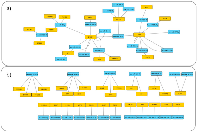
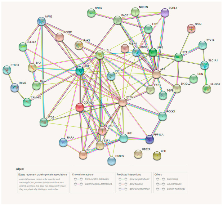
Alzheimer’s Disease (AD) is a progressive neurodegenerative disorder and the most common cause of dementia; however, early diagnosis of the disease is challenging. Research suggests that biomarkers found in blood, such as microRNAs (miRNA), may be promising for AD diagnostics. Experimental data on miRNA–target interactions (MTI) associated with AD are scattered across databases and publications, thus making the identification of promising miRNA biomarkers for AD difficult. In response to this, a list of experimentally validated AD-associated MTIs was obtained from miRTarBase. Cytoscape was used to create a visual MTI network. STRING software was used for protein–protein interaction analysis and mirPath was used for pathway enrichment analysis. Several targets regulated by multiple miRNAs were identified, including: BACE1, APP, NCSTN, SP1, SIRT1, and PTEN. The miRNA with the highest numbers of interactions in the network were: miR-9, miR-16, miR-34a, miR-106a, miR-107, miR-125b, miR-146, and miR-181c. The analysis revealed seven subnetworks, representing disease modules which have a potential for further biomarker development. The obtained MTI network is not yet complete, and additional studies are needed for the comprehensive understanding of the AD-associated miRNA targetome.
Overview
The claims and statistics of this paper haven’t been checked yet.
Run the check — free, no account needed. It reads the open-access text and checks the sources it cites.
Integrity signals
Registry
Cites
Statistics
Figures
Verification
Claims the paper makes
The claims and statistics of this paper haven’t been checked yet.
Run the check — free, no account needed. It reads the open-access text and checks the sources it cites.
Claims other papers make about this one
Each is checked against this paper’s own text: does it say what the citing paper says it does?
The check wasn’t confident either way, so it gives no verdict — never disputed.
In the citing paper
“miR-9-5p [42,43], miR-16-5p [101], miR-29b [47], miR-124 [59] and miR-206-3p [105] were related to the generation of Aβ.”
From MicroRNAs in Extracellular Vesicles of Alzheimer’s Disease (2023) · cites this paper as [42]
Experimental — accuracy not yet measuredIn the citing paper
“miR-9-5p targets several AD-related genes including BACE1, SIRT1, CAMKK2 [42] and TGFBIp [43].”
From MicroRNAs in Extracellular Vesicles of Alzheimer’s Disease (2023) · cites this paper as [42]
Self-check
Percentages, sums and reported differences re-computed from the paper’s own numbers.
The self-check needs the paper’s full text.
Citations
Cites
NIA‐AA Research Framework: Toward a biological definition of Alzheimer's disease
Jack, C. R., Bennett, D. A., Blennow, K., Carrillo, M. C., Dunn, B., Haeberlein, S. B., Holtzman, D. M., Jagust, W., Jessen, F., Karlawish, J., et al. (2018) · Alzheimer's & Dementia · doi:10.1016/j.jalz.2018.02.018 · Cited by 10,537
Lukiw, W. J., Zhao, Y., & Cui, J. G. (2008) · Journal of Biological Chemistry · doi:10.1074/jbc.m805371200 · Cited by 441
Inverse association between cancer and Alzheimer's disease: results from the Framingham Heart Study
Driver, J. A., Beiser, A., Au, R., Kreger, B. E., Splansky, G. L., Kurth, T., Kiel, D. P., Lu, K. P., Seshadri, S., & Wolf, P. A. (2012) · BMJ · doi:10.1136/bmj.e1442 · Cited by 425
Altered Insulin Signaling in Alzheimer’s Disease Brain – Special Emphasis on PI3K-Akt Pathway
Gabbouj, S., Ryhänen, S., Marttinen, M., Wittrahm, R., Takalo, M., Kemppainen, S., Martiskainen, H., Tanila, H., Haapasalo, A., Hiltunen, M., et al. (2019) · Frontiers in Neuroscience · doi:10.3389/fnins.2019.00629 · Cited by 401
Genes associated with Alzheimer's disease: an overview and current status
Giri, M., Lü, Y., & Zhang, M. (2016) · Clinical Interventions in Aging · doi:10.2147/cia.s105769 · Cited by 311
Wang, X., Liu, P., Zhu, H., Xu, Y., Ma, C., Dai, X., Huang, L., Liu, Y., Zhang, L., & Qin, C. (2009) · Brain Research Bulletin · doi:10.1016/j.brainresbull.2009.08.006 · Cited by 271
Wnt signaling deregulation in the aging and Alzheimer’s brain
Palomer, E., Buechler, J., & Salinas, P. C. (2019) · Frontiers in Cellular Neuroscience · doi:10.3389/fncel.2019.00227 · Cited by 237
Kirouac, L., Rajic, A. J., Cribbs, D. H., & Padmanabhan, J. (2017) · eneuro · doi:10.1523/eneuro.0149-16.2017 · Cited by 207
Zhang, T., Han, Y., Wang, J., Hou, D., Deng, H., Deng, Y. L., & Song, Z. (2018) · Frontiers in Aging Neuroscience · doi:10.3389/fnagi.2018.00176 · Cited by 38
MicroRNAs targeting Nicastrin regulate Aβ production and are affected by target site polymorphisms
Delay, C., Dorval, V., Fok, A., Grenier-Boley, B., Lambert, J.-C., Hsiung, G.-Y., & Hébert, S. S. (2014) · Frontiers in Molecular Neuroscience · doi:10.3389/fnmol.2014.00067 · Cited by 28 · cited 3×
“hsa-miR-24-3p NCSTN 23385 25100943 [30]”
Influential
Research works printed but not linked: 130
- Zhao, 2020, microRNA-Based Biomarkers in Alzheimer’s Disease (AD), Front. Neurosci., 14, 585432, 10.3389/fnins.2020.585432 · DOI
- Blennow, 2018, Biomarkers for Alzheimer’s disease: Current status and prospects for the future, J. Intern. Med., 284, 643, 10.1111/joim.12816 · DOI · Paper page
- Vos, 2016, GBD 2015 Disease and Injury Incidence and Prevalence Collaborators, Global, regional, and national incidence, prevalence, and years lived with disability for 310 diseases and injuries, 1990–2015: A systematic analysis for the Global Burden of Disease Study 2015, Lancet, 388, 1545, 10.1016/S0140-6736(16)31678-6 · DOI · Paper page
- Dubois, 2014, Advancing research diagnostic criteria for Alzheimer’s disease: The IWG-2 criteria, Lancet Neurol., 13, 614, 10.1016/S1474-4422(14)70090-0 · DOI · Paper page
- Palmqvist, 2019, Performance of Fully Automated Plasma Assays as Screening Tests for Alzheimer Disease–Related β-Amyloid Status, JAMA Neurol., 76, 1060, 10.1001/jamaneurol.2019.1632 · DOI · Paper page
- Brickman, 2021, Plasma p-tau181, p-tau217, and other blood-based Alzheimer’s disease biomarkers in a multi-ethnic, community study, Alzheimer’s Dement., 17, 1353, 10.1002/alz.12301 · DOI · Paper page
- Ossenkoppele, 2021, Tau PET correlates with different Alzheimer’s disease-related features compared to CSF and plasma p-tau biomarkers, EMBO Mol. Med., 13, e14398, 10.15252/emmm.202114398 · DOI
- Cullen, 2021, Plasma biomarkers of Alzheimer’s disease improve prediction of cognitive decline in cognitively unimpaired elderly populations, Nat. Commun., 12, 3555, 10.1038/s41467-021-23746-0 · DOI
- Karikari, 2020, Blood phosphorylated tau 181 as a biomarker for Alzheimer’s disease: A diagnostic performance and prediction modelling study using data from four prospective cohorts, Lancet Neurol., 19, 422, 10.1016/S1474-4422(20)30071-5 · DOI · Paper page
- Hrovatin, 2018, Classification of miRNA-related sequence variations, Epigenomics, 10, 463, 10.2217/epi-2017-0126 · DOI
- Friedman, 2008, Most mammalian mRNAs are conserved targets of microRNAs, Genome Res., 19, 92, 10.1101/gr.082701.108 · DOI · Paper page
- Hammond, 2015, An overview of microRNAs, Adv. Drug Deliv. Rev., 87, 3, 10.1016/j.addr.2015.05.001 · DOI
- Wang, 2019, MicroRNAs in Alzheimer’s Disease, Front. Genet., 10, 153, 10.3389/fgene.2019.00153 · DOI
- Vergallo, 2021, MiRNA-15b and miRNA-125b are associated with regional Aβ-PET and FDG-PET uptake in cognitively normal individuals with subjective memory complaints, Transl. Psychiatry, 11, 78, 10.1038/s41398-020-01184-8 · DOI
- Ballard, 2011, Alzheimer’s disease, Lancet, 377, 1019, 10.1016/S0140-6736(10)61349-9 · DOI · Paper page
- Ansari, 2019, miR-146a and miR-181a are involved in the progression of mild cognitive impairment to Alzheimer’s disease, Neurobiol. Aging, 82, 102, 10.1016/j.neurobiolaging.2019.06.005 · DOI
- Xie, 2016, Increased Serum miR-206 Level Predicts Conversion from Amnestic Mild Cognitive Impairment to Alzheimer’s Disease: A 5-Year Follow-up Study, J. Alzheimer’s Dis., 55, 509, 10.3233/JAD-160468 · DOI
- Henry, 2018, Inhibition of miR-155 Limits Neuroinflammation and Improves Functional Recovery After Experimental Traumatic Brain Injury in Mice, Neurotherapeutics, 16, 216, 10.1007/s13311-018-0665-9 · DOI
- Lukiw, 2012, NF-ĸB-regulated micro RNAs (miRNAs) in primary human brain cells, Exp. Neurol., 235, 484, 10.1016/j.expneurol.2011.11.022 · DOI · Paper page
- Jaber, 2019, Addressing Alzheimer’s Disease (AD) Neuropathology Using Anti-microRNA (AM) Strategies, Mol. Neurobiol., 56, 8101, 10.1007/s12035-019-1632-0 · DOI
- Abuelezz, 2021, MicroRNAs as Potential Orchestrators of Alzheimer’s Disease-Related Pathologies: Insights on Current Status and Future Possibilities, Front. Aging Neurosci., 13, 743573, 10.3389/fnagi.2021.743573 · DOI
- Chou, 2017, miRTarBase update 2018: A resource for experimentally validated microRNA-target interactions, Nucleic Acids Res., 46, D296, 10.1093/nar/gkx1067 · DOI
- Shannon, 2003, Cytoscape: A Software Environment for Integrated Models of Biomolecular Interaction Networks, Genome Res., 13, 2498, 10.1101/gr.1239303 · DOI · Paper page
- Szklarczyk, 2020, The STRING database in 2021: Customizable protein–protein networks, and functional characterization of user-uploaded gene/measurement sets, Nucleic Acids Res., 49, D605, 10.1093/nar/gkaa1074 · DOI · Paper page
- Vlachos, 2015, DIANA-miRPath v3.0: Deciphering microRNA function with experimental support, Nucleic Acids Res., 43, W460, 10.1093/nar/gkv403 · DOI
- Shioya, 2010, Aberrant microRNA expression in the brains of neurodegenerative diseases: miR-29a decreased in Alzheimer disease brains targets neurone navigator 3, Neuropathol. Appl. Neurobiol., 36, 320, 10.1111/j.1365-2990.2010.01076.x · DOI · Paper page
- Li, 2016, miR-98-5p Acts as a Target for Alzheimer’s Disease by Regulating Aβ Production Through Modulating SNX6 Expression, J. Mol. Neurosci., 60, 413, 10.1007/s12031-016-0815-7 · DOI
- Lei, 2015, Downregulated miR-29c correlates with increased BACE1 expression in sporadic Alzheimer’s disease, Int. J. Clin. Exp. Pathol., 8, 1565
- Zhang, 2013, Regulation of STAT3 by miR-106a is linked to cognitive impairment in ovariectomized mice, Brain Res., 1503, 43, 10.1016/j.brainres.2013.01.052 · DOI
- Villa, 2013, Expression of the Transcription Factor Sp1 and its Regulatory hsa-miR-29b in Peripheral Blood Mononuclear Cells from Patients with Alzheimer’s Disease, J. Alzheimer’s Dis., 35, 487, 10.3233/JAD-122263 · DOI
- Absalon, 2013, MiR-26b, Upregulated in Alzheimer’s Disease, Activates Cell Cycle Entry, Tau-Phosphorylation, and Apoptosis in Postmitotic Neurons, J. Neurosci., 33, 14645, 10.1523/JNEUROSCI.1327-13.2013 · DOI · Paper page
- Benito, 2014, Micro RNA-125b induces tau hyperphosphorylation and cognitive deficits in Alzheimer’s disease, EMBO J., 33, 1667, 10.15252/embj.201387576 · DOI · Paper page
- Yan, H., Xu, T., Zhao, H., Lee, K.-C., Wang, H.-Y., and Zhang, Y. (2013). Isoflurane Increases Neuronal Cell Death Vulnerability by Downregulating miR-214. PLoS ONE, 8. · DOI
- Ghanbari, 2016, Genome-wide identification of microRNA-related variants associated with risk of Alzheimer’s disease, Sci. Rep., 6, 28387, 10.1038/srep28387 · DOI
- Schonrock, 2011, Target Gene Repression Mediated by miRNAs miR-181c and miR-9 Both of Which Are Down-regulated by Amyloid-β, J. Mol. Neurosci., 46, 324, 10.1007/s12031-011-9587-2 · DOI
- Zhang, 2016, MicroRNA-146a represses LRP2 translation and leads to cell apoptosis in Alzheimer’s disease, FEBS Lett., 590, 2190, 10.1002/1873-3468.12229 · DOI
- Hebert, 2009, MicroRNA regulation of Alzheimer’s Amyloid precursor protein expression, Neurobiol. Dis., 33, 422, 10.1016/j.nbd.2008.11.009 · DOI · Paper page
- Song, 2009, MicroRNA-205 inhibits tumor cell migration through down-regulating the expression of the LDL receptor-related protein 1, Biochem. Biophys. Res. Commun., 388, 400, 10.1016/j.bbrc.2009.08.020 · DOI
- Li, 2016, miR-302 Attenuates Amyloid-β-Induced Neurotoxicity through Activation of Akt Signaling, J. Alzheimer’s Dis., 50, 1083, 10.3233/JAD-150741 · DOI
- Wang, 2010, miR-107 Regulates Granulin/Progranulin with Implications for Traumatic Brain Injury and Neurodegenerative Disease, Am. J. Pathol., 177, 334, 10.2353/ajpath.2010.091202 · DOI
- Jayadev, 2013, Presenilin 2 influences miR146 level and activity in microglia, J. Neurochem., 127, 592, 10.1111/jnc.12400 · DOI · Paper page
- Li, 2016, MicroRNA-613 regulates the expression of brain-derived neurotrophic factor in Alzheimer’s disease, Biosci. Trends, 10, 372, 10.5582/bst.2016.01127 · DOI
- Li, 2016, Neuroprotective Effect of Osthole on Neuron Synapses in an Alzheimer’s Disease Cell Model via Upregulation of MicroRNA-9, J. Mol. Neurosci., 60, 71, 10.1007/s12031-016-0793-9 · DOI · Paper page
- Parsi, 2015, Preclinical Evaluation of miR-15/107 Family Members as Multifactorial Drug Targets for Alzheimer’s Disease, Mol. Ther. Nucleic Acids, 4, e256, 10.1038/mtna.2015.33 · DOI · Paper page
- Jovicic, A., Jolissaint, J.F.Z., Moser, R., Santos, M.D.F.S.S., and Luthi-Carter, R. (2013). MicroRNA-22 (miR-22) Overexpression Is Neuroprotective via General Anti-Apoptotic Effects and May also Target Specific Huntington’s Disease-Related Mechanisms. PLoS ONE, 8. · DOI
- Wang, 2016, MicroRNA-146a suppresses ROCK1 allowing hyperphosphorylation of tau in Alzheimer’s disease, Sci. Rep., 6, 26697, 10.1038/srep26697 · DOI
- Pogue, 2010, Micro RNA-125b (miRNA-125b) function in astrogliosis and glial cell proliferation, Neurosci. Lett., 476, 18, 10.1016/j.neulet.2010.03.054 · DOI
- Zhang, 2016, MiR-299-5p regulates apoptosis through autophagy in neurons and ameliorates cognitive capacity in APPswe/PS1dE9 mice, Sci. Rep., 6, 24566, 10.1038/srep24566 · DOI
- Lau, 2013, Alteration of the micro RNA network during the progression of Alzheimer’s disease, EMBO Mol. Med., 5, 1613, 10.1002/emmm.201201974 · DOI · Paper page
- Agostini, 2011, Neuronal differentiation by TAp73 is mediated by microRNA-34a regulation of synaptic protein targets, Proc. Natl. Acad. Sci. USA, 108, 21093, 10.1073/pnas.1112061109 · DOI
- Liu, 2016, MicroRNA-26b is upregulated in a double transgenic mouse model of Alzheimer’s disease and promotes the expression of amyloid-β by targeting insulin-like growth factor 1, Mol. Med. Rep., 13, 2809, 10.3892/mmr.2016.4860 · DOI
- Ubhi, 2013, Widespread microRNA dysregulation in multiple system atrophy-disease-related alteration in miR-96, Eur. J. Neurosci., 39, 1026, 10.1111/ejn.12444 · DOI
- Wang, 2008, The Expression of MicroRNA miR-107 Decreases Early in Alzheimer’s Disease and May Accelerate Disease Progression through Regulation of β-Site Amyloid Precursor Protein-Cleaving Enzyme 1, J. Neurosci., 28, 1213, 10.1523/JNEUROSCI.5065-07.2008 · DOI · Paper page
- Liu, 2016, miR-106b inhibits tau phosphorylation at Tyr18 by targeting Fyn in a model of Alzheimer’s disease, Biochem. Biophys. Res. Commun., 478, 852, 10.1016/j.bbrc.2016.08.037 · DOI
- Vilardo, 2010, MicroRNA-101 Regulates Amyloid Precursor Protein Expression in Hippocampal Neurons, J. Biol. Chem., 285, 18344, 10.1074/jbc.M110.112664 · DOI
- Papadopoulou, 2008, Loss of microRNA cluster miR-29a/b-1 in sporadic Alzheimer’s disease correlates with increased BACE1/β-secretase expression, Proc. Natl. Acad. Sci. USA, 105, 6415, 10.1073/pnas.0710263105 · DOI · Paper page
- Zhao, Y., Alexandrov, P.N., Jaber, V., and Lukiw, W.J. (2016). Deficiency in the Ubiquitin Conjugating Enzyme UBE2A in Alzheimer’s Disease (AD) is Linked to Deficits in a Natural Circular miRNA-7 Sponge (circRNA; ciRS-7). Genes, 7. · DOI
- Pereira, 2016, Recombinant pre-miR-29b for Alzheimer’s disease therapeutics, Sci. Rep., 6, 19946, 10.1038/srep19946 · DOI
- Patel, 2008, MicroRNAs can regulate human APP levels, Mol. Neurodegener., 3, 10, 10.1186/1750-1326-3-10 · DOI
- Wang, 2015, MicroRNA-138 promotes tau phosphorylation by targeting retinoic acid receptor alpha, FEBS Lett., 589, 726, 10.1016/j.febslet.2015.02.001 · DOI
- Liu, 2016, MicroRNA-107 prevents amyloid-beta induced blood-brain barrier disruption and endothelial cell dysfunction by targeting Endophilin-1, Exp. Cell Res., 343, 248, 10.1016/j.yexcr.2016.03.026 · DOI
- Zheng, 2016, MicroRNA-511 Binds to FKBP5 mRNA, Which Encodes a Chaperone Protein, and Regulates Neuronal Differentiation, J. Biol. Chem., 291, 17897, 10.1074/jbc.M116.727941 · DOI
- Long, 2011, MicroRNA-101 downregulates Alzheimer’s amyloid-β precursor protein levels in human cell cultures and is differentially expressed, Biochem. Biophys. Res. Commun., 404, 889, 10.1016/j.bbrc.2010.12.053 · DOI
- Wu, 2016, The Protective Role of microRNA-200c in Alzheimer’s Disease Pathologies Is Induced by Beta Amyloid-Triggered Endoplasmic Reticulum Stress, Front. Mol. Neurosci., 9, 140, 10.3389/fnmol.2016.00140 · DOI
- Long, 2014, MicroRNA-339-5p Down-regulates Protein Expression of β-Site Amyloid Precursor Protein-Cleaving Enzyme 1 (BACE1) in Human Primary Brain Cultures and Is Reduced in Brain Tissue Specimens of Alzheimer Disease Subjects, J. Biol. Chem., 289, 5184, 10.1074/jbc.M113.518241 · DOI · Paper page
- Zong, 2011, miR-29c regulates BACE1 protein expression, Brain Res., 1395, 108, 10.1016/j.brainres.2011.04.035 · DOI · Paper page
- Zhang, 2016, MiR-195 dependent roles of mitofusin2 in the mitochondrial dysfunction of hippocampal neurons in SAMP8 mice, Brain Res., 1652, 135, 10.1016/j.brainres.2016.09.047 · DOI
- Stutzmann, 2005, Calcium Dysregulation, IP3 Signaling, and Alzheimer’s Disease, Neuroscientist, 11, 110, 10.1177/1073858404270899 · DOI
- Chang, 2012, Role of p53 in Neurodegenerative Diseases, Neurodegener. Dis., 9, 68, 10.1159/000329999 · DOI
- Zhang, 2014, Towards Understanding the Roles of Heparan Sulfate Proteoglycans in Alzheimer’s Disease, BioMed Res. Int., 2014, 516028
- Lehrer, 2018, Glioma and Alzheimer’s Disease, J. Alzheimer’s Dis. Rep., 2, 213, 10.3233/ADR-180084 · DOI
- Penzes, 2011, Impaired regulation of synaptic actin cytoskeleton in Alzheimer’s disease, Brain Res. Rev., 67, 184, 10.1016/j.brainresrev.2011.01.003 · DOI
- Vasco, 2018, The Phosphoinositide Signal Transduction Pathway in the Pathogenesis of Alzheimer’s Disease, Curr. Alzheimer Res., 15, 355, 10.2174/1567205014666170829100230 · DOI
- Greene, 2016, Tight junction modulation of the blood brain barrier: CNS delivery of small molecules, Tissue Barriers, 4, e1138017, 10.1080/21688370.2015.1138017 · DOI
- Berezin, 2014, Targeting of ECM molecules and their metabolizing enzymes and receptors for the treatment of CNS diseases, Prog. Brain Res., 214, 353, 10.1016/B978-0-444-63486-3.00015-3 · DOI
- Caltagarone, 2007, Focal adhesions regulate Aβ signaling and cell death in Alzheimer’s disease, Biochim. Biophys. Acta (BBA)—Mol. Basis Dis., 1772, 438, 10.1016/j.bbadis.2006.11.007 · DOI · Paper page
- Jayaraman, 2014, Alzheimer’s Disease and Type 2 Diabetes: Multiple Mechanisms Contribute to Interactions, Curr. Diabetes Rep., 14, 476, 10.1007/s11892-014-0476-2 · DOI
- Maiese, 2018, Forkhead Transcription Factors: Formulating a FOXO Target for Cognitive Loss, Curr. Neurovasc. Res., 14, 415, 10.2174/1567202614666171116102911 · DOI
- Frank, 1999, Increased cerebrospinal fluid cAMP levels in Alzheimer’s disease, Brain Res., 846, 265, 10.1016/S0006-8993(99)01981-2 · DOI
- Haase, 2013, Amyloid- Peptides Activate 1-Adrenergic Cardiovascular Receptors, Hypertension, 62, 966, 10.1161/HYPERTENSIONAHA.113.01348 · DOI
- Sevush, 1998, Platelet Activation in Alzheimer Disease, Arch. Neurol., 55, 530, 10.1001/archneur.55.4.530 · DOI
- Pereira, 2014, The role of endoplasmic reticulum in amyloid precursor protein processing and trafficking: Implications for Alzheimer’s disease, Biochim. Biophys. Acta (BBA)—Mol. Basis Dis., 1842, 1444, 10.1016/j.bbadis.2014.05.003 · DOI
- Mielke, 2010, Alterations of the Sphingolipid Pathway in Alzheimer’s Disease: New Biomarkers and Treatment Targets?, NeuroMol. Med., 12, 331, 10.1007/s12017-010-8121-y · DOI
- Akasaka-Manya, K., and Manya, H. (2020). The Role of APP O-Glycosylation in Alzheimer’s Disease. Biomolecules, 10. · DOI
- Zhu, 2002, The Role of Mitogen-Activated Protein Kinase Pathways in Alzheimer’s Disease, Neurosignals, 11, 270, 10.1159/000067426 · DOI
- McKay, 2019, Peri-Infarct Upregulation of the Oxytocin Receptor in Vascular Dementia, J. Neuropathol. Exp. Neurol., 78, 436, 10.1093/jnen/nlz023 · DOI
- Malik, 2015, Genetics of CD33 in Alzheimer’s disease and acute myeloid leukemia, Hum. Mol. Genet., 24, 3557, 10.1093/hmg/ddv092 · DOI
- Oddo, 2012, The role of mTOR signaling in Alzheimer disease, Front. Biosci., 4, 941, 10.2741/s310 · DOI
- Angeli, 2020, Altered Expression of Glial Gap Junction Proteins Cx43, Cx30, and Cx47 in the 5XFAD Model of Alzheimer’s Disease, Front. Neurosci., 14, 582934, 10.3389/fnins.2020.582934 · DOI
- Woo, 2011, Expression of ErbB4 in the neurons of Alzheimer’s disease brain and APP/PS1 mice, a model of Alzheimer’s disease, Anat. Cell Biol., 44, 116, 10.5115/acb.2011.44.2.116 · DOI
- Curtis, 2020, Mini-review: Role of the PI3K/Akt pathway and tyrosine phosphatases in Alzheimer’s disease susceptibility, Ann. Hum. Genet., 85, 1, 10.1111/ahg.12410 · DOI
- Monsonego, 2013, CD4 T cells in immunity and immunotherapy of Alzheimer’s disease, Immunology, 139, 438, 10.1111/imm.12103 · DOI
- Watson, 2011, The Neurotrophins and Their Role in Alzheimers Disease, Curr. Neuropharmacol., 9, 559, 10.2174/157015911798376190 · DOI
- Bernhardi, 2015, Role of TGFβ signaling in the pathogenesis of Alzheimer’s disease, Front. Cell. Neurosci., 9, 426
- Uddin, 2020, Estrogen Signaling in Alzheimer’s Disease: Molecular Insights and Therapeutic Targets for Alzheimer’s Dementia, Mol. Neurobiol., 57, 2654, 10.1007/s12035-020-01911-8 · DOI · Paper page
- Choi, 2021, Association between dementia and hepatitis B and C virus infection, Medicine, 100, e26476, 10.1097/MD.0000000000026476 · DOI
- Zhang, 2021, Roles and Mechanisms of Axon-Guidance Molecules in Alzheimer’s Disease, Mol. Neurobiol., 58, 3290, 10.1007/s12035-021-02311-2 · DOI
- Yamazaki, 2019, Selective loss of cortical endothelial tight junction proteins during Alzheimer’s disease progression, Brain, 142, 1077, 10.1093/brain/awz011 · DOI · Paper page
- Hong, 2020, TRP Channels as Emerging Therapeutic Targets for Neurodegenerative Diseases, Front. Physiol., 11, 238, 10.3389/fphys.2020.00238 · DOI
- Huh, 2016, The reemergence of long-term potentiation in aged Alzheimer’s disease mouse model, Sci. Rep., 6, 29152, 10.1038/srep29152 · DOI
- Nixon, 2005, Endosome function and dysfunction in Alzheimer’s disease and other neurodegenerative diseases, Neurobiol. Aging, 26, 373, 10.1016/j.neurobiolaging.2004.09.018 · DOI
- Wang, 2019, Therapeutic Potential of AMP-Activated Protein Kinase in Alzheimer’s Disease, J. Alzheimer’s Dis., 68, 33, 10.3233/JAD-181043 · DOI
- Sanders, 2020, Phosphodiesterase Inhibitors for Alzheimer’s Disease: A Systematic Review of Clinical Trials and Epidemiology with a Mechanistic Rationale, J. Alzheimer’s Dis. Rep., 4, 185, 10.3233/ADR-200191 · DOI
- Irwin, 2020, A Positive Feedback Loop of Hippo- and c-Jun-Amino-Terminal Kinase Signaling Pathways Regulates Amyloid-Beta-Mediated Neurodegeneration, Front. Cell Dev. Biol., 8, 117, 10.3389/fcell.2020.00117 · DOI
- Park, 2019, Dysregulated Fc gamma receptor–mediated phagocytosis pathway in Alzheimer’s disease: Network-based gene expression analysis, Neurobiol. Aging, 88, 24, 10.1016/j.neurobiolaging.2019.12.001 · DOI
- Maillet, 2003, Crosstalk between Rap1 and Rac regulates secretion of sAPPα, Nature, 5, 633
- Vogrinc, D., Goričar, K., Kunej, T., and Dolžan, V. (2021). Systematic Search for Novel Circulating Biomarkers Associated with Extracellular Vesicles in Alzheimer’s Disease: Combining Literature Screening and Database Mining Approaches. J. Pers. Med., 11. · DOI
- Das, 2017, Role of BACE1 in Alzheimer’s synaptic function, Transl. Neurodegener., 6, 23, 10.1186/s40035-017-0093-5 · DOI
- Wong, 2011, Amyloid Precursor Protein Processing and Alzheimer’s Disease, Annu. Rev. Neurosci., 34, 185, 10.1146/annurev-neuro-061010-113613 · DOI · Paper page
- Kaether, 2006, Assembly, Trafficking and Function of γ-Secretase, Neurodegener. Dis., 3, 275, 10.1159/000095267 · DOI
- Yoon, 2014, Differential roles of Sirt1 in HIF-1α and HIF-2α mediated hypoxic responses, Biochem. Biophys. Res. Commun., 444, 36, 10.1016/j.bbrc.2014.01.001 · DOI
- Kleszka, 2020, Hypoxia-inducible factor-2α is crucial for proper brain development, Sci. Rep., 10, 19146, 10.1038/s41598-020-75838-4 · DOI
- Elalla, 2013, The role of sirtuins in Alzheimer’s disease, Front. Aging Neurosci., 5, 16
- Citron, 2008, Transcription factor Sp1 dysregulation in Alzheimer’s disease, J. Neurosci. Res., 86, 2499, 10.1002/jnr.21695 · DOI · Paper page
- Knafo, 2016, PTEN recruitment controls synaptic and cognitive function in Alzheimer’s models, Nat. Neurosci., 19, 443, 10.1038/nn.4225 · DOI
- Frere, 2016, Targeting PTEN interactions for Alzheimer’s disease, Nat. Neurosci., 19, 416, 10.1038/nn.4248 · DOI
- Chen, 2021, Revisiting the Relationship between Alzheimer’s Disease and Cancer with a circRNA Perspective, Front. Cell Dev. Biol., 9, 647197, 10.3389/fcell.2021.647197 · DOI
- Ou, 2013, Does Alzheimer’s Disease Protect against Cancers? A Nationwide Population-Based Study, Neuroepidemiology, 40, 42, 10.1159/000341411 · DOI
- Liu, 2013, Transcriptional signaling pathways inversely regulated in Alzheimer’s disease and glioblastoma multiform, Sci. Rep., 3, 3467, 10.1038/srep03467 · DOI
- Musicco, 2013, Inverse occurrence of cancer and Alzheimer disease: A population-based incidence study, Neurology, 81, 322, 10.1212/WNL.0b013e31829c5ec1 · DOI · Paper page
- Rinaldi, 2021, The biological pathways of Alzheimer disease: A review, AIMS Neurosci., 8, 86, 10.3934/Neuroscience.2021005 · DOI
- Caruana, 2015, Longitudinal studies, J. Thorac. Dis., 7, E537
- Wang, 2018, The microRNA-1908 up-regulation in the peripheral blood cells impairs amyloid clearance by targeting ApoE, Int. J. Geriatr. Psychiatry, 33, 980, 10.1002/gps.4881 · DOI
- Deng, 2014, The miR-193a-3p regulated PSEN1 gene suppresses the multi-chemoresistance of bladder cancer, Biochim. Biophys. Acta (BBA)—Mol. Basis Dis., 1852, 520, 10.1016/j.bbadis.2014.12.014 · DOI
- Robinson, 2017, Multiplexing Biomarker Methods, Proteomics and Considerations for Alzheimer’s Disease, Proteom. Methods Neuropsychiatry Res., 974, 21, 10.1007/978-3-319-52479-5_2 · DOI
- Maoz, 2017, Alzheimer’s Disease and ncRNAs, Neuroepigenom. Aging Dis., 978, 337, 10.1007/978-3-319-53889-1_18 · DOI
- Lugli, G., Cohen, A.M., Bennett, D.A., Shah, R., Fields, C., Hernandez, A.G., and Smalheiser, N.R. (2015). Plasma Exosomal miRNAs in Persons with and without Alzheimer Disease: Altered Expression and Prospects for Biomarkers. PLoS ONE, 10. · DOI · Paper page
- Leidinger, 2013, A blood based 12-miRNA signature of Alzheimer disease patients, Genome Biol., 14, R78, 10.1186/gb-2013-14-7-r78 · DOI
- Zetterberg, 2019, Blood-based molecular biomarkers for Alzheimer’s disease, Mol. Brain, 12, 26, 10.1186/s13041-019-0448-1 · DOI
- Zhao, 2013, Regulation of TREM2 expression by an NF-ĸB-sensitive miRNA-34a, NeuroReport, 24, 318, 10.1097/WNR.0b013e32835fb6b0 · DOI
Cited by
Sorted by citations
MicroRNAs in Extracellular Vesicles of Alzheimer’s Disease
Li, W., & Zheng, Y. (2023) · Cells · doi:10.3390/cells12101378 · Cited by 43
Metadata
- Authors
- Turk, A., Kunej, T., & Peterlin, B.
- Affiliations
- Department of Animal Science, Biotechnical Faculty, University of Ljubljana, 1230 Domžale, Slovenia
- Clinical Institute of Genomic Medicine, University Medical Centre Ljubljana, 1000 Ljubljana, Slovenia
- ORCID
- Kunej, Tanja
- Venue
- Journal of Personalized Medicine
- Publisher
- MDPI AG
- Published
- 2021-12-02
- Volume
- 11
- Issue
- 12
- Pages
- 1275
- ISSN
- 2075-4426
- PMC
- PMC8708198
- Cited by
- 20
- References
- 10
- Language
- en
- Funders
- Slovenian Research Agency (P4-0220)
About these checks
Registry, retraction, Cites and statistics results are computed, not judged, with one exception: a printed reference that no index lists is read once by a model before it counts as a citation error. Claims are extracted by a model and kept only when their quote appears verbatim in the text; each cited source is then checked, with a model’s help, for whether it says what the claim relies on. Those verdicts can be wrong — read the claim’s quote and the cited source before relying on one.
Statistics and arithmetic are recomputed, not judged.
Confidence bands are shown only for fields calibrated on our annotated test set. None is calibrated yet, so no band is shown.
What counts as a claim ›How we measure confidence ›How we check this ›
Are you an author, or do you see a problem with this page? Request a review
