

The DOI matches its Crossref record; 40 of the 44 references it cites were found in a registry or an index; 39 of the 48 sources its claims cite were checked for support.
Optimization of non-coding regions for a non-modified mRNA COVID-19 vaccine
Open accessAbstract
The CVnCoV (CureVac) mRNA vaccine for severe acute respiratory syndrome coronavirus-2 (SARS-CoV-2) was recently evaluated in a phase 2b/3 efficacy trial in humans 1. CV2CoV is a second-generation mRNA vaccine containing non-modified nucleosides but with optimized non-coding regions and enhanced antigen expression. Here we report the results of a head-to-head comparison of the immunogenicity and protective efficacy of CVnCoV and CV2CoV in non-human primates. We immunized 18 cynomolgus macaques with two doses of 12 μg lipid nanoparticle-formulated CVnCoV or CV2CoV or with sham (n = 6 per group). Compared with CVnCoV, CV2CoV induced substantially higher titres of binding and neutralizing antibodies, memory B cell responses and T cell responses as well as more potent neutralizing antibody responses against SARS-CoV-2 variants, including the Delta variant. Moreover, CV2CoV was found to be comparably immunogenic to the BNT162b2 (Pfizer) vaccine in macaques. Although CVnCoV provided partial protection against SARS-CoV-2 challenge, CV2CoV afforded more robust protection with markedly lower viral loads in the upper and lower respiratory tracts. Binding and neutralizing antibody titres were correlated with protective efficacy. These data demonstrate that optimization of non-coding regions can greatly improve the immunogenicity and protective efficacy of a non-modified mRNA SARS-CoV-2 vaccine in non-human primates.
Overview
AI snapshot
Written from the paper’s full text.
TL;DR
In a head-to-head non-human primate study, a second-generation non-modified mRNA SARS-CoV-2 vaccine (CV2CoV) with optimized non-coding regions induced substantially higher antibody, memory B cell, and T cell responses and more robust protection against SARS-CoV-2 challenge than CVnCoV. CV2CoV was comparably immunogenic to BNT162b2 in macaques, and binding/neutralizing antibody titres correlated with protection.
Key findings
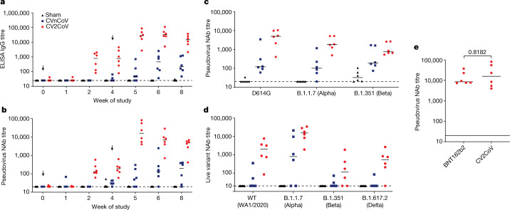
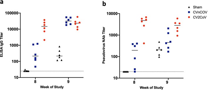
- CV2CoV induced higher binding and neutralizing antibodies than CVnCoV. At week 8, median RBD-binding titres were 14,827 (range 2,133–37,079) vs 214 (range 47–1,238) (P = 0.0043), and median pseudovirus neutralizing titres were 4,752 (range 414–6,793) vs 196 (range 20–405) (P = 0.0022).
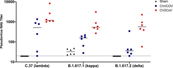
- CV2CoV elicited higher pseudovirus neutralizing titres against variants including D614G, Alpha, Beta, Lambda, Kappa, and Delta (e.g., Delta median 568 vs 36; Beta 755 vs 189; Alpha 1,813 vs 101; P values as reported). At week 5, CV2CoV neutralizing antibody responses were comparable to BNT162b2.
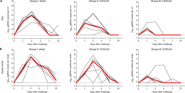
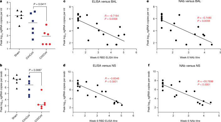
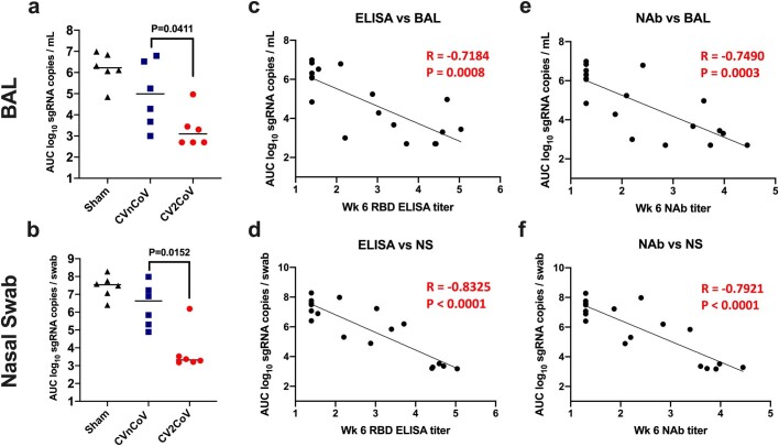
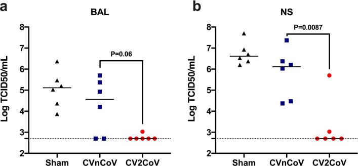
- CV2CoV gave lower peak viral loads after challenge: median BAL sgRNA 2.90 vs CVnCoV 4.92 vs sham 6.02 log10 copies per ml; nasal swab 3.17 vs 6.42 vs 7.35 log10 copies per swab. CV2CoV was lower than CVnCoV in BAL (P = 0.0411) and nasal swab (P = 0.0087); no detectable infectious virus by TCID50 in five of six CV2CoV animals on day 2.
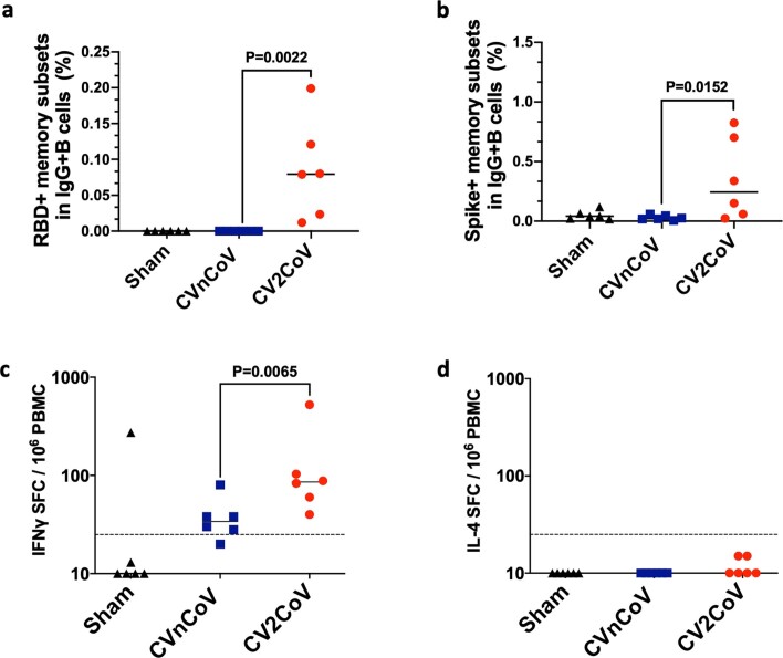
- Binding and neutralizing antibody titres inversely correlated with peak viral loads (BAL: P = 0.0008, R = −0.7148 and P = 0.0015, R = −0.6912; nasal: P < 0.0001, R = −0.8346 and P < 0.0001, R = −0.8766). CV2CoV also increased memory B cells (P = 0.022 and P = 0.0152) and IFNγ T cell responses (P = 0.0065); IL-4 was not detectable.
Methods
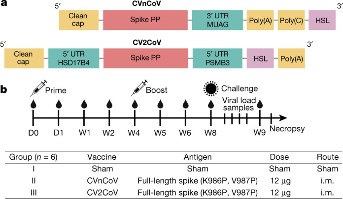
- Head-to-head non-human primate study: 18 cynomolgus macaques (both sexes, 3–20 years) randomized to 12 µg CVnCoV (n = 6), 12 µg CV2CoV (n = 6), or sham (n = 6); intramuscular prime on day 0, boost at week 4, challenge at week 8 with 1.0 × 10^5 TCID50 SARS-CoV-2 USA-WA1/2020 by intranasal and intratracheal routes; necropsy 10 days after challenge.
- Immunological and virological assays included RBD ELISA, pseudovirus and live-virus neutralization, B cell flow cytometry, IFNγ and IL-4 ELISPOT, sgRNA RT–PCR, TCID50, and histopathology; assays were performed with blinding.
- Statistics: two-tailed non-parametric Mann–Whitney U test for group comparisons; two-sided Spearman rank-correlation for correlates; P < 0.05 considered significant; GraphPad Prism v9.0.
Limitations
- Stated: the study was not specifically designed as a safety study; no adverse effects or unexpected/enhanced pathology were observed. Future studies are required to directly assess protective efficacy of CV2CoV against SARS-CoV-2 variants of concern in non-human primates. B cell germinal centre responses and durability of protective efficacy following CV2CoV vaccination remain to be determined.
- Stated: the challenge virus was SARS-CoV-2 WA1/2020, not a variant of concern.
- Evident from text: small group sizes (n = 6 per group), non-human primate model rather than humans, and the BNT162b2 comparison used leftover clinical product and only pseudovirus neutralizing titres at week 5; no human efficacy data for CV2CoV are reported.
Model-written summary. Check it against the paper before relying on it.
Integrity signals
Registry
Cites
Statistics
Figures
Verification
Claims the paper makes
30 Own result · 12 Partly supported · 6 Own method · 5 Own interpretation · 4 Supported · 3 No full text of the sources · 2 Sources not found · 1 Couldn’t confirm · 1 No basis given · accuracy not yet measured
The cited sources support only part of this claim — read each source’s verdict below.
In the paper
“Previous studies with rodents and non-human primates have demonstrated protection by CVnCoV2,23,24.”
§ Optimization of non-coding regions for a non-modified mRNA COVID-19 vaccine > Discussion
What the sources say
- Supportedcites [2] Hoffmann, D. et al. CVnCoV and CV2CoV protect human ACE2 transgenic mice from ancestral B BavPat1 and emerging B.1.351 SARS-CoV-2. Nat. Commun. 12, 4048 (2021). source
The Results report that CVnCoV protected human ACE2 transgenic mice from lethal SARS-CoV-2 challenge. The References also identify a study reporting CVnCoV protection in non-human primates (reference 24). (LLM, conf=0.91)
- Partly supportedcites [23] Rauch, S. et al. mRNA-based SARS-CoV-2 vaccine candidate CVnCoV induces high levels of virus-neutralising antibodies and mediates protection in rodents. NPJ Vaccines 6, 57 (2021). sourceExperimental — accuracy not yet measured
The Discussion reports that CVnCoV protected hamsters against SARS-CoV-2 challenge, supporting the rodent portion of the claim. It does not report protection by CVnCoV in non-human primates; its comparison with primate challenge models concerns other vaccine studies. No page numbers are available in our copy. (LLM, conf=0.88)
- Supportedcites [24] Rauch, S. et al. mRNA vaccine CVnCoV protects non-human primates from SARS-CoV-2 challenge infection. Preprint at https://doi.org/10.1101/2021.03.22.435960 (2020). source
The Results report that CVnCoV protected transgenic mice against lethal SARS-CoV-2 challenge. The References section identifies separate CVnCoV studies reporting protection in rodents (ref. 23) and non-human primates (ref. 24), supporting the sentence’s description of previous studies. No page numbers are available in our copy. (LLM, conf=0.90)
boosteddirect- Asserted: passes — The authors put it forward as true.
- Checkable: passes — It can be checked against evidence.
- Load-bearing: passes — The paper’s conclusions depend on it.
- Based: passes — The paper gives a reason why it holds.
- Supported
Every source the paper cites for this claim was checked and supports it.
In the paper
“CV2CoV has previously been shown to lead to higher antigen expression than CVnCoV in cell culture3.”
§ Optimization of non-coding regions for a non-modified mRNA COVID-19 vaccine > Discussion
What the sources say
- Supportedcites [3] Roth, N. et al. CV2CoV, an enhanced mRNA-based SARS-CoV-2 vaccine candidate, supports higher protein expression and improved immunogenicity in rats. Preprint at https://doi.org/10.1101/2021.05.13.443734 (2021). source
Section 3.1 (In vitro protein expression) reports higher SARS-CoV-2 spike protein expression in HeLa cells transfected with CV2CoV mRNA than in cells transfected with the same amount of CVnCoV mRNA. This directly supports the claim about higher antigen expression in cell culture. (LLM, conf=0.95)
plaindirect- Asserted: passes — The authors put it forward as true.
- Checkable: passes — It can be checked against evidence.
- Load-bearing: unclear — It is unclear whether the paper’s conclusions depend on it.
- Based: passes — The paper gives a reason why it holds.
- Supported
The cited sources support only part of this claim — read each source’s verdict below.
In the paper
“However, protection in macaques was primarily observed in the lower respiratory tract23,24.”
§ Optimization of non-coding regions for a non-modified mRNA COVID-19 vaccine > Discussion
What the sources say
- Supportedcites [23] Rauch, S. et al. mRNA-based SARS-CoV-2 vaccine candidate CVnCoV induces high levels of virus-neutralising antibodies and mediates protection in rodents. NPJ Vaccines 6, 57 (2021). source
The Discussion (page not provided) says that prior SARS-CoV-2 challenge models in non-human primates showed better protection in the lower than in the upper respiratory tract. This supports the claim about where protection in macaques was primarily observed; the paper reports it as a previous observation, not as a result of its own rodent experiments. (LLM, conf=0.90)
- Partly supportedcites [24] Rauch, S. et al. mRNA vaccine CVnCoV protects non-human primates from SARS-CoV-2 challenge infection. Preprint at https://doi.org/10.1101/2021.03.22.435960 (2020). sourceExperimental — accuracy not yet measured
The Results (page unavailable) report near-complete protection in the lower respiratory tract but residual viral replication in the upper respiratory tract of vaccinated mice. The paper mentions protection in non-human primates in a previous study, but does not say that protection in macaques was primarily in the lower respiratory tract. (LLM, conf=0.85)
plain- Asserted: passes — The authors put it forward as true.
- Checkable: passes — It can be checked against evidence.
- Load-bearing: unclear — It is unclear whether the paper’s conclusions depend on it.
- Based: passes — The paper gives a reason why it holds.
- Supported
Every source the paper cites for this claim was checked and supports it.
In the paper
“The neutralizing antibody titres induced by CV2CoV in this study also appear to be similar to those reported for other mRNA vaccines in macaques27,28.”
§ Optimization of non-coding regions for a non-modified mRNA COVID-19 vaccine > Discussion
What the sources say
- Supportedcites [27] Corbett, K. S. et al. Evaluation of the mRNA-1273 vaccine against SARS-CoV-2 in nonhuman primates. N. Engl. J. Med. 383, 1544–1555 (2020). source
The Methods (Rhesus Macaque Model; page unavailable) identify the animals as rhesus macaques vaccinated with mRNA-1273. The Results (Antibody Responses after mRNA-1273 Vaccination; page unavailable) report their neutralizing antibody titres. The citation thus supplies an example of titres reported for another mRNA vaccine in macaques; the comparison with CV2CoV is the citing authors’ own assessment. (LLM, conf=0.92)
- Supportedcites [28] Vogel, A. B. et al. BNT162b vaccines protect rhesus macaques from SARS-CoV-2. Nature 592, 283–289 (2021). source
The 'BNT162b-elicited immunogenicity in macaques' section reports neutralizing antibody titres induced by the mRNA vaccine candidates BNT162b1 and BNT162b2 in macaques. The similarity to CV2CoV is the citing authors’ comparison; this source supplies the reported titres for another mRNA vaccine. (LLM, conf=0.92)
hedged- Asserted: passes — The authors put it forward as true.
- Checkable: passes — It can be checked against evidence.
- Load-bearing: unclear — It is unclear whether the paper’s conclusions depend on it.
- Based: passes — The paper gives a reason why it holds.
- Supported
The cited sources support only part of this claim — read each source’s verdict below.
In the paper
“Consistent with prior observations from our laboratory and others15,16,22, these findings suggest that binding and neutralizing antibody titres are important correlates of protection for these SARS-CoV-2 vaccines in non-human primates.”
§ Optimization of non-coding regions for a non-modified mRNA COVID-19 vaccine > Protective efficacy
What the sources say
- Supportedcites [15] Yu, J. et al. DNA vaccine protection against SARS-CoV-2 in rhesus macaques. Science 369, 806–811 (2020). source
The Immune correlates of vaccine-induced protection section reports that both neutralizing antibody titres and ELISA binding-antibody titres inversely correlated with viral levels after challenge in vaccinated rhesus macaques. The binding-antibody correlation was reported for bronchoalveolar lavage samples; the paper identifies neutralizing antibody titres as an immune correlate of protection. (LLM, conf=0.90)
- Supportedcites [16] Mercado, N. B. et al. Single-shot Ad26 vaccine protects against SARS-CoV-2 in rhesus macaques. Nature 586, 583–588 (2020). source
The Immune Correlates of Protection section reports that binding ELISA titres and neutralizing antibody titres inversely correlated with viral RNA after challenge in vaccinated rhesus macaques. It concludes that serum antibody titres may be useful correlates of protection for SARS-CoV-2 vaccines, matching the citing sentence’s qualified claim. (LLM, conf=0.92)
- Partly supportedcites [22] McMahan, K. et al. Correlates of protection against SARS-CoV-2 in rhesus macaques. Nature 590, 630–634 (2021). sourceExperimental — accuracy not yet measured
The IgG Adoptive Transfer Protection Study reports protection thresholds for neutralizing titres and binding-antibody ELISA titres in rhesus macaques. The Discussion also says neutralizing titres correlated with protection in earlier DNA and Ad26 vaccine studies. This paper’s direct binding-titre evidence comes from transfer of convalescent IgG, however, rather than from vaccinated animals, so it does not directly establish both as correlates for the vaccines. (LLM, conf=0.83)
hedgedcorrelational- Asserted: passes — The authors put it forward as true.
- Checkable: passes — It can be checked against evidence.
- Load-bearing: passes — The paper’s conclusions depend on it.
- Based: passes — The paper gives a reason why it holds.
- Supported
The cited sources support only part of this claim — read each source’s verdict below.
In the paper
“Although the correlates of protection in this study were binding and neutralizing antibody titres34,35, it is likely that CD8+ T cells contribute to viral clearance in tissues36,37.”
§ Optimization of non-coding regions for a non-modified mRNA COVID-19 vaccine > Discussion
What the sources say
- Partly supportedcites [36] Lafon, E. et al. Potent SARS-CoV-2-specific T cell immunity and low anaphylatoxin levels correlate with mild disease progression in COVID-19 patients. Front. Immunol. 12, 684014 (2021). sourceExperimental — accuracy not yet measured
The Introduction (page unavailable) says CD8+ T cells play a critical role in viral clearance and associates early CD8+ T-cell immunity with clearance. The study measured cells from blood, however, and the cited passages do not establish clearance specifically in tissues. (LLM, conf=0.82)
- Supportedcites [37] Schmidt, M. E. & Varga, S. M. The CD8 T cell response to respiratory virus infections. Front. Immunol. 9, 678 (2018). source
The “Mechanisms of CD8 T Cell-Mediated Viral Clearance” section reports that CD8 T cells can mediate viral clearance in the lungs of mice with acute respiratory infections. It also describes human evidence linking airway CD8 T cells with lower respiratory viral loads, supporting the citing sentence’s hedged claim. (LLM, conf=0.92)
hedgedconditional- Asserted: passes — The authors put it forward as true.
- Checkable: passes — It can be checked against evidence.
- Load-bearing: unclear — It is unclear whether the paper’s conclusions depend on it.
- Based: passes — The paper gives a reason why it holds.
- Partly supported
The cited sources support only part of this claim — read each source’s verdict below.
In the paper
“Although our challenge virus in this study was SARS-CoV-2 WA1/2020, the neutralizing antibody titres elicited by CV2CoV to viral variants exceeded the values we previously reported as threshold titres for protection (50–100)17,19,22.”
§ Optimization of non-coding regions for a non-modified mRNA COVID-19 vaccine > Discussion
What the sources say
- Partly supportedcites [17] Chandrashekar, A. et al. SARS-CoV-2 infection protects against rechallenge in rhesus macaques. Science 369, 812–817 (2020). sourceExperimental — accuracy not yet measured
The Virology and immunology section reports neutralizing antibody titres of about 100 in infected rhesus macaques, which were subsequently protected against rechallenge. But the Discussion explicitly says the study could not determine immune correlates of protection; it does not establish 50–100 as a protective threshold. The cited paper also does not report CV2CoV titres against variants, which are results of the citing study rather than a finding attributed to this source. (LLM, conf=0.88)
- Not checked — no full text of the sourcecites [19] He, X. et al. Low-dose Ad26.COV2.S protection against SARS-CoV-2 challenge in rhesus macaques. Cell https://doi.org/10.1016/j.cell.2021.05.040 (2021). source
- Partly supportedcites [22] McMahan, K. et al. Correlates of protection against SARS-CoV-2 in rhesus macaques. Nature 590, 630–634 (2021). sourceExperimental — accuracy not yet measured
The IgG Adoptive Transfer Protection Study (page unavailable) reports a pseudovirus neutralizing-antibody threshold of approximately 50 in rhesus macaques. Its approximately 100 threshold is for RBD ELISA binding titres, not neutralizing-antibody titres. The paper therefore supports the lower end of the cited 50–100 range, but not the full range as neutralizing-antibody thresholds; it does not assess CV2CoV variant titres. (LLM, conf=0.86)
plaindirect- Asserted: passes — The authors put it forward as true.
- Checkable: passes — It can be checked against evidence.
- Load-bearing: passes — The paper’s conclusions depend on it.
- Based: passes — The paper gives a reason why it holds.
- Partly supported
Every source the paper cites for this claim was checked and supports it.
In the paper
“Moreover, the neutralizing antibody titres induced by BNT162b2 in our study (Fig. 2e) were comparable to those reported for BNT162b2 in a prior study28.”
§ Optimization of non-coding regions for a non-modified mRNA COVID-19 vaccine > Discussion
What the sources say
- Supportedcites [28] Vogel, A. B. et al. BNT162b vaccines protect rhesus macaques from SARS-CoV-2. Nature 592, 283–289 (2021). source
The ‘BNT162b-elicited immunogenicity in macaques’ section reports BNT162b2 neutralization titres, including peak geometric mean titres of 962 and 1,689 at the two dose levels. The source thus supplies the prior-study titres invoked by the citation; the comparison with the citing authors’ own results is their assessment. (LLM, conf=0.90)
plain- Asserted: passes — The authors put it forward as true.
- Checkable: passes — It can be checked against evidence.
- Load-bearing: unclear — It is unclear whether the paper’s conclusions depend on it.
- Based: passes — The paper gives a reason why it holds.
- Rests on an assumption: The BNT162b2 clinical vaccine obtained as leftover product from pharmacies retained immunogenicity comparable to the clinical vaccine.
- Supported
A result the paper reports from its own data — it cites no other source for it, so there is none to check. The quote shows it is in the paper.
In the paper
“Higher numbers of RBD-specific and spike-specific memory B cells were detected in the CV2CoV-vaccinated animals as compared with those vaccinated with CVnCoV at week 6 (P = 0.022 and P = 0.0152, respectively) (Extended Data Fig. 3a, b).”
§ Optimization of non-coding regions for a non-modified mRNA COVID-19 vaccine > Vaccine immunogenicity
plaincorrelational- Asserted: passes — The authors put it forward as true.
- Checkable: passes — It can be checked against evidence.
- Load-bearing: passes — The paper’s conclusions depend on it.
- Based: passes — The paper gives a reason why it holds.
A result the paper reports from its own data — it cites no other source for it, so there is none to check. The quote shows it is in the paper.
In the paper
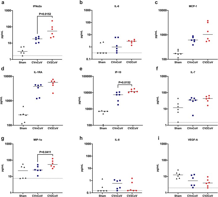
“CV2CoV induced higher levels of IFNα2a, IP-10 and MIP-1 than CVnCoV (P = 0.0152, P = 0.0152 and P = 0.0411, respectively; Extended Data Fig. 1).”
§ Optimization of non-coding regions for a non-modified mRNA COVID-19 vaccine > Vaccine immunogenicity
plaindirect- Asserted: passes — The authors put it forward as true.
- Checkable: passes — It can be checked against evidence.
- Load-bearing: unclear — It is unclear whether the paper’s conclusions depend on it.
- Based: passes — The paper gives a reason why it holds.
- It may state two separate claims as one. 29
- Probably not a substantial claim. 10
- The quote only partly states this claim. 1
The cited sources support only part of this claim — read each source’s verdict below.
In the paper
“CV2CoV is a second-generation mRNA vaccine containing non-modified nucleosides but with optimized non-coding regions and enhanced antigen expression.”
- Also stated in: § Main · plain
“CV2CoV is a second-generation mRNA vaccine that incorporates modifications of non-coding regions that were selected by empiric screening for improved antigen expression2,3.”
§ Optimization of non-coding regions for a non-modified mRNA COVID-19 vaccine > Abstract
What the sources say
- Partly supportedcites [2] Hoffmann, D. et al. CVnCoV and CV2CoV protect human ACE2 transgenic mice from ancestral B BavPat1 and emerging B.1.351 SARS-CoV-2. Nat. Commun. 12, 4048 (2021). sourceExperimental — accuracy not yet measured
Methods > Vaccination (page unavailable) describes CV2CoV as an mRNA vaccine without chemically modified nucleosides and specifies its untranslated regions. Results (page unavailable) says CV2CoV and CVnCoV encode the same protein but differ in their non-coding regions. Our copy does not establish that those regions are optimized or that they enhance antigen expression; its reported antibody responses do not directly measure antigen expression. (LLM, conf=0.84)
- Supportedcites [3] Roth, N. et al. CV2CoV, an enhanced mRNA-based SARS-CoV-2 vaccine candidate, supports higher protein expression and improved immunogenicity in rats. Preprint at https://doi.org/10.1101/2021.05.13.443734 (2021). source
The Introduction identifies CV2CoV as a next-generation, chemically unmodified mRNA vaccine with changes to its non-coding regions. Section 3.1 reports higher spike-protein expression from CV2CoV mRNA than from CVnCoV mRNA in transfected HeLa cells, supporting the enhanced-expression claim in that setting. (LLM, conf=0.94)
plain- Asserted: passes — The authors put it forward as true.
- Checkable: passes — It can be checked against evidence.
- Load-bearing: passes — The paper’s conclusions depend on it.
- Based: passes — The paper gives a reason why it holds.
It may state two separate claims as one.
- Also stated in: § Main · plain
The cited sources support only part of this claim — read each source’s verdict below.
In the paper
“Both vaccines encode the same full-length, pre-fusion stabilized severe acute respiratory syndrome coronavirus-2 (SARS-CoV-2) spike protein13,14 and are encapsulated in lipid nanoparticles (LNPs) with identical composition.”
§ Optimization of non-coding regions for a non-modified mRNA COVID-19 vaccine > Main
What the sources say
- Partly supportedcites [13] Pallesen, J. et al. Immunogenicity and structures of a rationally designed prefusion MERS-CoV spike antigen. Proc. Natl Acad. Sci. USA 114, E7348–E7357 (2017). sourceExperimental — accuracy not yet measured
The Results (p. 3) describe a two-proline design that stabilizes MERS-CoV spike in its prefusion form and report analogous effects for SARS-CoV spike. The paper does not establish that the two vaccines encode the same full-length SARS-CoV-2 spike. The citation is attached to the spike-protein description, not the subsequent LNP-composition statement. (LLM, conf=0.87)
- Partly supportedcites [14] Kirchdoerfer, R. N. et al. Stabilized coronavirus spikes are resistant to conformational changes induced by receptor recognition or proteolysis. Sci. Rep. 8, 15701 (2018). sourceExperimental — accuracy not yet measured
The Introduction (page unavailable) describes prefusion stabilization of coronavirus spike proteins using two proline mutations, and the Results examine a stabilized SARS-CoV spike ectodomain. The cited paper does not establish that both vaccines encode the same full-length SARS-CoV-2 spike protein. The LNP-composition clause follows the citations and is not judged as attributed to this source. (LLM, conf=0.87)
plain- Asserted: passes — The authors put it forward as true.
- Checkable: passes — It can be checked against evidence.
- Load-bearing: passes — The paper’s conclusions depend on it.
- Based: passes — The paper gives a reason why it holds.
It may state two separate claims as one.
- Partly supported
The cited sources support only part of this claim — read each source’s verdict below.
In the paper
“Both CVnCoV and CV2CoV are based on RNActive technology4–7 and consist of non-chemically modified sequence-engineered mRNA without pseudouridine6–12.”
- Also stated in: § Discussion · plain
“Both CVnCoV and CV2CoV contain only non-modified nucleosides with no pseudouridine or derivates, and CV2CoV has previously been shown to lead to higher antigen expression than CVnCoV in cell culture3.”
§ Optimization of non-coding regions for a non-modified mRNA COVID-19 vaccine > Main
What the sources say
- Not checked — no full text of the sourcecites [4] Hoerr, I., Obst, R., Rammensee, H. G. & Jung, G. In vivo application of RNA leads to induction of specific cytotoxic T lymphocytes and antibodies. Eur. J. Immunol. 30, 1–7 (2000). source
- Partly supportedcites [5] Fotin-Mleczek, M. et al. Highly potent mRNA based cancer vaccines represent an attractive platform for combination therapies supporting an improved therapeutic effect. J. Gene Med. 14, 428–439 (2012). sourceExperimental — accuracy not yet measured
The RNActive technology section (page unavailable) describes CureVac’s RNActive mRNA vaccines, and Results and Discussion describes sequence engineering of their coding and noncoding regions. The paper does not identify CVnCoV or CV2CoV or establish that either uses this technology; it also does not state that their mRNA is chemically unmodified or lacks pseudouridine. (LLM, conf=0.85)
- Not checked — no full text of the sourcecites [6] Fotin-Mleczek, M. et al. Messenger RNA-based vaccines with dual activity induce balanced TLR-7 dependent adaptive immune responses and provide antitumor activity. J. Immunother. 34, 1–15 (2011). source
- Partly supportedcites [7] Kubler, H. et al. Self-adjuvanted mRNA vaccination in advanced prostate cancer patients: a first-in-man phase I/IIa study. J. Immunother. Cancer 3, 26 (2015). sourceExperimental — accuracy not yet measured
The Background and Methods (RNActive® technology; page not provided) describe RNActive as an mRNA-vaccine platform using free and protamine-complexed mRNA. Our copy studies CV9103, however, and does not identify CVnCoV or CV2CoV or establish that their mRNA is sequence-engineered, non-chemically modified, or free of pseudouridine. (LLM, conf=0.79)
- Partly supportedcites [8] Lutz, J. et al. Unmodified mRNA in LNPs constitutes a competitive technology for prophylactic vaccines. NPJ Vaccines 2, 29 (2017). sourceExperimental — accuracy not yet measured
The Materials and methods section (‘mRNA vaccines’; page not provided) says the vaccines studied used the RNActive platform, lacked chemically modified nucleosides, and underwent sequence optimization. That supports the description of the technology, including the absence of pseudouridine, but the paper discusses rabies and influenza vaccines and does not identify CVnCoV or CV2CoV or establish their composition. (LLM, conf=0.88)
- Partly supportedcites [9] Stitz, L. et al. A thermostable messenger RNA based vaccine against rabies. PLoS Negl. Trop. Dis. 11, e0006108 (2017). sourceExperimental — accuracy not yet measured
The Introduction and Methods describe a rabies vaccine based on RNActive technology, including optimization of its mRNA sequence. They do not identify CVnCoV or CV2CoV or establish that either uses this technology. The passages also do not say whether their mRNA is chemically unmodified or lacks pseudouridine. (LLM, conf=0.87)
- Partly supportedcites [10] Schnee, M. et al. An mRNA vaccine encoding rabies virus glycoprotein induces protection against lethal infection in mice and correlates of protection in adult and newborn pigs. PLoS Negl. Trop. Dis. 10, e0004746 (2016). sourceExperimental — accuracy not yet measured
Materials and Methods (mRNA- and protein-based vaccines; page unavailable) says the rabies mRNA vaccines used the RNActive platform, had GC-enriched coding sequences and enhanced UTRs, and contained no chemically modified nucleotides. That supports the technology description, including the absence of pseudouridine, for the vaccines studied here. The paper does not identify CVnCoV or CV2CoV or establish that either has those properties. (LLM, conf=0.86)
- Not checked — no full text of the sourcecites [11] Petsch, B. et al. Protective efficacy of in vitro synthesized, specific mRNA vaccines against influenza A virus infection. Nat. Biotechnol. 30, 1210–1216 (2012). source
- Partly supportedcites [12] Aldrich, C. et al. Proof-of-concept of a low-dose unmodified mRNA-based rabies vaccine formulated with lipid nanoparticles in human volunteers: a phase 1 trial. Vaccine 39, 1310–1318 (2021). sourceExperimental — accuracy not yet measured
The Introduction (page unavailable) identifies RNActive® as CureVac’s mRNA platform and says the vaccine approach is being applied to SARS-CoV-2. It does not identify CVnCoV or CV2CoV or establish that either uses sequence-engineered mRNA without pseudouridine. Searches for those vaccine names and the claimed mRNA features found no supporting passage. (LLM, conf=0.83)
plain- Asserted: passes — The authors put it forward as true.
- Checkable: passes — It can be checked against evidence.
- Load-bearing: passes — The paper’s conclusions depend on it.
- Based: passes — The paper gives a reason why it holds.
It may state two separate claims as one.
- Also stated in: § Discussion · plain
The cited sources support only part of this claim — read each source’s verdict below.
In the paper
“CV2CoV has been engineered with different non-coding regions flanking the open reading frame, which have previously been shown to improve transgene expression3 and protection against SARS-CoV-2 in ACE2-transgenic mice2.”
§ Optimization of non-coding regions for a non-modified mRNA COVID-19 vaccine > Main
What the sources say
- Partly supportedcites [2] Hoffmann, D. et al. CVnCoV and CV2CoV protect human ACE2 transgenic mice from ancestral B BavPat1 and emerging B.1.351 SARS-CoV-2. Nat. Commun. 12, 4048 (2021). sourceExperimental — accuracy not yet measured
The Methods (Vaccination; page unavailable) describe CV2CoV’s different non-coding regions, and the Discussion reports that CV2CoV protected hACE2-transgenic mice against disease caused by both tested SARS-CoV-2 variants. The paper does not show that those regions improved protection relative to CVnCoV: the Discussion reports full protection with both vaccines. The transgene-expression claim is attached to a different citation and is not assessed here. (LLM, conf=0.84)
- Could not verifycites [3] Roth, N. et al. CV2CoV, an enhanced mRNA-based SARS-CoV-2 vaccine candidate, supports higher protein expression and improved immunogenicity in rats. Preprint at https://doi.org/10.1101/2021.05.13.443734 (2021). sourceExperimental — accuracy not yet measured
The citation to this paper is attached to the claim of protection against SARS-CoV-2 in ACE2-transgenic mice. Sections 2.3–2.4 describe experiments in Wistar rats, and the Introduction mentions protective efficacy in non-human primates; no passage found reports protection in ACE2-transgenic mice. The paper does describe different non-coding regions and increased protein expression, but those are not the protection claim attached to this citation. (LLM, conf=0.76)
plaindirect- Asserted: passes — The authors put it forward as true.
- Checkable: passes — It can be checked against evidence.
- Load-bearing: passes — The paper’s conclusions depend on it.
- Based: passes — The paper gives a reason why it holds.
It may state two separate claims as one.
- Partly supported
The cited sources that could be checked couldn’t confirm or dispute this claim.
In the paper
“Following challenge, we observed anamnestic binding and neutralizing antibody responses in all CVnCoV-vaccinated animals and in a subset of the CV2CoV-vaccinated animals16 (Extended Data Fig. 6).”
§ Optimization of non-coding regions for a non-modified mRNA COVID-19 vaccine > Protective efficacy
What the sources say
- Could not verifycites [16] Mercado, N. B. et al. Single-shot Ad26 vaccine protects against SARS-CoV-2 in rhesus macaques. Nature 586, 583–588 (2020). sourceExperimental — accuracy not yet measured
The Methods (Animals and study design) list Ad26 vaccine groups, not CVnCoV or CV2CoV. The Immune Responses in Vaccinated Animals Following Challenge section reports increased neutralizing antibodies in most Ad26-vaccinated animals but no anamnestic neutralizing response in the Ad26-S.PP group. Searches for CVnCoV and CV2CoV found no passage addressing responses in those groups; the Ad26 findings do not establish or contradict the claim about different vaccines. (LLM, conf=0.78)
plain- Asserted: passes — The authors put it forward as true.
- Checkable: passes — It can be checked against evidence.
- Load-bearing: passes — The paper’s conclusions depend on it.
- Based: passes — The paper gives a reason why it holds.
It may state two separate claims as one.
- Could not verify
The cited sources support only part of this claim — read each source’s verdict below.
In the paper
“Previous mRNA vaccine clinical trials have demonstrated onset of protective efficacy after the first dose with improved protection after the boost immunization25,26.”
§ Optimization of non-coding regions for a non-modified mRNA COVID-19 vaccine > Discussion
What the sources say
- Supportedcites [25] Polack, F. P. et al. Safety and efficacy of the BNT162b2 mRNA Covid-19 vaccine. N. Engl. J. Med. 383, 2603–2615 (2020). source
The Discussion reports early partial protection after the first dose and higher observed efficacy after the second dose. The Results > Efficacy section gives 52% efficacy between doses and 95% from seven days after dose two. The paper cautions that the trial was not designed to assess a single-dose regimen. (LLM, conf=0.93)
- Partly supportedcites [26] Baden, L. R. et al. Efficacy and safety of the mRNA-1273 SARS-CoV-2 vaccine. N. Engl. J. Med. 384, 403–416 (2021). sourceExperimental — accuracy not yet measured
The Discussion (p. 11) reports fewer symptomatic COVID-19 cases after one dose of mRNA-1273, but cautions that the trial was not designed to evaluate single-dose efficacy. The Efficacy section (p. 9) establishes strong efficacy for the two-dose regimen, but does not demonstrate that protection improved specifically because of the second dose. (LLM, conf=0.85)
boosteddirect- Asserted: passes — The authors put it forward as true.
- Checkable: passes — It can be checked against evidence.
- Load-bearing: fails — The paper’s conclusions may not depend on it.
- Based: passes — The paper gives a reason why it holds.
Probably not a substantial claim. The paper’s conclusions may not depend on it. It may state two separate claims as one.
- Supported
Every source the paper cites for this claim was checked and supports it.
In the paper
“We previously reported that depletion of CD8+ T cells partially abrogated protective efficacy against SARS-CoV-2 re-challenge in convalescent macaques22.”
§ Optimization of non-coding regions for a non-modified mRNA COVID-19 vaccine > Discussion
What the sources say
- Supportedcites [22] McMahan, K. et al. Correlates of protection against SARS-CoV-2 in rhesus macaques. Nature 590, 630–634 (2021). source
The Discussion states that CD8 depletion partially abrogated protection of convalescent macaques against SARS-CoV-2 re-challenge. The CD8 Depletion Study reports breakthrough virus in CD8-depleted animals compared with sham controls, supporting that conclusion. (LLM, conf=0.95)
plaindirect- Asserted: passes — The authors put it forward as true.
- Checkable: passes — It can be checked against evidence.
- Load-bearing: fails — The paper’s conclusions may not depend on it.
- Based: passes — The paper gives a reason why it holds.
Probably not a substantial claim. The paper’s conclusions may not depend on it.
- Supported
The cited sources support only part of this claim — read each source’s verdict below.
In the paper
“As previously reported for other vaccines29–33, the neutralizing antibody titres against certain SARS-CoV-2 variants, such as the B.1.351 (Beta) and B.1.617.2 (Delta) variants, were lower than those against the parental strain WA1/2020.”
§ Optimization of non-coding regions for a non-modified mRNA COVID-19 vaccine > Discussion
What the sources say
- Partly supportedcites [29] Liu, C. et al. Reduced neutralization of SARS-CoV-2 B.1.617 by vaccine and convalescent serum. Cell https://doi.org/10.1016/j.cell.2021.06.020 (2021). sourceExperimental — accuracy not yet measured
The Results section on vaccine serum reports lower neutralizing titres against B.1.617.2 than against Victoria, and the Introduction reports reduced titres against B.1.351. The paper uses Victoria, an early Wuhan-related strain, as its comparator—not the specifically named WA1/2020 strain. (LLM, conf=0.87)
- Could not verifycites [30] Haas, E. J. et al. Impact and effectiveness of mRNA BNT162b2 vaccine against SARS-CoV-2 infections and COVID-19 cases, hospitalisations, and deaths following a nationwide vaccination campaign in Israel: an observational study using national surveillance data. Lancet 397, 1819–1829 (2021). sourceExperimental — accuracy not yet measured
The Discussion reports effectiveness against B.1.1.7 and says effectiveness against B.1.351 could not be estimated. Its mention of lower neutralising antibodies compares one vaccine dose with two, not variants with WA1/2020. Searches for Delta, WA1/2020, and variant-specific neutralising titres found no passage addressing the claimed comparison; page numbers were not available in our copy. (LLM, conf=0.76)
- Partly supportedcites [31] Wu, K. et al. Serum neutralizing activity elicited by mRNA-1273 vaccine. N. Engl. J. Med. 384, 1468–1470 (2021). sourceExperimental — accuracy not yet measured
In the main text (page not specified), the paper reports lower neutralizing titers against B.1.351 in serum from mRNA-1273 recipients, using the original Wuhan-Hu-1 isolate as a reference. It does not report results for B.1.617.2 (Delta) or name WA1/2020 as the comparator. (LLM, conf=0.88)
- Partly supportedcites [32] Wibmer, C. K. et al. SARS-CoV-2 501Y.V2 escapes neutralization by South African COVID-19 donor plasma. Nat. Med. 27, 622–625 (2021). sourceExperimental — accuracy not yet measured
The Figure 2 passage reports reduced neutralization of 501Y.V2 (Beta) relative to the original Wuhan-1 D614G lineage using convalescent plasma; the vaccine discussion also reports reduced neutralization of Beta by vaccinee sera. The paper does not address Delta, and its tested comparator is Wuhan-1 D614G, not the specified WA1/2020 strain. (LLM, conf=0.88)
- Partly supportedcites [33] Wall, E. C. et al. Neutralising antibody activity against SARS-CoV-2 VOCs B.1.617.2 and B.1.351 by BNT162b2 vaccination. Lancet 397, 2331–2333 (2021). sourceExperimental — accuracy not yet measured
The main text reports neutralising antibody titres 5.8-fold lower against B.1.617.2 and 4.9-fold lower against B.1.351 than against its Wild-type strain. It describes that comparator as having the original spike sequence but does not identify it as WA1/2020, so the named comparator is not established by our copy. (LLM, conf=0.83)
plain- Asserted: passes — The authors put it forward as true.
- Checkable: passes — It can be checked against evidence.
- Load-bearing: fails — The paper’s conclusions may not depend on it.
- Based: passes — The paper gives a reason why it holds.
Probably not a substantial claim. The paper’s conclusions may not depend on it.
- Partly supported
The cited sources support only part of this claim — read each source’s verdict below.
In the paper
“Memory B cells might contribute to the durability of antibody responses38,39; B cell germinal centre responses and the durability of protective efficacy following CV2CoV vaccination remain to be determined.”
§ Optimization of non-coding regions for a non-modified mRNA COVID-19 vaccine > Discussion
What the sources say
- Supportedcites [38] Abayasingam, A. et al. Long-term persistence of RBD+ memory B cells encoding neutralizing antibodies in SARS-CoV-2 infection. Cell Rep. Med. 2, 100228 (2021). source
The Results (‘RBD-specific memory B cells’) say that a durable memory B-cell response capable of generating neutralizing antibodies on demand could mitigate declining neutralizing activity. The Discussion reports that memory B cells persisted for up to six months after infection and could make neutralizing antibodies despite falling antibody titers. This supports the citing sentence’s hedged claim, though it does not establish that memory B cells maintain circulating antibody titers. (LLM, conf=0.88)
- Partly supportedcites [39] Dan, J. M. et al. Immunological memory to SARS-CoV-2 assessed for up to 8 months after infection. Science 371, https://doi.org/10.1126/science.abf4063 (2021). sourceExperimental — accuracy not yet measured
The Concluding remarks report durable Spike IgG titers and persistent memory B cells 5–8 months after infection. They do not establish that memory B cells contribute to the durability of circulating antibody responses; the paper instead describes antibody levels as reflecting production by short- and then long-lived plasma cells. (LLM, conf=0.84)
hedgedconditional- Asserted: passes — The authors put it forward as true.
- Checkable: passes — It can be checked against evidence.
- Load-bearing: fails — The paper’s conclusions may not depend on it.
- Based: passes — The paper gives a reason why it holds.
Probably not a substantial claim. The paper’s conclusions may not depend on it.
- Supported
A result the paper reports from its own data — it cites no other source for it, so there is none to check. The quote shows it is in the paper.
In the paper
“The sham controls had peak medians of 6.02 (range, 4.62–6.81) log10-transformed sgRNA copies per ml in the BAL and 7.35 (range, 5.84–8.09) log10-transformed sgRNA copies per swab in the nasal swab samples on day 2 (Fig. 3).”
§ Optimization of non-coding regions for a non-modified mRNA COVID-19 vaccine > Protective efficacy
plain- Asserted: passes — The authors put it forward as true.
- Checkable: passes — It can be checked against evidence.
- Load-bearing: passes — The paper’s conclusions depend on it.
- Based: passes — The paper gives a reason why it holds.
It may state two separate claims as one.
A result the paper reports from its own data — it cites no other source for it, so there is none to check. The quote shows it is in the paper.
In the paper
“At week 2, binding antibody titres were detected only with CV2CoV and not with CVnCoV, with median values of 25 (range, 25–25) and 799 (range, 82–2,010) for CVnCoV and CV2CoV, respectively (Fig. 2a).”
§ Optimization of non-coding regions for a non-modified mRNA COVID-19 vaccine > Vaccine immunogenicity
plain- Asserted: passes — The authors put it forward as true.
- Checkable: passes — It can be checked against evidence.
- Load-bearing: passes — The paper’s conclusions depend on it.
- Based: passes — The paper gives a reason why it holds.
It may state two separate claims as one.
A result the paper reports from its own data — it cites no other source for it, so there is none to check. The quote shows it is in the paper.
In the paper
“At week 2, neutralizing antibodies were detected only with CV2CoV and not with CVnCoV, with median values of 20 (range, 20–20) and 131 (range, 62–578) for CVnCoV and CV2CoV, respectively (Fig. 2b).”
§ Optimization of non-coding regions for a non-modified mRNA COVID-19 vaccine > Vaccine immunogenicity
plain- Asserted: passes — The authors put it forward as true.
- Checkable: passes — It can be checked against evidence.
- Load-bearing: passes — The paper’s conclusions depend on it.
- Based: passes — The paper gives a reason why it holds.
It may state two separate claims as one.
A result the paper reports from its own data — it cites no other source for it, so there is none to check. The quote shows it is in the paper.
In the paper
“At peak immunity at week 5, the neutralizing antibody responses induced by CV2CoV were comparable to those induced by BNT162b2 (Fig. 2e).”
- Also stated in: § Abstract · plain
“Moreover, CV2CoV was found to be comparably immunogenic to the BNT162b2 (Pfizer) vaccine in macaques.”
§ Optimization of non-coding regions for a non-modified mRNA COVID-19 vaccine > Vaccine immunogenicity
plainn = 6- Asserted: passes — The authors put it forward as true.
- Checkable: passes — It can be checked against evidence.
- Load-bearing: passes — The paper’s conclusions depend on it.
- Based: passes — The paper gives a reason why it holds.
The quote only partly states this claim.
- Rests on an assumption: The BNT162b2 clinical vaccine obtained as leftover product from pharmacies retained immunogenicity comparable to the clinical vaccine.
- Also stated in: § Abstract · plain
A result the paper reports from its own data — it cites no other source for it, so there is none to check. The quote shows it is in the paper.
In the paper
“The live-virus neutralizing antibody responses elicited by CV2CoV were higher than those elicited by CVnCoV against the WA1/2020 and B.1.617.2 (Delta) strains (P = 0.0466 and 0.0152, respectively), with similar trends for B.1.1.7 (Alpha) and B.1.351 (Beta) (P = 0.0628 and 0.1450, respectively) (Fig. 2d).”
§ Optimization of non-coding regions for a non-modified mRNA COVID-19 vaccine > Vaccine immunogenicity
plaindirect- Asserted: passes — The authors put it forward as true.
- Checkable: passes — It can be checked against evidence.
- Load-bearing: passes — The paper’s conclusions depend on it.
- Based: passes — The paper gives a reason why it holds.
It may state two separate claims as one.
A result the paper reports from its own data — it cites no other source for it, so there is none to check. The quote shows it is in the paper.
In the paper
“By week 8, the binding antibody titres had increased in the CVnCoV group but were still >50 times lower than those in the CV2CoV group (P = 0.0043), with median values of 214 (range, 47–1,238) and 14,827 (range, 2,133–37,079), respectively.”
§ Optimization of non-coding regions for a non-modified mRNA COVID-19 vaccine > Vaccine immunogenicity
plaindirect- Asserted: passes — The authors put it forward as true.
- Checkable: passes — It can be checked against evidence.
- Load-bearing: passes — The paper’s conclusions depend on it.
- Based: passes — The paper gives a reason why it holds.
It may state two separate claims as one.
A result the paper reports from its own data — it cites no other source for it, so there is none to check. The quote shows it is in the paper.
In the paper
“One week after the week 4 boost, the neutralizing antibody titres were increased, with median values of 55 (range, 20–302) and 15,827 (range, 3,985–81,081) for CVnCoV and CV2CoV, respectively.”
§ Optimization of non-coding regions for a non-modified mRNA COVID-19 vaccine > Vaccine immunogenicity
plaindirect- Asserted: passes — The authors put it forward as true.
- Checkable: passes — It can be checked against evidence.
- Load-bearing: passes — The paper’s conclusions depend on it.
- Based: passes — The paper gives a reason why it holds.
It may state two separate claims as one.
A result the paper reports from its own data — it cites no other source for it, so there is none to check. The quote shows it is in the paper.
In the paper
“One week after the week-4 boost, the antibody titres were increased in both groups, with medians of 48 (range, 75–710) and 28,407 (range, 2,714–86,541) for CVnCoV and CV2CoV, respectively (Fig. 2a).”
§ Optimization of non-coding regions for a non-modified mRNA COVID-19 vaccine > Vaccine immunogenicity
plaindirect- Asserted: passes — The authors put it forward as true.
- Checkable: passes — It can be checked against evidence.
- Load-bearing: passes — The paper’s conclusions depend on it.
- Based: passes — The paper gives a reason why it holds.
It may state two separate claims as one.
A result the paper reports from its own data — it cites no other source for it, so there is none to check. The quote shows it is in the paper.
In the paper
“IFNγ responses were detected in both groups but were higher in the CV2CoV group (P = 0.0065) (Extended Data Fig. 3c).”
§ Optimization of non-coding regions for a non-modified mRNA COVID-19 vaccine > Vaccine immunogenicity
plaincorrelational- Asserted: passes — The authors put it forward as true.
- Checkable: passes — It can be checked against evidence.
- Load-bearing: passes — The paper’s conclusions depend on it.
- Based: passes — The paper gives a reason why it holds.
It may state two separate claims as one.
A result the paper reports from its own data — it cites no other source for it, so there is none to check. The quote shows it is in the paper.
In the paper
“By week 8, the neutralizing antibody titres had increased in the CVnCoV group but were still >20 times lower than those in the CV2CoV group (P = 0.0022), with median values of 196 (range, 20–405) and 4,752 (range, 414–6,793), respectively.”
§ Optimization of non-coding regions for a non-modified mRNA COVID-19 vaccine > Vaccine immunogenicity
plaindirect- Asserted: passes — The authors put it forward as true.
- Checkable: passes — It can be checked against evidence.
- Load-bearing: passes — The paper’s conclusions depend on it.
- Based: passes — The paper gives a reason why it holds.
It may state two separate claims as one.
A result the paper reports from its own data — it cites no other source for it, so there is none to check. The quote shows it is in the paper.
In the paper
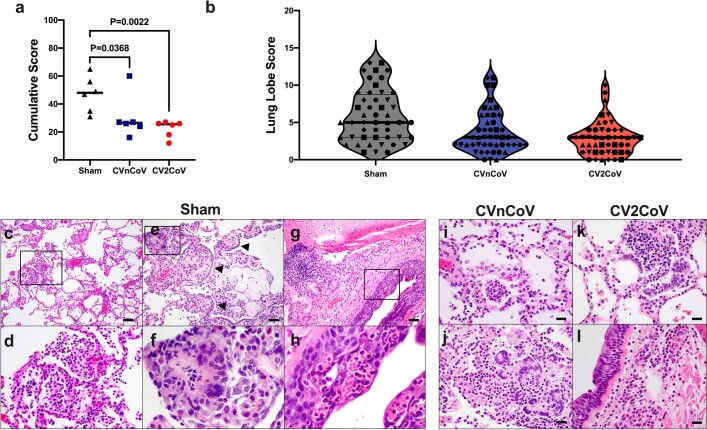
“The pathological lesions in vaccinated animals were similar to those observed for animals in the sham group (Extended Data Fig. 7i–l) but were overall fewer in number and more focal in distribution.”
§ Optimization of non-coding regions for a non-modified mRNA COVID-19 vaccine > Protective efficacy
plaincorrelational- Asserted: passes — The authors put it forward as true.
- Checkable: passes — It can be checked against evidence.
- Load-bearing: passes — The paper’s conclusions depend on it.
- Based: passes — The paper gives a reason why it holds.
It may state two separate claims as one.
A result the paper reports from its own data — it cites no other source for it, so there is none to check. The quote shows it is in the paper.
In the paper
“Animals in the sham group also had more lung lobes affected (Extended Data Fig. 7b) and more extensive lung lesions, with a greater proportion of lung lobes showing evidence of interstitial inflammation, alveolar inflammatory infiltrates and type II pneumocyte hyperplasia (Extended Data Fig. 7c–h).”
§ Optimization of non-coding regions for a non-modified mRNA COVID-19 vaccine > Protective efficacy
plaincorrelational- Asserted: passes — The authors put it forward as true.
- Checkable: passes — It can be checked against evidence.
- Load-bearing: passes — The paper’s conclusions depend on it.
- Based: passes — The paper gives a reason why it holds.
It may state two separate claims as one.
A result the paper reports from its own data — it cites no other source for it, so there is none to check. The quote shows it is in the paper.
In the paper
“In the present study, the prime immunization with CV2CoV induced binding and neutralizing antibodies in all macaques by week 2, and these responses had increased substantially by 1 week after the boost immunization.”
§ Optimization of non-coding regions for a non-modified mRNA COVID-19 vaccine > Discussion
plaindirect- Asserted: passes — The authors put it forward as true.
- Checkable: passes — It can be checked against evidence.
- Load-bearing: passes — The paper’s conclusions depend on it.
- Based: passes — The paper gives a reason why it holds.
It may state two separate claims as one.
A result the paper reports from its own data — it cites no other source for it, so there is none to check. The quote shows it is in the paper.
In the paper
“The CV2CoV-immunized animals exhibited peak medians of 2.90 (range, 1.70–4.64) log10-transformed sgRNA copies per ml in the BAL and 3.17 (range, 2.59–5.63) log10-transformed sgRNA copies per swab in the nasal swab samples (Fig. 3), with resolution of sgRNA levels in the BAL samples by day 2 in most animals and by day 4 in all animals.”
§ Optimization of non-coding regions for a non-modified mRNA COVID-19 vaccine > Protective efficacy
plain- Asserted: passes — The authors put it forward as true.
- Checkable: passes — It can be checked against evidence.
- Load-bearing: passes — The paper’s conclusions depend on it.
- Based: passes — The paper gives a reason why it holds.
It may state two separate claims as one.
A result the paper reports from its own data — it cites no other source for it, so there is none to check. The quote shows it is in the paper.
In the paper
“The CVnCoV-immunized animals showed peak medians of 4.92 (range, 2.40–6.61) log10-transformed sgRNA copies per ml in the BAL and 6.42 (range, 4.46–7.81) log10-transformed sgRNA copies per swab in the nasal swab samples (Fig. 3).”
§ Optimization of non-coding regions for a non-modified mRNA COVID-19 vaccine > Protective efficacy
plain- Asserted: passes — The authors put it forward as true.
- Checkable: passes — It can be checked against evidence.
- Load-bearing: passes — The paper’s conclusions depend on it.
- Based: passes — The paper gives a reason why it holds.
It may state two separate claims as one.
A result the paper reports from its own data — it cites no other source for it, so there is none to check. The quote shows it is in the paper.
In the paper
“In contrast to CVnCoV, CV2CoV induced >10-fold-higher neutralizing antibody responses against multiple viral variants and provided >3 log reductions in sgRNA copies per ml in BAL and >4 log reductions in sgRNA copies per swab in nasal swab samples compared with sham controls.”
§ Optimization of non-coding regions for a non-modified mRNA COVID-19 vaccine > Discussion
plaindirect- Asserted: passes — The authors put it forward as true.
- Checkable: passes — It can be checked against evidence.
- Load-bearing: passes — The paper’s conclusions depend on it.
- Based: passes — The paper gives a reason why it holds.
It may state two separate claims as one.
In the paper
“Viral replication was largely resolved by day 10 in the animals vaccinated with CVnCoV and CV2CoV, and those with sham treatment had higher cumulative lung pathology scores19 (CVnCoV animals compared with sham controls, P = 0.0368; CV2CoV animals compared with sham controls, P = 0.0022) (Extended Data Fig. 7a).”
§ Optimization of non-coding regions for a non-modified mRNA COVID-19 vaccine > Protective efficacy
What the sources say
- Not checked — no full text of the sourcecites [19] He, X. et al. Low-dose Ad26.COV2.S protection against SARS-CoV-2 challenge in rhesus macaques. Cell https://doi.org/10.1016/j.cell.2021.05.040 (2021). source
plaincorrelational- Asserted: passes — The authors put it forward as true.
- Checkable: passes — It can be checked against evidence.
- Load-bearing: passes — The paper’s conclusions depend on it.
- Based: passes — The paper gives a reason why it holds.
It may state two separate claims as one.
A result the paper reports from its own data — it cites no other source for it, so there is none to check. The quote shows it is in the paper.
In the paper
“CV2CoV induced both antigen-specific memory B cell responses and T cell responses.”
§ Optimization of non-coding regions for a non-modified mRNA COVID-19 vaccine > Discussion
plaindirect- Asserted: passes — The authors put it forward as true.
- Checkable: passes — It can be checked against evidence.
- Load-bearing: passes — The paper’s conclusions depend on it.
- Based: passes — The paper gives a reason why it holds.
It may state two separate claims as one.
In the paper
“In this trial, the observed vaccine efficacy against symptomatic coronavirus disease 2019 (COVID-19) was approximately 48% and 53% in the overall study population and in a subgroup of participants 18–60 years of age, respectively1.”
§ Optimization of non-coding regions for a non-modified mRNA COVID-19 vaccine > Main
What the sources say
- Not checked — no full text of the sourcecites [1] Kremsner, P. G. et al. Efficacy and safety of the CVnCoV SARS-CoV-2 mRNA vaccine candidate: results from Herald, a phase 2b/3, randomised, observer-blinded, placebo-controlled clinical trial in ten countries in Europe and Latin America. Lancet Infect. Dis. https://doi.org/10.1016/S1473-3099(21)00677–0 (2021). source
plain- Asserted: passes — The authors put it forward as true.
- Checkable: passes — It can be checked against evidence.
- Load-bearing: unclear — It is unclear whether the paper’s conclusions depend on it.
- Based: passes — The paper gives a reason why it holds.
It may state two separate claims as one.
A conclusion the paper draws from its own results — it cites no other source for it, so there is none to check. The quote shows it is in the paper.
In the paper
“These data suggest that optimization of non-coding elements of the mRNA backbone can substantially improve the immunogenicity and protective efficacy of mRNA vaccines.”
§ Optimization of non-coding regions for a non-modified mRNA COVID-19 vaccine > Discussion
hedgeddirect- Asserted: passes — The authors put it forward as true.
- Checkable: passes — It can be checked against evidence.
- Load-bearing: passes — The paper’s conclusions depend on it.
- Based: passes — The paper gives a reason why it holds.
It may state two separate claims as one.
A conclusion the paper draws from its own results — it cites no other source for it, so there is none to check. The quote shows it is in the paper.
In the paper
“These data demonstrate that optimization of non-coding regions can greatly improve the immunogenicity and protective efficacy of a non-modified mRNA SARS-CoV-2 vaccine in non-human primates.”
- Also stated in: § Discussion · plain
“In summary, our data show that optimization of non-coding regions in a SARS-CoV-2 mRNA vaccine can substantially improve its immunogenicity against multiple viral variants and can enhance its protective efficacy against SARS-CoV-2 challenge in macaques.”
§ Optimization of non-coding regions for a non-modified mRNA COVID-19 vaccine > Abstract
boosteddirectn = 6- Asserted: passes — The authors put it forward as true.
- Checkable: passes — It can be checked against evidence.
- Load-bearing: passes — The paper’s conclusions depend on it.
- Based: passes — The paper gives a reason why it holds.
It may state two separate claims as one.
- Also stated in: § Discussion · plain
A conclusion the paper draws from its own results — it cites no other source for it, so there is none to check. The quote shows it is in the paper.
In the paper
“CV2CoV elicited substantially greater humoral and cellular immune responses and provided significantly improved protective efficacy against SARS-CoV-2 challenge as compared with CVnCoV in macaques.”
§ Optimization of non-coding regions for a non-modified mRNA COVID-19 vaccine > Discussion
plaindirect- Asserted: passes — The authors put it forward as true.
- Checkable: passes — It can be checked against evidence.
- Load-bearing: passes — The paper’s conclusions depend on it.
- Based: passes — The paper gives a reason why it holds.
It may state two separate claims as one.
What the paper did — it cites no source for this, so there is none to check. The quote shows it is in the paper.
In the paper
“All animal studies were conducted in compliance with all relevant local, state and federal regulations and were approved by the Bioqual Institutional Animal Care and Use Committee.”
§ Optimization of non-coding regions for a non-modified mRNA COVID-19 vaccine > Methods > Animals and study design
plain- Asserted: passes — The authors put it forward as true.
- Checkable: passes — It can be checked against evidence.
- Load-bearing: fails — The paper’s conclusions may not depend on it.
- Based: passes — The paper gives a reason why it holds.
Probably not a substantial claim. The paper’s conclusions may not depend on it. It may state two separate claims as one.
What the paper did — it cites no source for this, so there is none to check. The quote shows it is in the paper.
In the paper
“The immunological assays were performed with blinding.”
- Also stated in: § B cell immunophenotyping · plain
“The immunological assays were performed with blinding.”
§ Optimization of non-coding regions for a non-modified mRNA COVID-19 vaccine > Methods > ELISA
plain- Asserted: passes — The authors put it forward as true.
- Checkable: passes — It can be checked against evidence.
- Load-bearing: fails — The paper’s conclusions may not depend on it.
- Based: passes — The paper gives a reason why it holds.
Probably not a substantial claim. The paper’s conclusions may not depend on it.
- Also stated in: § B cell immunophenotyping · plain
The paper states this without a source or data behind it here, so there is nothing to check it against. The quote shows it is in the paper.
In the paper
“LNPs are composed of ionizable amino lipid, phospholipid and cholesterol and PEGylated lipid; the compositions for CVnCoV and CV2CoV are identical.”
§ Optimization of non-coding regions for a non-modified mRNA COVID-19 vaccine > Methods > mRNA vaccines
plain- Asserted: passes — The authors put it forward as true.
- Checkable: passes — It can be checked against evidence.
- Load-bearing: fails — The paper’s conclusions may not depend on it.
- Based: fails — The paper may give no reason why it holds.
Probably not a substantial claim. The paper’s conclusions may not depend on it. The paper may give no reason why it holds. It may state two separate claims as one.
What the paper did — it cites no source for this, so there is none to check. The quote shows it is in the paper.
In the paper
“Blinded histopathological evaluation was performed by a board-certified veterinary pathologist (A.J.M.).”
§ Optimization of non-coding regions for a non-modified mRNA COVID-19 vaccine > Methods > Histopathology
plain- Asserted: passes — The authors put it forward as true.
- Checkable: passes — It can be checked against evidence.
- Load-bearing: fails — The paper’s conclusions may not depend on it.
- Based: passes — The paper gives a reason why it holds.
Probably not a substantial claim. The paper’s conclusions may not depend on it.
What the paper did — it cites no source for this, so there is none to check. The quote shows it is in the paper.
In the paper
“Immunological and virological assays were performed with blinding.”
§ Optimization of non-coding regions for a non-modified mRNA COVID-19 vaccine > Methods > Animals and study design
plain- Asserted: passes — The authors put it forward as true.
- Checkable: passes — It can be checked against evidence.
- Load-bearing: fails — The paper’s conclusions may not depend on it.
- Based: passes — The paper gives a reason why it holds.
Probably not a substantial claim. The paper’s conclusions may not depend on it.
What the paper did — it cites no source for this, so there is none to check. The quote shows it is in the paper.
In the paper
“Eighteen cynomolgus macaques of both sexes between the ages of 3 and 20 years were randomly assigned to three groups.”
§ Optimization of non-coding regions for a non-modified mRNA COVID-19 vaccine > Methods > Animals and study design
plain- Asserted: passes — The authors put it forward as true.
- Checkable: passes — It can be checked against evidence.
- Load-bearing: passes — The paper’s conclusions depend on it.
- Based: passes — The paper gives a reason why it holds.
Probably not a substantial claim.
The paper relies on this without support.
The BNT162b2 clinical vaccine obtained as leftover product from pharmacies retained immunogenicity comparable to the clinical vaccine.
“We also compared the pseudovirus neutralizing antibody titres induced in macaques by two immunizations with 12 μg of CV2CoV to those induced by two immunizations with 30 μg of the Pfizer BNT162b2 clinical vaccine obtained as leftover product from pharmacies.”
Claims other papers make about this one
Each is checked against this paper’s own text: does it say what the citing paper says it does?
The source backs part of the claim, not all of it — or backs it for a different population, condition or comparison.
In the citing paper
“The optimised non-coding regions of mRNA SARS-CoV-2 vaccine CV2CoV have previously been shown to improve homologous and heterologous neutralising antibody responses in rats and non-human primates, compared with CVnCoV and other modified mRNA vaccines10,11.”
From Efficacy of an unmodified bivalent mRNA vaccine against SARS-CoV-2 variants in female small animal models (2023) · cites this paper as [11]
Experimental — accuracy not yet measuredThe source backs part of the claim, not all of it — or backs it for a different population, condition or comparison.
In the citing paper
“The rat-derived data translated very well to improved immunogenicity and protective efficacy in a non-human primate challenge model for SARS-CoV-210,11 and our cross-neutralization results emphasize the broad antigen coverage induced by the bivalent vaccine.”
From Efficacy of an unmodified bivalent mRNA vaccine against SARS-CoV-2 variants in female small animal models (2023) · cites this paper as [11]
Experimental — accuracy not yet measuredThe cited source says what the claim attributes to it.
In the citing paper
“mRNA vaccines9,10 have been recognized as viable tools to limit the spread of COVID-19 owing to their scalable production, safety and efficacy1–3.”
From Algorithm for optimized mRNA design improves stability and immunogenicity (2023) · cites this paper as [3]
The cited source says what the claim attributes to it.
In the citing paper
“and UTR engineering has a profound effect on protein expression3.”
From Algorithm for optimized mRNA design improves stability and immunogenicity (2023) · cites this paper as [3]
The cited source says what the claim attributes to it.
In the citing paper
“Protection induced by CV2CoV was reported to be superior to CVnCoV in a non-human primate SARS-CoV-2 challenge model11.”
From Efficacy of an unmodified bivalent mRNA vaccine against SARS-CoV-2 variants in female small animal models (2023) · cites this paper as [11]
The cited source says what the claim attributes to it.
In the citing paper
“CV2CoV has now entered clinical testing (NCT05260437) and has been shown to induce high neutralizing antibodies in rats10 and non-human primates11, with titers comparable to those seen with Comirnaty, a licensed mRNA COVID-19 vaccine.”
From Efficacy of an unmodified bivalent mRNA vaccine against SARS-CoV-2 variants in female small animal models (2023) · cites this paper as [11]
In the citing paper
“However, CureVac further engineered the UTRs of the mRNA and showed a superior immunological profile in preclinical tests,136,137”
From COVID-19 mRNA vaccines: Platforms and current developments (2022) · cites this paper as [136]
Self-check
Percentages, sums and reported differences re-computed from the paper’s own numbers.
Each contradiction between two passages listed here was confirmed by an independent check.
Rated minor: it would not touch a result — a value no result rests on, a p-value that stays on the same side of the significance level, or a conflict that is clerical, resolved elsewhere in the paper, or outside its own findings.
In the paper
§ Optimization of non-coding regions for a non-modified mRNA COVID-19 vaccine > Vaccine immunogenicity
“We also compared the pseudovirus neutralizing antibody titres induced in macaques by two immunizations with 12 μg of CV2CoV to those induced by two immunizations with 30 μg of the Pfizer BNT162b2 clinical vaccine obtained as leftover product from pharmacies.”
§ Optimization of non-coding regions for a non-modified mRNA COVID-19 vaccine > Vaccine immunogenicity
“e, Sera collected from non-human primates immunized with 12 µg of CVnCoV or 30 µg of BNT162b2 on day 35 (week 5) after boosting were analysed for pseudovirus neutralizing antibody titres against the ancestral WA/2020 (WT) strain.”
Rated minor: it would not touch a result — a value no result rests on, a p-value that stays on the same side of the significance level, or a conflict that is clerical, resolved elsewhere in the paper, or outside its own findings.
In the paper
§ Optimization of non-coding regions for a non-modified mRNA COVID-19 vaccine > Vaccine immunogenicity
“with medians of 48 (range, 75–710) and 28,407 (range, 2,714–86,541) for CVnCoV and CV2CoV, respectively (Fig. 2a).”
§ Optimization of non-coding regions for a non-modified mRNA COVID-19 vaccine > Vaccine immunogenicity
“One week after the week-4 boost, the antibody titres were increased in both groups”
Citations
Cites
Safety and Efficacy of the BNT162b2 mRNA Covid-19 Vaccine
Polack, F. P., Thomas, S. J., Kitchin, N., Absalon, J., Gurtman, A., Lockhart, S., Perez, J. L., Pérez Marc, G., Moreira, E. D., Zerbini, C., et al. (2020) · New England Journal of Medicine · doi:10.1056/nejmoa2034577 · Cited by 14,592
Efficacy and Safety of the mRNA-1273 SARS-CoV-2 Vaccine
Baden, L. R., El Sahly, H. M., Essink, B., Kotloff, K., Frey, S., Novak, R., Diemert, D., Spector, S. A., Rouphael, N., Creech, C. B., et al. (2021) · New England Journal of Medicine · doi:10.1056/nejmoa2035389 · Cited by 10,119
Immunological memory to SARS-CoV-2 assessed for up to 8 months after infection.
Dan, J. M., Mateus, J., Kato, Y., Hastie, K. M., Yu, E. D., Faliti, C. E., Grifoni, A., Ramirez, S. I., Haupt, S., Frazier, A., et al. (2021) · Science · doi:10.1126/science.abf4063 · Cited by 2,656
Haas, E. J., Angulo, F. J., McLaughlin, J. M., Anis, E., Singer, S. R., Khan, F., Brooks, N., Smaja, M., Mircus, G., Pan, K., et al. (2021) · The Lancet · doi:10.1016/s0140-6736(21)00947-8 · Cited by 1,564
Correlates of protection against symptomatic and asymptomatic SARS-CoV-2 infection
Feng, S., Phillips, D. J., White, T., Sayal, H., Aley, P. K., Bibi, S., Dold, C., Fuskova, M., Gilbert, S. C., Hirsch, I., et al. (2021) · Nature Medicine · doi:10.1038/s41591-021-01540-1 · Cited by 1,163
SARS-CoV-2 501Y.V2 escapes neutralization by South African COVID-19 donor plasma
Wibmer, C. K., Ayres, F., Hermanus, T., Madzivhandila, M., Kgagudi, P., Oosthuysen, B., Lambson, B. E., de Oliveira, T., Vermeulen, M., van der Berg, K., et al. (2021) · Nature Medicine · doi:10.1038/s41591-021-01285-x · Cited by 1,147
Immunogenicity and structures of a rationally designed prefusion MERS-CoV spike antigen
Pallesen, J., Wang, N., Corbett, K. S., Wrapp, D., Kirchdoerfer, R. N., Turner, H. L., Cottrell, C. A., Becker, M. M., Wang, L., Shi, W., et al. (2017) · Proceedings of the National Academy of Sciences · doi:10.1073/pnas.1707304114 · Cited by 1,073
Correlates of Protection Against SARS-CoV-2 in Rhesus Macaques
McMahan, K., Yu, J., Mercado, N. B., Loos, C., Tostanoski, L. H., Chandrashekar, A., Liu, J., Peter, L., Atyeo, C., Zhu, A., et al. (2021) · Nature · doi:10.1038/s41586-020-03041-6 · Cited by 1,068
Self-citationEvaluation of the mRNA-1273 Vaccine against SARS-CoV-2 in Nonhuman Primates
Corbett, K. S., Flynn, B., Foulds, K. E., Francica, J. R., Boyoglu-Barnum, S., Werner, A. P., Flach, B., O’Connell, S., Bock, K. W., Minai, M., et al. (2020) · New England Journal of Medicine · doi:10.1056/nejmoa2024671 · Cited by 1,066
Self-citationDNA vaccine protection against SARS-CoV-2 in rhesus macaques
Yu, J., Tostanoski, L. H., Peter, L., Mercado, N. B., McMahan, K., Mahrokhian, S. H., Nkolola, J. P., Liu, J., Li, Z., Chandrashekar, A., et al. (2020) · Science · doi:10.1126/science.abc6284 · Cited by 997
Self-citationSARS-CoV-2 infection protects against rechallenge in rhesus macaques
Chandrashekar, A., Liu, J., Martinot, A. J., McMahan, K., Mercado, N. B., Peter, L., Tostanoski, L. H., Yu, J., Maliga, Z., Nekorchuk, M., et al. (2020) · Science · doi:10.1126/science.abc4776 · Cited by 816
Self-citationSingle-Shot Ad26 Vaccine Protects Against SARS-CoV-2 in Rhesus Macaques
Mercado, N. B., Zahn, R., Wegmann, F., Loos, C., Chandrashekar, A., Yu, J., Liu, J., Peter, L., McMahan, K., Tostanoski, L. H., et al. (2020) · Nature · doi:10.1038/s41586-020-2607-z · Cited by 789
Self-citationRapid COVID-19 vaccine development
Graham, B. S. (2020) · Science · doi:10.1126/science.abb8923 · Cited by 771
BNT162b vaccines protect rhesus macaques from SARS-CoV-2
Vogel, A. B., Kanevsky, I., Che, Y., Swanson, K. A., Muik, A., Vormehr, M., Kranz, L. M., Walzer, K. C., Hein, S., Güler, A., et al. (2021) · Nature · doi:10.1038/s41586-021-03275-y · Cited by 694
Reduced neutralization of SARS-CoV-2 B.1.617 by vaccine and convalescent serum
Liu, C., Ginn, H. M., Dejnirattisai, W., Supasa, P., Wang, B., Tuekprakhon, A., Nutalai, R., Zhou, D., Mentzer, A. J., Zhao, Y., et al. (2021) · Cell · doi:10.1016/j.cell.2021.06.020 · Cited by 669
Neutralising antibody activity against SARS-CoV-2 VOCs B.1.617.2 and B.1.351 by BNT162b2 vaccination
Wall, E. C., Wu, M., Harvey, R., Kelly, G., Warchal, S., Sawyer, C., Daniels, R., Hobson, P., Hatipoglu, E., Ngai, Y., et al. (2021) · The Lancet · doi:10.1016/s0140-6736(21)01290-3 · Cited by 491
Kirchdoerfer, R. N., Wang, N., Pallesen, J., Wrapp, D., Turner, H. L., Cottrell, C. A., Corbett, K. S., Graham, B. S., McLellan, J. S., & Ward, A. B. (2018) · Scientific Reports · doi:10.1038/s41598-018-34171-7 · Cited by 466
Petsch, B., Schnee, M., Vogel, A. B., Lange, E., Hoffmann, B., Voss, D., Schlake, T., Thess, A., Kallen, K.-J., Stitz, L., et al. (2012) · Nature Biotechnology · doi:10.1038/nbt.2436 · Cited by 464
Self-citationSerum Neutralizing Activity Elicited by mRNA-1273 Vaccine
Wu, K., Werner, A. P., Koch, M., Choi, A., Narayanan, E., Stewart-Jones, G. B. E., Colpitts, T., Bennett, H., Boyoglu-Barnum, S., Shi, W., et al. (2021) · New England Journal of Medicine · doi:10.1056/nejmc2102179 · Cited by 409
The CD8 T Cell Response to Respiratory Virus Infections
Schmidt, M. E., & Varga, S. M. (2018) · Frontiers in Immunology · doi:10.3389/fimmu.2018.00678 · Cited by 371
Fotin-Mleczek, M., Duchardt, K. M., Lorenz, C., Pfeiffer, R., Ojkić-Zrna, S., Probst, J., & Kallen, K.-J. (2011) · Journal of Immunotherapy · doi:10.1097/cji.0b013e3181f7dbe8 · Cited by 335
Kübler, H., Scheel, B., Gnad-Vogt, U., Miller, K., Schultze-Seemann, W., vom Dorp, F., Parmiani, G., Hampel, C., Wedel, S., Trojan, L., et al. (2015) · Journal for Immunotherapy of Cancer · doi:10.1186/s40425-015-0068-y · Cited by 287
Unmodified mRNA in LNPs constitutes a competitive technology for prophylactic vaccines
Lutz, J., Lazzaro, S., Habbeddine, M., Schmidt, K. E., Baumhof, P., Mui, B. L., Tam, Y. K., Madden, T. D., Hope, M. J., Heidenreich, R., et al. (2017) · npj Vaccines · doi:10.1038/s41541-017-0032-6 · Cited by 257
Schnee, M., Vogel, A. B., Voss, D., Petsch, B., Baumhof, P., Kramps, T., & Stitz, L. (2016) · PLOS Neglected Tropical Diseases · doi:10.1371/journal.pntd.0004746 · Cited by 212
Self-citationAldrich, C., Leroux–Roels, I., Huang, K. B., Bica, M. A., Loeliger, E., Schoenborn-Kellenberger, O., Walz, L., Leroux-Roels, G., von Sonnenburg, F., & Oostvogels, L. (2021) · Vaccine · doi:10.1016/j.vaccine.2020.12.070 · Cited by 204
Chimeric spike mRNA vaccines protect against Sarbecovirus challenge in mice
Martinez, D. R., Schäfer, A., Leist, S. R., la Cruz, G. D., West, A., Atochina‐Vasserman, E. N., Lindesmith, L. C., Pardi, N., Parks, R., Barr, M., et al. (2021) · Science · doi:10.1126/science.abi4506 · Cited by 175
Self-citationKremsner, P. G., Ahuad Guerrero, R. A., Arana-Arri, E., Aroca Martinez, G. J., Bonten, M., Chandler, R., Corral, G., De Block, E. J. L., Ecker, L., Gabor, J. J., et al. (2022) · The Lancet Infectious Diseases · doi:10.1016/s1473-3099(21)00677-0 · Cited by 172
Rauch, S., Roth, N., Schwendt, K., Fotin-Mleczek, M., Mueller, S. O., & Petsch, B. (2021) · npj Vaccines · doi:10.1038/s41541-021-00311-w · Cited by 165
Self-citationA thermostable messenger RNA based vaccine against rabies
Stitz, L., Vogel, A., Schnee, M., Voss, D., Rauch, S., Mutzke, T., Ketterer, T., Kramps, T., & Petsch, B. (2017) · PLOS Neglected Tropical Diseases · doi:10.1371/journal.pntd.0006108 · Cited by 117
Self-citationFotin‐Mleczek, M., Zanzinger, K., Heidenreich, R., Lorenz, C., Thess, A., Duchardt, K. M., & Kallen, K. (2012) · The Journal of Gene Medicine · doi:10.1002/jgm.2605 · Cited by 103
Comparison of Subgenomic and Total RNA in SARS-CoV-2-Challenged Rhesus Macaques
Dagotto, G., Mercado, N. B., Martinez, D. R., Hou, Y. J., Nkolola, J. P., Carnahan, R. H., Crowe, J. E., Baric, R. S., & Barouch, D. H. (2021) · Journal of Virology · doi:10.1128/jvi.02370-20 · Cited by 103
Self-citationAbayasingam, A., Balachandran, H., Agapiou, D., Hammoud, M., Rodrigo, C., Keoshkerian, E., Li, H., Brasher, N. A., Christ, D., Rouet, R., et al. (2021) · Cell Reports Medicine · doi:10.1016/j.xcrm.2021.100228 · Cited by 78
Low-dose Ad26.COV2.S protection against SARS-CoV-2 challenge in rhesus macaques
He, X., Chandrashekar, A., Zahn, R., Wegmann, F., Yu, J., Mercado, N. B., McMahan, K., Martinot, A. J., Piedra-Mora, C., Beecy, S., et al. (2021) · Cell · doi:10.1016/j.cell.2021.05.040 · Cited by 65
Self-citationCVnCoV protects human ACE2 transgenic mice from ancestral B BavPat1 and emerging B.1.351 SARS-CoV-2
Hoffmann, D., Corleis, B., Rauch, S., Roth, N., Mühe, J., Halwe, N. J., Ulrich, L., Fricke, C., Schön, J., Kraft, A., et al. (2021) · doi:10.1101/2021.03.22.435960 · Cited by 48
Self-citationLafon, E., Diem, G., Witting, C., Zaderer, V., Bellmann-Weiler, R. M., Reindl, M., Bauer, A., Griesmacher, A., Fux, V., Hoermann, G., et al. (2021) · Frontiers in Immunology · doi:10.3389/fimmu.2021.684014 · Cited by 46
Roth, N., Schön, J., Hoffmann, D., Thran, M., Thess, A., Mueller, S. O., Petsch, B., & Rauch, S. (2021) · doi:10.1101/2021.05.13.443734 · Cited by 18
Self-citationKremsner, P. G., Guerrero, R. A. A., Arana, E., Aroca Martinez, G. J., Bonten, M. J. M., Chandler, R., Corral, G., De Block, E. J. L., Ecker, L., Gabor, J. J., et al. (2021) · doi:10.2139/ssrn.3911826 · Cited by 17
Kremsner, P. G., Guerrero, R. A. A., Arana‐Arri, E., et al. (2022) · Kölner Universitäts PublikationsServer (Universität zu Köln) · Cited by 2
Preference of respiratory phases to perform reaction time tasks.
Philipp, M., & Santibanez, G. (1988) · PubMed · Cited by 2
Hoerr, I. (2000) · doi:10.1002/1521-4141(200001)30:1
Research works printed but not linked: 2
- He, X. et al. Low-dose Ad26.COV2.S protection against SARS-CoV-2 challenge in rhesus macaques. Cell 184, 3467–3473 (2021). · DOI · Paper page
- Rauch, S. et al. mRNA vaccine CVnCoV protects non-human primates from SARS-CoV-2 challenge infection. Preprint at https://doi.org/10.1101/2021.03.22.435960 (2020). · DOI · Paper page
Cited by
Sorted by citations
Mendes, B. B., Conniot, J., Avital, A., Yao, D., Jiang, X., Zhou, X., Sharf-Pauker, N., Xiao, Y., Adir, O., Liang, H., et al. (2022) · Nature Reviews Methods Primers · doi:10.1038/s43586-022-00104-y · Cited by 719
Advances in COVID-19 mRNA vaccine development
Fang, E., Liu, X., Li, M., Zhang, Z., Song, L., Zhu, B., Wu, X., Liu, J., Zhao, D., & Li, Y. (2022) · Signal Transduction and Targeted Therapy · doi:10.1038/s41392-022-00950-y · Cited by 601
Self-citationUnlocking the promise of mRNA therapeutics
Rohner, E., Yang, R., Foo, K. S., Goedel, A., & Chien, K. R. (2022) · Nature Biotechnology · doi:10.1038/s41587-022-01491-z · Cited by 612
The landscape of mRNA nanomedicine
Huang, X., Kong, N., Zhang, X., Cao, Y., Langer, R., & Tao, W. (2022) · Nature Medicine · doi:10.1038/s41591-022-02061-1 · Cited by 511
Innate immune mechanisms of mRNA vaccines
Verbeke, R., Hogan, M. J., Loré, K., & Pardi, N. (2022) · Immunity · doi:10.1016/j.immuni.2022.10.014 · Cited by 535
Metadata
- Authors
- Gebre, M. S., Rauch, S., Roth, N., Yu, J., Chandrashekar, A., Mercado, N. B., He, X., Liu, J., McMahan, K., Martinot, A., Martinez, D. R., Giffin, V., Hope, D., Patel, S., Sellers, D., Sanborn, O., Barrett, J., Liu, X., Cole, A. C., Pessaint, L., Valentin, D., Flinchbaugh, Z., Yalley-Ogunro, J., Muench, J., Brown, R., Cook, A., Teow, E., Andersen, H., Lewis, M. G., Boon, A. C. M., Baric, R. S., Mueller, S. O., Petsch, B., & Barouch, D. H.
- Affiliations
- Beth Israel Deaconess Medical Center (US)
- CureVac (Germany) (DE)
- Tufts University (US)
- University of North Carolina at Chapel Hill (US)
- Bioqual (US)
- Washington University in St. Louis (US)
- Corresponding author
- Rauch, Susanne
- Barouch, Dan H.
- ORCID
- Gebre, Makda S.
- Rauch, Susanne
- Yu, Jingyou
- Chandrashekar, Abishek
- Mercado, Noe B.
- He, Xuan
- Liu, Jinyan
- McMahan, Katherine
- Martinot, Amanda
- Martinez, David R.
- Giffin, Victoria
- Hope, David
- Patel, Shivani
- Sellers, Daniel
- Barrett, Julia
- Liu, Xiaowen
- Cole, Andrew C.
- Pessaint, Laurent
- Muench, Jeanne
- Brown, Renita
- Cook, Anthony
- Andersen, Hanne
- Lewis, Mark G.
- Boon, Adrianus C. M.
- Baric, Ralph S.
- Mueller, Stefan O.
- Petsch, Benjamin
- Barouch, Dan H.
- Venue
- Nature
- Published
- 2021-11-18
- Volume
- 601
- Issue
- 7893
- Pages
- 410-414
- ISSN
- 0028-0836 (ISSN-L)
- 1476-4687
- PMC
- PMC8770133
- OpenAlex
- W3212637969
- Cited by
- 127
- References
- 44
- Citations by year
- 2026: 9 · 2025: 22 · 2024: 32 · 2023: 30 · 2022: 31 · 2021: 3
- FWCI
- 7.17
- Type
- journal article
- Language
- en
- Open access
- hybrid
- doi.org
- Locations
- Nature (publishedVersion, cc-by)
- PubMed (publishedVersion)
- PubMed Central (submittedVersion, cc-by)
- Digital Commons@Becker (Washington University School of Medicine) (submittedVersion, cc-by)
- RePEc: Research Papers in Economics (submittedVersion)
- ImmPort
- Funders
- Ragon Institute of MGH, MIT and Harvard
- Massachusetts Consortium on Pathogen Readiness
- Bundesministerium für Bildung und Forschung (01KI20703)
- National Institutes of Health (CA260476)
- National Cancer Institute (U01CA260476)
- Topics
- Health Sciences › Medicine › Infectious Diseases › SARS-CoV-2 and COVID-19 Research
- Life Sciences › Biochemistry, Genetics and Molecular Biology › Molecular Biology › vaccines and immunoinformatics approaches
- Health Sciences › Medicine › Infectious Diseases › SARS-CoV-2 detection and testing
- Keywords
- mRNA vaccines, SARS-CoV-2, COVID-19 vaccines, immunogenicity, neutralizing antibodies, memory B cells, T cell response, Delta variant, viral load, protective efficacy, lipid nanoparticle formulation, non-human primates, cynomolgus macaques
- MeSH terms
- Immunogenicity, Vaccine, COVID-19, SARS-CoV-2, COVID-19 Vaccines, mRNA Vaccines, BNT162 Vaccine, Memory B Cells, Animals, Antibodies, Viral, Female, Macaca fascicularis, Male, Nucleosides, Respiratory System, T-Lymphocytes, Vaccines, Synthetic, Viral Load, Antibodies, Neutralizing
About these checks
Registry, retraction, Cites and statistics results are computed, not judged, with one exception: a printed reference that no index lists is read once by a model before it counts as a citation error. Claims are extracted by a model and kept only when their quote appears verbatim in the text; each cited source is then checked, with a model’s help, for whether it says what the claim relies on. Those verdicts can be wrong — read the claim’s quote and the cited source before relying on one.
A language model drew the claims from the paper on 2026-10-11, under version 3 of the claim definition (model deepseek-v4.1-flash, prompt extract-v3).
A second model labelled each claim on 2026-10-11 (model jev-1.13-20260917, definition revision gold-guideline-v0.2).
Cited sources are checked with a language model’s help (gpt-6-sol).
Statistics and arithmetic are recomputed, not judged.
Confidence bands are shown only for fields calibrated on our annotated test set. None is calibrated yet, so no band is shown.
What counts as a claim ›How we measure confidence ›How we check this ›
Are you an author, or do you see a problem with this page? Request a review
