

The DOI matches its Crossref record; 228 of the 228 references it cites were found in a registry or an index.
Harnessing neuroplasticity for clinical applications
Open accessAbstract
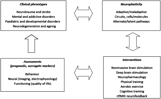
Neuroplasticity can be defined as the ability of the nervous system to respond to intrinsic or extrinsic stimuli by reorganizing its structure, function and connections. Major advances in the understanding of neuroplasticity have to date yielded few established interventions. To advance the translation of neuroplasticity research towards clinical applications, the National Institutes of Health Blueprint for Neuroscience Research sponsored a workshop in 2009. Basic and clinical researchers in disciplines from central nervous system injury/stroke, mental/addictive disorders, paediatric/developmental disorders and neurodegeneration/ageing identified cardinal examples of neuroplasticity, underlying mechanisms, therapeutic implications and common denominators. Promising therapies that may enhance training-induced cognitive and motor learning, such as brain stimulation and neuropharmacological interventions, were identified, along with questions of how best to use this body of information to reduce human disability. Improved understanding of adaptive mechanisms at every level, from molecules to synapses, to networks, to behaviour, can be gained from iterative collaborations between basic and clinical researchers. Lessons can be gleaned from studying fields related to plasticity, such as development, critical periods, learning and response to disease. Improved means of assessing neuroplasticity in humans, including biomarkers for predicting and monitoring treatment response, are needed. Neuroplasticity occurs with many variations, in many forms, and in many contexts. However, common themes in plasticity that emerge across diverse central nervous system conditions include experience dependence, time sensitivity and the importance of motivation and attention. Integration of information across disciplines should enhance opportunities for the translation of neuroplasticity and circuit retraining research into effective clinical therapies.
Overview
The claims and statistics of this paper haven’t been checked yet.
Run the check — free, no account needed. It reads the open-access text and checks the sources it cites.
Integrity signals
Registry
Cites
Statistics
Figures
Verification
Claims the paper makes
The claims and statistics of this paper haven’t been checked yet.
Run the check — free, no account needed. It reads the open-access text and checks the sources it cites.
Claims other papers make about this one
Each is checked against this paper’s own text: does it say what the citing paper says it does?
The source backs part of the claim, not all of it — or backs it for a different population, condition or comparison.
In the citing paper
“yielding larger and long-lasting functional improvements that modify the impaired system (Cramer et al., 2011).”
From Cognitive Neuroscience of Attention Deficit Hyperactivity Disorder (ADHD) and Its Clinical Translation (2018) · cites this paper as Cramer et al., 2011
Experimental — accuracy not yet measuredThe cited source says what the claim attributes to it.
In the citing paper
“while these same external factors change the ways our brains operate, including neuronal structures and functions, as indicated by studies into neuroplasticity (Fuchs and Flügge 2014;Cramer et al. 2011).”
From Authenticity and Love Drugs: Insights from Psychedelic Therapy (2026) · cites this paper as Cramer et al. 2011
In the citing paper
“In the nervous system, integrins are essential molecules for neuroplasticity, i.e. the ability to adapt to internal and external stimuli by reorganizing its structure, function and connections [49].”
From Gene expression profiles underlying aggressive behavior in the prefrontal cortex of cattle (2021) · cites this paper as [49]
Self-check
Percentages, sums and reported differences re-computed from the paper’s own numbers.
The self-check needs the paper’s full text.
Citations
Cites
1 retracted
Persson, J., & Reuter‐Lorenz, P. A. (2008) · Psychological Science · doi:10.1111/j.1467-9280.2008.02172.x · Cited by 109
Self-citationRetractedKoob, G. F., & Volkow, N. D. (2010) · Neuropsychopharmacology · doi:10.1038/npp.2009.110 · Cited by 5,340
Deep Brain Stimulation for Treatment-Resistant Depression
Mayberg, H. S., Lozano, A. M., Voon, V., McNeely, H. E., Seminowicz, D., Hamani, C., Schwalb, J. M., & Kennedy, S. H. (2005) · Neuron · doi:10.1016/j.neuron.2005.02.014 · Cited by 4,059
Self-citationShankar, G. M., Li, S., Mehta, T. H., Garcia-Munoz, A., Shepardson, N. E., Smith, I., Brett, F. M., Farrell, M. A., Rowan, M. J., Lemere, C. A., et al. (2008) · Nature Medicine · doi:10.1038/nm1782 · Cited by 3,685
Be smart, exercise your heart: Exercise effects on brain and cognition
Hillman, C. H., Erickson, K. I., & Kramer, A. F. (2008) · Nature Reviews Neuroscience · doi:10.1038/nrn2298 · Cited by 3,563
Self-citationThe Singularity Is near: When Humans Transcend Biology
Mead, W. R., & Kurzweil, R. (2006) · Foreign Affairs · doi:10.2307/20031996 · Cited by 3,418
AN IMPLICIT TECHNOLOGY OF GENERALIZATION 1
Stokes, T. F., & Baer, D. M. (1977) · Journal of Applied Behavior Analysis · doi:10.1901/jaba.1977.10-349 · Cited by 3,351
The adaptive brain: aging and neurocognitive scaffolding
Park, D. C., & Reuter-Lorenz, P. (2009) · Annual Review of Psychology · doi:10.1146/annurev.psych.59.103006.093656 · Cited by 2,790
Self-citationExercise: a behavioral intervention to enhance brain health and plasticity
Cotman, C. (2002) · Trends in Neurosciences · doi:10.1016/s0166-2236(02)02143-4 · Cited by 2,739
Aerobic Exercise Training Increases Brain Volume in Aging Humans
Colcombe, S. J., Erickson, K. I., Scalf, P. E., Kim, J. S., Prakash, R., McAuley, E., Elavsky, S., Marquez, D. X., Hu, L., & Kramer, A. F. (2006) · The Journals of Gerontology Series A: Biological Sciences and Medical Sciences · doi:10.1093/gerona/61.11.1166 · Cited by 2,134
Self-citation
Research works printed but not linked: 15
- Belanger, 2007, Journal of Neuropsychiatry and Clinical Neurosciences, 19, 5, 10.1176/appi.neuropsych.19.1.5 · DOI
- 2002, 111, 761
- 2004, 44, 411
- 2004, 124, 71
- 2009, PSYCHOL SCI PUBLIC INT, 9, 1
- 2004, Neurotoxicity research, 6, 223
- 2009, 56, 169
- Kennedy, 2007, American Journal of Psychiatry, 164, 778, 10.1176/appi.ajp.164.5.778 · DOI · Paper page
- 2008, NEUROPHARMACOL REV, 33, 134
- 2005, 45, 651
- Safiulina, 2010, 2, 1
- 2009, 46, 379
- 2009, 50, 30
- 2004, 47, 33
- Wolf, 2002, The neurologist, 8, 325, 10.1097/00127893-200211000-00001 · DOI
Cited by
Sorted by citations
Psychedelics promote structural and functional neural plasticity
Ly, C., Greb, A. C., Cameron, L. P., Wong, J. M., Barragan, E. V., Wilson, P. C., Burbach, K. F., Soltanzadeh Zarandi, S., Sood, A., Paddy, M. R., et al. (2018) · Cell Reports · doi:10.1016/j.celrep.2018.05.022 · Cited by 1,207
Colucci-D’Amato, L., Speranza, L., & Volpicelli, F. (2020) · International Journal of Molecular Sciences · doi:10.3390/ijms21207777 · Cited by 991
Ten years of theta burst stimulation in humans: Established knowledge, unknowns and prospects
Suppa, A., Huang, Y.-Z., Funke, K., Ridding, M. C., Cheeran, B., Di Lazzaro, V., Ziemann, U., & Rothwell, J. C. (2016) · Brain Stimulation · doi:10.1016/j.brs.2016.01.006 · Cited by 609
Rubia, K. (2018) · Frontiers in Human Neuroscience · doi:10.3389/fnhum.2018.00100 · Cited by 384
“…improve task performance (Kuo and Nitsche, 2012), presumably via boosting training-induced plasticity through the addition of stimulation-induced plasticity (Ziemann and Siebner, 2008), yielding larger and long-lasting functional improvements that modify the impaired system (Cramer et al., 2011).”
BackgroundTowards an understanding of psychedelic-induced neuroplasticity
Calder, A. E., & Hasler, G. (2023) · Neuropsychopharmacology · doi:10.1038/s41386-022-01389-z · Cited by 313
Neurotrophic factors and neuroplasticity pathways in the pathophysiology and treatment of depression
Levy, M. J. F., Boulle, F., Steinbusch, H. W., van den Hove, D. L. A., Kenis, G., & Lanfumey, L. (2018) · Psychopharmacology · doi:10.1007/s00213-018-4950-4 · Cited by 308
From Serotonin to Neuroplasticity: Evolvement of Theories for Major Depressive Disorder
Liu, B., Liu, J., Wang, M., Zhang, Y., & Li, L. (2017) · Frontiers in Cellular Neuroscience · doi:10.3389/fncel.2017.00305 · Cited by 249
Psychedelics in Psychiatry: Neuroplastic, Immunomodulatory, and Neurotransmitter Mechanisms
Inserra, A., De Gregorio, D., & Gobbi, G. (2021) · Pharmacological Reviews · doi:10.1124/pharmrev.120.000056 · Cited by 232
Activity-dependent neural plasticity from bench to bedside
Ganguly, K., & Poo, M.-M. (2013) · Neuron · doi:10.1016/j.neuron.2013.10.028 · Cited by 202
Donse, L., Padberg, F., Sack, A. T., Rush, A. J., & Arns, M. (2018) · Brain Stimulation · doi:10.1016/j.brs.2017.11.004 · Cited by 183
Tolahunase, M. R., Sagar, R., Faiq, M., & Dada, R. (2018) · Restorative Neurology and Neuroscience · doi:10.3233/rnn-170810 · Cited by 167
Spike-Timing-Dependent Plasticity in Primate Corticospinal Connections Induced during Free Behavior
Nishimura, Y., Perlmutter, S. I., Eaton, R. W., & Fetz, E. E. (2013) · Neuron · doi:10.1016/j.neuron.2013.08.028 · Cited by 145
Self-citationResilience: The Science of Mastering Life's Greatest Challenges
Southwick, S., & Charney, D. (2018) · doi:10.1017/9781108349246 · Cited by 153
Richmond, L. L., Wolk, D., Chein, J., & Olson, I. R. (2014) · Journal of Cognitive Neuroscience · doi:10.1162/jocn_a_00657 · Cited by 143
Meaningful Task-Specific Training (MTST) for stroke rehabilitation:A randomized controlled trial
Narayan Arya, K., Verma, R., Garg, R. K., Sharma, V. P., Agarwal, M., & Aggarwal, G. G. (2012) · Topics in Stroke Rehabilitation · doi:10.1310/tsr1903-193 · Cited by 136
The Dynamic Brain: Neuroplasticity and Mental Health
Kays, J. L., Hurley, R. A., & Taber, K. H. (2012) · The Journal of Neuropsychiatry and Clinical Neurosciences · doi:10.1176/appi.neuropsych.12050109 · Cited by 128
New developments in the understanding and management of persistent pain
Flor, H. (2012) · Current Opinion in Psychiatry · doi:10.1097/yco.0b013e3283503510 · Cited by 125
Does Psychedelic Therapy Have a Transdiagnostic Action and Prophylactic Potential?
Kočárová, R., Horáček, J., & Carhart-Harris, R. (2021) · Frontiers in Psychiatry · doi:10.3389/fpsyt.2021.661233 · Cited by 96
Solomon, M., Yoon, J. H., Ragland, J. D., Niendam, T. A., Lesh, T. A., Fairbrother, W., & Carter, C. S. (2014) · Biological Psychiatry · doi:10.1016/j.biopsych.2013.08.036 · Cited by 78
Kang, M. J. Y., Hawken, E., & Vazquez, G. H. (2022) · Frontiers in Psychiatry · doi:10.3389/fpsyt.2022.860882 · Cited by 77
Fisher, M., Loewy, R., Hardy, K., Schlosser, D., & Vinogradov, S. (2013) · Annual Review of Clinical Psychology · doi:10.1146/annurev-clinpsy-032511-143134 · Cited by 71
Self-citationCarey, L., Walsh, A., Adikari, A., Goodin, P., Alahakoon, D., De Silva, D., Ong, K.-L., Nilsson, M., & Boyd, L. (2019) · Neural Plasticity · doi:10.1155/2019/5232374 · Cited by 68
Deckersbach, T., Das, S. K., Urban, L. E., Salinardi, T., Batra, P., Rodman, A. M., Arulpragasam, A. R., Dougherty, D. D., & Roberts, S. B. (2014) · Nutrition & Diabetes · doi:10.1038/nutd.2014.26 · Cited by 55
Cognitive Remediation in Schizophrenia
Kaneko, Y., & Keshavan, M. (2012) · Clinical Psychopharmacology and Neuroscience · doi:10.9758/cpn.2012.10.3.125 · Cited by 51
Chen, H., Dong, G., & Li, K. (2023) · Frontiers in Psychiatry · doi:10.3389/fpsyt.2023.1094583 · Cited by 47
Li, X., Zhao, J., Li, Z., Zhang, L., & Huo, Z. (2020) · Neural Plasticity · doi:10.1155/2020/8850653 · Cited by 35
Gene expression profiles underlying aggressive behavior in the prefrontal cortex of cattle
Eusebi, P. G., Sevane, N., O’Rourke, T., Pizarro, M., Boeckx, C., & Dunner, S. (2021) · BMC Genomics · doi:10.1186/s12864-021-07505-5 · Cited by 24
Fang, P., Gao, Y., Li, Y., Li, C., Zhang, T., Wu, L., Zhu, Y., & Xie, Y. (2024) · NeuroImage · doi:10.1016/j.neuroimage.2024.120785 · Cited by 18
“It embodies the capability of the brain to reorganize itself by forming new neural connections (Cramer et al., 2011).”
BackgroundTamayo-Serrano, P., Garbaya, S., Bouakaz, S., & Blazevic, P. (2020) · IEEE Systems, Man, and Cybernetics Magazine · doi:10.1109/msmc.2020.3002519 · Cited by 16
Noga, B. R., & Guest, J. D. (2021) · Current Opinion in Neurology · doi:10.1097/wco.0000000000000999 · Cited by 14
Quiles, V., Ferrero, L., Iáñez, E., Ortiz, M., Cano, J. M., & Azorín, J. M. (2022) · Applied Sciences · doi:10.3390/app12010415 · Cited by 10
Wynn, J. K., & Green, M. F. (2024) · Clinical EEG and Neuroscience · doi:10.1177/15500594241252897 · Cited by 3
Wang, W., Wen, X., Zhang, Y., Wang, H., Hu, L., Tan, C., & Liu, Z. (2026) · Frontiers in Human Neuroscience · doi:10.3389/fnhum.2026.1924031
Authenticity and Love Drugs: Insights from Psychedelic Therapy
Thomas, O., & Silva, F. (2026) · AJOB Neuroscience · doi:10.1080/21507740.2025.2517007
Metadata
- Authors
- Cramer, S. C., Sur, M., Dobkin, B. H., O'Brien, C., Sanger, T. D., Trojanowski, J. Q., Rumsey, J. M., Hicks, R., Cameron, J., Chen, D., Chen, W. G., Cohen, L. G., deCharms, C., Duffy, C. J., Eden, G. F., Fetz, E. E., Filart, R., Freund, M., Grant, S. J., Haber, S., Kalivas, P. W., Kolb, B., Kramer, A. F., Lynch, M., Mayberg, H. S., McQuillen, P. S., Nitkin, R., Pascual-Leone, A., Reuter-Lorenz, P., Schiff, N., Sharma, A., Shekim, L., Stryker, M., Sullivan, E. V., & Vinogradov, S.
- Affiliations
- University of California, Irvine (US)
- University of California, Los Angeles (US)
- University of Southern California (US)
- National Institutes of Health (US)
- Oregon Health & Science University (US)
- Omneuron (United States) (US)
- National Institute of Mental Health (US)
- Medical University of South Carolina (US)
- University of Lethbridge (CA)
- University of Illinois Urbana-Champaign (US)
- Emory University (US)
- University of Colorado Boulder (US)
- University of California, San Francisco (US)
- Corresponding author
- Cramer, S. C.
- ORCID
- Venue
- Brain
- Publisher
- Oxford University Press (OUP)
- Published
- 2011-04-10
- Volume
- 134
- Issue
- 6
- Pages
- 1591-1609
- ISSN
- 0006-8950 (ISSN-L)
- 1460-2156
- PMC
- PMC3102236
- OpenAlex
- W2107022859
- Cited by
- 1,259
- References
- 228
- Citations by year
- 2026: 92 · 2025: 116 · 2024: 93 · 2023: 89 · 2022: 99 · 2021: 93 · 2020: 130 · 2019: 80 · 2018: 53 · 2017: 79 · 2016: 69 · 2015: 69 · 2014: 85 · 2013: 58 · 2012: 43
- FWCI
- 34.98
- Type
- journal article
- Language
- en
- Open access
- bronze
- doi.org
- Locations
- Brain (publishedVersion)
- PubMed Central (publishedVersion, cc-by-nc)
- PubMed (publishedVersion)
- DSpace@MIT (Massachusetts Institute of Technology) (submittedVersion, cc-by-nc)
- eScholarship (California Digital Library) (submittedVersion)
- Funders
- U.S. Department of Health and Human Services
- Pfizer
- GlaxoSmithKline
- Gilead Sciences
- National Institutes of Health
- Allergan
- National Institute on Alcohol Abuse and Alcoholism (R37AA010723, K05AA017168)
- National Institute on Aging (R01AG036682, R37AG025667)
- Eunice Kennedy Shriver National Institute of Child Health and Human Development (R01HD069381)
- Topics
- Life Sciences › Neuroscience › Neurology › Transcranial Magnetic Stimulation Studies
- Life Sciences › Neuroscience › Cellular and Molecular Neuroscience › Neuroscience and Neuropharmacology Research
- Life Sciences › Neuroscience › Developmental Neuroscience › Neurogenesis and neuroplasticity mechanisms
- Keywords
- neuroplasticity, clinical translation, brain stimulation, stroke, central nervous system injury, mental illness, addiction, neurodegeneration, aging, experience-dependent plasticity, critical period, treatment response biomarkers, disability reduction
- MeSH terms
- Aging, Animals, Brain Diseases, Cognition Disorders, Humans, Neuronal Plasticity, Recovery of Function, Stroke, Biomedical Research
About these checks
Registry, retraction, Cites and statistics results are computed, not judged, with one exception: a printed reference that no index lists is read once by a model before it counts as a citation error. Claims are extracted by a model and kept only when their quote appears verbatim in the text; each cited source is then checked, with a model’s help, for whether it says what the claim relies on. Those verdicts can be wrong — read the claim’s quote and the cited source before relying on one.
Statistics and arithmetic are recomputed, not judged.
Confidence bands are shown only for fields calibrated on our annotated test set. None is calibrated yet, so no band is shown.
What counts as a claim ›How we measure confidence ›How we check this ›
Are you an author, or do you see a problem with this page? Request a review
