

Mapping Human Laryngeal Motor Cortex during Vocalization
Open accessPublished version of a preprint
The DOI matches its Crossref record; 103 of the 103 references it cites were found in a registry or an index.
Abstract
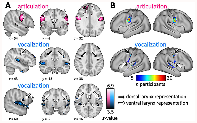
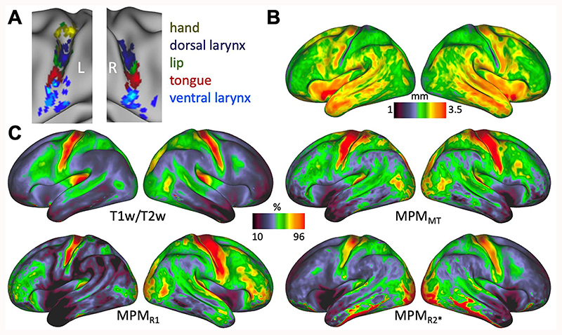
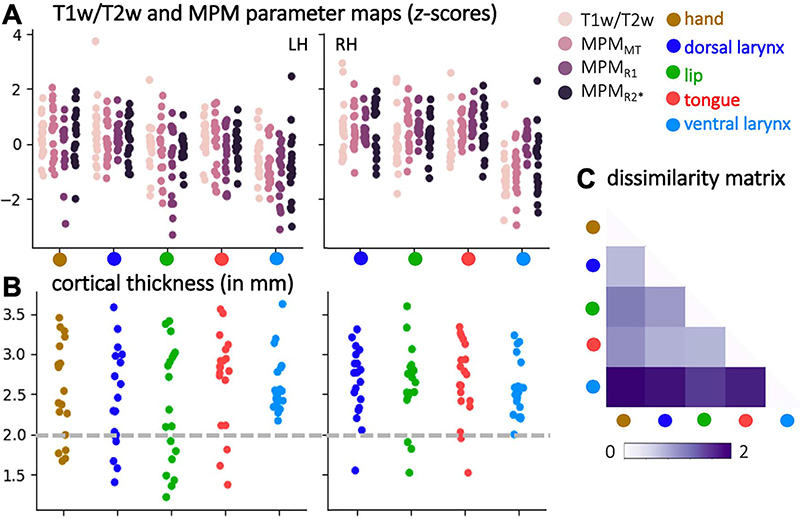
The representations of the articulators involved in human speech production are organized somatotopically in primary motor cortex. The neural representation of the larynx, however, remains debated. Both a dorsal and a ventral larynx representation have been previously described. It is unknown, however, whether both representations are located in primary motor cortex. Here, we mapped the motor representations of the human larynx using functional magnetic resonance imaging and characterized the cortical microstructure underlying the activated regions. We isolated brain activity related to laryngeal activity during vocalization while controlling for breathing. We also mapped the articulators (the lips and tongue) and the hand area. We found two separate activations during vocalization—a dorsal and a ventral larynx representation. Structural and quantitative neuroimaging revealed that myelin content and cortical thickness underlying the dorsal, but not the ventral larynx representation, are similar to those of other primary motor representations. This finding confirms that the dorsal larynx representation is located in primary motor cortex and that the ventral one is not. We further speculate that the location of the ventral larynx representation is in premotor cortex, as seen in other primates. It remains unclear, however, whether and how these two representations differentially contribute to laryngeal motor control.
Overview
The claims and statistics of this paper haven’t been checked yet.
Free: it reads the open-access text and checks the first 5 claims that cite a source we can check. Create a free account to check every claim.
Integrity signals
Registry
Cites
Statistics
Figures


Verification
Claims the paper makes
The claims and statistics of this paper haven’t been checked yet.
Free: it reads the open-access text and checks the first 5 claims that cite a source we can check. Create a free account to check every claim.
Claims other papers make about this one
Each is checked against this paper’s own text: does it say what the citing paper says it does?
The source backs part of the claim, not all of it — or backs it for a different population, condition or comparison.
In the citing paper
“Subsequent work discovered that SMC populations encode articulatory kinematic trajectories and vocal-tract configurations43 during continuous speech24,39,44,45 (Fig. 1; Mugler et al. 2018, Chartier et al. 2018 and Dichter et al. 2018).”
From The speech neuroprosthesis (2024) · cites this paper as [39]
Experimental — accuracy not yet measuredThe cited source says what the claim attributes to it.
In the citing paper
“In healthy speakers, neural representations in the SMC (comprising the precentral and postcentral gyri) encode articulatory movements of the orofacial musculature22,24,30.”
From A high-performance neuroprosthesis for speech decoding and avatar control (2023) · cites this paper as [30]
Experimental — accuracy not yet measuredThe cited source says what the claim attributes to it.
In the citing paper
“many studies have already found that multiple myelin markers can provide complementary information (Callaghan, Freund, Draganski, Anderson, Cappelletti, Chowdhury, Diedrichsen, Fitzgerald, Smittenaar, Helms, Lutti, Weiskopf, 2014, Eichert et al., 2020, Kolind, Laule, Vavasour, Li, Traboulsee, Mädler, Moore, Mackay, 2008, Lipp, Jones, Bells, Sgarlata, Foster, Stickland, Davidson, Tallantyre, Rober”
From Can MRI measure myelin? Systematic review, qualitative assessment, and meta-analysis of studies validating microstructural imaging with myelin histology (2021) · cites this paper as Eichert et al., 2020
Self-check
Percentages, sums and reported differences re-computed from the paper’s own numbers.
The self-check needs the paper’s full text.
Citations
Cites
Fitting Linear Mixed-Effects Models Using lme4
Bates, D., Mächler, M., Bolker, B., & Walker, S. (2015) · Journal of Statistical Software · doi:10.18637/jss.v067.i01 · Cited by 86,835
Jenkinson, M., Bannister, P., Brady, M., & Smith, S. (2002) · NeuroImage · doi:10.1006/nimg.2002.1132 · Cited by 11,888
Jenkinson, M., Beckmann, C. F., Behrens, T. E. J., Woolrich, M. W., & Smith, S. M. (2012) · NeuroImage · doi:10.1016/j.neuroimage.2011.09.015 · Cited by 11,314
Fast robust automated brain extraction
Smith, S. M. (2002) · Human Brain Mapping · doi:10.1002/hbm.10062 · Cited by 10,496
Fischl, B. (2012) · NeuroImage · doi:10.1016/j.neuroimage.2012.01.021 · Cited by 9,526
The organization of the human cerebellum estimated by intrinsic functional connectivity
Buckner, R. L., Krienen, F. M., Castellanos, A., Diaz, J. C., & Yeo, B. T. T. (2011) · Journal of Neurophysiology · doi:10.1152/jn.00339.2011 · Cited by 7,304
A global optimisation method for robust affine registration of brain images
Jenkinson, M., & Smith, S. (2001) · Medical Image Analysis · doi:10.1016/s1361-8415(01)00036-6 · Cited by 6,872
Measuring the thickness of the human cerebral cortex from magnetic resonance images
Fischl, B., & Dale, A. M. (2000) · Proceedings of the National Academy of Sciences · doi:10.1073/pnas.200033797 · Cited by 5,818
The minimal preprocessing pipelines for the Human Connectome Project
Glasser, M. F., Sotiropoulos, S. N., Wilson, J. A., Coalson, T. S., Fischl, B., Andersson, J. L., Xu, J., Jbabdi, S., Webster, M., Polimeni, J. R., et al. (2013) · NeuroImage · doi:10.1016/j.neuroimage.2013.04.127 · Cited by 5,253
PENFIELD, W., & BOLDREY, E. (1937) · Brain · doi:10.1093/brain/60.4.389 · Cited by 4,188
Temporal Autocorrelation in Univariate Linear Modeling of FMRI Data
Woolrich, M. W., Ripley, B. D., Brady, M., & Smith, S. M. (2001) · NeuroImage · doi:10.1006/nimg.2001.0931 · Cited by 2,852
The Adolescent Brain Cognitive Development (ABCD) study: Imaging acquisition across 21 sites
Casey, B. J., Cannonier, T., Conley, M. I., Cohen, A. O., Barch, D. M., Heitzeg, M. M., Soules, M. E., Teslovich, T., Dellarco, D. V., Garavan, H., et al. (2018) · Developmental Cognitive Neuroscience · doi:10.1016/j.dcn.2018.03.001 · Cited by 2,204
Multilevel linear modelling for FMRI group analysis using Bayesian inference
Woolrich, M. W., Behrens, T. E. J., Beckmann, C. F., Jenkinson, M., & Smith, S. M. (2004) · NeuroImage · doi:10.1016/j.neuroimage.2003.12.023 · Cited by 1,728
Motor Areas of the Medial Wall: A Review of Their Location and Functional Activation
Picard, N., & Strick, P. L. (1996) · Cerebral Cortex · doi:10.1093/cercor/6.3.342 · Cited by 1,712
Function in the human connectome: task-fMRI and individual differences in behavior
Barch, D. M., Burgess, G. C., Harms, M. P., Petersen, S. E., Schlaggar, B. L., Corbetta, M., Glasser, M. F., Curtiss, S., Dixit, S., Feldt, C., et al. (2013) · NeuroImage · doi:10.1016/j.neuroimage.2013.05.033 · Cited by 1,529
Mapping Human Cortical Areas In Vivo Based on Myelin Content as Revealed by T1- and T2-Weighted MRI
Glasser, M. F., & Van Essen, D. C. (2011) · The Journal of Neuroscience · doi:10.1523/jneurosci.2180-11.2011 · Cited by 1,447
A probabilistic MR atlas of the human cerebellum
Diedrichsen, J., Balsters, J. H., Flavell, J., Cussans, E., & Ramnani, N. (2009) · NeuroImage · doi:10.1016/j.neuroimage.2009.01.045 · Cited by 1,359
Sikweyiya, Y., & Jewkes, R. (2012) · PLoS ONE · doi:10.1371/journal.pone.0035495 · Cited by 1,310
Positron Emission Tomographic Studies of the Processing of Singe Words
Petersen, S. E., Fox, P. T., Posner, M. I., Mintun, M., & Raichle, M. E. (1989) · Journal of Cognitive Neuroscience · doi:10.1162/jocn.1989.1.2.153 · Cited by 1,289
Cluster-extent based thresholding in fMRI analyses: Pitfalls and recommendations
Woo, C.-W., Krishnan, A., & Wager, T. D. (2014) · NeuroImage · doi:10.1016/j.neuroimage.2013.12.058 · Cited by 1,253 · cited 2×
“Interpreting group-level 109 results, however, might obscure subject-specific features and inter-individual variability, which 110 limits sensitivity and functional resolution (Bennett and Miller 2010; Nieto-Castañón and 111 Fedorenko 2012; Bouchard et al. 2013; Woo et al. 2014).”
BackgroundThe Cerebral Cortex of Man: A Clinical Study of Localization of Function
(1968) · JAMA: The Journal of the American Medical Association · doi:10.1001/jama.1968.03150020096041 · Cited by 1,214
Neural pathways underlying vocal control
Jürgens, U. (2002) · Neuroscience & Biobehavioral Reviews · doi:10.1016/s0149-7634(01)00068-9 · Cited by 852
E2F1 Regulates Cellular Growth by mTORC1 Signaling
Real, S., Meo-Evoli, N., Espada, L., & Tauler, A. (2011) · PLoS ONE · doi:10.1371/journal.pone.0016163 · Cited by 814
Fast, automated, N‐dimensional phase‐unwrapping algorithm
Jenkinson, M. (2003) · Magnetic Resonance in Medicine · doi:10.1002/mrm.10354 · Cited by 695
Functional organization of human sensorimotor cortex for speech articulation
Bouchard, K. E., Mesgarani, N., Johnson, K., & Chang, E. F. (2013) · Nature · doi:10.1038/nature11911 · Cited by 686 · cited 4×
“These studies also show activity in both a dorsal and a ventral region during speech production (Bouchard et al. 2013; Chang et al. 2013; Toyoda et al. 2014; Breshears et al. 2015; Dichter et al. 2018).”
BackgroundInfluentialHow reliable are the results from functional magnetic resonance imaging?
Bennett, C. M., & Miller, M. B. (2010) · Annals of the New York Academy of Sciences · doi:10.1111/j.1749-6632.2010.05446.x · Cited by 577
Devlin, J. T., Matthews, P. M., & Rushworth, M. F. S. (2003) · Journal of Cognitive Neuroscience · doi:10.1162/089892903321107837 · Cited by 576
OBSERVATIONS ON THE EXCITABLE CORTEX OF THE CHIMPANZEE, ORANG‐UTAN, AND GORILLA
Leyton, A. S. F., & Sherrington, C. S. (1917) · Quarterly Journal of Experimental Physiology · doi:10.1113/expphysiol.1917.sp000240 · Cited by 568
An fMRI investigation of syllable sequence production
Bohland, J. W., & Guenther, F. H. (2006) · NeuroImage · doi:10.1016/j.neuroimage.2006.04.173 · Cited by 542
Quantitative multi-parameter mapping of R1, PD*, MT, and R2* at 3T: a multi-center validation
Weiskopf, N., Suckling, J., Williams, G., Correia, M. M., Inkster, B., Tait, R., Ooi, C., Bullmore, E. T., & Lutti, A. (2013) · Frontiers in Neuroscience · doi:10.3389/fnins.2013.00095 · Cited by 538
Convergent transcriptional specializations in the brains of humans and song-learning birds
Pfenning, A. R., Hara, E., Whitney, O., Rivas, M. V., Wang, R., Roulhac, P. L., Howard, J. T., Wirthlin, M., Lovell, P. V., Ganapathy, G., et al. (2014) · Science · doi:10.1126/science.1256846 · Cited by 463
Trends and properties of human cerebral cortex: Correlations with cortical myelin content
Glasser, M. F., Goyal, M. S., Preuss, T. M., Raichle, M. E., & Van Essen, D. C. (2014) · NeuroImage · doi:10.1016/j.neuroimage.2013.03.060 · Cited by 456
Using high-resolution quantitative mapping of R1 as an index of cortical myelination
Lutti, A., Dick, F., Sereno, M. I., & Weiskopf, N. (2014) · NeuroImage · doi:10.1016/j.neuroimage.2013.06.005 · Cited by 346 · cited 4×
“The use of high-resolution quantitative neuroimaging allows us to characterize cortical architecture noninvasively (Weiskopf et al. 2013; Lutti et al. 2014).”
BackgroundMethodInfluentialEvolution of vocal learning and spoken language
Jarvis, E. D. (2019) · Science · doi:10.1126/science.aax0287 · Cited by 304
Gennatas, E. D., Avants, B. B., Wolf, D. H., Satterthwaite, T. D., Ruparel, K., Ciric, R., Hakonarson, H., Gur, R. E., & Gur, R. C. (2017) · The Journal of Neuroscience · doi:10.1523/jneurosci.3550-16.2017 · Cited by 302
Foerster, O. (1931) · The Lancet · Cited by 301
Draganski, B., Ashburner, J., Hutton, C., Kherif, F., Frackowiak, R. S. J., Helms, G., & Weiskopf, N. (2011) · NeuroImage · doi:10.1016/j.neuroimage.2011.01.052 · Cited by 294
Callaghan, M. F., Freund, P., Draganski, B., Anderson, E., Cappelletti, M., Chowdhury, R., Diedrichsen, J., FitzGerald, T. H. B., Smittenaar, P., Helms, G., et al. (2014) · Neurobiology of Aging · doi:10.1016/j.neurobiolaging.2014.02.008 · Cited by 292 · cited 3×
“The three MPM parameter maps have been shown to correlate to different degrees with myelin content in white matter, subcortical structures and grey matter (Draganski et al. 2011; Callaghan et al. 2014; Lutti et al. 2014; Bagnato et al. 2018).”
BackgroundA larynx area in the human motor cortex
Brown, S., Ngan, E., & Liotti, M. (2008) · Cerebral Cortex · doi:10.1093/cercor/bhm131 · Cited by 286 · cited 2×
“Using the neutral vowel schwa or glottal stops instead (Gick 2002; Loucks et al. 2007; Brown et al. 2008; Grabski et al. 2012; Belyk et al. 2018), however, might have caused pharynx activity that would have been more difficult to dissociate.”
BackgroundNeural correlates of voluntary breathing in humans
McKay, L. C., Evans, K. C., Frackowiak, R. S. J., & Corfield, D. R. (2003) · Journal of Applied Physiology · doi:10.1152/japplphysiol.00641.2002 · Cited by 280
Identification of calcification with MRI using susceptibility‐weighted imaging: A case study
Wu, Z., Mittal, S., Kish, K., Yu, Y., Hu, J., & Haacke, E. M. (2009) · Journal of Magnetic Resonance Imaging · doi:10.1002/jmri.21617 · Cited by 260
Empirical approaches to the study of language evolution
Fitch, W. T. (2017) · Psychonomic Bulletin & Review · doi:10.3758/s13423-017-1236-5 · Cited by 259
Human cortical sensorimotor network underlying feedback control of vocal pitch
Chang, E. F., Niziolek, C. A., Knight, R. T., Nagarajan, S. S., & Houde, J. F. (2013) · Proceedings of the National Academy of Sciences · doi:10.1073/pnas.1216827110 · Cited by 258
hMRI – A toolbox for quantitative MRI in neuroscience and clinical research
Tabelow, K., Balteau, E., Ashburner, J., Callaghan, M. F., Draganski, B., Helms, G., Kherif, F., Leutritz, T., Lutti, A., Phillips, C., et al. (2019) · NeuroImage · doi:10.1016/j.neuroimage.2019.01.029 · Cited by 256
Nieto-Castañón, A., & Fedorenko, E. (2012) · NeuroImage · doi:10.1016/j.neuroimage.2012.06.065 · Cited by 247 · cited 1×
“Interpreting group-level results, however, might obscure subject-specific features and inter-individual variability, which limits sensitivity and functional resolution (Bennett and Miller 2010; Nieto-Castañón and Fedorenko 2012; Bouchard et al. 2013; Woo et al. 2014).”
BackgroundPreibisch, C., & Deichmann, R. (2009) · Magnetic Resonance in Medicine · doi:10.1002/mrm.21776 · Cited by 230
Ackermann, H., Hage, S. R., & Ziegler, W. (2014) · Behavioral and Brain Sciences · doi:10.1017/s0140525x13003099 · Cited by 215
The Cerebral Cortex of Man: Clinical Study of Localization of Function
&NA; (1950) · Academic Medicine · doi:10.1097/00001888-195009000-00037 · Cited by 214
The somatotopy of speech: phonation and articulation in the human motor cortex
Brown, S., Laird, A. R., Pfordresher, P. Q., Thelen, S. M., Turkeltaub, P., & Liotti, M. (2009) · Brain and Cognition · doi:10.1016/j.bandc.2008.12.006 · Cited by 210
Overt and imagined singing of an Italian aria
Kleber, B., Birbaumer, N., Veit, R., Trevorrow, T., & Lotze, M. (2007) · NeuroImage · doi:10.1016/j.neuroimage.2007.02.053 · Cited by 186
The Control of Vocal Pitch in Human Laryngeal Motor Cortex
Dichter, B. K., Breshears, J. D., Leonard, M. K., & Chang, E. F. (2018) · Cell · doi:10.1016/j.cell.2018.05.016 · Cited by 183 · cited 6×
“We suggest that pitch control is facilitated by the dorsal larynx representation, which is located in primary motor cortex (Kleber et al. 2013; Dichter et al. 2018; Finkel et al. 2019).”
BackgroundInfluentialCentral nervous system control of the laryngeal muscles in humans
Ludlow, C. L. (2005) · Respiratory Physiology & Neurobiology · doi:10.1016/j.resp.2005.04.015 · Cited by 175
Neurons Controlling Voluntary Vocalization in the Macaque Ventral Premotor Cortex
Coudé, G., Ferrari, P. F., Rodà, F., Maranesi, M., Borelli, E., Veroni, V., Monti, F., Rozzi, S., & Fogassi, L. (2011) · PLoS ONE · doi:10.1371/journal.pone.0026822 · Cited by 170
Grabski, K., Lamalle, L., Vilain, C., Schwartz, J., Vallée, N., Tropres, I., Baciu, M., Le Bas, J., & Sato, M. (2012) · Human Brain Mapping · doi:10.1002/hbm.21363 · Cited by 170
The laryngeal motor cortex: its organization and connectivity
Simonyan, K. (2014) · Current Opinion in Neurobiology · doi:10.1016/j.conb.2014.05.006 · Cited by 169
Clustered functional MRI of overt speech production
Sörös, P., Sokoloff, L. G., Bose, A., McIntosh, A. R., Graham, S. J., & Stuss, D. T. (2006) · NeuroImage · doi:10.1016/j.neuroimage.2006.02.046 · Cited by 168
Optimization and validation of methods for mapping of the radiofrequency transmit field at 3T
Lutti, A., Hutton, C., Finsterbusch, J., Helms, G., & Weiskopf, N. (2010) · Magnetic Resonance in Medicine · doi:10.1002/mrm.22421 · Cited by 166
Evans, K. C., Shea, S. A., & Saykin, A. J. (1999) · The Journal of Physiology · doi:10.1111/j.1469-7793.1999.00383.x · Cited by 165
Grydeland, H., Vértes, P. E., Váša, F., Romero-Garcia, R., Whitaker, K., Alexander-Bloch, A. F., Bjørnerud, A., Patel, A. X., Sederevičius, D., Tamnes, C. K., et al. (2019) · Cerebral Cortex · doi:10.1093/cercor/bhy330 · Cited by 164
Ramsay, S. C., Adams, L., Murphy, K., Corfield, D. R., Grootoonk, S., Bailey, D. L., Frackowiak, R. S., & Guz, A. (1993) · The Journal of Physiology · doi:10.1113/jphysiol.1993.sp019503 · Cited by 162
Loucks, T. M. J., Poletto, C. J., Simonyan, K., Reynolds, C. L., & Ludlow, C. L. (2007) · NeuroImage · doi:10.1016/j.neuroimage.2007.01.049 · Cited by 150
Brain evolution by brain pathway duplication
Chakraborty, M., & Jarvis, E. D. (2015) · Philosophical Transactions of the Royal Society B: Biological Sciences · doi:10.1098/rstb.2015.0056 · Cited by 150 · cited 1×
“Additional support for a duplication theory also comes from genetic profiling analyses comparing gene expressions in song-learning birds and humans (Pfenning et al. 2014; Chakraborty and Jarvis 2015).”
BackgroundCerebral areas associated with motor control of speech in humans
Murphy, K., Corfield, D. R., Guz, A., Fink, G. R., Wise, R. J. S., Harrison, J., & Adams, L. (1997) · Journal of Applied Physiology · doi:10.1152/jappl.1997.83.5.1438 · Cited by 149
Robust and Fast Whole Brain Mapping of the RF Transmit Field B1 at 7T
Lutti, A., Stadler, J., Josephs, O., Windischberger, C., Speck, O., Bernarding, J., Hutton, C., & Weiskopf, N. (2012) · PLoS ONE · doi:10.1371/journal.pone.0032379 · Cited by 146
The origins of the vocal brain in humans
Belyk, M., & Brown, S. (2017) · Neuroscience & Biobehavioral Reviews · doi:10.1016/j.neubiorev.2017.03.014 · Cited by 144 · cited 6×
“These various findings have led to the proposal of an evolutionary “duplication and migration” hypothesis that the larynx motor cortex (LMC) comprises two structures located dorsally and ventrally in the human brain (Belyk and Brown 2017; Jarvis 2019; reviewed in Mars et al. 2018).”
BackgroundResultInfluentialThe classic supplementary motor area is formed by two independent areas.
Rizzolatti, G., Luppino, G., & Matelli, M. (1996) · PubMed · Cited by 140
Creation of an Open-Access, Mutation-Defined Fibroblast Resource for Neurological Disease Research
Wray, S., Self, M., NINDS Parkinson's Disease iPSC Consortium, NINDS Huntington's Disease iPSC Consortium, NINDS ALS iPSC Consortium, Lewis, P. A., Taanman, J.-W., Ryan, N. S., Mahoney, C. J., Liang, Y., et al. (2012) · PLoS ONE · doi:10.1371/journal.pone.0043099 · Cited by 138
Efferent subcortical projections of the laryngeal motorcortex in the rhesus monkey
Simonyan, K., & Jürgens, U. (2003) · Brain Research · doi:10.1016/s0006-8993(03)02548-4 · Cited by 137
Kleber, B., Zeitouni, A. G., Friberg, A., & Zatorre, R. J. (2013) · Journal of Neuroscience · doi:10.1523/jneurosci.4418-12.2013 · Cited by 130
Features of Cortically Evoked Swallowing in the Awake Primate (Macaca fascicularis)
Martin, R. E., Kemppainen, P., Masuda, Y., Yao, D., Murray, G. M., & Sessle, B. J. (1999) · Journal of Neurophysiology · doi:10.1152/jn.1999.82.3.1529 · Cited by 129
Simonyan, K., Ostuni, J. L., Ludlow, C. L., & Horwitz, B. (2009) · Journal of Neuroscience · doi:10.1523/jneurosci.4897-09.2009 · Cited by 127
Fiez, J. A., & Raichle, M. E. (1997) · International review of neurobiology · doi:10.1016/s0074-7742(08)60354-2 · Cited by 118
Functional Properties of Neurons in the Primate Tongue Primary Motor Cortex During Swallowing
Martin, R. E., Murray, G. M., Kemppainen, P., Masuda, Y., & Sessle, B. J. (1997) · Journal of Neurophysiology · doi:10.1152/jn.1997.78.3.1516 · Cited by 115
Cortical motor representation of the laryngeal muscles inMacaca mulatta
Hast, M. H., Fischer, J. M., Wetzel, A. B., & Thompson, V. E. (1974) · Brain Research · doi:10.1016/0006-8993(74)91046-4 · Cited by 114
Ku¨nzle, H. (1978) · Neuroscience · doi:10.1016/0306-4522(78)90151-3 · Cited by 112
Kumar, V. V., Croxson, P. L., & Simonyan, K. (2016) · Journal of Neuroscience · doi:10.1523/jneurosci.3914-15.2016 · Cited by 108
A general linear relaxometry model of R1 using imaging data
Callaghan, M. F., Helms, G., Lutti, A., Mohammadi, S., & Weiskopf, N. (2014) · Magnetic Resonance in Medicine · doi:10.1002/mrm.25210 · Cited by 100
On the elicitability of vocalization from the cortical larynx area
Jürgens, U. (1974) · Brain Research · doi:10.1016/0006-8993(74)90853-1 · Cited by 97
Chang, E. F., Breshears, J. D., Raygor, K. P., Lau, D., Molinaro, A. M., & Berger, M. S. (2017) · Journal of Neurosurgery · doi:10.3171/2015.10.jns151087 · Cited by 92
Body Topography Parcellates Human Sensory and Motor Cortex
Kuehn, E., Dinse, J., Jakobsen, E., Long, X., Schäfer, A., Bazin, P., Villringer, A., Sereno, M. I., & Margulies, D. S. (2017) · Cerebral Cortex · doi:10.1093/cercor/bhx026 · Cited by 79
Weiskopf, N., Callaghan, M. F., Josephs, O., Lutti, A., & Mohammadi, S. (2014) · Frontiers in Neuroscience · doi:10.3389/fnins.2014.00278 · Cited by 75
Cortico-cortical projections of the motorcortical larynx area in the rhesus monkey
Simonyan, K., & Jürgens, U. (2002) · Brain Research · doi:10.1016/s0006-8993(02)02960-8 · Cited by 72
A probabilistic map of the human ventral sensorimotor cortex using electrical stimulation
Breshears, J. D., Molinaro, A. M., & Chang, E. F. (2015) · Journal of Neurosurgery · doi:10.3171/2014.11.jns14889 · Cited by 67
Carey, D., Krishnan, S., Callaghan, M. F., Sereno, M. I., & Dick, F. (2017) · Cerebral Cortex · doi:10.1093/cercor/bhw393 · Cited by 63 · cited 1×
“…distinct activation peaks that followed the predicted somatotopic organization in the mid-portion of the central sulcus with the tongue representation more ventral than the lip representation in both hemispheres (Figure 3A) (Penfield and Rasmussen 1950; Grabski et al. 2012; Carey et al. 2017).”
BackgroundUntangling the R2* contrast in multiple sclerosis: A combined MRI-histology study at 7.0 Tesla
Bagnato, F. R., Hametner, S., Boyd, E. C., Endmayr, V., Shi, Y., Ikonomidou, V. N., Chen, G., Pawate, S., Lassmann, H., Smith, S. A., et al. (2018) · PLoS ONE · doi:10.1371/journal.pone.0193839 · Cited by 63
Speech map in the human ventral sensory-motor cortex
Conant, D., Bouchard, K. E., & Chang, E. F. (2014) · Current Opinion in Neurobiology · doi:10.1016/j.conb.2013.08.015 · Cited by 62
Morphological and functional variability in central and subcentral motor cortex of the human brain
Eichert, N., Watkins, K. E., Mars, R. B., & Petrides, M. (2021) · Brain Structure and Function · doi:10.1007/s00429-020-02180-w · Cited by 58
Self-citationCortical Sensorimotor Control in Vocalization: A Functional Magnetic Resonance Imaging Study
Olthoff, A., Baudewig, J., Kruse, E., & Dechent, P. (2008) · The Laryngoscope · doi:10.1097/mlg.0b013e31817fd40f · Cited by 52
An X-Ray Investigation of Pharyngeal Constriction in American English Schwa
Gick, B. (2002) · Phonetica · doi:10.1159/000056204 · Cited by 48
Human primary motor cortex shows hemispheric specialization for speech
Terumitsu, M., Fujii, Y., Suzuki, K., Kwee, I. L., & Nakada, T. (2006) · Neuroreport · doi:10.1097/01.wnr.0000224778.97399.c4 · Cited by 45 · cited 3×
“In addition to activation of the dorsal larynx representation, multiple neuroimaging studies reported activation in a region that we refer to as ventral larynx representation (Terumitsu et al. 2006; Olthoff et al. 2008; Grabski et al. 2012).”
BackgroundResultConnectivity and the search for specializations in the language-capable brain
Mars, R. B., Eichert, N., Jbabdi, S., Verhagen, L., & Rushworth, M. F. S. (2018) · Current Opinion in Behavioral Sciences · doi:10.1016/j.cobeha.2017.11.001 · Cited by 41
Self-citationCortical representation of tympanic membrane movements due to pressure variation: An fMRI study
Job, A., Paucod, J., O’Beirne, G. A., & Delon‐Martin, C. (2010) · Human Brain Mapping · doi:10.1002/hbm.21063 · Cited by 35
Evidence of the voice-related cortical potential: An electroencephalographic study
Galgano, J., & Froud, K. (2008) · NeuroImage · doi:10.1016/j.neuroimage.2008.03.019 · Cited by 32
Synthetic quantitative MRI through relaxometry modelling
Callaghan, M. F., Mohammadi, S., & Weiskopf, N. (2016) · NMR in Biomedicine · doi:10.1002/nbm.3658 · Cited by 31
Neuromagnetic detection of the laryngeal area: Sensory-evoked fields to air-puff stimulation
Miyaji, H., Hironaga, N., Umezaki, T., Hagiwara, K., Shigeto, H., Sawatsubashi, M., Tobimatsu, S., & Komune, S. (2013) · NeuroImage · doi:10.1016/j.neuroimage.2013.11.008 · Cited by 30
Toyoda, G., Brown, E. C., Matsuzaki, N., Kojima, K., Nishida, M., & Asano, E. (2013) · Clinical Neurophysiology · doi:10.1016/j.clinph.2013.11.008 · Cited by 26
Somatotopy of the extrinsic laryngeal muscles in the human sensorimotor cortex
Belyk, M., & Brown, S. (2014) · Behavioural Brain Research · doi:10.1016/j.bbr.2014.05.048 · Cited by 26
Exploring the Effects of Working for Endowments on Behaviour in Standard Economic Games
Harrison, F., & Mouden, C. E. (2011) · PLoS ONE · doi:10.1371/journal.pone.0027623 · Cited by 23
Finkel, S., Veit, R., Lotze, M., Friberg, A., Vuust, P., Soekadar, S. R., Birbaumer, N., & Kleber, B. (2019) · Human Brain Mapping · doi:10.1002/hbm.24515 · Cited by 21
How does human motor cortex regulate vocal pitch in singers?
Belyk, M., Lee, Y. S., & Brown, S. (2018) · Royal Society Open Science · doi:10.1098/rsos.172208 · Cited by 18
Galgano, J., Pantazatos, S. P., Allen, K., Yanagihara, T. K., & Hirsch, J. (2018) · Brain Research · doi:10.1016/j.brainres.2018.11.040 · Cited by 11
Morphological and functional variability in central and subcentral motor cortex of the human brainPreprint
Eichert, N., Watkins, K. E., Mars, R. B., et al. (2020) · bioRxiv (Cold Spring Harbor Laboratory) · doi:10.1101/2020.03.17.995035 · Cited by 10
Self-citationResponse by Chancellor E. A. Burnett
(1931) · Journal of Animal Science · doi:10.1093/ansci/1931.1.309 · Cited by 6
Research works printed but not linked: 4
- Bailey, 1950, The isocortex of the chimpanzee
- Brodmann, 1905, Beitrage zur histologischen Lokalisation der Grosshirnrinde: III. Mitteilung. Die Rindenfelder der niederen Affen, J Psychol Neurol, 4, 177
- Fiez, 1997, The Cerebellum and Cognition (International Review of Neurobiology), 233
- Foerster, 1936, Symptomatologie der Erkrankungen des Grosshirns. Motorische Felder und Bahnen. Sensible corticale Felder, Handbuch der Neurologie 6
Cited by
A somato-cognitive action network alternates with effector regions in motor cortex
Gordon, E. M., Chauvin, R. J., Van, A. N., Rajesh, A., Nielsen, A., Newbold, D. J., Lynch, C. J., Seider, N. A., Krimmel, S. R., Scheidter, K. M., et al. (2023) · Nature · doi:10.1038/s41586-023-05964-2 · Cited by 592
A high-performance neuroprosthesis for speech decoding and avatar control
Metzger, S. L., Littlejohn, K. T., Silva, A. B., Moses, D. A., Seaton, M. P., Wang, R., Dougherty, M. E., Liu, J. R., Wu, P., Berger, M. A., et al. (2023) · Nature · doi:10.1038/s41586-023-06443-4 · Cited by 522
Lazari, A., & Lipp, I. (2021) · NeuroImage · doi:10.1016/j.neuroimage.2021.117744 · Cited by 197
A Neurosurgical Functional Dissection of the Middle Precentral Gyrus during Speech Production
Silva, A. B., Liu, J. R., Zhao, L., Levy, D. F., Scott, T. L., & Chang, E. F. (2022) · The Journal of Neuroscience · doi:10.1523/jneurosci.1614-22.2022 · Cited by 111
“Notably, the functional localization of the dLMC and vLMC has been confirmed by a recent fMRI study that used novel analysis techniques to account for individual variation and breathing that may have complicated earlier imaging attempts (Eichert et al., 2020).”
ResultSilva, A. B., Littlejohn, K. T., Liu, J. R., Moses, D. A., & Chang, E. F. (2024) · Nature Reviews Neuroscience · doi:10.1038/s41583-024-00819-9 · Cited by 103
“Phonemes are defined as the smallest perceptually distinct units of sound that form a language (39 unstressed phonemes exist in English) 32,33 .”
Influential
Metadata
- Authors
- Eichert, N., Papp, D., Mars, R. B., & Watkins, K. E.
- Affiliations
- John Radcliffe Hospital (GB)
- Radboud University Nijmegen (NL)
- University of Oxford (GB)
- Corresponding author
- Eichert, Nicole
- Venue
- Cerebral Cortex
- Publisher
- Oxford University Press (OUP)
- Published
- 2020-07-30
- Volume
- 30
- Issue
- 12
- Pages
- 6254-6269
- ISSN
- 1047-3211 (ISSN-L)
- 1460-2199
- PMC
- PMC7610685
- OpenAlex
- W3036586088
- Semantic Scholar
- 9b3730b3f7f1b6da87f00543aaf66a2274f0418a
- Cited by
- 89
- References
- 102
- Citations by year
- 2026: 15 · 2025: 15 · 2024: 13 · 2023: 24 · 2022: 8 · 2021: 10 · 2020: 4
- FWCI
- 6.18
- Type
- journal article
- Language
- en
- Open access
- hybrid
- doi.org
- Licence
- Locations
- Cerebral Cortex (publishedVersion, cc-by)
- bioRxiv (Cold Spring Harbor Laboratory) (acceptedVersion, cc-by-nc)
- PubMed (publishedVersion)
- PubMed Central (acceptedVersion, cc-by)
- Radboud Repository (Radboud University) (submittedVersion)
- Radboud Repository (Radboud University) (submittedVersion, other-oa)
- Funders
- Neuroscience at the University of Oxford (203730/Z/16/Z)
- Wellcome Centre for Integrative Neuroimaging
- Wellcome Trust (203139/Z/16/Z)
- Biotechnology and Biological Sciences Research Council (BB/N019814/1)
- Netherlands Organization for Scientific Research NWO (452-13-015)
- Topics
- Life Sciences › Neuroscience › Cognitive Neuroscience › Neurobiology of Language and Bilingualism
- Life Sciences › Neuroscience › Cognitive Neuroscience › Tactile and Sensory Interactions
- Social Sciences › Psychology › Experimental and Cognitive Psychology › Phonetics and Phonology Research
- Keywords
- larynx, primary motor cortex, vocalizations, somatotopic mapping, myelin content, cortical thickness, premotor cortex, speech articulators, laryngeal motor control
- MeSH terms
- Adolescent, Adult, Brain Mapping, Female, Humans, Larynx, Magnetic Resonance Imaging, Male, Motor Cortex, Speech, Young Adult
About these checks
Registry, retraction, Cites and statistics results are computed, not judged, with one exception: a printed reference that no index lists is read once by a model before it counts as a citation error. Claims are extracted by a model and kept only when their quote appears verbatim in the text; each cited source is then checked, with a model’s help, for whether it says what the claim relies on. Those verdicts can be wrong — read the claim’s quote and the cited source before relying on one.
Statistics and arithmetic are recomputed, not judged.
Confidence bands are shown only for fields calibrated on our annotated test set. None is calibrated yet, so no band is shown.
What counts as a claim ›How we measure confidence ›How we check this ›
Are you an author, or do you see a problem with this page? Request a review
Data sources
Bibliographic data: Semantic Scholar, OpenAlex, Crossref.
Contains information from the Semantic Scholar Academic Graph Datasets, made available under the ODC Attribution License (ODC-BY 1.0).
Semantic Scholar’s data is described in: Kinney, R., et al. (2023). The Semantic Scholar Open Data Platform. arXiv:2301.10140.
