

AlphaFold Protein Structure Database in 2024: providing structure coverage for over 214 million protein sequences
Open accessThe DOI matches its Crossref record; 29 of the 29 references it cites were found in a registry or an index; 18 of the 18 sources its claims cite were checked for support.
Abstract
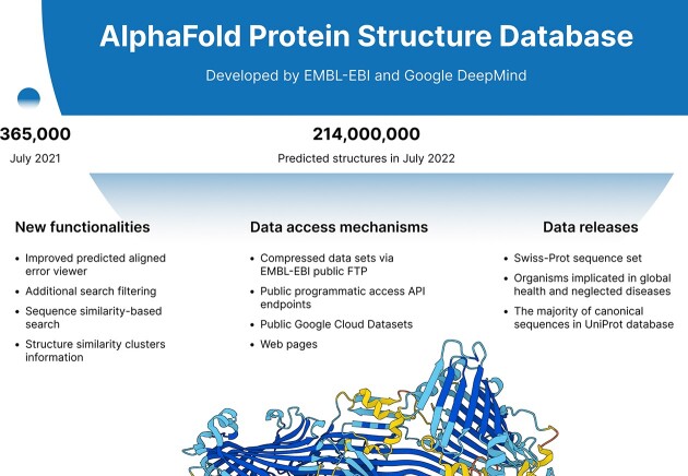
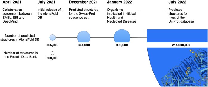
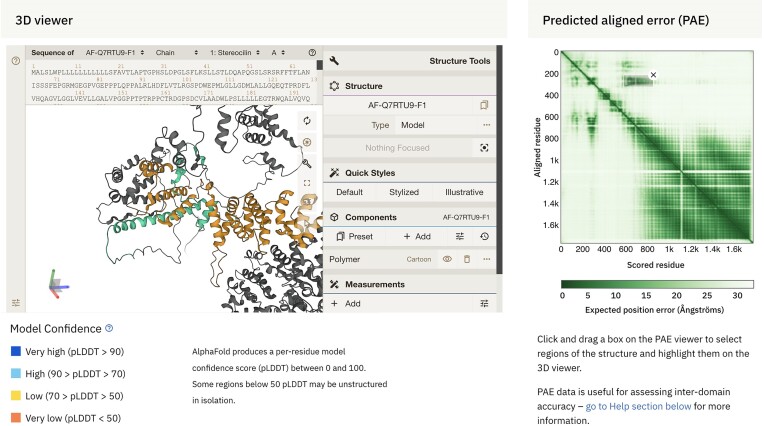
The AlphaFold Database Protein Structure Database (AlphaFold DB, https://alphafold.ebi.ac.uk) has significantly impacted structural biology by amassing over 214 million predicted protein structures, expanding from the initial 300k structures released in 2021. Enabled by the groundbreaking AlphaFold2 artificial intelligence (AI) system, the predictions archived in AlphaFold DB have been integrated into primary data resources such as PDB, UniProt, Ensembl, InterPro and MobiDB. Our manuscript details subsequent enhancements in data archiving, covering successive releases encompassing model organisms, global health proteomes, Swiss-Prot integration, and a host of curated protein datasets. We detail the data access mechanisms of AlphaFold DB, from direct file access via FTP to advanced queries using Google Cloud Public Datasets and the programmatic access endpoints of the database. We also discuss the improvements and services added since its initial release, including enhancements to the Predicted Aligned Error viewer, customisation options for the 3D viewer, and improvements in the search engine of AlphaFold DB.
Overview
CoChat Summary
TL;DR
AlphaFold DB has grown from ~300k to over 214 million predicted protein structures as of September 2023, with expanded access, search, and visualization features. This 2024 update describes data archiving, access mechanisms, integration with core resources, and new clustering/BLAST/PAE viewer capabilities.
Key findings
- AlphaFold DB expanded from approximately 300k structures in 2021 to over 214 million predicted protein structures as of September 2023; the initial July 2021 release housed over 360,000 structures for 20 model organism proteomes.
- Predictions are integrated into resources including PDB, UniProt, Ensembl, InterPro and MobiDB, and are accessible via FTP, Google Cloud Public Datasets (~23 TiB), API, and the website; PAE data is unavailable from EMBL-EBI public FTP.
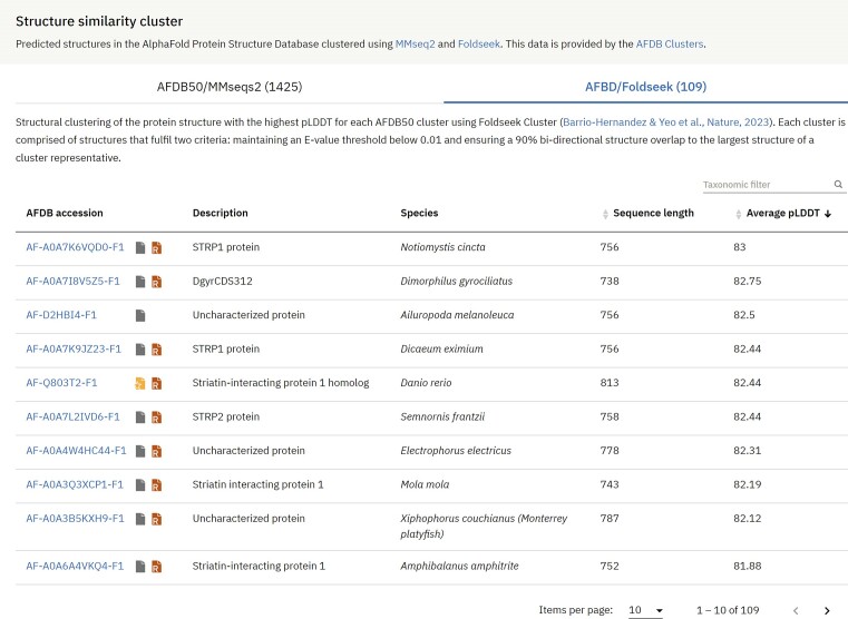
- Structure similarity clustering used MMseqs2 to cluster 214 million UniProtKB sequences into 52 million clusters, then Foldseek secondary clustering yielded 18.8 million clusters, finalizing 2.30 million robust clusters each with at least a pair of structures.
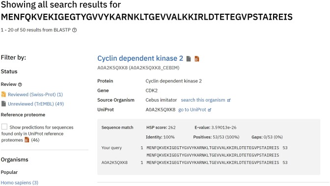
- New features include BLAST sequence similarity search, improved search filters for reviewed/unreviewed and reference proteome status, enhanced PAE viewer–Mol* interactivity, and a November 2022 update fixing a numerical bug that affected ~4% of total structure predictions.
Methods
- Descriptive database update/report with no primary experimental design; data generation is carried out by Google DeepMind, with predictions stored in PDB, mmCIF (modelCIF), and binaryCIF formats plus JSON metadata and confidence metrics pLDDT and PAE.
- Archiving uses successive releases from UniProt reference proteomes (e.g., 2021_02, 2021_04), Swiss-Prot, WHO global health proteomes, and MANE select; as of September 2023, EMBL-EBI FTP hosts TAR files for proteomes of 48 organisms.
- Access and analysis infrastructure includes FTP, Google Cloud Public Datasets, an API keyed on UniProt accessions, Apache-Solr search, BLAST on Google Cloud Platform, and Foldseek/MMseqs2 clustering.
Limitations
- Stated: sequences are not covered if they are less than 16 amino acids; >2700 aa for Swiss-Prot/proteome sequences or >1280 aa for other UniProt sequences; contain non-standard amino acids; are not in the UniProt ‘one sequence per gene’ FASTA file; or are viral proteins; these limitations are under discussion.
- Stated: some sequences may be outdated compared to UniProt due to less frequent AlphaFold DB releases; PAE data is unavailable from EMBL-EBI public FTP; full Google Cloud dataset download requires a Google account and can take roughly 2.5 days at 1 Gbps.
- Evident from the text: all archived structures are predicted models rather than experimental structures, and users must interpret pLDDT/PAE cautiously (e.g., pLDDT <50 likely disordered, 50–70 lower confidence); the database also currently lacks isoform and multimeric predicted structures, which are listed as future work.
Model-written summary. Check it against the paper before relying on it.
Integrity signals
Registry
Cites
Statistics
Figures

Verification
Claims the paper makes
11 Valid · 1 Not valid · 0 Not checked · 28 Own results and interpretations
Every source the paper cites for this claim was checked and supports it.
In the paper
“The new generation of predicted protein structure models has demonstrated remarkable accuracy, which could mitigate the continually widening gap between known protein sequences and experimentally determined protein structures (7).”
§ AlphaFold Protein Structure Database in 2024: providing structure coverage for over 214 million protein sequences > Introduction
What the sources say
- Supportedcites [7] Varadi M., Bordin N., Orengo C., Velankar S. The opportunities and challenges posed by the new generation of deep learning-based protein structure predictors. Curr. Opin. Struct. Biol. 2023; 79: 102543. 36807079 PMC7618866 source
The Introduction says prediction models made a major advance in accuracy and can match or sometimes surpass experimentally determined structures. It also describes a growing gap between known protein sequences and determined structures; the section on new challenges says predicted structures have greatly increased coverage of protein sequence space. These passages support both the accuracy claim and the proposed potential to narrow that gap (page numbers unavailable). (LLM, conf=0.93)
boosted- Asserted: passes — The authors put it forward as true.
- Checkable: passes — It can be checked against evidence.
- Load-bearing: passes — The paper’s conclusions depend on it.
- Based: passes — The paper gives a reason why it holds.
- Supported
Every source the paper cites for this claim was checked and supports it.
In the paper
“In response to frequent requests from the user community since the AlphaFold DB launch, we have made significant strides to implement a sequence-based similarity search feature. We have incorporated the Basic Local Alignment Search Tool (BLAST) (21) into our Google Cloud Platform infrastructure to achieve this.”
§ AlphaFold Protein Structure Database in 2024: providing structure coverage for over 214 million protein sequences > Implementation > Data access
What the sources say
- Supportedcites [21] Camacho C., Coulouris G., Avagyan V., Ma N., Papadopoulos J., Bealer K., Madden T.L. BLAST+: architecture and applications. BMC Bioinf. 2009; 10: 421. PMC2803857 20003500 source
The Background identifies BLAST as a sequence similarity search program that matches a query sequence against a database. In the citing sentence, the citation identifies the tool incorporated; the AlphaFold DB implementation is the citing authors’ own report, not a claim attributed to this source. (LLM, conf=0.91)
plainstates a cause- Asserted: passes — The authors put it forward as true.
- Checkable: passes — It can be checked against evidence.
- Load-bearing: unclear — It is unclear whether the paper’s conclusions depend on it.
- Based: passes — The paper gives a reason why it holds.
- Supported
Every source the paper cites for this claim was checked and supports it.
In the paper
“The impact of the predicted structures is further enhanced by their seamless integration into several core data resources, including PDBe-KB (17), UniProt (6), Ensembl (16), InterPro (15), Genecards (24) and MobiDB (25), among others.”
§ AlphaFold Protein Structure Database in 2024: providing structure coverage for over 214 million protein sequences > Conclusions and outlook
What the sources say
- Supportedcites [17] consortium P.D.B.-K.B. PDBe-KB: collaboratively defining the biological context of structural data. Nucleic Acids Res. 2022; 50: D534– D542. 34755867 PMC8728252 source
Citation (17) is attached specifically to PDBe-KB, rather than to the other resources in the sentence. The Introduction identifies PDBe-KB as a resource that integrates structural and functional annotations, supporting its identification here. (LLM, conf=0.90)
- Supportedcites [6] U.P. Consortium UniProt: the Universal Protein Knowledgebase in 2023. Nucleic Acids Res. 2023; 51: D523– D531. 36408920 PMC9825514 source
The citation is attached to UniProt in a list of resources, and the cited paper is a description of UniProt. Its Data integration and Interactive visualisations in entry pages sections state that UniProt incorporates AlphaFold-predicted structures into protein entries alongside other data; the supplied passages do not give page numbers. (LLM, conf=0.93)
- Supportedcites [16] Cunningham F., Allen J.E., Allen J., Alvarez-Jarreta J., Amode M.R., Armean I.M., Austine-Orimoloye O., Azov A.G., Barnes I., Bennett R. et al. Ensembl 2022. Nucleic Acids Res. 2022; 50: D988– D995. 34791404 PMC8728283 source
The citation identifies Ensembl as one of the resources in the list. In “Developing a new website” (page not provided), the paper says Ensembl’s Entity Viewer provides access to protein information, including 3D structures from PDBe. (LLM, conf=0.86)
plainstates a cause- Asserted: passes — The authors put it forward as true.
- Checkable: passes — It can be checked against evidence.
- Load-bearing: unclear — It is unclear whether the paper’s conclusions depend on it.
- Based: passes — The paper gives a reason why it holds.
- Supported
Every source the paper cites for this claim was checked and supports it.
In the paper
“Tackling this challenge, Steinegger et al. recently unveiled the Foldseek Cluster, a state-of-the-art structural-alignment-based clustering algorithm designed for vast datasets (22). They applied this novel method to the AlphaFold DB archive, making the derived structure similarity clusters available to the research community.”
§ AlphaFold Protein Structure Database in 2024: providing structure coverage for over 214 million protein sequences > Implementation > Data access
What the sources say
- Supportedcites [22] Barrio-Hernandez I., Yeo J., Jänes J., Mirdita M., Gilchrist C.L.M., Wein T., Varadi M., Velankar S., Beltrao P., Steinegger M. Clustering-predicted structures at the scale of the known protein universe. Nature. 2023; 622: 637– 645. 37704730 PMC10584675 source
The Abstract says the authors used Foldseek cluster to cluster the AlphaFold database. The Methods section (“Web server”) says they developed a server for users to explore the clusters, their members and related clusters, supporting the claim that the results were made available to researchers. (LLM, conf=0.93)
plainstates a cause- Asserted: passes — The authors put it forward as true.
- Checkable: passes — It can be checked against evidence.
- Load-bearing: passes — The paper’s conclusions depend on it.
- Based: passes — The paper gives a reason why it holds.
- Supported
- Probably not a substantial claim. 30
- It may state two separate claims as one. 15
- The quote only partly states this claim. 1
Claims other papers make about this one
Each is checked against this paper’s own text: does it say what the citing paper says it does?
The cited source says what the claim attributes to it.
In the citing paper
“houses over 200 million pre-run AF2 predictions”
From The power and pitfalls of AlphaFold2 for structure prediction beyond rigid globular proteins (2024) · cites this paper as [7]
Citations
Cites
Highly accurate protein structure prediction with AlphaFold
Jumper, J., Evans, R., Pritzel, A., Green, T., Figurnov, M., Ronneberger, O., Tunyasuvunakool, K., Bates, R., Žídek, A., Potapenko, A., et al. (2021) · Nature · doi:10.1038/s41586-021-03819-2 · Cited by 43,764
Self-citationBLAST+: architecture and applications
Camacho, C., Coulouris, G., Avagyan, V., Ma, N., Papadopoulos, J., Bealer, K., & Madden, T. L. (2009) · BMC Bioinformatics · doi:10.1186/1471-2105-10-421 · Cited by 22,074
UniProt: the Universal Protein Knowledgebase in 2023
The UniProt Consortium, Bateman, A., Martin, M.-J., Orchard, S., Magrane, M., Ahmad, S., Alpi, E., Bowler-Barnett, E. H., Britto, R., Bye-A-Jee, H., et al. (2023) · Nucleic Acids Research · doi:10.1093/nar/gkac1052 · Cited by 7,210
Evolutionary-scale prediction of atomic level protein structure with a language model
Lin, Z., Akin, H., Rao, R., Hie, B., Zhu, Z., Lu, W., Smetanin, N., Verkuil, R., Kabeli, O., Shmueli, Y., et al. (2022) · doi:10.1101/2022.07.20.500902 · Cited by 5,990 · cited 1×
“In the wake of the latest breakthroughs in prediction software, new databases such as the AlphaFold Protein Structure Database and the ESM Atlas ( 14 ) have emerged.”
MethodThe GeneCards suite: from gene data mining to disease genome sequence analyses
Stelzer, G., Rosen, N., Plaschkes, I., Zimmerman, S., Twik, M., Fishilevich, S., Stein, T. I., Nudel, R., Lieder, I., Mazor, Y., et al. (2016) · Current Protocols in Bioinformatics · doi:10.1002/cpbi.5 · Cited by 5,550 · cited 1×
“The impact of the predicted structures is further enhanced by their seamless integration into several core data resources, including PDBe-KB ( 17 ), UniProt ( 6 ), Ensembl ( 16 ), InterPro ( 15 ), Genecards ( 24 ) and MobiDB ( 25 ), among others.”
BackgroundAccurate prediction of protein structures and interactions using a 3-track network
Baek, M., DiMaio, F., Anishchenko, I., Dauparas, J., Ovchinnikov, S., Lee, G. R., Wang, J., Cong, Q., Kinch, L. N., Schaeffer, R. D., et al. (2021) · doi:10.1101/2021.06.14.448402 · Cited by 5,549
Paysan-Lafosse, T., Blum, M., Chuguransky, S., Grego, T., Pinto, B. L., Salazar, G. A., Bileschi, M. L., Bork, P., Bridge, A., Colwell, L., et al. (2023) · Nucleic Acids Research · doi:10.1093/nar/gkac993 · Cited by 2,399
Cunningham, F., Allen, J. E., Allen, J., Alvarez-Jarreta, J., Amode, M. R., Armean, I. M., Austine-Orimoloye, O., Azov, A. G., Barnes, I., Bennett, R., et al. (2022) · Nucleic Acids Research · doi:10.1093/nar/gkab1049 · Cited by 1,923
Mol* Viewer: modern web app for 3D visualization and analysis of large biomolecular structures
Sehnal, D., Bittrich, S., Deshpande, M., Svobodová, R., Berka, K., Bazgier, V., Velankar, S., Burley, S. K., Koča, J., & Rose, A. S. (2021) · Nucleic Acids Research · doi:10.1093/nar/gkab314 · Cited by 1,154 · cited 1×
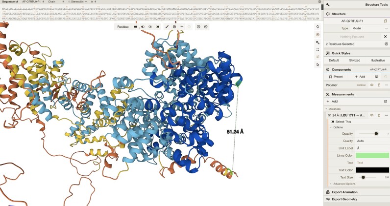
“We improved the visualisation of non-consecutive regions and the interactivity between the PAE viewer and the 3D molecular graphics viewer, Mol* ( 23 ) (Figure 5 ).”
MethodSelf-citationMariani, V., Biasini, M., Barbato, A., & Schwede, T. (2013) · Bioinformatics · doi:10.1093/bioinformatics/btt473 · Cited by 1,138 · cited 1×
“AlphaFold yields a per-residue estimate of its confidence, known as pLDDT, ranging from 0 to 100, indicating the tool’s predicted score on the lDDT-C α metric ( 19 ).”
Method
Cited by
Items 1,201–1,300 of 2,174
Singh, S., Singh, R., & Sharma, S. (2026) · BioData Mining · doi:10.1186/s13040-026-00577-7 · Cited by 2
“The AlphaFold Protein Structure Database, which contains nearly 214 million predicted protein structures [19], was recently released by the DeepMind group and the European Bioinformatics Institute.”
Kiyota, K., Shiraishi, H., Ohno, S., Soga, Y., Shimizu, N., Yatsuka, H., Inoue, M., Takeno, T., Hongxia, W., Witaningrum, A. M., et al. (2025) · Biochemical and Biophysical Research Communications · doi:10.1016/j.bbrc.2025.152378 · Cited by 1
Harnessing population genetics and animal models to uncover the genetics of skin scarring
Peña, O. A., Cañadas‐Garre, M., Collins, I., Timpson, N. J., & Martin, P. B. (2025) · Journal of Plastic Reconstructive & Aesthetic Surgery · doi:10.1016/j.bjps.2025.11.010 · Cited by 1
“Other resources, such as https://www.uniprot.org, 75 using recent advances in protein structure prediction, such as https://www.alphafold.ebi.ac.uk, 76 provide additional support for orthology relationships.”
ProteoCast: a web server to predict, validate, and interpret missense variant effects
(2026) · doi:10.1016/j.jmb.2026.169657 · Cited by 1
“While pLMs are independent of MSAs and could in principle help decrypt IDR sequences, they currently offer no enhanced predictive power in IDRs 12 .”
Rajalakshmi, E., Chandrasekhar, B., Saranya, E., et al. (2025) · Apmis · doi:10.1111/apm.70088 · Cited by 1
Jiménez-Carretero, M., Pozo-Gualda, T., Lázaro, M., et al. (2025) · International Journal of Biological Macromolecules · doi:10.1016/j.ijbiomac.2025.142165 · Cited by 1
Bautista, A. P. R., Terrill, J. R., Duong, M. N., Angelica, G., Tsioutsias, I., James, C. P., Lafoux, A., Huchet, C., & Arthur, P. G. (2025) · Methods · doi:10.1016/j.ymeth.2025.07.010 · Cited by 2
Clark‐ElSayed, A., Nayvelt, K. E., Ishida, S., d’Oelsnitz, S., & Ellington, A. D. (2025) · Current Protocols · doi:10.1002/cpz1.70218 · Cited by 5
Madugula, S. S., Jayasinghe‐Arachchige, V. M., Radler, C. N., Wang, S., & Liu, J. (2025) · Journal of Molecular Biology · doi:10.1016/j.jmb.2025.169538 · Cited by 3
Gracia, L., Hughes, E. R., Middleton, D. R., Mueller, K. D., Portillo, J., Sharma, A., Davey, L., Panzetta, M. E., Chanin, R. B., Bhatt, A. S., et al. (2026) · mBio · doi:10.1128/mbio.01048-26 · Cited by 2
“1) cps1 in Table 1 using a combination of NCBI annotations, glycosyltransfer ase assignments with dbCAN3 (90), and comparisons of predicted protein structures to PDB using Phyre2 (91) and AlphaFold (92, 93).”
Green, R., Richards, H., Ozbilek, D., Tyrrell, F., Barton, V., Zhang, Z., Lovell, S. C., Gifford, D. R., Lagator, M., McBain, A. J., et al. (2025) · Molecular Biology and Evolution · doi:10.1093/molbev/msaf182 · Cited by 4
Knutson, B. A., & Rothblum, L. (2025) · Genes · doi:10.3390/genes16010061 · Cited by 1
Sornsenee, P., Kooltheat, N., Pattaranggoon, N. C., Surachat, K., Monteil, A., & Romyasamit, C. (2025) · Life · doi:10.3390/life15121866 · Cited by 2
“The 3D structures for both the receptor, human tyrosinase (UniProt: P14679), and the seven microbial ligands were determined using AlphaFold prediction models [38,39].”
Jain, A., Mayeen, N. F., Meadow, M. E., Kalandadze, N., Swovick, K., Welle, K. A., Hryhorenko, J. R., & Ghaemmaghami, S. (2026) · Cell Reports · doi:10.1016/j.celrep.2026.117620 · Cited by 1
Tang, P., Guo, S., Zhang, P., et al. (2026) · BMC Pharmacology and Toxicology · doi:10.1186/s40360-026-01098-z · Cited by 1
“The 3D configurations of PTGS2 protein was sourced from the AlphaFold Protein Structure Database ( h t t p s : / / a l p h a f o l d . e b i . a c . u k / ) [32].”
Yonezawa, S., & Bono, H. U. (2026) · Bioinformatics Advances · doi:10.1093/bioadv/vbag013
“…and structural searches were conducted using Foldseek (3Di + AA and Foldseek-TM method) against the complete AFDB data-base (van Kempen et al. 2024, Varadi et al. 2024); rice–human hits were retained (Supplementary Table S7, available as supplementary material at Bioinformatics Advances online)…”
InfluentialThe genetic repertoire of the deep sea: from sequence to structure and functionPreprint
Guo, Y., Wang, Z., Li, D., Wang, L., Lan, H., Guo, F., Zhao, Z., Liu, Z., Meng, L., Shen, X., et al. (2026) · bioRxiv (Cold Spring Harbor Laboratory) · doi:10.64898/2026.02.03.703410 · Cited by 1
Completion of partial structures using Patterson maps with the CrysFormer machine-learning model
Pan, T., Dramko, E., Miller, M. D., Kyrillidis, A., & Phillips, G. N., Jr. (2025) · Acta Crystallographica Section D Structural Biology · doi:10.1107/s2059798325009659 · Cited by 2
Exploring proteins and protein–ligand complexes through residue interaction networks
Begue, S. C., Leonardi, E., Minervini, G., & Tosatto, S. C. E. (2026) · Nature Protocols · doi:10.1038/s41596-026-01334-0 · Cited by 2
KHNYN is a manganese-dependent endoribonuclease required for ZAP-mediated antiviral restriction
Youle, R. L., Lista, M. J., Brudenell, E. L., Thompson, B., Bouton, C. R., Morris, E. R., Neil, S. J. D., Swanson, C. M., & Taylor, I. A. (2025) · Nucleic Acids Research · doi:10.1093/nar/gkaf1360 · Cited by 2
“Published and predicted protein structures were obtained from the RCSB PDB [ 61 ] and the Al-phaFold DB [ 62 , 63 ].”
Tao, K., Luan, Y., & Li, J. (2026) · Environmental Science Nano · doi:10.1039/d5en01079a · Cited by 3
Computational Methods for the Discovery and Optimization of TAAR1 and TAAR5 Ligands
Scarano, N., Espinoza, S., Brullo, C., & Cichero, E. (2024) · International Journal of Molecular Sciences · doi:10.3390/ijms25158226 · Cited by 3
“In 2024, the same group explored an SAR rationalization of a series of amino-oxazoline TAAR1 agonists produced by Roche [50] by docking in the AlphaFold predicted structure of h TAAR1 [62,63].”
BackgroundMethodInfluentialOliw, E. H. (2025) · Archives of Biochemistry and Biophysics · doi:10.1016/j.abb.2025.110574 · Cited by 1
Diacylglycerol kinases: Molecular mechanism of cellular and physiological functions
Kimura, T., & Epand, R. M. (2026) · Progress in Lipid Research · doi:10.1016/j.plipres.2025.101373 · Cited by 2
Kim, C., Tagmount, A., Zhu, Z., Wilson, F., Li, D., Ostrov, D. A., Brad Barbazuk, W., Bacher, R., & Vulpe, C. D. (2026) · Archives of Toxicology · doi:10.1007/s00204-025-04294-8 · Cited by 1
Structural basis for heteromeric assembly and subthreshold activation of human M-channel
Wang, Y., Yang, H. L., Qu, Y., Li, J., Li, X., Hou, W., Wu, K., Xie, G., Wang, X., Ye, Y., et al. (2026) · Vita · doi:10.15302/vita.2026.05.0032 · Cited by 2
Meng, L., Wang, X., Zhang, F., et al. (2025) · Naunyn-Schmiedeberg s Archives of Pharmacology · doi:10.1007/s00210-025-04138-3 · Cited by 1
“(John et al. 2021; Varadi et al. 2023, 2021).”
Banu, K., Khan, M. A., Li, S., Reynolds-Gagliano, M., Wang, X., Mobley, R. J., Barnett, K. R., Wielgosz, M. M., Bauler, M. N., Wincek, C., et al. (2026) · Science Advances · doi:10.1126/sciadv.aeb3503 · Cited by 1
Distinct modes of coupling between VCP, an essential unfoldase, and deubiquitinasesPreprint
Vostal, L. E., Dahan, N. E., Zhang, W., Reynolds, M. J., Chait, B. T., & Kapoor, T. M. (2024) · bioRxiv (Cold Spring Harbor Laboratory) · doi:10.1101/2024.09.08.611915 · Cited by 1
“The additional density was rigid body fit with a model for the VCPIP1 UBX-L domain (AF-Q96JH7-F1) (43, 44) (Fig.”
BackgroundMethodSchlembach, M. C., & Wrublewski, D. (2025) · Science & Technology Libraries · doi:10.1080/0194262x.2025.2468333 · Cited by 4
“DeepMind also provided the software as an open access resource, enabling scientists worldwide to design and synthesize novel protein designs (Jumper et al. 2021; Komaroff 2021; Varadi et al. 2024).”
Chen, Y., Wei, S., Wang, Q., Hameed, M. S., Bao, J., Yan, J., Keserű, G. M., & Han, X. (2025) · European Journal of Medicinal Chemistry · doi:10.1016/j.ejmech.2025.118109 · Cited by 2
SEFP: Structure-Based Enzyme Function Prediction
Zhang, Z., Yu, G., Deng, Z., Luo, C., Cai, C., Zhang, W., Hu, F., Choi, K., Wei, Z., Wang, L., et al. (2025) · IEEE Transactions on Computational Biology and Bioinformatics · doi:10.1109/tcbbio.2025.3552846 · Cited by 1
“We addressed this by collecting enzyme structures for each enzyme from the AlphaFold Protein Structure Database (AFDB) [31], resulting in a version of the HEC-Net dataset with structural information.”
A bacterial toxin-antitoxin system involved in an unusual response to genotoxic stress
Lin, J. D., Nicholson, B. L., & Ensminger, A. W. (2025) · EMBO Reports · doi:10.1038/s44319-025-00545-y · Cited by 1
“EV4A) are highly similar to the AlphaFold (Jumper et al, 2021; Varadi et al, 2024) predicted structure of GndR (Fig.”
Inhibitor fluorination pattern tunes chemically induced protein dimerizationPreprint
Schwegler, E., Harder, J.-M., Preuss, M. D., Guhl, C., Zhang, S., Wagner, A., Bader, N., Stallforth, P., Lakemeyer, M., Schindelin, H., et al. (2025) · bioRxiv (Cold Spring Harbor Laboratory) · doi:10.1101/2025.07.23.666362 · Cited by 1
“Comparison of available crystal structures for oxidized 47 and inhibitor-bound 23 T. brucei Tpx WT , the AlphaFold2 model of the oxidized enzyme 48,49 and our MD simulations revealed substantial structural plasticity for the W39 side chain.”
Sutana, P., Luetragoon, T., Buakaew, W., Daowtak, K., Nilsri, N., Thongsri, Y., Potup, P., Léon, C., & Usuwanthim, K. (2025) · International Journal of Molecular Sciences · doi:10.3390/ijms26188771 · Cited by 2
Investigating the Context‐Dependent Phase Separation of Human HOX Transcription Factors
Karmakar, S., Manglam, J., Kant, K., & De, S. (2025) · Proteins Structure Function and Bioinformatics · doi:10.1002/prot.70009 · Cited by 1
“Recent advances in artificial intelligence (AI) and machine learning (ML) have enhanced predictions of protein structure [30], biomolecular interactions [31] including formation of complex heterotypic biomolecular condensates [32].”
ARP2/3 regulates cell surface dynamics of CeTOCA-1 in C. elegans zygotes
Nageswaran, A., Mitra, P., Rai, M., Anand, D., Pratap, Y., & Padmanabhan, A. (2025) · Journal of Biosciences · doi:10.1007/s12038-025-00526-2 · Cited by 1
A methyltransferase molecular switch unlocks para -quinone methide generation and oligomerization
Ma, C., Zhang, Z., Wang, W., et al. (2025) · Chemical Science · doi:10.1039/d5sc06912b · Cited by 1
Dai, Y., Yu, J., He, G., Mo, L., Wen, Y., Guan, R., Ye, X., Guo, Y., Lu, J., Chen, J., et al. (2025) · Phytomedicine · doi:10.1016/j.phymed.2025.157223 · Cited by 2
Saif, A., Baruah, P., Wazid, S. W., Panigrahi, A., Han, J.-H., Ahmed, M. A., & Quan, F.-S. (2025) · Infection, Genetics and Evolution · doi:10.1016/j.meegid.2025.105733 · Cited by 1
Scilinkbert: a BERT-style language model for understanding scientific texts with citations
Yu, J.-Y., Yang, D., & Lee, K.-H. (2025) · The Journal of Supercomputing · doi:10.1007/s11227-025-07838-y · Cited by 2
“Recent advances have shown AI’s ability to accelerate drug discovery [5, 24, 26, 41], predict protein structures [1, 17, 36], automate theorem proving [15, 31, 35], and even conduct physical experiments [34].”
Amin, Z., Mohiddin, F. A., Ali, A., Shikari, A. B., Nabi, S. U., Bhat, T. A., Gupta, V., Wani, F. J., Parveen, S., Mir, M. S., et al. (2025) · Journal of Agriculture and Food Research · doi:10.1016/j.jafr.2025.102092 · Cited by 3
Flexible iron: disorder in the ironome brings order to protein structure and function
Uversky, V. N., & Ferreira, G. C. (2025) · Frontiers in Molecular Biosciences · doi:10.3389/fmolb.2025.1537164 · Cited by 1
Structural and functional insights from detailed computational analysis of Plasmodium repetome
Kaur, S., Kumar, V., Singh, K., Behl, A., Arpna, Mishra, P. C., & Hora, R. (2026) · Journal of Genetics · doi:10.1007/s12041-025-01516-x · Cited by 1
ProxiCapture Reveals Context-Dependent CRBN Interactome Landscape of Molecular Glue Degraders
(2026) · doi:10.64898/2026.01.05.697692 · Cited by 1
“…we applied a recently developed method [29] in which the structural assessment software MASTER [38] is used to screen the AF2 Human genome library [39] for all proteins that contain a G-loop with a similar backbone (root-mean-squared-deviation (RMSD) cut off 1.5 Å) to the published G-loop degron…”
InfluentialInvestigation of the histone deacetylase inhibitor potential of phorbazole analogues
Reierth, I. A. N., Guttormsen, Y., Andersen, J. H., Hansen, E. H., Abdelhady, A. M., Langer, M. K., Haug, B. E., Bayer, A., & Petit, G. A. (2025) · Bioorganic & Medicinal Chemistry · doi:10.1016/j.bmc.2025.118489 · Cited by 1
Characterization of DNA/RNA binding properties of CspA protein from Mycobacterium tuberculosis
Pankratova, P. Y., Lekontseva, N. V., & Никулин, А. Д. (2025) · In Silico Research in Biomedicine · doi:10.1016/j.insi.2025.100033 · Cited by 1
Clinical description, genetic analysis and characterization of two NLRP12 heterozygous VUS variants
Lizama-Muñoz, A., Gutiérrez‐Bautista, J. F., García-Aznar, J. M., Perea, I., Bernal, M., & López‐Nevot, M. Á. (2026) · Frontiers in Immunology · doi:10.3389/fimmu.2026.1755711 · Cited by 1
“The native structure of NLRP12was modeled based on the AlphaFold-predicted structure (AF-P59046-F1) (64, 65).”
Farrow, J. M., Wells, G., & Pesci, E. C. (2026) · PLoS ONE · doi:10.1371/journal.pone.0350814 · Cited by 1
“Since the structure of DtpC has not been solved experimentally, we used the predicted structure of A. baumannii DtpC that is available in the AlphaFold Protein Structure Database [38,39].”
Assessment of Alphafold Protein Models for Small-Molecule Ligand Docking versus Co-Folding
Maidanik, H., Lazou, M., Bajaj, R., Sarwar, R., Ziaee, O., Vajda, S., & Joseph‐McCarthy, D. (2026) · Journal of Chemical Information and Modeling · doi:10.1021/acs.jcim.6c01038 · Cited by 3
Mapping of Nuclear Localization Signal in Secreted Liver-Specific Protein 2 of Plasmodium falciparum
Shrikondawar, A. N., Chennoju, K., Ghosh, D. K., & Ranjan, A. (2024) · ACS Infectious Diseases · doi:10.1021/acsinfecdis.4c00715 · Cited by 1
Lindstad, L. J., Mrozek, P. M., Boehlich, G. J., Leivers, S., Pope, P. B., La Rosa, S. L., & Westereng, B. (2025) · doi:10.64898/2025.12.12.693890 · Cited by 3
Regulation of Vacuole Fusion in Stomata by Dephosphorylation of the HOPS subunit VPS39
Pullen, A.-M., Billings, G., Hodgens, C., White, G., Akpa, B. S., & Rojas-Pierce, M. (2025) · doi:10.1101/2025.10.02.680005 · Cited by 1
Beyond prediction: Why experimental structural biology remains essential in plants
Palayam, M., & Shabek, N. (2026) · Current Opinion in Plant Biology · doi:10.1016/j.pbi.2026.102937 · Cited by 1
Reddy, P. R., Ridha, F., & Gromiha, M. M. (2025) · Journal of Molecular Biology · doi:10.1016/j.jmb.2025.169593 · Cited by 3
Verma, A., Goel, A., Koner, N., Gunasekaran, G., & Radha, V. (2024) · doi:10.1101/2024.04.07.588436 · Cited by 2
Diakov, A., Sure, F., Ilyaskin, A. V., & Korbmacher, C. (2025) · Pflügers Archiv - European Journal of Physiology · doi:10.1007/s00424-025-03099-z · Cited by 2
“Alpha-Fold modeling of human αENaC (AlphaFold ID: P37088) places S594 immediately C-terminal to the second transmembrane helix [27, 66], albeit with a low confidence score.”
Heyder, C., Murray, C. A., Granith, P. K. E., Makhkamov, M., Bourget, E., Cain, I., Davidson, B., Doone, T.-J., Korasadowicz, J., Hull, N., et al. (2026) · Endocrinology · doi:10.1210/endocr/bqag089 · Cited by 1
Differential tolerance for SEA domain misfolding encodes a MAPK pathway-specific responsePreprint
Priyadarshini, A., & Cullen, P. J. (2026) · bioRxiv (Cold Spring Harbor Laboratory) · doi:10.64898/2026.05.06.723240 · Cited by 1
Abid, M. S. R., Naldrett, M. J., Alvarez, S., Eichhorn, C. D., Andrews, M. T., & Checco, J. W. (2025) · ACS Chemical Neuroscience · doi:10.1021/acschemneuro.4c00635 · Cited by 3
Characterization of a Rice GH5_11 Gene Associated with Endosperm and Seed Traits
Gistelinck, K., Madder, Z., Verbeke, I., & Damme, E. J. M. V. (2025) · Plants · doi:10.3390/plants14223428 · Cited by 1
“Protein structures were obtained from the AlphaFold protein structure database [78,79].”
Artificial intelligence-based epitope discovery of Mpox virus: Rational vaccine design
Adugna, A., Abebaw, D., Admasu, A., Tegegne, B. A., Teffera, Z. H., Hibstu, T., Baye, G., Ashenef, B., Mengistu, E. F., & Jemal, M. (2025) · Vaccine X · doi:10.1016/j.jvacx.2025.100686 · Cited by 4
Grill-Walcher, S., Breitwieser, A., Afzal, M., Pum, D., Bonvin, A. M. J. J., Oostenbrink, C., & Schäffer, C. (2026) · Journal of Molecular Graphics and Modelling · doi:10.1016/j.jmgm.2026.109413 · Cited by 1
Xu, X., Wang, X., Xu, L., et al. (2025) · Bioorganic Chemistry · doi:10.1016/j.bioorg.2025.109198 · Cited by 1
Trapala, J., Álvarez‐Añorve, L. I., Vasquez-Martínez, N., Chavira-Suárez, E., Vásquez-Bochm, L. X., Matuz‐Mares, D., Cortés‐Benítez, F., & González‐Andrade, M. (2026) · ChemMedChem · doi:10.1002/cmdc.70446 · Cited by 1
“The PDB file was downloaded from the following link: https://alphafold.ebi.ac.uk/entry/P18031 [24, 25].”
AlphaFold in protein structure and function prediction
Kurgan, L., & Yang, J.-Y. (2025) · Computational and Structural Biotechnology Journal · doi:10.1016/j.csbj.2025.11.064 · Cited by 2
“The introduction of AlphaFold2 [1], its subsequent AlphaFold3 version [2], and the public release of hundreds of millions of protein structures predicted by this landmark method [3,4] have fundamentally changed the fields of structural biology and protein bioinformatics.”
Argyle, M., Chipman, D., Woolley, A. C., Bundy, B. C., & Corte, D. D. (2026) · SynBio · doi:10.3390/synbio4010005 · Cited by 3
“The AlphaFold Database now provides access to over 214 million predicted protein structures; although not all predictions are accurate, the models include confidence scores that enable assessment of structural reliability [23].”
Cruz‐Mirón, R., Swale, C., Liang, Q., Yamaryo‐Botté, Y., Arnold, C.-S., Hakimi, M., Botté, C. Y., Bougdour, A., & Saeij, J. P. J. (2026) · Cell Reports · doi:10.1016/j.celrep.2026.117299 · Cited by 1
Canè, C., Casciaro, B., Vetrano, C., et al. (2025) · International Journal of Molecular Sciences · doi:10.3390/ijms26209940 · Cited by 1
“The PDB file for the protein with UniProt code Q9I5H3 was obtained from the Al-phaFold Protein Structure Database [32,33], the NMR structure of Esc(1-21)-1c peptide of sequence GIFSKLAGKKIKN d LLI d SGLKG was kindly provided by Ghosh et al. [34], while the PDB structure of the DNA duplex,…”
Decoding protein dynamicity in DNA ligase activity through deep learning-based structural ensemblesPreprint
Mishra, N., Callaghan, S., & Briney, B. S. (2024) · bioRxiv (Cold Spring Harbor Laboratory) · doi:10.1101/2024.11.07.622521 · Cited by 1
Evolutionary conservation and innovations of RNA polymerase II transcription elongation factorsPreprint
Francette, A. M., Grover, A., Clark, N., & Arndt, K. M. (2026) · bioRxiv (Cold Spring Harbor Laboratory) · doi:10.64898/2026.01.11.698887 · Cited by 1
FusionProt: Fusing Sequence and Structural Information for Unified Protein Representation LearningPreprint
Kalifa, D., Singer, U., & Radinsky, K. (2025) · bioRxiv (Cold Spring Harbor Laboratory) · doi:10.1101/2025.08.06.668973 · Cited by 7
“Despite the vast number of proteins in nature, the number of known 3D structures remains limited [48].”
Diverse polysaccharide production and biofilm formation abilities of clinical Klebsiella pneumoniae
IV, R. B., Cella, E., Azarian, T., Rendueles, O., & Fleeman, R. (2024) · doi:10.21203/rs.3.rs-4630973/v1 · Cited by 29
“The AlphaFold Protein Structure Database was used to predict and generate the �mbriae structures, last accessed on March 2024 48, 49 .”
MethodRajan, H., Devipriya, S. P., & Bhat, S. G. (2026) · Journal of Hazardous Materials Plastics · doi:10.1016/j.hazmp.2026.100066 · Cited by 1
Conformational dynamics of the bacterial E3 ligase SspH1
Kennedy, C. R., Esposito, D., Huber, J., House, D., & Rittinger, K. (2025) · Journal of Biological Chemistry · doi:10.1016/j.jbc.2025.110671 · Cited by 1
“S1 B ) (32–34), and found in other bacterial effectors such as SifA, though its function in E3 ligase activity has not been investigated thus far.”
Yang, X. H., Hu, Z., Zhou, T., et al. (2025) · ChemBioChem · doi:10.1002/cbic.202500296 · Cited by 1
Franzotti, C. L. P., Palópoli, N., Palma, J., & Pierdominici‐Sottile, G. (2025) · Frontiers in Chemical Biology · doi:10.3389/fchbi.2025.1538350 · Cited by 1
Crystal structure and mutagenesis of a nucleic acid–binding BRCT domain in human PARP4
Frigon, L., & Pascal, J. M. (2025) · Journal of Biological Chemistry · doi:10.1016/j.jbc.2025.110277 · Cited by 1
Walunjkar, N., Lai, T. Y., Akhter, N., Miller, J. H., Bettinger, J. Q., Marcus, E., Phizicky, E. M., Ghaemmaghami, S., & Fay, J. C. (2024) · doi:10.1101/2024.12.09.627561 · Cited by 3
TooT-PLM-P2S: Incorporating Secondary Structure Information into Protein Language ModelsPreprint
Ghazikhani, H., & Butler, G. (2024) · bioRxiv (Cold Spring Harbor Laboratory) · doi:10.1101/2024.08.13.607781 · Cited by 1
Tryptoline Stereoprobe Elaboration Identifies Inhibitors of the GRPEL1-HSPA9 Chaperone ComplexPreprint
Hayward, R. E., Berkeley, R. F., Gao, Z., Garhammer, M., Morizono, M. A., Njomen, E., Li, H., DeMeester, K. E., Cociorva, V., Herzik, M. A., et al. (2025) · bioRxiv (Cold Spring Harbor Laboratory) · doi:10.1101/2025.10.20.683548 · Cited by 1
Cai, X., Liu, Y., Li, J., Zhao, Z., Yao, X., Zhai, Z., Liucong, M., Liu, Y., Wang, X., Mochizuki, K., et al. (2026) · Science China Life Sciences · doi:10.1007/s11427-025-3103-x · Cited by 1
“Structural information for the Rib1 protein was predicted using AlphaFold and retrieved from UniProt (UniProt Consortium, 2025; Varadi et al., 2024).”
Guillén-Samander, A., Perepelkina, N., Horáčková, V., et al. (2025) · bioRxiv (Cold Spring Harbor Laboratory) · doi:10.1101/2025.07.25.666630 · Cited by 1
“Structures were downloaded from the AlphaFold public database 96,97 , for the shuttle-like LTPs, or predicted using AlphaFold3 41 , for the BLTPs, and analysed using PyMOL 98 .”
BCI inhibits MKP3 by targeting the kinase-binding domain and disrupting ERK2 interaction
Qiu, S.-J., Zhang, Y., Gong, W., Ding, Y.-H., Wu, J., Wang, Z., & Yao, H. (2025) · Journal of Biological Chemistry · doi:10.1016/j.jbc.2025.110570 · Cited by 3
“Notably, AlphaFold 2 (AF2) predicts a high-activity conformation for MKP3 (22, 23), differing from its crystal structure (Fig.”
Kamli, H. M., Shubaili, A. A. Y., Yousif, A. A. M., et al. (2026) · Molecular Diversity · doi:10.1007/s11030-025-11430-0 · Cited by 1
“Protein sequences were downloaded using the UniProt database (The UniProt Consortium, 2023), and structural models were searched in the AlphaFold Protein Structure Database [32] or modeled de novo using SwissSidechain modeling capabilities.”
Liao, K., Fu, Z., Liu, X., Yu, X., Jin, L., Cheng, J., Yang, D., Ai, K., Liu, Z., Guo, D., et al. (2025) · Frontiers in Immunology · doi:10.3389/fimmu.2025.1592231 · Cited by 1
Deciphering the Causes of IbfA-Mediated Abortive Infection in the P22-like Phage UAB_Phi20
López-Pérez, J., Cortés, P., Campoy, S., Erill, I., & Llagostera, M. (2025) · International Journal of Molecular Sciences · doi:10.3390/ijms26104918 · Cited by 1
“Furthermore, a sequence-based search with the entire IbfA sequence against the AlphaFold database [41,42] yielded six proteins annotated as members of the AAA-family ATPases or ATP-binding cassette domain-containing proteins, showing a 100% identity match.”
The Cryo-EM structure of TRPC1 and TRPC5 heterotetramerPreprint
Chen, Y., Che, T., Cheng, X., et al. (2024) · bioRxiv (Cold Spring Harbor Laboratory) · doi:10.1101/2024.11.03.621705 · Cited by 1
Raja, S., Kumar, P., S, H., et al. (2025) · bioRxiv (Cold Spring Harbor Laboratory) · doi:10.1101/2025.04.17.649292 · Cited by 1
Konstantinou, E. K., Panagiotopoulos, A. A., & Dimitriou, M. (2025) · Current Issues in Molecular Biology · doi:10.3390/cimb47090777 · Cited by 2
“The structure of THRA receptor was extracted from AlphaFold database (https:// alphafold.ebi.ac.uk/, accessed on 15 September 2025) [18] (Code: AF-P10827-F1) in the pdb format and was introduced in the GalaxyWEB server (https://galaxy.seoklab.org/, accessed on 15 September 2025) [19], together with…”
Guillén-Samander, A., Ahmed, Y., Ribeiro-Holbein, A., Cubillán-Marín, J., Höhn, K., Rocha‐Roa, C., Dröge, J., Panitzsch, L. S., Burda, P., Gilberger, T., et al. (2026) · bioRxiv (Cold Spring Harbor Laboratory) · doi:10.64898/2026.09.11.750981
“Structures of the identified LTPs were downloaded from the AlphaFold public database 60,93 and analysed using PyMOL 94 and UCSF ChimeraX 95 (developed by the Resource for Biocomputing, Visualization, and Informatics at the University of California, San Francisco, with support from National…”
de Dios, R., Forsyth, V. S., Harkova, L. G., Schache, K. J., Hsueh, B. Y., Strat, H., Riley, B., Rubio, A., Pérez‐Pulido, A. J., Waters, C. M., et al. (2026) · npj Biofilms and Microbiomes · doi:10.1038/s41522-026-00968-y · Cited by 5
Kong, Z., Zhu, Y., Xu, Y., Yin, M., Hou, T., Wu, J., Xu, H., & Hsieh, C. (2026) · bioRxiv (Cold Spring Harbor Laboratory) · doi:10.64898/2026.02.14.705870 · Cited by 1
“Leveraging the structure PDB files predicted by OmegaFold, we use the FoldSeek [20] easy-search tool to identify the most similar proteins in the AlphaFold SwissProt [49] database and calculated the mean TM-Score [61] (Table 1).”
Wang, H., Pereira, J. H., DeGiovanni, A. M., et al. (2025) · bioRxiv (Cold Spring Harbor Laboratory) · doi:10.1101/2025.07.18.665579 · Cited by 1
Teng, J., Zhao, X., Yan, Q., Yang, J., Pei, F., Zhang, X., Duan, C., Chen, Z., Xu, Q., Liu, Y., et al. (2026) · Genetics Selection Evolution · doi:10.1186/s12711-025-01028-3 · Cited by 5
“Since the 3D structure of the AHNAK protein is not available in the AlphaFold database [64], we only analyzed the MTX1 and GHR proteins (see Additional file 1 Figure S6).”
Vollmer, V., Arnaud, C., Thelen, S., Schwarz, V., Hanley, P. J., & Bähler, M. (2025) · Molecular Biology of the Cell · doi:10.1091/mbc.e25-02-0048 · Cited by 1
(2025) · doi:10.1093/nar/gkaf1041 · Cited by 1
“Exploring structural insights using AlphaFold [ 41 ], we found PinQ has all the structural features of an SSR [ 42 ], including a DNA binding site, arm region, and catalytic domain (Fig.”
Karemera, K. J., Váradi, G., Bende, G., Merber, R., Dán, K., Papp, C., Farkas, A., Maróti, G., Tóth, G. K., Borics, A., et al. (2026) · Probiotics and Antimicrobial Proteins · doi:10.1007/s12602-025-10890-y · Cited by 2
Metadata
- Authors
- Varadi, M., Bertoni, D., Magana, P., Paramval, U., Pidruchna, I., Radhakrishnan, M., Tsenkov, M., Nair, S., Mirdita, M., Yeo, J., Kovalevskiy, O., Tunyasuvunakool, K., Laydon, A., Žídek, A., Tomlinson, H., Hariharan, D., Abrahamson, J., Green, T., Jumper, J., Birney, E., Steinegger, M., Hassabis, D., & Velankar, S.
- Affiliations
- European Molecular Biology Laboratory, European Bioinformatics Institute , Hinxton, UK
- European Bioinformatics Institute (GB)
- Seoul National University (KR)
- Google DeepMind (United Kingdom) (GB)
- Corresponding author
- Hassabis, Demis
- Velankar, Sameer
- ORCID
- Venue
- Nucleic Acids Research
- Publisher
- Oxford University Press (OUP)
- Published
- 2023-11-02
- Volume
- 52
- Issue
- D1
- Pages
- D368-D375
- ISSN
- 0305-1048 (ISSN-L)
- 1362-4962
- PMC
- PMC10767828
- OpenAlex
- W4388464011
- Cited by
- 2,052
- References
- 29
- Citations by year
- 2026: 724 · 2025: 1015 · 2024: 307 · 2023: 5
- FWCI
- 181.98
- Type
- journal article
- Language
- en
- Open access
- gold
- doi.org
- Licence
- Locations
- Nucleic Acids Research (publishedVersion, cc-by)
- PubMed Central (publishedVersion, cc-by)
- PubMed (publishedVersion)
- Seoul National University Open Repository (Seoul National University) (submittedVersion)
- Funders
- Google DeepMind
- National Research Foundation of Korea (2019R1A6A1A10073437, 2020M3A9G7103933, 2021R1C1C102065, 2021M3A9I4021220, RS-2023-00250470)
- Samsung DS Research Fund
- Seoul National University
- Topics
- Life Sciences › Biochemistry, Genetics and Molecular Biology › Molecular Biology › Protein Structure and Dynamics
- Life Sciences › Biochemistry, Genetics and Molecular Biology › Structural Biology › Enzyme Structure and Function
- Life Sciences › Biochemistry, Genetics and Molecular Biology › Molecular Biology › Machine Learning in Bioinformatics
- Keywords
- predicted protein structures, AlphaFold2, protein structure prediction, protein sequence coverage, data archiving, database access, search engines
- MeSH terms
- Amino Acid Sequence, Artificial Intelligence, Proteins, Protein Structure, Secondary, Proteome, Databases, Protein, Search Engine
About these checks
Registry, retraction, Cites and statistics results are computed, not judged, with one exception: a printed reference that no index lists is read once by a model before it counts as a citation error. Claims are extracted by a model and kept only when their quote appears verbatim in the text; each cited source is then checked, with a model’s help, for whether it says what the claim relies on. Those verdicts can be wrong — read the claim’s quote and the cited source before relying on one.
The claim verdicts this report counts are experimental: their accuracy is not yet measured.
A language model drew the claims from the paper on 2026-10-10, under version 3 of the claim definition (model deepseek-v4.1-flash, prompt extract-v3).
A second model labelled each claim on 2026-10-10 (model jev-1.13-20260917, definition revision gold-guideline-v0.2).
Cited sources are checked with a language model’s help (gpt-6-sol).
Statistics and arithmetic are recomputed, not judged.
Confidence bands are shown only for fields calibrated on our annotated test set. None is calibrated yet, so no band is shown.
What counts as a claim ›How we measure confidence ›How we check this ›
Are you an author, or do you see a problem with this page? Request a review
Data sources
Bibliographic data: Semantic Scholar, OpenAlex, Crossref.
Contains information from the Semantic Scholar Academic Graph Datasets, made available under the ODC Attribution License (ODC-BY 1.0).
Semantic Scholar’s data is described in: Kinney, R., et al. (2023). The Semantic Scholar Open Data Platform. arXiv:2301.10140.
