

The DOI matches its Crossref record; 47 of the 47 references it cites were found in a registry or an index; none of the 77 sources its claims cite is checked for support yet.
Trimethylamine‐N‐Oxide Induces Vascular Inflammation by Activating the NLRP3 Inflammasome Through the SIRT3‐SOD2‐mtROS Signaling Pathway
Open accessAbstract
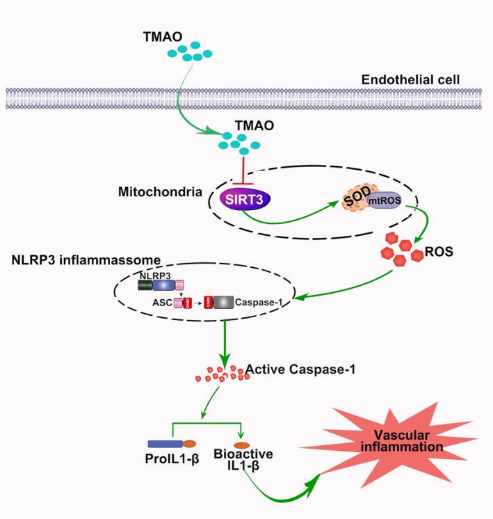
Background: Trimethylamine‐N‐oxide ( TMAO ) has recently been identified as a novel and independent risk factor for promoting atherosclerosis through inducing vascular inflammation. However, the exact mechanism is currently unclear. Studies have established a central role of nucleotide‐binding oligomerization domain–like receptor family pyrin domain–containing 3 ( NLRP 3) inflammasome in the pathogenesis of vascular inflammation. Here, we examined the potential role of the NLRP 3 inflammasome in TMAO ‐induced vascular inflammation in vitro and in vivo and the underlying mechanisms. Methods and Results: Experiments using liquid chromatography‐tandem mass spectrometry, Western blot, and fluorescent probes showed that TMAO ‐induced inflammation in human umbilical vein endothelial cells ( HUVEC s) and aortas from ApoE −/− mice. Moreover, TMAO promoted NLRP 3 and activated caspase‐1 p20 expression and caspase‐1 activity in vitro and in vivo. Notably, a caspase‐1 inhibitor ( YVAD ), an NLRP 3 inhibitor ( MCC 950), as well as NLRP 3 short interfering RNA attenuated TMAO ‐induced activation of the NLRP 3 inflammasome, subsequently leading to suppression of inflammation in HUVEC s. TMAO additionally stimulated reactive oxygen species ( ROS ) generation, in particular, mitochondrial ROS , while inhibiting manganese superoxide dismutase 2 (SOD2) activation and sirtuin 3 ( SIRT 3) expression in HUVEC s and aortas from ApoE −/− mice. TMAO ‐induced endothelial NLRP 3 inflammasome activation was ameliorated by the mitochondrial ROS scavenger Mito‐ TEMPO , or SIRT 3 overexpression in HUVEC s. Conversely, TMAO failed to further inhibit SOD2 and activate the NLRP 3 inflammasome or induce inflammation in SIRT 3 short interfering RNA –treated HUVEC s and aortas from SIRT 3 −/− mice. Conclusions: TMAO promoted vascular inflammation by activating the NLRP 3 inflammasome, and the NLRP 3 inflammasome activation in part was mediated through inhibition of the SIRT 3‐ SOD 2–mitochondrial ROS signaling pathway.
Overview
AI snapshot
Written from the paper’s full text.
TL;DR
TMAO promotes vascular inflammation by activating the NLRP3 inflammasome, mediated in part through inhibition of the SIRT3-SOD2-mtROS signaling pathway. In HUVECs and mouse aortas, TMAO increased NLRP3/caspase-1 p20 and inflammatory markers, while SIRT3 overexpression or mtROS scavenging attenuated these effects.
Key findings
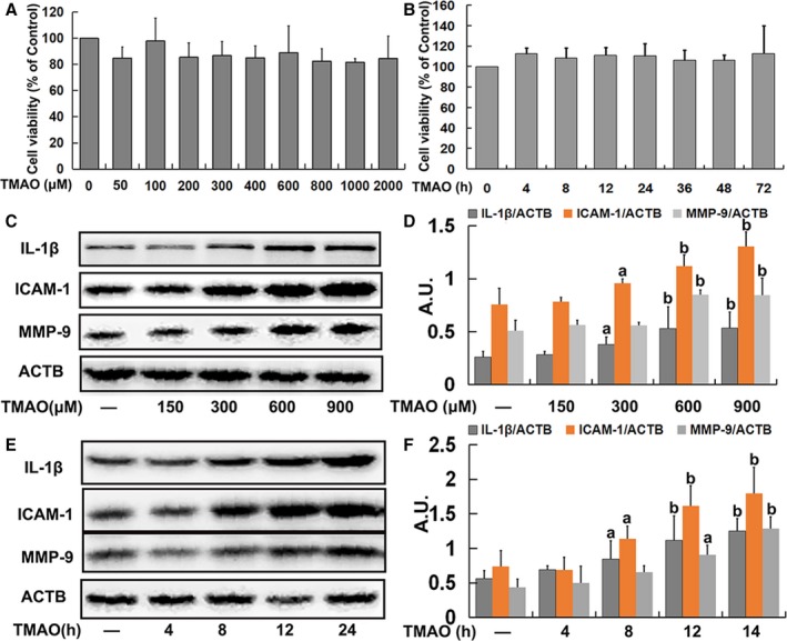
- TMAO (600 μmol/L for 24 hours) increased IL-1β, ICAM-1, and MMP-9 expression and monocyte adhesion in HUVECs without significantly affecting cell viability; TMAO also promoted NLRP3 and caspase-1 p20 expression and caspase-1 activity in concentration- and time-dependent manners.
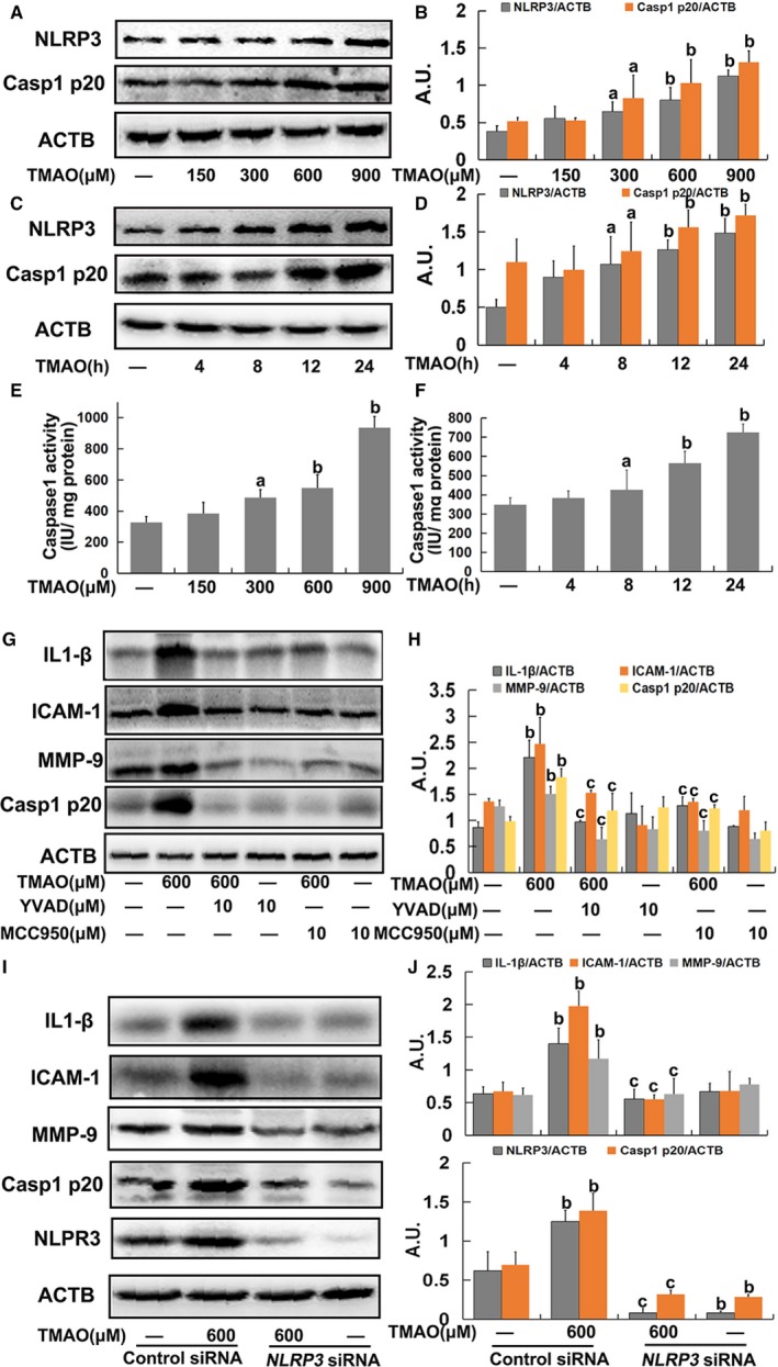
- NLRP3 inhibition with MCC950 (10 μmol/L), caspase-1 inhibition with YVAD (10 μmol/L), or NLRP3 siRNA attenuated TMAO-induced NLRP3/caspase-1 p20 and inflammatory protein expression, as well as monocyte adhesion.
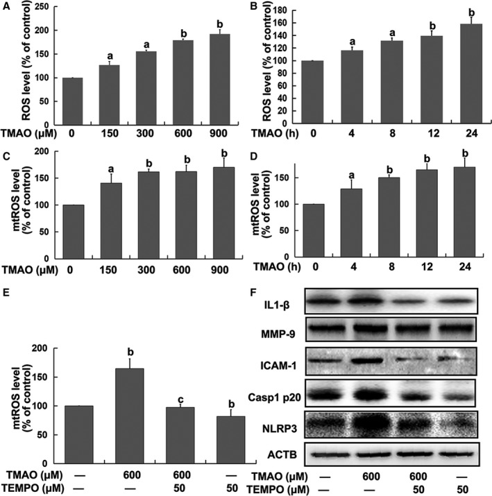
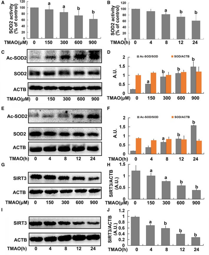
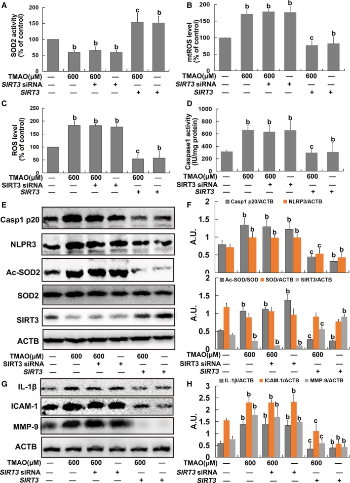
- TMAO increased total ROS and mitochondrial ROS (mtROS); the mtROS scavenger Mito-TEMPO (TEMPO, 50 μmol/L) inhibited TMAO-induced NLRP3 inflammasome activation and inflammation. TMAO suppressed SOD2 activity and SIRT3 expression and increased SOD2 acetylation; SIRT3 siRNA mimicked TMAO effects, whereas SIRT3 overexpression diminished them.
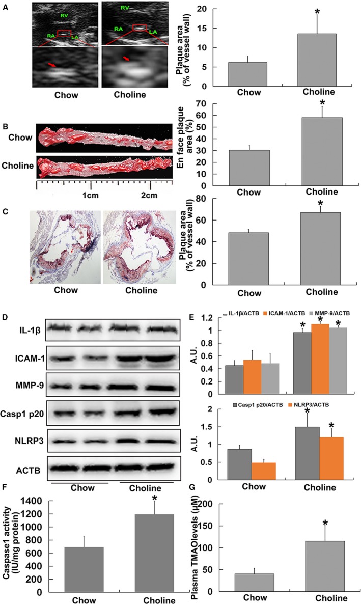
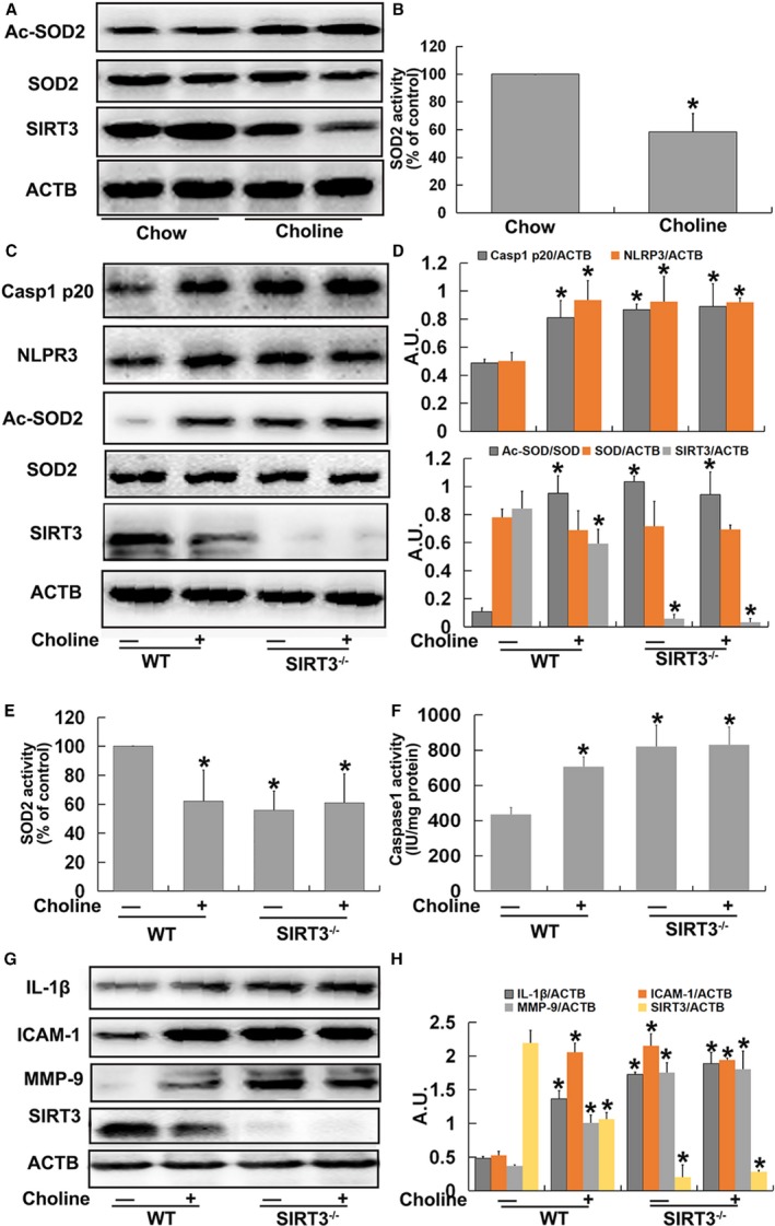
- In vivo, 1% choline for 4 months in ApoE−/− mice increased plasma TMAO, atherosclerotic lesion areas in whole aorta and aortic root, and aortic NLRP3, caspase-1 p20, caspase-1 activity, IL-1β, ICAM-1, and MMP-9; SIRT3−/− mice showed similar inflammasome activation even on chow diet.
Methods
- In vitro: HUVECs were treated with TMAO (0, 150, 300, 600, 900 μmol/L) for 24 hours or 600 μmol/L for 0, 4, 12, and 24 hours; interventions included YVAD (10 μmol/L), MCC950 (10 μmol/L), TEMPO (50 μmol/L), NLRP3 siRNA, SIRT3 siRNA, and SIRT3 overexpression plasmid. Assays included Western blot, caspase-1 activity, SOD2 activity, DCFH-DA/MitoSOX ROS detection, and monocyte adhesion.
- In vivo: 8-week-old female 129/SvJ wild-type, ApoE−/−, and SIRT3−/− mice were fed standard chow or 1% choline-supplemented chow for 4 months; n=10 per group. Analyses included plasma TMAO by liquid chromatography-tandem mass spectrometry, aortic Western blot, caspase-1 and SOD2 activity, ultrasound of aortic sinus, and Oil-red O lesion quantification.
- Statistics: data presented as means±SE of 3 experiments; t test and 1-way ANOVA with Tukey-Kramer post hoc test, significance at P<0.05.
Limitations
- Stated by the paper: the 600 μmol/L TMAO concentration used for in vitro time-course and mechanism studies was higher than that in humans; the exact role of NF-κB in TMAO-induced NLRP3 inflammasome activation needs further studies.
- Evident from the text: in vivo experiments used dietary choline as a TMAO precursor rather than direct TMAO administration, and only female mice were studied; no human intervention data are presented.
Model-written summary. Check it against the paper before relying on it.
Integrity signals
Registry
Cites
Statistics
Figures
Verification
Claims the paper makes
69 Own result · 52 Not checked yet · 20 Own interpretation · 4 Own method
A result the paper reports from its own data — it cites no other source for it, so there is none to check. The quote shows it is in the paper.
In the paper
“TMAO was tolerated by endothelial cells and exerted no significant effects on cell viability”
- Also stated in: § TMAO Induced Inflammation in Endothelial Cells · hedged
“These results indicated that TMAO triggered inflammation with no significant effects on cell viability in endothelial cells.”
§ Trimethylamine‐N‐Oxide Induces Vascular Inflammation by Activating the NLRP3 Inflammasome Through the SIRT3‐SOD2‐mtROS Signaling Pathway > Results > TMAO Induced Inflammation in Endothelial Cells
plain- Asserted: passes — The authors put it forward as true.
- Checkable: passes — It can be checked against evidence.
- Load-bearing: passes — The paper’s conclusions depend on it.
- Based: passes — The paper gives a reason why it holds.
- Also stated in: § TMAO Induced Inflammation in Endothelial Cells · hedged
A result the paper reports from its own data — it cites no other source for it, so there is none to check. The quote shows it is in the paper.
In the paper
“Additionally, SIRT3 siRNA had similar effects on IL‐1β, ICAM‐1, and MMP‐9 expression as TMAO.”
§ Trimethylamine‐N‐Oxide Induces Vascular Inflammation by Activating the NLRP3 Inflammasome Through the SIRT3‐SOD2‐mtROS Signaling Pathway > Results > TMAO Induced mtROS Accumulation via the SIRT3‐SOD2 Pathway in Endothelial Cells
plaincorrelational- Asserted: passes — The authors put it forward as true.
- Checkable: passes — It can be checked against evidence.
- Load-bearing: passes — The paper’s conclusions depend on it.
- Based: passes — The paper gives a reason why it holds.
A result the paper reports from its own data — it cites no other source for it, so there is none to check. The quote shows it is in the paper.
In the paper
“acetylation levels of SOD2 were markedly increased by TMAO in a dose‐ and time‐dependent manner.”
§ Trimethylamine‐N‐Oxide Induces Vascular Inflammation by Activating the NLRP3 Inflammasome Through the SIRT3‐SOD2‐mtROS Signaling Pathway > Results > TMAO Induced mtROS Accumulation via the SIRT3‐SOD2 Pathway in Endothelial Cells
plaindirect- Asserted: passes — The authors put it forward as true.
- Checkable: passes — It can be checked against evidence.
- Load-bearing: passes — The paper’s conclusions depend on it.
- Based: passes — The paper gives a reason why it holds.
A result the paper reports from its own data — it cites no other source for it, so there is none to check. The quote shows it is in the paper.
In the paper
“However, the TMAO‐induced increase in IL‐1β, ICAM‐1, and MMP‐9 was markedly attenuated on transfection with SIRT3 overexpression plasmid (Figure 5G and 5H).”
§ Trimethylamine‐N‐Oxide Induces Vascular Inflammation by Activating the NLRP3 Inflammasome Through the SIRT3‐SOD2‐mtROS Signaling Pathway > Results > TMAO Induced mtROS Accumulation via the SIRT3‐SOD2 Pathway in Endothelial Cells
plaindirect- Asserted: passes — The authors put it forward as true.
- Checkable: passes — It can be checked against evidence.
- Load-bearing: passes — The paper’s conclusions depend on it.
- Based: passes — The paper gives a reason why it holds.
A result the paper reports from its own data — it cites no other source for it, so there is none to check. The quote shows it is in the paper.
In the paper
“Transfection with SIRT3 siRNA alone led to decreased SOD2 activity and increased total ROS and mtROS levels followed by an increase in caspase‐1 activity and NLRP3 and caspase‐1 p20 expression, similar to the results obtained with TMAO and SIRT3 siRNA cotreatment (Figure 5A through 5F).”
§ Trimethylamine‐N‐Oxide Induces Vascular Inflammation by Activating the NLRP3 Inflammasome Through the SIRT3‐SOD2‐mtROS Signaling Pathway > Results > TMAO Induced mtROS Accumulation via the SIRT3‐SOD2 Pathway in Endothelial Cells
plaindirect- Asserted: passes — The authors put it forward as true.
- Checkable: passes — It can be checked against evidence.
- Load-bearing: passes — The paper’s conclusions depend on it.
- Based: passes — The paper gives a reason why it holds.
A result the paper reports from its own data — it cites no other source for it, so there is none to check. The quote shows it is in the paper.
In the paper
“Conversely, transfection with the SIRT3 overexpression plasmid significantly diminished the effects of TMAO on SOD2 activity, mtROS/ROS generation, and NLRP3 inflammasome activation (Figure 5A through 5F).”
§ Trimethylamine‐N‐Oxide Induces Vascular Inflammation by Activating the NLRP3 Inflammasome Through the SIRT3‐SOD2‐mtROS Signaling Pathway > Results > TMAO Induced mtROS Accumulation via the SIRT3‐SOD2 Pathway in Endothelial Cells
plaindirect- Asserted: passes — The authors put it forward as true.
- Checkable: passes — It can be checked against evidence.
- Load-bearing: passes — The paper’s conclusions depend on it.
- Based: passes — The paper gives a reason why it holds.
A result the paper reports from its own data — it cites no other source for it, so there is none to check. The quote shows it is in the paper.
In the paper
“Compared with chow‐fed ApoE−/− mice, the atherosclerotic lesion areas in whole aorta and aortic root, as indices of AS severity, were significantly higher in mice fed choline (Figure 6A through 6C).”
§ Trimethylamine‐N‐Oxide Induces Vascular Inflammation by Activating the NLRP3 Inflammasome Through the SIRT3‐SOD2‐mtROS Signaling Pathway > Results > TMAO Induced Vascular Inflammation via NLRP3 Inflammasome Activation In Vivo
plaincorrelational- Asserted: passes — The authors put it forward as true.
- Checkable: passes — It can be checked against evidence.
- Load-bearing: passes — The paper’s conclusions depend on it.
- Based: passes — The paper gives a reason why it holds.
A result the paper reports from its own data — it cites no other source for it, so there is none to check. The quote shows it is in the paper.
In the paper
“Transfection with SIRT3 siRNA alone led to decreased SOD2 activity and increased total ROS and mtROS levels followed by an increase in caspase‐1 activity and NLRP3 and caspase‐1 p20 expression, similar to the results obtained with TMAO and SIRT3 siRNA cotreatment (Figure 5A through 5F).”
§ Trimethylamine‐N‐Oxide Induces Vascular Inflammation by Activating the NLRP3 Inflammasome Through the SIRT3‐SOD2‐mtROS Signaling Pathway > Results > TMAO Induced mtROS Accumulation via the SIRT3‐SOD2 Pathway in Endothelial Cells
plaindirect- Asserted: passes — The authors put it forward as true.
- Checkable: passes — It can be checked against evidence.
- Load-bearing: passes — The paper’s conclusions depend on it.
- Based: passes — The paper gives a reason why it holds.
A result the paper reports from its own data — it cites no other source for it, so there is none to check. The quote shows it is in the paper.
In the paper
“TMAO additionally stimulated reactive oxygen species (ROS) generation, in particular, mitochondrial ROS, while inhibiting manganese superoxide dismutase 2 (SOD2) activation and sirtuin 3 (SIRT3) expression in HUVECs and aortas from ApoE−/− mice.”
§ Trimethylamine‐N‐Oxide Induces Vascular Inflammation by Activating the NLRP3 Inflammasome Through the SIRT3‐SOD2‐mtROS Signaling Pathway > Abstract > Methods and Results
plaindirect- Asserted: passes — The authors put it forward as true.
- Checkable: passes — It can be checked against evidence.
- Load-bearing: passes — The paper’s conclusions depend on it.
- Based: passes — The paper gives a reason why it holds.
A result the paper reports from its own data — it cites no other source for it, so there is none to check. The quote shows it is in the paper.
In the paper
“Compared with chow‐fed ApoE−/− mice, the atherosclerotic lesion areas in whole aorta and aortic root, as indices of AS severity, were significantly higher in mice fed choline (Figure 6A through 6C).”
§ Trimethylamine‐N‐Oxide Induces Vascular Inflammation by Activating the NLRP3 Inflammasome Through the SIRT3‐SOD2‐mtROS Signaling Pathway > Results > TMAO Induced Vascular Inflammation via NLRP3 Inflammasome Activation In Vivo
plaincorrelational- Asserted: passes — The authors put it forward as true.
- Checkable: passes — It can be checked against evidence.
- Load-bearing: passes — The paper’s conclusions depend on it.
- Based: passes — The paper gives a reason why it holds.
- It may state two separate claims as one. 27
- Probably not a substantial claim. 34
A result the paper reports from its own data — it cites no other source for it, so there is none to check. The quote shows it is in the paper.
In the paper
“Experiments using liquid chromatography‐tandem mass spectrometry, Western blot, and fluorescent probes showed that TMAO‐induced inflammation in human umbilical vein endothelial cells (HUVECs) and aortas from ApoE−/− mice.”
- Also stated in: § TMAO Induced Inflammation in Endothelial Cells · hedged
“These results indicated that TMAO triggered inflammation with no significant effects on cell viability in endothelial cells.”
§ Trimethylamine‐N‐Oxide Induces Vascular Inflammation by Activating the NLRP3 Inflammasome Through the SIRT3‐SOD2‐mtROS Signaling Pathway > Abstract > Methods and Results
plaindirect- Asserted: passes — The authors put it forward as true.
- Checkable: passes — It can be checked against evidence.
- Load-bearing: passes — The paper’s conclusions depend on it.
- Based: passes — The paper gives a reason why it holds.
It may state two separate claims as one.
- Also stated in: § TMAO Induced Inflammation in Endothelial Cells · hedged
A result the paper reports from its own data — it cites no other source for it, so there is none to check. The quote shows it is in the paper.
In the paper
“Pretreatment with MCC950 (10 μmol/L) or YVAD (10 μmol/L) markedly inhibited TMAO‐induced NLRP3 and caspase‐1 p20 expression, followed by decreased expression of IL‐1β, ICAM‐1, and MMP9 in endothelial cells (Figure 2G and 2H).”
§ Trimethylamine‐N‐Oxide Induces Vascular Inflammation by Activating the NLRP3 Inflammasome Through the SIRT3‐SOD2‐mtROS Signaling Pathway > Results > TMAO Induced Inflammation via NLRP3 Inflammasome Activation in Endothelial Cells
plaindirect- Asserted: passes — The authors put it forward as true.
- Checkable: passes — It can be checked against evidence.
- Load-bearing: passes — The paper’s conclusions depend on it.
- Based: passes — The paper gives a reason why it holds.
It may state two separate claims as one.
A result the paper reports from its own data — it cites no other source for it, so there is none to check. The quote shows it is in the paper.
In the paper
“pretreatment with TEMPO (50 μmol/L) markedly inhibited TMAO‐induced activation of the NLRP3 inflammasome followed by decreased expression of IL‐1β, ICAM‐1, and MMP‐9 (Figure 3E and 3F)”
§ Trimethylamine‐N‐Oxide Induces Vascular Inflammation by Activating the NLRP3 Inflammasome Through the SIRT3‐SOD2‐mtROS Signaling Pathway > Results > mtROS Played a Key Role in TMAO‐Induced Activation of the NLRP3 Inflammasome in Endothelial Cells
plaindirect- Asserted: passes — The authors put it forward as true.
- Checkable: passes — It can be checked against evidence.
- Load-bearing: passes — The paper’s conclusions depend on it.
- Based: passes — The paper gives a reason why it holds.
It may state two separate claims as one.
A result the paper reports from its own data — it cites no other source for it, so there is none to check. The quote shows it is in the paper.
In the paper
“TMAO‐induced endothelial NLRP3 inflammasome activation was ameliorated by the mitochondrial ROS scavenger Mito‐TEMPO, or SIRT3 overexpression in HUVECs.”
§ Trimethylamine‐N‐Oxide Induces Vascular Inflammation by Activating the NLRP3 Inflammasome Through the SIRT3‐SOD2‐mtROS Signaling Pathway > Abstract > Methods and Results
plaindirect- Asserted: passes — The authors put it forward as true.
- Checkable: passes — It can be checked against evidence.
- Load-bearing: passes — The paper’s conclusions depend on it.
- Based: passes — The paper gives a reason why it holds.
It may state two separate claims as one.
A result the paper reports from its own data — it cites no other source for it, so there is none to check. The quote shows it is in the paper.
In the paper
“Conversely, TMAO failed to further inhibit magnesium SOD2 and activate the NLRP3 inflammasome or induce inflammation in SIRT3 short interfering RNA–treated HUVECs and aortas from SIRT3−/− mice.”
§ Trimethylamine‐N‐Oxide Induces Vascular Inflammation by Activating the NLRP3 Inflammasome Through the SIRT3‐SOD2‐mtROS Signaling Pathway > Abstract > Methods and Results
plain- Asserted: passes — The authors put it forward as true.
- Checkable: passes — It can be checked against evidence.
- Load-bearing: passes — The paper’s conclusions depend on it.
- Based: passes — The paper gives a reason why it holds.
It may state two separate claims as one.
A result the paper reports from its own data — it cites no other source for it, so there is none to check. The quote shows it is in the paper.
In the paper
“Moreover, TMAO promoted NLRP3 and activated caspase‐1 p20 expression and caspase‐1 activity in vitro and in vivo.”
§ Trimethylamine‐N‐Oxide Induces Vascular Inflammation by Activating the NLRP3 Inflammasome Through the SIRT3‐SOD2‐mtROS Signaling Pathway > Abstract > Methods and Results
plaindirect- Asserted: passes — The authors put it forward as true.
- Checkable: passes — It can be checked against evidence.
- Load-bearing: passes — The paper’s conclusions depend on it.
- Based: passes — The paper gives a reason why it holds.
It may state two separate claims as one.
None of the sources cited for this claim have been checked yet.
In the paper
“Following decades of research, the most well‐characterized mechanism underlying activation of the NLRP3 inflammasome is reactive oxygen species (ROS), particularly mitochondrial ROS (mtROS), generation.”
§ Trimethylamine‐N‐Oxide Induces Vascular Inflammation by Activating the NLRP3 Inflammasome Through the SIRT3‐SOD2‐mtROS Signaling Pathway > Introduction
What the sources say
- Not checked yetcites [14] Zhou RB, Yazdi AS, Menu P, Tschopp J. A role for mitochondria in NLRP3 inflammasome activation. Nature. 2011; 469: 221– 225. 21124315 source
- Not checked yetcites [15] Alfonso‐Loeches S, Urena‐Peralta JR, Morillo‐Bargues MJ, Oliver‐De La Cruz J, Guerri C. Role of mitochondria ROS generation in ethanol‐induced NLRP3 inflammasome activation and cell death in astroglial cells. Front Cell Neurosci. 2014; 8: 216– 234. 25136295 PMC4118026 source
boosteddirect- Asserted: passes — The authors put it forward as true.
- Checkable: passes — It can be checked against evidence.
- Load-bearing: passes — The paper’s conclusions depend on it.
- Based: passes — The paper gives a reason why it holds.
Probably not a substantial claim.
None of the sources cited for this claim have been checked yet.
In the paper
“Oxidized low‐density lipoprotein and cholesterol crystals activate the inflammasome, thereby leading to the processing and secretion of the proinflammatory cytokines IL‐1β and IL‐18 and ultimately triggering vascular inflammation via upregulation of adhesion molecules.9, 10”
§ Trimethylamine‐N‐Oxide Induces Vascular Inflammation by Activating the NLRP3 Inflammasome Through the SIRT3‐SOD2‐mtROS Signaling Pathway > Introduction
What the sources say
- Not checked yetcites [9] Yin Y, Pastrana JL, Li XY, Huang X, Mallilankaraman K, Choi ET, Madesh M, Wang H, Yang XF. Inflammasomes: sensors of metabolic stresses for vascular inflammation. Front Biosci (Landmark Ed). 2013; 18: 638– 649. 23276949 PMC3590813 source
- Not checked yetcites [10] Duewell P, Kono H, Rayner KJ, Sirois CM, Vladimer G, Bauernfeind FG, Abela GS, Franchi L, Nunez G, Schnurr M, Espevik T, Lien E, Fitzgerald KA, Rock KL, Moore KJ, Wright SD, Hornung V, Latz E. NLRP3 inflammasomes are required for atherogenesis and activated by cholesterol crystals. Nature. 2010; 464: 1357– 1361. 20428172 PMC2946640 source
plaindirect- Asserted: passes — The authors put it forward as true.
- Checkable: passes — It can be checked against evidence.
- Load-bearing: fails — The paper’s conclusions may not depend on it.
- Based: passes — The paper gives a reason why it holds.
Probably not a substantial claim. The paper’s conclusions may not depend on it. It may state two separate claims as one.
None of the sources cited for this claim have been checked yet.
In the paper
“Accumulating evidence indicates that AS is the result of a prolonged and excessive inflammatory processes in the vascular wall, which often begins with inflammatory changes in the endothelium and is characterized by the expression of adhesion molecules.2, 3”
§ Trimethylamine‐N‐Oxide Induces Vascular Inflammation by Activating the NLRP3 Inflammasome Through the SIRT3‐SOD2‐mtROS Signaling Pathway > Introduction
What the sources say
- Not checked yetcites [2] Maskrey BH, Megson IL, Whitfield PD, Rossi AG. Mechanisms of resolution of inflammation: a focus on cardiovascular disease. Arterioscler Thromb Vasc Biol. 2011; 31: 1001– 1006. 21508346 source
- Not checked yetcites [3] Wong BW, Meredith A, Lin D, McManus BM. The biological role of inflammation in atherosclerosis. Can J Cardiol. 2012; 28: 631– 641. 22985787 source
hedgeddirect- Asserted: passes — The authors put it forward as true.
- Checkable: passes — It can be checked against evidence.
- Load-bearing: passes — The paper’s conclusions depend on it.
- Based: passes — The paper gives a reason why it holds.
Probably not a substantial claim.
None of the sources cited for this claim have been checked yet.
In the paper
“Several studies have suggested that ablation of NLRP3 protects endothelial cells from inflammatory damage, leading to attenuation of AS.12, 13”
§ Trimethylamine‐N‐Oxide Induces Vascular Inflammation by Activating the NLRP3 Inflammasome Through the SIRT3‐SOD2‐mtROS Signaling Pathway > Introduction
What the sources say
- Not checked yetcites [12] Wu JJ, Xu XS, Li Y, Kou JP, Huang F, Liu BL, Liu K. Quercetin, luteolin and epigallocatechin gallate alleviate TXNIP and NLRP3‐mediated inflammation and apoptosis with regulation of AMPK in endothelial cells. Eur J Pharmacol. 2014; 745: 59– 68. 25446924 source
- Not checked yetcites [13] Chen Y, Wang L, Pitzer AL, Li X, Li PL, Zhang Y. Contribution of redox‐dependent activation of endothelial NLRP3 inflammasomes to hyperglycemia‐induced endothelial dysfunction. J Mol Med. 2016; 94: 1335– 1347. 27783111 PMC5512566 source
hedgeddirect- Asserted: passes — The authors put it forward as true.
- Checkable: passes — It can be checked against evidence.
- Load-bearing: passes — The paper’s conclusions depend on it.
- Based: passes — The paper gives a reason why it holds.
Probably not a substantial claim. It may state two separate claims as one.
In the paper
“In view of the finding that SOD2, the primary mitochondrial oxidative scavenger, plays a crucial role in regulation of mtROS,29”
§ Trimethylamine‐N‐Oxide Induces Vascular Inflammation by Activating the NLRP3 Inflammasome Through the SIRT3‐SOD2‐mtROS Signaling Pathway > Results > TMAO Induced mtROS Accumulation via the SIRT3‐SOD2 Pathway in Endothelial Cells
What the sources say
- Not checked yetcites [29] Tao R, Vassilopoulos A, Parisiadou L, Yan Y, Gius D. Regulation of MnSOD enzymatic activity by SIRT3 connects the mitochondrial acetylome signaling networks to aging and carcinogenesis. Antioxid Redox Signal. 2014; 20: 1646– 1654. 23886445 PMC3942696 source
plaindirect- Asserted: passes — The authors put it forward as true.
- Checkable: passes — It can be checked against evidence.
- Load-bearing: passes — The paper’s conclusions depend on it.
- Based: passes — The paper gives a reason why it holds.
Probably not a substantial claim. It may state two separate claims as one.
In the paper
“More recently, TMAO has been shown to promote vascular inflammation, leading to AS.7”
§ Trimethylamine‐N‐Oxide Induces Vascular Inflammation by Activating the NLRP3 Inflammasome Through the SIRT3‐SOD2‐mtROS Signaling Pathway > Introduction
What the sources say
- Not checked yetcites [7] Seldin MM, Meng YH, Qi HX, Zhu WF, Wang ZE, Hazen SL, Lusis AJ, Shih DM. Trimethylamine N‐oxide promotes vascular inflammation through signaling of mitogen‐activated protein kinase and nuclear factor‐κB. J Am Heart Assoc. 2016; 5: e002767. DOI: 10.1161/JAHA.115.002767. PMC4802459 26903003 source
plaindirect- Asserted: passes — The authors put it forward as true.
- Checkable: passes — It can be checked against evidence.
- Load-bearing: passes — The paper’s conclusions depend on it.
- Based: passes — The paper gives a reason why it holds.
It may state two separate claims as one.
In the paper
“The NLRP3 inflammasome plays a key role in the pathogenesis of diverse inflammatory diseases, including AS, type 2 diabetes mellitus, and gout.9”
§ Trimethylamine‐N‐Oxide Induces Vascular Inflammation by Activating the NLRP3 Inflammasome Through the SIRT3‐SOD2‐mtROS Signaling Pathway > Introduction
What the sources say
- Not checked yetcites [9] Yin Y, Pastrana JL, Li XY, Huang X, Mallilankaraman K, Choi ET, Madesh M, Wang H, Yang XF. Inflammasomes: sensors of metabolic stresses for vascular inflammation. Front Biosci (Landmark Ed). 2013; 18: 638– 649. 23276949 PMC3590813 source
plaindirect- Asserted: passes — The authors put it forward as true.
- Checkable: passes — It can be checked against evidence.
- Load-bearing: passes — The paper’s conclusions depend on it.
- Based: passes — The paper gives a reason why it holds.
Probably not a substantial claim.
In the paper
“In 2016, Seldin and co‐workers7 showed that TMAO promotes vascular inflammation through signaling of mitogen‐activated protein kinase and nuclear factor‐κB (NF‐κB).”
§ Trimethylamine‐N‐Oxide Induces Vascular Inflammation by Activating the NLRP3 Inflammasome Through the SIRT3‐SOD2‐mtROS Signaling Pathway > Introduction
What the sources say
- Not checked yetcites [7] Seldin MM, Meng YH, Qi HX, Zhu WF, Wang ZE, Hazen SL, Lusis AJ, Shih DM. Trimethylamine N‐oxide promotes vascular inflammation through signaling of mitogen‐activated protein kinase and nuclear factor‐κB. J Am Heart Assoc. 2016; 5: e002767. DOI: 10.1161/JAHA.115.002767. PMC4802459 26903003 source
plaindirect- Asserted: passes — The authors put it forward as true.
- Checkable: passes — It can be checked against evidence.
- Load-bearing: fails — The paper’s conclusions may not depend on it.
- Based: passes — The paper gives a reason why it holds.
Probably not a substantial claim. The paper’s conclusions may not depend on it.
In the paper
“Xiao et al11 additionally reported that atheroprone flow induces the NLRP3 inflammasome in endothelium through serol regulatory element binding protein 2 activation.”
§ Trimethylamine‐N‐Oxide Induces Vascular Inflammation by Activating the NLRP3 Inflammasome Through the SIRT3‐SOD2‐mtROS Signaling Pathway > Introduction
What the sources say
- Not checked yetcites [11] Xiao H, Lu M, Lin TY, Chen Z, Chen G, Wang WC, Marin T, Shentu TP, Wen L, Gongol B, Sun W, Liang X, Chen J, Huang HD, Pedra JHF, Johnson DA, Shyy JYJ. Sterol regulatory element binding protein 2 activation of NLRP3 inflammasome in endothelium mediates hemodynamic‐induced atherosclerosis susceptibility. Circulation. 2013; 128: 632– 642. 23838163 PMC3798034 source
plaindirect- Asserted: passes — The authors put it forward as true.
- Checkable: passes — It can be checked against evidence.
- Load-bearing: fails — The paper’s conclusions may not depend on it.
- Based: passes — The paper gives a reason why it holds.
Probably not a substantial claim. The paper’s conclusions may not depend on it.
None of the sources cited for this claim have been checked yet.
In the paper
“Previous studies suggest that TMAO partially contributes to the development of AS by regulating the major pathway of cholesterol metabolism and the bile acid synthetic pathway.4, 5, 6”
§ Trimethylamine‐N‐Oxide Induces Vascular Inflammation by Activating the NLRP3 Inflammasome Through the SIRT3‐SOD2‐mtROS Signaling Pathway > Introduction
What the sources say
- Not checked yetcites [4] Wang Z, Klipfell E, Bennett BJ, Koeth R, Levison BS, Dugar B, Feldstein AE, Britt EB, Fu X, Chung YM, Wu Y, Schauer P, Smith JD, Allayee H, Tang WH, DiDonato JA, Lusis AJ, Hazen SL. Gut flora metabolism of phosphatidylcholine promotes cardiovascular disease. Nature. 2011; 472: 57– 63. 21475195 PMC3086762 source
- Not checked yetcites [5] Tang WH, Wang Z, Levison BS, Koeth RA, Britt EB, Fu X, Wu Y, Hazen SL. Intestinal microbial metabolism of phosphatidylcholine and cardiovascular risk. N Engl J Med. 2013; 368: 1575– 1584. 23614584 PMC3701945 source
- Not checked yetcites [6] Koeth RA, Wang Z, Levison BS, Buffa JA, Org E, Sheehy BT, Britt EB, Fu X, Wu Y, Li L, Smith JD, DiDonato JA, Chen J, Li H, Wu GD, Lewis JD, Warrier M, Brown JM, Krauss RM, Tang WH, Bushman FD, Lusis AJ, Hazen SL. Intestinal microbiota metabolism of l‐carnitine, a nutrient in red meat, promotes atherosclerosis. Nat Med. 2013; 19: 576– 585. 23563705 PMC3650111 source
hedgedconditional- Asserted: passes — The authors put it forward as true.
- Checkable: passes — It can be checked against evidence.
- Load-bearing: fails — The paper’s conclusions may not depend on it.
- Based: passes — The paper gives a reason why it holds.
Probably not a substantial claim. The paper’s conclusions may not depend on it.
In the paper
“SOD2 activity is known to be tightly regulated via acetylation at its lysine residues.20”
§ Trimethylamine‐N‐Oxide Induces Vascular Inflammation by Activating the NLRP3 Inflammasome Through the SIRT3‐SOD2‐mtROS Signaling Pathway > Results > TMAO Induced mtROS Accumulation via the SIRT3‐SOD2 Pathway in Endothelial Cells
What the sources say
- Not checked yetcites [20] Tao R, Coleman MC, Pennington JD, Ozden O, Park SH, Jiang H, Kim HS, Flynn CR, Hill S, Hayes McDonald W, Olivier AK, Spitz DR, Gius D. Sirt3‐mediated deacetylation of evolutionarily conserved lysine 122 regulates MnSOD activity in response to stress. Mol Cell. 2010; 40: 893– 904. 21172655 PMC3266626 source
boosteddirect- Asserted: passes — The authors put it forward as true.
- Checkable: passes — It can be checked against evidence.
- Load-bearing: passes — The paper’s conclusions depend on it.
- Based: passes — The paper gives a reason why it holds.
Probably not a substantial claim.
In the paper
“This increased innate immunity in endothelium synergizes with hyperlipidemia to cause topographical distribution of atherosclerotic lesions.11”
§ Trimethylamine‐N‐Oxide Induces Vascular Inflammation by Activating the NLRP3 Inflammasome Through the SIRT3‐SOD2‐mtROS Signaling Pathway > Introduction
What the sources say
- Not checked yetcites [11] Xiao H, Lu M, Lin TY, Chen Z, Chen G, Wang WC, Marin T, Shentu TP, Wen L, Gongol B, Sun W, Liang X, Chen J, Huang HD, Pedra JHF, Johnson DA, Shyy JYJ. Sterol regulatory element binding protein 2 activation of NLRP3 inflammasome in endothelium mediates hemodynamic‐induced atherosclerosis susceptibility. Circulation. 2013; 128: 632– 642. 23838163 PMC3798034 source
plaindirect- Asserted: passes — The authors put it forward as true.
- Checkable: passes — It can be checked against evidence.
- Load-bearing: fails — The paper’s conclusions may not depend on it.
- Based: passes — The paper gives a reason why it holds.
Probably not a substantial claim. The paper’s conclusions may not depend on it.
In the paper
“mtROS levels were evaluated using MitoSOX™ Red, a highly selective fluorescent probe for detection of O2∙− generated within mitochondria.28”
§ Trimethylamine‐N‐Oxide Induces Vascular Inflammation by Activating the NLRP3 Inflammasome Through the SIRT3‐SOD2‐mtROS Signaling Pathway > Results > mtROS Played a Key Role in TMAO‐Induced Activation of the NLRP3 Inflammasome in Endothelial Cells
What the sources say
- Not checked yetcites [28] Chowdhury SK, Dobrowsky RT, Fernyhough P. Nutrient excess and altered mitochondrial proteome and function contribute to neurodegeneration in diabetes. Mitochondrion. 2011; 11: 845– 854. 21742060 PMC3375692 source
plain- Asserted: passes — The authors put it forward as true.
- Checkable: passes — It can be checked against evidence.
- Load-bearing: passes — The paper’s conclusions depend on it.
- Based: passes — The paper gives a reason why it holds.
Probably not a substantial claim.
None of the sources cited for this claim have been checked yet.
In the paper
“The majority of NLRP3 activators increase mtROS levels, and chemical scavengers of mtROS effectively inhibit NLRP3 activation.14, 15”
§ Trimethylamine‐N‐Oxide Induces Vascular Inflammation by Activating the NLRP3 Inflammasome Through the SIRT3‐SOD2‐mtROS Signaling Pathway > Introduction
What the sources say
- Not checked yetcites [14] Zhou RB, Yazdi AS, Menu P, Tschopp J. A role for mitochondria in NLRP3 inflammasome activation. Nature. 2011; 469: 221– 225. 21124315 source
- Not checked yetcites [15] Alfonso‐Loeches S, Urena‐Peralta JR, Morillo‐Bargues MJ, Oliver‐De La Cruz J, Guerri C. Role of mitochondria ROS generation in ethanol‐induced NLRP3 inflammasome activation and cell death in astroglial cells. Front Cell Neurosci. 2014; 8: 216– 234. 25136295 PMC4118026 source
plaindirect- Asserted: passes — The authors put it forward as true.
- Checkable: passes — It can be checked against evidence.
- Load-bearing: passes — The paper’s conclusions depend on it.
- Based: passes — The paper gives a reason why it holds.
It may state two separate claims as one.
None of the sources cited for this claim have been checked yet.
In the paper
“More importantly, SIRT3 can directly bind and deacetylate SOD2, leading to increased SOD2 activity and subsequently exerting significant effects on mtROS homeostasis and NLRP3 inflammasome activation.18, 19, 20”
§ Trimethylamine‐N‐Oxide Induces Vascular Inflammation by Activating the NLRP3 Inflammasome Through the SIRT3‐SOD2‐mtROS Signaling Pathway > Introduction
What the sources say
- Not checked yetcites [18] Zhao WY, Zhang L, Sui MX, Zhu YH, Zeng L. Protective effects of sirtuin 3 in a murine model of sepsis‐induced acute kidney injury. Sci Rep. 2016; 6: 33201. 27620507 PMC5020492 source
- Not checked yetcites [19] Traba J, Kwarteng‐Siaw M, Okoli TC, Li J, Huffstutler RD, Bray A, Waclawiw MA, Han K, Pelletier M, Sauve AA, Siegel RM, Sack MN. Fasting and refeeding differentially regulate NLRP3 inflammasome activation in human subjects. J Clin Invest. 2015; 125: 4592– 4600. 26529255 PMC4665779 source
- Not checked yetcites [20] Tao R, Coleman MC, Pennington JD, Ozden O, Park SH, Jiang H, Kim HS, Flynn CR, Hill S, Hayes McDonald W, Olivier AK, Spitz DR, Gius D. Sirt3‐mediated deacetylation of evolutionarily conserved lysine 122 regulates MnSOD activity in response to stress. Mol Cell. 2010; 40: 893– 904. 21172655 PMC3266626 source
plaindirect- Asserted: passes — The authors put it forward as true.
- Checkable: passes — It can be checked against evidence.
- Load-bearing: passes — The paper’s conclusions depend on it.
- Based: passes — The paper gives a reason why it holds.
It may state two separate claims as one.
What the paper did — it cites no source for this, so there is none to check. The quote shows it is in the paper.
In the paper
“To reduce variability, image parameters remained constant throughout the experiment.”
§ Trimethylamine‐N‐Oxide Induces Vascular Inflammation by Activating the NLRP3 Inflammasome Through the SIRT3‐SOD2‐mtROS Signaling Pathway > Materials and Methods > Ultrasound Detection of Aortic Root Lesions
plaindirect- Asserted: passes — The authors put it forward as true.
- Checkable: passes — It can be checked against evidence.
- Load-bearing: fails — The paper’s conclusions may not depend on it.
- Based: passes — The paper gives a reason why it holds.
Probably not a substantial claim. The paper’s conclusions may not depend on it.
What the paper did — it cites no source for this, so there is none to check. The quote shows it is in the paper.
In the paper
“The ethics review board at the Third Military Medical University approved this study, and informed consent was obtained from all patients before participation.”
§ Trimethylamine‐N‐Oxide Induces Vascular Inflammation by Activating the NLRP3 Inflammasome Through the SIRT3‐SOD2‐mtROS Signaling Pathway > Materials and Methods > Cell Culture and Treatment
plain- Asserted: passes — The authors put it forward as true.
- Checkable: passes — It can be checked against evidence.
- Load-bearing: fails — The paper’s conclusions may not depend on it.
- Based: passes — The paper gives a reason why it holds.
Probably not a substantial claim. The paper’s conclusions may not depend on it. It may state two separate claims as one.
What the paper did — it cites no source for this, so there is none to check. The quote shows it is in the paper.
In the paper
“All examinations were performed by an experienced operator, and all measurements were repeated 3 times at the same site.”
§ Trimethylamine‐N‐Oxide Induces Vascular Inflammation by Activating the NLRP3 Inflammasome Through the SIRT3‐SOD2‐mtROS Signaling Pathway > Materials and Methods > Ultrasound Detection of Aortic Root Lesions
plain- Asserted: passes — The authors put it forward as true.
- Checkable: passes — It can be checked against evidence.
- Load-bearing: fails — The paper’s conclusions may not depend on it.
- Based: passes — The paper gives a reason why it holds.
Probably not a substantial claim. The paper’s conclusions may not depend on it. It may state two separate claims as one.
What the paper did — it cites no source for this, so there is none to check. The quote shows it is in the paper.
In the paper
“Animal experiments were carried out in strict accordance with the recommendations in the Guide for the Care and Use of Laboratory Animals by the National Institutes of Health and approved by the Animal Care and Use Committee of the Third Military Medical University (Chongqing, China; Approval SYXC‐2014‐00112).”
§ Trimethylamine‐N‐Oxide Induces Vascular Inflammation by Activating the NLRP3 Inflammasome Through the SIRT3‐SOD2‐mtROS Signaling Pathway > Materials and Methods > Animals and Treatment
plain- Asserted: passes — The authors put it forward as true.
- Checkable: passes — It can be checked against evidence.
- Load-bearing: fails — The paper’s conclusions may not depend on it.
- Based: passes — The paper gives a reason why it holds.
Probably not a substantial claim. The paper’s conclusions may not depend on it.
In the paper
“These results support a role for TMAO in the activation of inflammatory pathways in cells of the vasculature, leading to endothelial cell leukocyte recruitment and AS.”
§ Trimethylamine‐N‐Oxide Induces Vascular Inflammation by Activating the NLRP3 Inflammasome Through the SIRT3‐SOD2‐mtROS Signaling Pathway > Discussion
What the sources say
- Not checked yetcites [7] Seldin MM, Meng YH, Qi HX, Zhu WF, Wang ZE, Hazen SL, Lusis AJ, Shih DM. Trimethylamine N‐oxide promotes vascular inflammation through signaling of mitogen‐activated protein kinase and nuclear factor‐κB. J Am Heart Assoc. 2016; 5: e002767. DOI: 10.1161/JAHA.115.002767. PMC4802459 26903003 source
hedgeddirect- Asserted: passes — The authors put it forward as true.
- Checkable: passes — It can be checked against evidence.
- Load-bearing: passes — The paper’s conclusions depend on it.
- Based: passes — The paper gives a reason why it holds.
It may state two separate claims as one.
None of the sources cited for this claim have been checked yet.
In the paper
“IL‐1β is a major atheroprone factor that plays a significant role in promoting the development of lipid plaques and destabilizing plaques by upregulating adhesion molecules, including vascular cell adhesion molecule‐1 and ICAM‐1.31, 32, 33”
§ Trimethylamine‐N‐Oxide Induces Vascular Inflammation by Activating the NLRP3 Inflammasome Through the SIRT3‐SOD2‐mtROS Signaling Pathway > Discussion
What the sources say
- Not checked yetcites [31] De Nardo D, Latz E. NLRP3 inflammasomes link inflammation and metabolic disease. Trends Immunol. 2011; 32: 373– 379. 21733753 PMC3151541 source
- Not checked yetcites [32] Connat JL. [ Inflammasome and cardiovascular diseases]. Ann Cardiol Angeiol (Paris). 2011; 60: 48– 54. 20800829 source
- Not checked yetcites [33] Alcocer‐Gomez E, Cordero MD. NLRP3 inflammasome: common nexus between depression and cardiovascular diseases. Nat Rev Cardiol. 2017; 14: 124. 28054580 source
plaindirect- Asserted: passes — The authors put it forward as true.
- Checkable: passes — It can be checked against evidence.
- Load-bearing: fails — The paper’s conclusions may not depend on it.
- Based: passes — The paper gives a reason why it holds.
Probably not a substantial claim. The paper’s conclusions may not depend on it. It may state two separate claims as one.
In the paper
“Moreover, recent studies have demonstrated that sterol regulatory element binding protein 2 activation of NLRP3 inflammasome in endothelium mediates hemodynamic‐induced AS susceptibility,11”
§ Trimethylamine‐N‐Oxide Induces Vascular Inflammation by Activating the NLRP3 Inflammasome Through the SIRT3‐SOD2‐mtROS Signaling Pathway > Discussion
What the sources say
- Not checked yetcites [11] Xiao H, Lu M, Lin TY, Chen Z, Chen G, Wang WC, Marin T, Shentu TP, Wen L, Gongol B, Sun W, Liang X, Chen J, Huang HD, Pedra JHF, Johnson DA, Shyy JYJ. Sterol regulatory element binding protein 2 activation of NLRP3 inflammasome in endothelium mediates hemodynamic‐induced atherosclerosis susceptibility. Circulation. 2013; 128: 632– 642. 23838163 PMC3798034 source
boosteddirect- Asserted: passes — The authors put it forward as true.
- Checkable: passes — It can be checked against evidence.
- Load-bearing: fails — The paper’s conclusions may not depend on it.
- Based: passes — The paper gives a reason why it holds.
Probably not a substantial claim. The paper’s conclusions may not depend on it.
In the paper
“AS is decreased in several rodent models lacking NLRP3 or IL‐1 molecules, whereas in contrast, mice deficient in the IL‐1 receptor antagonist show increased AS.10”
§ Trimethylamine‐N‐Oxide Induces Vascular Inflammation by Activating the NLRP3 Inflammasome Through the SIRT3‐SOD2‐mtROS Signaling Pathway > Discussion
What the sources say
- Not checked yetcites [10] Duewell P, Kono H, Rayner KJ, Sirois CM, Vladimer G, Bauernfeind FG, Abela GS, Franchi L, Nunez G, Schnurr M, Espevik T, Lien E, Fitzgerald KA, Rock KL, Moore KJ, Wright SD, Hornung V, Latz E. NLRP3 inflammasomes are required for atherogenesis and activated by cholesterol crystals. Nature. 2010; 464: 1357– 1361. 20428172 PMC2946640 source
plaindirect- Asserted: passes — The authors put it forward as true.
- Checkable: passes — It can be checked against evidence.
- Load-bearing: unclear — It is unclear whether the paper’s conclusions depend on it.
- Based: passes — The paper gives a reason why it holds.
It may state two separate claims as one.
In the paper
“Recent studies have shown that physiological levels of TMAO can induce expression of cytokines and adhesion molecules, at least in part via the NF‐κB signaling pathway in vascular endothelial and smooth muscle cells, both in vivo and in vitro.7”
§ Trimethylamine‐N‐Oxide Induces Vascular Inflammation by Activating the NLRP3 Inflammasome Through the SIRT3‐SOD2‐mtROS Signaling Pathway > Discussion
What the sources say
- Not checked yetcites [7] Seldin MM, Meng YH, Qi HX, Zhu WF, Wang ZE, Hazen SL, Lusis AJ, Shih DM. Trimethylamine N‐oxide promotes vascular inflammation through signaling of mitogen‐activated protein kinase and nuclear factor‐κB. J Am Heart Assoc. 2016; 5: e002767. DOI: 10.1161/JAHA.115.002767. PMC4802459 26903003 source
plainconditional- Asserted: passes — The authors put it forward as true.
- Checkable: passes — It can be checked against evidence.
- Load-bearing: fails — The paper’s conclusions may not depend on it.
- Based: passes — The paper gives a reason why it holds.
Probably not a substantial claim. The paper’s conclusions may not depend on it.
None of the sources cited for this claim have been checked yet.
In the paper
“Researchers have confirmed that TMAO contributes to the development of AS, in part, by affecting cholesterol metabolism through regulating the major pathway for cholesterol elimination from the body and the bile acid synthetic pathway at multiple levels.4, 6”
§ Trimethylamine‐N‐Oxide Induces Vascular Inflammation by Activating the NLRP3 Inflammasome Through the SIRT3‐SOD2‐mtROS Signaling Pathway > Discussion
What the sources say
- Not checked yetcites [4] Wang Z, Klipfell E, Bennett BJ, Koeth R, Levison BS, Dugar B, Feldstein AE, Britt EB, Fu X, Chung YM, Wu Y, Schauer P, Smith JD, Allayee H, Tang WH, DiDonato JA, Lusis AJ, Hazen SL. Gut flora metabolism of phosphatidylcholine promotes cardiovascular disease. Nature. 2011; 472: 57– 63. 21475195 PMC3086762 source
- Not checked yetcites [6] Koeth RA, Wang Z, Levison BS, Buffa JA, Org E, Sheehy BT, Britt EB, Fu X, Wu Y, Li L, Smith JD, DiDonato JA, Chen J, Li H, Wu GD, Lewis JD, Warrier M, Brown JM, Krauss RM, Tang WH, Bushman FD, Lusis AJ, Hazen SL. Intestinal microbiota metabolism of l‐carnitine, a nutrient in red meat, promotes atherosclerosis. Nat Med. 2013; 19: 576– 585. 23563705 PMC3650111 source
boosteddirect- Asserted: passes — The authors put it forward as true.
- Checkable: passes — It can be checked against evidence.
- Load-bearing: fails — The paper’s conclusions may not depend on it.
- Based: passes — The paper gives a reason why it holds.
Probably not a substantial claim. The paper’s conclusions may not depend on it.
None of the sources cited for this claim have been checked yet.
In the paper
“whereas ablation of NLRP3 protects endothelial cells from inflammatory damage, subsequently attenuating AS.12, 13”
§ Trimethylamine‐N‐Oxide Induces Vascular Inflammation by Activating the NLRP3 Inflammasome Through the SIRT3‐SOD2‐mtROS Signaling Pathway > Discussion
What the sources say
- Not checked yetcites [12] Wu JJ, Xu XS, Li Y, Kou JP, Huang F, Liu BL, Liu K. Quercetin, luteolin and epigallocatechin gallate alleviate TXNIP and NLRP3‐mediated inflammation and apoptosis with regulation of AMPK in endothelial cells. Eur J Pharmacol. 2014; 745: 59– 68. 25446924 source
- Not checked yetcites [13] Chen Y, Wang L, Pitzer AL, Li X, Li PL, Zhang Y. Contribution of redox‐dependent activation of endothelial NLRP3 inflammasomes to hyperglycemia‐induced endothelial dysfunction. J Mol Med. 2016; 94: 1335– 1347. 27783111 PMC5512566 source
plaindirect- Asserted: passes — The authors put it forward as true.
- Checkable: passes — It can be checked against evidence.
- Load-bearing: unclear — It is unclear whether the paper’s conclusions depend on it.
- Based: passes — The paper gives a reason why it holds.
It may state two separate claims as one.
In the paper
“The NLRP3 inflammasome is an IL‐1β family cytokine‐activating protein complex consisting of the pattern recognition receptor NLRP3, adaptor protein apoptotic speck‐like protein, and inactive pro‐caspase‐1, and involved in the regulation of innate immunity and inflammatory response.30”
§ Trimethylamine‐N‐Oxide Induces Vascular Inflammation by Activating the NLRP3 Inflammasome Through the SIRT3‐SOD2‐mtROS Signaling Pathway > Discussion
What the sources say
- Not checked yetcites [30] Gross O, Thomas CJ, Guarda G, Tschopp J. The inflammasome: an integrated view. Immunol Rev. 2011; 243: 136– 151. 21884173 source
plain- Asserted: passes — The authors put it forward as true.
- Checkable: passes — It can be checked against evidence.
- Load-bearing: fails — The paper’s conclusions may not depend on it.
- Based: passes — The paper gives a reason why it holds.
Probably not a substantial claim. The paper’s conclusions may not depend on it. It may state two separate claims as one.
None of the sources cited for this claim have been checked yet.
In the paper
“Primed macrophages secrete large amounts of IL‐1β in response to cholesterol crystals in an NLRP3 inflammasome‐dependent manner in mouse and human cells.34, 35”
§ Trimethylamine‐N‐Oxide Induces Vascular Inflammation by Activating the NLRP3 Inflammasome Through the SIRT3‐SOD2‐mtROS Signaling Pathway > Discussion
What the sources say
- Not checked yetcites [34] Rajamaki K, Lappalainen J, Oorni K, Valimaki E, Matikainen S, Kovanen PT, Eklund KK. Cholesterol crystals activate the NLRP3 inflammasome in human macrophages: a novel link between cholesterol metabolism and inflammation. PLoS One. 2010; 5: e11765. 20668705 PMC2909263 source
- Not checked yetcites [35] Abdul‐Muneer PM, Alikunju S, Mishra V, Schuetz H, Szlachetka AM, Burnham EL, Haorah J. Activation of NLRP3 inflammasome by cholesterol crystals in alcohol consumption induces atherosclerotic lesions. Brain Behav Immun. 2017; 62: 291– 305. 28232172 PMC6378699 source
plaindirect- Asserted: passes — The authors put it forward as true.
- Checkable: passes — It can be checked against evidence.
- Load-bearing: fails — The paper’s conclusions may not depend on it.
- Based: passes — The paper gives a reason why it holds.
Probably not a substantial claim. The paper’s conclusions may not depend on it.
In the paper
“Several endogenous metabolic stress molecules, such as oligomers of islet amyloid polypeptide, glucose, ceramid, oxidized LDL, and cholesterol crystals, are sensed by NLRP3, stimulating NLRP3 inflammasome complex assembly and, in turn, leading to activation of caspase‐1 and ultimately to the processing and secretion of the proinflammatory cytokines IL‐1β and IL‐18.9”
§ Trimethylamine‐N‐Oxide Induces Vascular Inflammation by Activating the NLRP3 Inflammasome Through the SIRT3‐SOD2‐mtROS Signaling Pathway > Discussion
What the sources say
- Not checked yetcites [9] Yin Y, Pastrana JL, Li XY, Huang X, Mallilankaraman K, Choi ET, Madesh M, Wang H, Yang XF. Inflammasomes: sensors of metabolic stresses for vascular inflammation. Front Biosci (Landmark Ed). 2013; 18: 638– 649. 23276949 PMC3590813 source
plaindirect- Asserted: passes — The authors put it forward as true.
- Checkable: passes — It can be checked against evidence.
- Load-bearing: fails — The paper’s conclusions may not depend on it.
- Based: passes — The paper gives a reason why it holds.
Probably not a substantial claim. The paper’s conclusions may not depend on it. It may state two separate claims as one.
None of the sources cited for this claim have been checked yet.
In the paper
“Inflammation participates centrally in all stages of AS from the initial lesion to end‐stage thrombotic complications.”
§ Trimethylamine‐N‐Oxide Induces Vascular Inflammation by Activating the NLRP3 Inflammasome Through the SIRT3‐SOD2‐mtROS Signaling Pathway > Discussion
What the sources say
- Not checked yetcites [2] Maskrey BH, Megson IL, Whitfield PD, Rossi AG. Mechanisms of resolution of inflammation: a focus on cardiovascular disease. Arterioscler Thromb Vasc Biol. 2011; 31: 1001– 1006. 21508346 source
- Not checked yetcites [3] Wong BW, Meredith A, Lin D, McManus BM. The biological role of inflammation in atherosclerosis. Can J Cardiol. 2012; 28: 631– 641. 22985787 source
plaindirect- Asserted: passes — The authors put it forward as true.
- Checkable: passes — It can be checked against evidence.
- Load-bearing: fails — The paper’s conclusions may not depend on it.
- Based: passes — The paper gives a reason why it holds.
Probably not a substantial claim. The paper’s conclusions may not depend on it.
In the paper
“It has also been found that the mtROS‐dependent activation of the endothelial NLRP3 inflammasome by hyperglycemia may be an important initiating mechanism for endothelial dysfunction,13”
§ Trimethylamine‐N‐Oxide Induces Vascular Inflammation by Activating the NLRP3 Inflammasome Through the SIRT3‐SOD2‐mtROS Signaling Pathway > Discussion
What the sources say
- Not checked yetcites [13] Chen Y, Wang L, Pitzer AL, Li X, Li PL, Zhang Y. Contribution of redox‐dependent activation of endothelial NLRP3 inflammasomes to hyperglycemia‐induced endothelial dysfunction. J Mol Med. 2016; 94: 1335– 1347. 27783111 PMC5512566 source
hedgedconditional- Asserted: passes — The authors put it forward as true.
- Checkable: passes — It can be checked against evidence.
- Load-bearing: fails — The paper’s conclusions may not depend on it.
- Based: passes — The paper gives a reason why it holds.
Probably not a substantial claim. The paper’s conclusions may not depend on it.
In the paper
“Recently, Zhao and colleagues18 demonstrated a protective role of SIRT3 against mitochondrial damage in the kidney through activating SOD2 and decreasing ROS production, subsequently inhibiting the NRLP3 inflammasome and downregulating IL‐1β and IL‐18.”
§ Trimethylamine‐N‐Oxide Induces Vascular Inflammation by Activating the NLRP3 Inflammasome Through the SIRT3‐SOD2‐mtROS Signaling Pathway > Discussion
What the sources say
- Not checked yetcites [18] Zhao WY, Zhang L, Sui MX, Zhu YH, Zeng L. Protective effects of sirtuin 3 in a murine model of sepsis‐induced acute kidney injury. Sci Rep. 2016; 6: 33201. 27620507 PMC5020492 source
boosteddirect- Asserted: passes — The authors put it forward as true.
- Checkable: passes — It can be checked against evidence.
- Load-bearing: fails — The paper’s conclusions may not depend on it.
- Based: passes — The paper gives a reason why it holds.
Probably not a substantial claim. The paper’s conclusions may not depend on it. It may state two separate claims as one.
In the paper
“Sirtuins are NAD+‐dependent enzymes implicated in a range of physiological and pathophysiological conditions, including AS, diabetes mellitus, cancer, longevity, and neurodegeneration, through deacetylation of numerous substrates.42”
§ Trimethylamine‐N‐Oxide Induces Vascular Inflammation by Activating the NLRP3 Inflammasome Through the SIRT3‐SOD2‐mtROS Signaling Pathway > Discussion
What the sources say
- Not checked yetcites [42] Haigis MC, Sinclair DA. Mammalian sirtuins: biological insights and disease relevance. Annu Rev Pathol. 2010; 5: 253– 295. 20078221 PMC2866163 source
plaincorrelational- Asserted: passes — The authors put it forward as true.
- Checkable: passes — It can be checked against evidence.
- Load-bearing: fails — The paper’s conclusions may not depend on it.
- Based: passes — The paper gives a reason why it holds.
Probably not a substantial claim. The paper’s conclusions may not depend on it.
In the paper
“Traba et al19 additionally confirmed that a 24‐hour fast in human subjects activates SIRT3 biology, thereby increasing SOD2 activity, in turn conferring resistance to NLRP3 inflammasome activation via blunting of the mtROS level.19”
§ Trimethylamine‐N‐Oxide Induces Vascular Inflammation by Activating the NLRP3 Inflammasome Through the SIRT3‐SOD2‐mtROS Signaling Pathway > Discussion
What the sources say
- Not checked yetcites [19] Traba J, Kwarteng‐Siaw M, Okoli TC, Li J, Huffstutler RD, Bray A, Waclawiw MA, Han K, Pelletier M, Sauve AA, Siegel RM, Sack MN. Fasting and refeeding differentially regulate NLRP3 inflammasome activation in human subjects. J Clin Invest. 2015; 125: 4592– 4600. 26529255 PMC4665779 source
boosteddirect- Asserted: passes — The authors put it forward as true.
- Checkable: passes — It can be checked against evidence.
- Load-bearing: unclear — It is unclear whether the paper’s conclusions depend on it.
- Based: passes — The paper gives a reason why it holds.
It may state two separate claims as one.
None of the sources cited for this claim have been checked yet.
In the paper
“Conversely, treatment with ROS inhibitors has been shown to suppress cadmium‐, silica‐, and asbestos‐induced NLRP3 inflammasome activation.37, 38”
§ Trimethylamine‐N‐Oxide Induces Vascular Inflammation by Activating the NLRP3 Inflammasome Through the SIRT3‐SOD2‐mtROS Signaling Pathway > Discussion
What the sources say
- Not checked yetcites [37] Dostert C, Petrilli V, Van Bruggen R, Steele C, Mossman BT, Tschopp J. Innate immune activation through Nalp3 inflammasome sensing of asbestos and silica. Science. 2008; 320: 674– 677. 18403674 PMC2396588 source
- Not checked yetcites [38] Chen H, Lu Y, Cao Z, Ma Q, Pi H, Fang Y, Yu Z, Hu H, Zhou Z. Cadmium induces NLRP3 inflammasome‐dependent pyroptosis in vascular endothelial cells. Toxicol Lett. 2016; 246: 7– 16. 26809137 source
plaindirect- Asserted: passes — The authors put it forward as true.
- Checkable: passes — It can be checked against evidence.
- Load-bearing: fails — The paper’s conclusions may not depend on it.
- Based: passes — The paper gives a reason why it holds.
Probably not a substantial claim. The paper’s conclusions may not depend on it.
None of the sources cited for this claim have been checked yet.
In the paper
“Specifically, SIRT3 directly binds and deacetylates SOD2, thereby increasing SOD2 activity and subsequently leading to a significant effect on mtROS homeostasis, which plays a key protective role in high glucose–,43 angiotensin II–,44 and hypoxia45‐induced endothelial dysfunction.”
§ Trimethylamine‐N‐Oxide Induces Vascular Inflammation by Activating the NLRP3 Inflammasome Through the SIRT3‐SOD2‐mtROS Signaling Pathway > Discussion
What the sources say
- Not checked yetcites [43] Liu G, Cao M, Xu Y, Li Y. SIRT3 protects endothelial cells from high glucose‐induced cytotoxicity. Int J Clin Exp Pathol. 2015; 8: 353– 360. 25755722 PMC4348913
- Not checked yetcites [44] Liu H, Chen T, Li N, Wang S, Bu P. Role of SIRT3 in angiotensin II‐induced human umbilical vein endothelial cells dysfunction. BMC Cardiovasc Disord. 2015; 15: 81– 88. 26223796 PMC4520206 source
- Not checked yetcites [45] Tseng AH, Wu LH, Shieh SS, Wang DL. SIRT3 interactions with FOXO3 acetylation, phosphorylation and ubiquitinylation mediate endothelial cell responses to hypoxia. Biochem J. 2014; 464: 157– 168. 25162939 source
plaindirect- Asserted: passes — The authors put it forward as true.
- Checkable: passes — It can be checked against evidence.
- Load-bearing: passes — The paper’s conclusions depend on it.
- Based: passes — The paper gives a reason why it holds.
It may state two separate claims as one.
A conclusion the paper draws from its own results — it cites no other source for it, so there is none to check. The quote shows it is in the paper.
In the paper
“Data from the present study showed that TMAO activated the NLRP3 inflammasome and IL‐1β release, subsequently causing inflammation, both in vitro in cultured HUVECs and in vivo in aortas from ApoE−/− mice, and ultimately promoting AS.”
§ Trimethylamine‐N‐Oxide Induces Vascular Inflammation by Activating the NLRP3 Inflammasome Through the SIRT3‐SOD2‐mtROS Signaling Pathway > Discussion
boosteddirect- Asserted: passes — The authors put it forward as true.
- Checkable: passes — It can be checked against evidence.
- Load-bearing: passes — The paper’s conclusions depend on it.
- Based: passes — The paper gives a reason why it holds.
It may state two separate claims as one.
In the paper
“SIRT3, a mitochondrial sirtuin, serves as a primary regulator of mitochondrial protein acetylation, which is involved in the regulation of energy production and mtROS homeostasis.16”
§ Trimethylamine‐N‐Oxide Induces Vascular Inflammation by Activating the NLRP3 Inflammasome Through the SIRT3‐SOD2‐mtROS Signaling Pathway > Discussion
What the sources say
- Not checked yetcites [16] Bause AS, Haigis MC. SIRT3 regulation of mitochondrial oxidative stress. Exp Gerontol. 2013; 48: 634– 639. 22964489 source
plaindirect- Asserted: passes — The authors put it forward as true.
- Checkable: passes — It can be checked against evidence.
- Load-bearing: passes — The paper’s conclusions depend on it.
- Based: passes — The paper gives a reason why it holds.
Probably not a substantial claim. It may state two separate claims as one.
In the paper
“and endothelial senescence could be mediated through mtROS and NLRP3 inflammasome signaling pathways.41”
§ Trimethylamine‐N‐Oxide Induces Vascular Inflammation by Activating the NLRP3 Inflammasome Through the SIRT3‐SOD2‐mtROS Signaling Pathway > Discussion
What the sources say
- Not checked yetcites [41] Sun C, Fan S, Wang X, Lu J, Zhang Z, Wu D, Shan Q, Zheng Y. Purple sweet potato color inhibits endothelial premature senescence by blocking the NLRP3 inflammasome. J Nutr Biochem. 2015; 26: 1029– 1040. 26164602 source
hedgedconditional- Asserted: passes — The authors put it forward as true.
- Checkable: passes — It can be checked against evidence.
- Load-bearing: fails — The paper’s conclusions may not depend on it.
- Based: passes — The paper gives a reason why it holds.
Probably not a substantial claim. The paper’s conclusions may not depend on it.
In the paper
“Inflammasome activators, such as uric acid crystals, enhance ROS production, thereby promoting association of thioredoxin‐interacting protein with NLRP3 and activation of the inflammasome.36”
§ Trimethylamine‐N‐Oxide Induces Vascular Inflammation by Activating the NLRP3 Inflammasome Through the SIRT3‐SOD2‐mtROS Signaling Pathway > Discussion
What the sources say
- Not checked yetcites [36] Zhou R, Tardivel A, Thorens B, Choi I, Tschopp J. Thioredoxin‐interacting protein links oxidative stress to inflammasome activation. Nat Immunol. 2010; 11: 136– 140. 20023662 source
plaindirect- Asserted: passes — The authors put it forward as true.
- Checkable: passes — It can be checked against evidence.
- Load-bearing: fails — The paper’s conclusions may not depend on it.
- Based: passes — The paper gives a reason why it holds.
Probably not a substantial claim. The paper’s conclusions may not depend on it. It may state two separate claims as one.
In the paper
“Previous studies have ascertained that ROS generation is the most common pathway in inflammasome assembly.”
§ Trimethylamine‐N‐Oxide Induces Vascular Inflammation by Activating the NLRP3 Inflammasome Through the SIRT3‐SOD2‐mtROS Signaling Pathway > Discussion
What the sources say
- Not checked yetcites [36] Zhou R, Tardivel A, Thorens B, Choi I, Tschopp J. Thioredoxin‐interacting protein links oxidative stress to inflammasome activation. Nat Immunol. 2010; 11: 136– 140. 20023662 source
boosteddirect- Asserted: passes — The authors put it forward as true.
- Checkable: passes — It can be checked against evidence.
- Load-bearing: fails — The paper’s conclusions may not depend on it.
- Based: passes — The paper gives a reason why it holds.
Probably not a substantial claim. The paper’s conclusions may not depend on it.
None of the sources cited for this claim have been checked yet.
In the paper
“The process often begins with inflammatory changes in the endothelium, characterized by expression of adhesion molecules.2, 3”
§ Trimethylamine‐N‐Oxide Induces Vascular Inflammation by Activating the NLRP3 Inflammasome Through the SIRT3‐SOD2‐mtROS Signaling Pathway > Discussion
What the sources say
- Not checked yetcites [2] Maskrey BH, Megson IL, Whitfield PD, Rossi AG. Mechanisms of resolution of inflammation: a focus on cardiovascular disease. Arterioscler Thromb Vasc Biol. 2011; 31: 1001– 1006. 21508346 source
- Not checked yetcites [3] Wong BW, Meredith A, Lin D, McManus BM. The biological role of inflammation in atherosclerosis. Can J Cardiol. 2012; 28: 631– 641. 22985787 source
plaindirect- Asserted: passes — The authors put it forward as true.
- Checkable: passes — It can be checked against evidence.
- Load-bearing: fails — The paper’s conclusions may not depend on it.
- Based: passes — The paper gives a reason why it holds.
Probably not a substantial claim. The paper’s conclusions may not depend on it.
None of the sources cited for this claim have been checked yet.
In the paper
“These results clearly indicate that the NLRP3 inflammasome plays a key role in the pathogenesis of vascular inflammation, thereby contributing to AS development.”
§ Trimethylamine‐N‐Oxide Induces Vascular Inflammation by Activating the NLRP3 Inflammasome Through the SIRT3‐SOD2‐mtROS Signaling Pathway > Discussion
What the sources say
- Not checked yetcites [10] Duewell P, Kono H, Rayner KJ, Sirois CM, Vladimer G, Bauernfeind FG, Abela GS, Franchi L, Nunez G, Schnurr M, Espevik T, Lien E, Fitzgerald KA, Rock KL, Moore KJ, Wright SD, Hornung V, Latz E. NLRP3 inflammasomes are required for atherogenesis and activated by cholesterol crystals. Nature. 2010; 464: 1357– 1361. 20428172 PMC2946640 source
- Not checked yetcites [11] Xiao H, Lu M, Lin TY, Chen Z, Chen G, Wang WC, Marin T, Shentu TP, Wen L, Gongol B, Sun W, Liang X, Chen J, Huang HD, Pedra JHF, Johnson DA, Shyy JYJ. Sterol regulatory element binding protein 2 activation of NLRP3 inflammasome in endothelium mediates hemodynamic‐induced atherosclerosis susceptibility. Circulation. 2013; 128: 632– 642. 23838163 PMC3798034 source
- Not checked yetcites [12] Wu JJ, Xu XS, Li Y, Kou JP, Huang F, Liu BL, Liu K. Quercetin, luteolin and epigallocatechin gallate alleviate TXNIP and NLRP3‐mediated inflammation and apoptosis with regulation of AMPK in endothelial cells. Eur J Pharmacol. 2014; 745: 59– 68. 25446924 source
boosteddirect- Asserted: passes — The authors put it forward as true.
- Checkable: passes — It can be checked against evidence.
- Load-bearing: passes — The paper’s conclusions depend on it.
- Based: passes — The paper gives a reason why it holds.
It may state two separate claims as one.
In the paper
“Additionally, it has been demonstrated that NF‐κB–mediated signaling is also involved in the NLRP3 inflammasome activation by regulating the transcription of inflammasome‐related components, including inactive NLRP3, pro‐IL‐1β, and pro‐IL‐18.46”
§ Trimethylamine‐N‐Oxide Induces Vascular Inflammation by Activating the NLRP3 Inflammasome Through the SIRT3‐SOD2‐mtROS Signaling Pathway > Discussion
What the sources say
- Not checked yetcites [46] Shao BZ, Xu ZQ, Han BZ, Su DF, Liu C. NLRP3 inflammasome and its inhibitors: a review. Front Pharmacol. 2015; 6: 262– 271. 26594174 PMC4633676 source
boosteddirect- Asserted: passes — The authors put it forward as true.
- Checkable: passes — It can be checked against evidence.
- Load-bearing: fails — The paper’s conclusions may not depend on it.
- Based: passes — The paper gives a reason why it holds.
Probably not a substantial claim. The paper’s conclusions may not depend on it.
The authors state this as an assumption.
Lower concentrations of TMAO would have some impact, although it may be hard to detect experimentally.
“AS is a chronic disease that develops over many years, and one can assume that lower concentrations of TMAO would have some impact, although it may be hard to detect experimentally.”
The paper relies on this without support.
Mitochondria are the major source of cellular ROS.
“Importantly, mitochondria are the major source of cellular ROS.”
The paper relies on this without support.
Blood levels of TMAO are causatively linked to AS risk.
“Given that blood levels of TMAO are causatively linked to AS risk, the next obvious question is the mechanism by which circulating TMAO promotes AS, which is under active investigation.”
Claims other papers make about this one
Each is checked against this paper’s own text: does it say what the citing paper says it does?
In the citing paper
“nowadays there is convincing evidence suggesting an association between TMAO and inflammation [8,9,10,11,12]”
From Implication of Trimethylamine N-Oxide (TMAO) in Disease: Potential Biomarker or New Therapeutic Target (2018) · cites this paper as [9]
In the citing paper
“Other experiments performed in HUVEC and mice also revealed a link between TMAO administration and inflammation by activation of NLRP3 inflammasome. One of these studies suggests that this activation occurs through the inhibition of SIRT3-SOD2-mitochondrial ROS signalling pathway.”
From Implication of Trimethylamine N-Oxide (TMAO) in Disease: Potential Biomarker or New Therapeutic Target (2018) · cites this paper as [9]
In the citing paper
“It has also been shown to promote platelet reactivity and thrombosis potential14, 83–85, vascular inflammation and inflammasome activation86–88, heightened heart failure89–91 and CKD92–95 related phenotypes in animal models (Figure 2).”
From Gut microbiota and cardiovascular disease. (2020) · cites this paper as [87]
In the citing paper
“Recent investigations have suggested TMAO can impact inflammation via priming and activation of the NLRP3 inflammasome in endothelial cells, as well as the arterial vascular wall in mice, involving mitochondrial reactive oxygen species (ROS) production, thioredoxin interacting protein (TXNIP) and lysosomal destabilization87, 130, 131.”
From Gut microbiota and cardiovascular disease. (2020) · cites this paper as [87]
Self-check
Percentages, sums and reported differences re-computed from the paper’s own numbers.
Each contradiction between two passages listed here was confirmed by an independent check.
Rated minor: it would not touch a result — a value no result rests on, a p-value that stays on the same side of the significance level, or a conflict that is clerical, resolved elsewhere in the paper, or outside its own findings.
In the paper
§ Trimethylamine‐N‐Oxide Induces Vascular Inflammation by Activating the NLRP3 Inflammasome Through the SIRT3‐SOD2‐mtROS Signaling Pathway > Results > TMAO Induced Vascular NLRP3 Inflammasome Activation in a SIRT3‐Dependent Manner In Vivo
“choline promoted SIRT3 expression and SOD2 acetylation as well as inhibition of SOD2 activity in ApoE−/− mice.”
§ Trimethylamine‐N‐Oxide Induces Vascular Inflammation by Activating the NLRP3 Inflammasome Through the SIRT3‐SOD2‐mtROS Signaling Pathway > Discussion
“Data from the present study showed that TMAO markedly suppressed SIRT3 expression and SOD2 activity in vitro and in vivo.”
Rated minor: it would not touch a result — a value no result rests on, a p-value that stays on the same side of the significance level, or a conflict that is clerical, resolved elsewhere in the paper, or outside its own findings.
In the paper
§ Trimethylamine‐N‐Oxide Induces Vascular Inflammation by Activating the NLRP3 Inflammasome Through the SIRT3‐SOD2‐mtROS Signaling Pathway > Results > TMAO Induced Inflammation in Endothelial Cells
“a fixed dose of TMAO (600 μmol/L) at different time points (4, 8, 12, 24, 36, 48, and 72 hours), following which cell viability was determined.”
§ Trimethylamine‐N‐Oxide Induces Vascular Inflammation by Activating the NLRP3 Inflammasome Through the SIRT3‐SOD2‐mtROS Signaling Pathway > Materials and Methods > Cell Viability Measurements
“a fixed concentration (600 μmol/L) for the indicated time periods (0, 4, 8, 12, 24, 36, and 72 hours).”
Citations
Cites
A role for mitochondria in nlrp3 inflammasome activation
Zhou, R., Yazdi, A. S., Menu, P., & Tschopp, J. (2011) · Nature · doi:10.1038/nature09663 · Cited by 5,602
Gut flora metabolism of phosphatidylcholine promotes cardiovascular disease
Wang, Z., Klipfell, E., Bennett, B. J., Koeth, R., Levison, B. S., DuGar, B., Feldstein, A. E., Britt, E. B., Fu, X., Chung, Y.-M., et al. (2011) · Nature · doi:10.1038/nature09922 · Cited by 5,597
Global mortality, disability, and the contribution of risk factors: Global Burden of Disease Study
Murray, C. J., & Lopez, A. D. (1997) · The Lancet · doi:10.1016/s0140-6736(96)07495-8 · Cited by 4,492 · cited 1×
“T he incidence of cardiovascular diseases such as atherosclerosis (AS) is increasing globally, creating a serious and expensive public health issue.(1) Accumulating evidence indicates that AS is the result of a prolonged and”
BackgroundIntestinal microbiota metabolism of l-carnitine, a nutrient in red meat, promotes atherosclerosis
Koeth, R. A., Wang, Z., Levison, B. S., Buffa, J. A., Org, E., Sheehy, B. T., Britt, E. B., Fu, X., Wu, Y., Li, L., et al. (2013) · Nature Medicine · doi:10.1038/nm.3145 · Cited by 4,386
NLRP3 inflammasomes are required for atherogenesis and activated by cholesterol crystals
Duewell, P., Kono, H., Rayner, K. J., Sirois, C. M., Vladimer, G., Bauernfeind, F. G., Abela, G. S., Franchi, L., Nuñez, G., Schnurr, M., et al. (2010) · Nature · doi:10.1038/nature08938 · Cited by 4,026
Intestinal microbial metabolism of phosphatidylcholine and cardiovascular risk
Tang, W. H. W., Wang, Z., Levison, B. S., Koeth, R. A., Britt, E. B., Fu, X., Wu, Y., & Hazen, S. L. (2013) · New England Journal of Medicine · doi:10.1056/nejmoa1109400 · Cited by 3,400 · cited 9×
“001).(5) Within the cohort examined, the hazard ratio for TMAO was significantly higher than that for traditional risk factors such as LDL cholesterol.”
BackgroundInfluentialThioredoxin-interacting protein links oxidative stress to inflammasome activation
Zhou, R., Tardivel, A., Thorens, B., Choi, I., & Tschopp, J. (2010) · Nature Immunology · doi:10.1038/ni.1831 · Cited by 2,708
Innate Immune Activation Through Nalp3 Inflammasome Sensing of Asbestos and Silica
Dostert, C., Pétrilli, V., Van Bruggen, R., Steele, C., Mossman, B. T., & Tschopp, J. (2008) · Science · doi:10.1126/science.1156995 · Cited by 2,658
Mammalian Sirtuins: Biological Insights and Disease Relevance
Haigis, M. C., & Sinclair, D. A. (2010) · Annual Review of Pathology: Mechanisms of Disease · doi:10.1146/annurev.pathol.4.110807.092250 · Cited by 2,085
Calorie restriction reduces oxidative stress by SIRT3-mediated SOD2 activation
Qiu, X., Brown, K., Hirschey, M. D., Verdin, E., & Chen, D. (2010) · Cell Metabolism · doi:10.1016/j.cmet.2010.11.015 · Cited by 1,310
Cited by
Sorted by citations
Gut microbiota and cardiovascular disease.
Witkowski, M., Weeks, T. L., & Hazen, S. L. (2020) · Circulation Research · doi:10.1161/circresaha.120.316242 · Cited by 1,051
Gut Microbiota as a Trigger for Metabolic Inflammation in Obesity and Type 2 Diabetes
Scheithauer, T. P. M., Rampanelli, E., Nieuwdorp, M., Vallance, B. A., Verchere, C. B., van Raalte, D. H., & Herrema, H. (2020) · Frontiers in Immunology · doi:10.3389/fimmu.2020.571731 · Cited by 715
Janeiro, M., Ramírez, M., Milagro, F., Martínez, J., & Solas, M. (2018) · Nutrients · doi:10.3390/nu10101398 · Cited by 631
How the immune system shapes atherosclerosis: roles of innate and adaptive immunity
Roy, P., Orecchioni, M., & Ley, K. F. (2021) · Nature reviews. Immunology · doi:10.1038/s41577-021-00584-1 · Cited by 610
Intestinal Microbiota in Cardiovascular Health and Disease
Tang, W. H. W., Bäckhed, F., Landmesser, U., & Hazen, S. L. (2019) · Journal of the American College of Cardiology · doi:10.1016/j.jacc.2019.03.024 · Cited by 524
Mitochondrial Sirtuin 3: New emerging biological function and therapeutic target
Zhang, J., Xiang, H., Liu, J., Chen, Y., He, R.-R., & Liu, B. (2020) · Theranostics · doi:10.7150/thno.45922 · Cited by 503
Gut microbiota derived metabolites in cardiovascular health and disease
Wang, Z., & Zhao, Y. (2018) · Protein & Cell · doi:10.1007/s13238-018-0549-0 · Cited by 491
The gut microbiota-derived metabolite trimethylamine N-oxide is elevated in Alzheimer’s disease
Vogt, N. M., Romano, K. A., Darst, B. F., Engelman, C. D., Johnson, S. C., Carlsson, C. M., Asthana, S., Blennow, K., Zetterberg, H., Bendlin, B. B., et al. (2018) · Alzheimer's Research & Therapy · doi:10.1186/s13195-018-0451-2 · Cited by 471
Trimethylamine N-Oxide Binds and Activates PERK to Promote Metabolic Dysfunction
Chen, S., Henderson, A., Petriello, M. C., Romano, K. A., Gearing, M., Miao, J., Schell, M., Sandoval-Espinola, W. J., Tao, J., Sha, B., et al. (2019) · Cell Metabolism · doi:10.1016/j.cmet.2019.08.021 · Cited by 427
Immune Cells in the BBB Disruption After Acute Ischemic Stroke: Targets for Immune Therapy?
Qiu, Y., Zhang, C., Chen, A., Wang, H.-L., Zhou, Y., Li, Y., & Hu, B. (2021) · Frontiers in Immunology · doi:10.3389/fimmu.2021.678744 · Cited by 414
Precision Nutrition and the Microbiome, Part I: Current State of the Science
Mills, S., Stanton, C., Lane, J. A., Smith, G., & Ross, R. P. (2019) · Nutrients · doi:10.3390/nu11040923 · Cited by 426
Wu, K., Yuan, Y., Yu, H., Dai, X., Wang, S., Sun, Z., Wang, F., Fei, H., Lin, Q., Jiang, H., et al. (2020) · Blood · doi:10.1182/blood.2019003990 · Cited by 345
Li, R., Xin, T., Li, D., Wang, C., Zhu, H., & Zhou, H. (2018) · Redox Biology · doi:10.1016/j.redox.2018.07.011 · Cited by 353
Yang, S., Li, X., Yang, F., Zhao, R., Pan, X., Liang, J., Tian, L., Li, X., Liu, L., Xing, Y., et al. (2019) · Frontiers in Pharmacology · doi:10.3389/fphar.2019.01360 · Cited by 360
Mirji, G., Worth, A., Bhat, S. A., El Sayed, M., Kannan, T., Goldman, A. R., Tang, H.-Y., Liu, Q., Auslander, N., Dang, C. V., et al. (2022) · Science Immunology · doi:10.1126/sciimmunol.abn0704 · Cited by 327
Haghikia, A., Li, X. S., Liman, T., Bledau, N., Schmidt, D., Zimmermann, F., Kränkel, N., Widera, C., Sonnenschein, K., Haghikia, A., et al. (2018) · Arteriosclerosis Thrombosis and Vascular Biology · doi:10.1161/atvbaha.118.311023 · Cited by 317
TMAO: how gut microbiota contributes to heart failure
Zhang, Y., Wang, Y., Ke, B., & Du, J. (2020) · Translational research · doi:10.1016/j.trsl.2020.08.007 · Cited by 311
Zhang, X., Li, Y., Yang, P., Liu, X., Lu, L., Chen, Y., Zhong, X., Li, Z., Liu, H., Ou, C., et al. (2020) · Arteriosclerosis, Thrombosis, and Vascular Biology · doi:10.1161/atvbaha.119.313414 · Cited by 291
Self-citationBrunt, V. E., Gioscia‐Ryan, R. A., Casso, A. G., VanDongen, N. S., Ziemba, B. P., Sapinsley, Z. J., Richey, J. J., Zigler, M. C., Neilson, A. P., Davy, K. P., et al. (2020) · Hypertension · doi:10.1161/hypertensionaha.120.14759 · Cited by 294
Livshits, G., & Kalinkovich, A. (2019) · Ageing Research Reviews · doi:10.1016/j.arr.2019.100980 · Cited by 245
Gut microbiota dependent trimethylamine N-oxide aggravates angiotensin II–induced hypertension
Jiang, S., Shui, Y., Cui, Y., Tang, C., Wang, X., Qiu, X., Hu, W., Fei, L., Li, Y., Zhang, S., et al. (2021) · Redox Biology · doi:10.1016/j.redox.2021.102115 · Cited by 237
Liu, Y., & Dai, M. (2020) · Mediators of Inflammation · doi:10.1155/2020/4634172 · Cited by 235
Microbial-derived metabolites as a risk factor of age-related cognitive decline and dementia
Connell, E., Le Gall, G., Pontifex, M. G., Sami, S., Cryan, J. F., Clarke, G., Müller, M., & Vauzour, D. (2022) · Molecular Neurodegeneration · doi:10.1186/s13024-022-00548-6 · Cited by 208
Brunt, V. E., LaRocca, T. J., Bazzoni, A. E., Sapinsley, Z. J., Miyamoto-Ditmon, J., Gioscia-Ryan, R. A., Neilson, A. P., Link, C. D., & Seals, D. R. (2021) · GeroScience · doi:10.1007/s11357-020-00257-2 · Cited by 197
Interactions between polysaccharides and gut microbiota: A metabolomic and microbial review
Zhang, D., Liu, J., Cheng, H., Wang, H., Tan, Y., Feng, W., & Peng, C. Y. (2022) · Food Research International · doi:10.1016/j.foodres.2022.111653 · Cited by 206
Zheng, J., Shi, L., Liang, F., Xu, W., Li, T., Gao, L., Sun, Z., Yu, J., & Zhang, J. (2018) · Frontiers in Neuroscience · doi:10.3389/fnins.2018.00414 · Cited by 173
Trimethylamine N-Oxide (TMAO), Diet and Cardiovascular Disease
Thomas, M. S., & Fernández, M. L. (2021) · Current Atherosclerosis Reports · doi:10.1007/s11883-021-00910-x · Cited by 192
Wang, D., Cao, L., Zhou, X., Wang, G., Ma, Y., Hao, X., & Fan, H. (2022) · Journal of Hazardous Materials · doi:10.1016/j.jhazmat.2022.129381 · Cited by 186
Wang, K., Lv, Q., Miao, Y., Qiao, S., Dai, Y., & Wei, Z. (2018) · Biochemical Pharmacology · doi:10.1016/j.bcp.2018.07.039 · Cited by 182
Corcoran, S. E., Halai, R., & Cooper, M. A. (2021) · Pharmacological Reviews · doi:10.1124/pharmrev.120.000171 · Cited by 180
SIRT3: A New Regulator of Cardiovascular Diseases
Sun, W., Liu, C., Chen, Q., Liu, N., Yan, Y., & Liu, B. (2018) · Oxidative Medicine and Cellular Longevity · doi:10.1155/2018/7293861 · Cited by 172
Lifestyle modifications for preventing and treating heart failure
Aggarwal, M., Bozkurt, B., Panjrath, G., Aggarwal, B., Ostfeld, R. J., Barnard, N. D., Gaggin, H., Freeman, A. M., Allen, K., Madan, S., et al. (2018) · Journal of the American College of Cardiology · doi:10.1016/j.jacc.2018.08.2160 · Cited by 162
Benson, T. W., Conrad, K. A., Li, X. S., Wang, Z., Helsley, R. N., Schugar, R. C., Coughlin, T. M., Wadding-Lee, C., Fleifil, S., Russell, H., et al. (2023) · Circulation · doi:10.1161/circulationaha.122.060573 · Cited by 166
Trimethylamine N-Oxide Exacerbates Cardiac Fibrosis via Activating the NLRP3 Inflammasome
Li, X., Geng, J., Zhao, J., Ni, Q., Zhao, C., Zheng, Y., Chen, X., & Wang, L. (2019) · Frontiers in Physiology · doi:10.3389/fphys.2019.00866 · Cited by 164
Inhibition of microbiota-dependent TMAO production attenuates chronic kidney disease in mice
Zhang, W., Miikeda, A., Zuckerman, J. E., Jia, X., Charugundla, S., Zhou, Z., Kaczor‐Urbanowicz, K. E., Magyar, C. E., Guo, F., Wang, Z., et al. (2021) · Scientific Reports · doi:10.1038/s41598-020-80063-0 · Cited by 148
Saaoud, F., Liu, L., Xu, K., Cueto, R., Shao, Y., Lu, Y., Sun, Y., Snyder, N. W., Wu, S., Yang, L., et al. (2022) · JCI Insight · doi:10.1172/jci.insight.158183 · Cited by 156
Sirtuins and Insulin Resistance
Zhou, S., Tang, X., & Chen, H. (2018) · Frontiers in Endocrinology · doi:10.3389/fendo.2018.00748 · Cited by 155
Reactive oxygen species in hypertension
Camargo, L. L., Rios, F. J. O., Montezano, A. C. I., & Touyz, R. M. (2024) · Nature Reviews Cardiology · doi:10.1038/s41569-024-01062-6 · Cited by 162
Li, P., & Chang, M. (2021) · International Journal of Molecular Sciences · doi:10.3390/ijms22147688 · Cited by 143
Cheng, H., Liu, J., Tan, Y., Feng, W., & Peng, C. Y. (2021) · Journal of Pharmaceutical Analysis · doi:10.1016/j.jpha.2021.10.003 · Cited by 140
Yu, L., Meng, G., Huang, B. S., Zhou, X., Stavrakis, S., Wang, M., Li, X., Zhou, L., Wang, Y., Wang, M., et al. (2018) · International Journal of Cardiology · doi:10.1016/j.ijcard.2017.11.071 · Cited by 131
Man, A. W. C., Zhou, Y., Xia, N., & Li, H. (2020) · Nutrients · doi:10.3390/nu12103054 · Cited by 138
Role and Effective Therapeutic Target of Gut Microbiota in Heart Failure
Jia, Q., Li, H., Zhou, H., Zhang, X., Zhang, A., Xie, Y., Li, Y., Lv, S., & Zhang, J. (2019) · Cardiovascular Therapeutics · doi:10.1155/2019/5164298 · Cited by 119
The role of the gut microbiota in health and cardiovascular diseases
Wang, L., Wang, S., Zhang, Q., He, C., Fu, C., & Wei, Q. (2022) · Molecular Biomedicine · doi:10.1186/s43556-022-00091-2 · Cited by 132
Microbial Metabolites and Gut Immunology
Takeuchi, T., Nakanishi, Y., & Ohno, H. (2024) · Annual Review of Immunology · doi:10.1146/annurev-immunol-090222-102035 · Cited by 143
Xu, L., Zhang, Y., Li, X., Li, C., Hu, H., Hu, J., Liu, M., Yang, Q. Z., Gu, P., Li, J., et al. (2025) · iMeta · doi:10.1002/imt2.270 · Cited by 143
Ren, P., Wu, D., Appel, R., Zhang, L., Zhang, C., Luo, W., Robertson, A. A. B., Cooper, M. A., Coselli, J. S., Milewicz, D. M., et al. (2020) · Journal of the American Heart Association · doi:10.1161/jaha.119.014044 · Cited by 128
Zhang, Y., Bai, X., Shen, K., Luo, L., Zhao, M., Xu, C., Jia, Y., Xiao, D., Li, Y., Gao, X., et al. (2022) · Cells · doi:10.3390/cells11162568 · Cited by 121
Fang, Q., Zheng, B., Liu, N., Liu, J., Liu, W., Huang, X., Zeng, X., Chen, L., Li, Z., & Ouyang, D. (2021) · Frontiers in Physiology · doi:10.3389/fphys.2021.682482 · Cited by 116
Penna, C. R. R., & Pagliaro, P. (2025) · Antioxidants · doi:10.3390/antiox14030256 · Cited by 142
“TMAO causes increased production of ROS at the mitochondrial level, inhibition of SOD2 and SIRT3 in ECs with consequent ED and inflammatory state following activation of the NLPR3 inflammasome (Figure 2) [194].”
Tan, Y., Sheng, Z., Zhou, P., Liu, C., Zhao, H., Song, L., Li, J., Zhou, J., Chen, Y., Wang, L., et al. (2019) · Circulation: Cardiovascular Interventions · doi:10.1161/circinterventions.118.007281 · Cited by 113
Simó, C., & García‐Cañas, V. (2020) · Food & Function · doi:10.1039/d0fo01237h · Cited by 112
Witkowski, M., Witkowski, M., Friebel, J., Buffa, J. A., Li, X. S., Wang, Z., Sangwan, N., Li, L., DiDonato, J. A., Tizian, C., et al. (2021) · Cardiovascular Research · doi:10.1093/cvr/cvab263 · Cited by 122
Gut microbiota-derived metabolites in CRC progression and causation
Dalal, N., Jalandra, R., Bayal, N., Yadav, A. K., Harshulika, Sharma, M., Makharia, G. K., Kumar, P., Singh, R. K., Solanki, P. R., et al. (2021) · Journal of Cancer Research and Clinical Oncology · doi:10.1007/s00432-021-03729-w · Cited by 105
Dietary Strategies for Management of Metabolic Syndrome: Role of Gut Microbiota Metabolites
Croci, S., D'Apolito, L. I., Gasperi, V., Catani, M. V., & Savini, I. (2021) · Nutrients · doi:10.3390/nu13051389 · Cited by 111
Praveenraj, S. S., Sonali, S., Anand, N., Hediyal, T. A., Vichitra, C., Kalyan, M., Kanna, P. V., Chandana, K. A., Paneyala, S., Mahalakshmi, A. M., et al. (2022) · Molecular Neurobiology · doi:10.1007/s12035-022-02990-5 · Cited by 116
Progress in Nonalcoholic Fatty Liver Disease: SIRT Family Regulates Mitochondrial Biogenesis
Zeng, C., & Chen, M. (2022) · Biomolecules · doi:10.3390/biom12081079 · Cited by 108
Self-citationGut Metabolite Trimethylamine-N-Oxide in Atherosclerosis: From Mechanism to Therapy
Wang, B., Qiu, J., Lian, J., Yang, X., & Zhou, J. (2021) · Frontiers in Cardiovascular Medicine · doi:10.3389/fcvm.2021.723886 · Cited by 109
Zhang, J., Liang, R., Wang, L., & Yang, B. (2018) · Journal of Ethnopharmacology · doi:10.1016/j.jep.2018.10.004 · Cited by 94
Chan, C. W. H., Law, B. M. H., Waye, M. M. Y., Chan, J. Y., So, W. K. W., & Chow, K. M. (2019) · Journal of Cancer · doi:10.7150/jca.31737 · Cited by 96
Gut Microbiota and Atherosclerosis—Focusing on the Plaque Stability
Shen, X., Li, L., Sun, Z., Zang, G., Zhang, L., Shao, C., & Wang, Z. (2021) · Frontiers in Cardiovascular Medicine · doi:10.3389/fcvm.2021.668532 · Cited by 103
Gut-Heart Axis: The Role of Gut Microbiota and Metabolites in Heart Failure
Snelson, M., Muralitharan, R. R., Liu, C., Markó, L., Forslund, S. K., Marques, F. Z., & Tang, W. H. W. (2025) · Circulation Research · doi:10.1161/circresaha.125.325516 · Cited by 118
Lee, Y., Nemet, I., Wang, Z., Lai, H. T. M., Otto, M. C. D. O., Lemaître, R. N., Fretts, A. M., Sotoodehnia, N., Budoff, M. J., DiDonato, J. A., et al. (2021) · Journal of the American Heart Association · doi:10.1161/jaha.120.020646 · Cited by 95
Mosterd, C. M., Kanbay, M., van den Born, B. H., van Raalte, D. H., & Rampanelli, E. (2021) · Best Practice & Research Clinical Endocrinology & Metabolism · doi:10.1016/j.beem.2021.101484 · Cited by 96
Gut microbiota and microbiota-derived metabolites in cardiovascular diseases
Chen, X., Zhang, H., Ren, S., Ding, Y.-N., Remex, N. S., Bhuiyan, M. S., Qu, J.-H., & Tang, X. (2023) · Chinese Medical Journal · doi:10.1097/cm9.0000000000002206 · Cited by 101
“In contrast, suppression of NLRP3 reverses the proin ammatory and proatherogenic phenotype of TMAO.”
BackgroundShanmugham, M., Bellanger, S., & Leo, C. H. (2023) · Pharmaceuticals · doi:10.3390/ph16040504 · Cited by 103
Bai, R., Lang, Y., Shao, J., Deng, Y., Refuhati, R., & Cui, L. (2021) · ASN NEURO · doi:10.1177/17590914211018100 · Cited by 86
Wang, Q., Guo, M., Liu, Y., Xu, M., Shi, L., Li, X., Zhao, J., Zhang, H., Wang, G., & Chen, W. (2022) · Nutrients · doi:10.3390/nu14061222 · Cited by 97
TMAO as a potential biomarker and therapeutic target for chronic kidney disease: A review
Ye, Z., Lulu, C., Xiangchang, Z., Fang, Q., Binjie, Z., Chunyang, L., Rao, T., & Dongsheng, O. (2022) · Frontiers in Pharmacology · doi:10.3389/fphar.2022.929262 · Cited by 91
Serum levels of Trimethylamine-N-oxide in patients with ischemic stroke
Rexidamu, M., Li, H., Jin, H., & Huang, J. (2019) · Bioscience Reports · doi:10.1042/bsr20190515 · Cited by 84
Emerging role of SIRT3 in mitochondrial dysfunction and cardiovascular diseases
Wu, J., Zeng, Z., Zhang, W., Deng, Z., Wan, Y., Zhang, Y., An, S. L., Huang, Q., & Chen, Z. (2018) · Free Radical Research · doi:10.1080/10715762.2018.1549732 · Cited by 83
Zhou, Y., Zhang, Y., Jin, S., Lv, J., Li, M., & Feng, N. (2024) · Biomedicine & Pharmacotherapy · doi:10.1016/j.biopha.2024.117031 · Cited by 94
Intestinal microbial metabolites in human metabolism and type 2 diabetes
Herrema, H., & Niess, J. H. (2020) · Diabetologia · doi:10.1007/s00125-020-05268-4 · Cited by 83
Wang, M., Wang, Z., Lee, Y., Lai, H. T. M., Otto, M. C. D. O., Lemaître, R. N., Fretts, A. M., Sotoodehnia, N., Budoff, M. J., DiDonato, J. A., et al. (2022) · Arteriosclerosis Thrombosis and Vascular Biology · doi:10.1161/atvbaha.121.316533 · Cited by 85
Yang, L., Zhang, H., & Chen, P. (2018) · Nitric Oxide · doi:10.1016/j.niox.2018.09.005 · Cited by 70
Trimethylamine-N-Oxide (TMAO) as a Rising-Star Metabolite: Implications for Human Health
Caradonna, E., Abate, F., Schiano, E., Paparella, F., Ferrara, F., Vanoli, E., Difruscolo, R., Goffredo, V. M., Amato, B., Setacci, C., et al. (2025) · Metabolites · doi:10.3390/metabo15040220 · Cited by 93
“Xiaolei Sun et al. demonstrated that in human umbilical vein endothelial cells, trimethylamine-N-oxide (TMAO) induces inflammation and endothelial dysfunction by activating the inflammasome thioredoxin-interacting protein-associated NLRP3 (TXNIP-NLRP3) [41].”
Nanto-Hara, F., Kanemitsu, Y., Fukuda, S., Kikuchi, K., Asaji, K., Saigusa, D., Iwasaki, T., Ho, H., Mishima, E., Suzuki, T., et al. (2019) · Nephrology Dialysis Transplantation · doi:10.1093/ndt/gfz126 · Cited by 76
Trimethylamine N -oxide: heart of the microbiota–CVD nexus?
Naghipour, S., Cox, A. J., Peart, J. N., Toit, E. F. D., & Headrick, J. P. (2020) · Nutrition Research Reviews · doi:10.1017/s0954422420000177 · Cited by 77
Role of the Microbiome in Gut-Heart-Kidney Cross Talk
Glorieux, G., Nigám, S. K., Vanholder, R. C., & Verbeke, F. (2023) · Circulation Research · doi:10.1161/circresaha.123.321763 · Cited by 80
Role of the Gut Microbiome in the Development of Atherosclerotic Cardiovascular Disease
Samarraie, A. A., Pichette, M., & Rousseau, G. (2023) · International Journal of Molecular Sciences · doi:10.3390/ijms24065420 · Cited by 78
Oktaviono, Y. H., Lamara, A. D., Saputra, P. B. T., Arnindita, J. N., Pasahari, D., Saputra, M. E., & Suasti, N. M. A. (2023) · Biomolecules and Biomedicine · doi:10.17305/bb.2023.8893 · Cited by 92
Microbiota in Gut‐Heart Axis: Metabolites and Mechanisms in Cardiovascular Disease
Kondapalli, N. B., Katari, V., Dalal, K. K., Paruchuri, S. M., & Thodeti, C. K. (2025) · Comprehensive physiology · doi:10.1002/cph4.70024 · Cited by 98
Inflammation and gut dysbiosis as drivers of CKD–MBD
Evenepoel, P., Stenvinkel, P., Shanahan, C. M., & Pacifici, R. (2023) · Nature Reviews Nephrology · doi:10.1038/s41581-023-00736-7 · Cited by 82
Zhou, S., Xue, J., Shan, J., Hong, Y., Zhu, W., Nie, Z., Zhang, Y., Ji, N., Luo, X., Zhang, T., et al. (2022) · Nutrients · doi:10.3390/nu14163338 · Cited by 74
Chan, M. M.-Y., Yang, X., Wang, H., Saaoud, F., Sun, Y., & Fong, D. (2019) · Nutrients · doi:10.3390/nu11081821 · Cited by 68
Roles of Gut Microbial Metabolites in Diabetic Kidney Disease
Fang, Q., Liu, N., Zheng, B., Guo, F., Zeng, X., Huang, X., & Ouyang, D. (2021) · Frontiers in Endocrinology · doi:10.3389/fendo.2021.636175 · Cited by 71
Dong, X., He, Y., Feng, Y., Zhao, Y., Cheng, J. S., Xiao, J., Yu, W., Zhao, J., Sai, Y., Dan, G., et al. (2021) · Clinical and Translational Medicine · doi:10.1002/ctm2.312 · Cited by 63
Self-citationIlyas, A., Wijayasinghe, Y. S., Khan, I., Samaloty, N. M. E., Adnan, M., Dar, T. A., Poddar, N. K., Singh, L. R., Sharma, H., & Khan, S. (2022) · Frontiers in Molecular Biosciences · doi:10.3389/fmolb.2022.964624 · Cited by 73
Gut microbiota in dementia. Critical review of novel findings and their potential application
Łuc, M., Misiak, B., Pawłowski, M., Stańczykiewicz, B., Zabłocka, A., Szcześniak, D. M., Pałęga, A., & Rymaszewska, J. (2020) · Progress in Neuro-Psychopharmacology and Biological Psychiatry · doi:10.1016/j.pnpbp.2020.110039 · Cited by 69
Shih, D. M., Zhu, W., Schugar, R. C., Meng, Y., Jia, X., Miikeda, A., Wang, Z., Zieger, M., Lee, R., Graham, M., et al. (2019) · Arteriosclerosis, Thrombosis, and Vascular Biology · doi:10.1161/atvbaha.119.312592 · Cited by 67
Yang, G., Lin, C., Yang, Y., Li, Y., Wang, P., Wen, X., Pan, M., Zhao, H., Ho, C., & Li, S. (2019) · Journal of Agricultural and Food Chemistry · doi:10.1021/acs.jafc.9b01270 · Cited by 66
Wu, J., Deng, Z., Sun, M., Zhang, W., Yang, Y., Zeng, Z., Wu, J., Zhang, Q., Liu, Y., Chen, Z., et al. (2019) · Laboratory Investigation · doi:10.1038/s41374-019-0332-8 · Cited by 62
Targeting Trimethylamine N-Oxide: A New Therapeutic Strategy for Alleviating Atherosclerosis
Jing, L., Zhang, H., Xiang, Q., Shen, L., Guo, X., Zhai, C., & Hu, H. (2022) · Frontiers in Cardiovascular Medicine · doi:10.3389/fcvm.2022.864600 · Cited by 69
Wang, N., Hao, Y., & Fu, L. (2022) · Nutrients · doi:10.3390/nu14193955 · Cited by 66
Balancing the Equation: A Natural History of Trimethylamine and Trimethylamine-N-oxide
Loo, R. L., Chan, Q. M., Nicholson, J. K., & Holmes, E. C. (2022) · Journal of Proteome Research · doi:10.1021/acs.jproteome.1c00851 · Cited by 63
Macpherson, M. E., Hov, J. R., Ueland, T., Dahl, T. B., Kummen, M., Otterdal, K., Holm, K., Berge, R. K., Mollnes, T. E., Trøseid, M., et al. (2020) · Frontiers in Immunology · doi:10.3389/fimmu.2020.574500 · Cited by 60
Elevated plasma trimethylamine-N-oxide levels are associated with diabetic retinopathy
Liu, W., Wang, C., Xia, Y., Xia, W. T., Liu, G., Ren, C., Gu, Y., Li, X., & Lu, P. (2020) · Acta Diabetologica · doi:10.1007/s00592-020-01610-9 · Cited by 66
Mitochondrial dysfunction and oxidative stress in diabetic wound
Jiang, G., Jiang, T., Chen, J., Yao, H., Mao, R., Yang, X., Chen, Z., & Li, W. (2023) · Journal of Biochemical and Molecular Toxicology · doi:10.1002/jbt.23407 · Cited by 65
The Nutritional Supplement L-Alpha Glycerylphosphorylcholine Promotes Atherosclerosis
Wang, Z., Hazen, J. E., Jia, X., Org, E., Zhao, Y., Osborn, L. J., Nimer, N. A., Buffa, J. A., Culley, M. K., Krajcik, D., et al. (2021) · International Journal of Molecular Sciences · doi:10.3390/ijms222413477 · Cited by 52
The dietary source of trimethylamine N-oxide and clinical outcomes: an unexpected liaison
Evans, M., Dai, L., Avesani, C. M., Kublickiene, K. R., & Stenvinkel, P. (2023) · Clinical Kidney Journal · doi:10.1093/ckj/sfad095 · Cited by 68
Metadata
- Authors
- Chen, M., Zhu, X., Ran, L., Lang, H., Yi, L., & Mi, M.
- Affiliations
- Research Center for Nutrition and Food Safety, Institute of Military Preventive Medicine, Third Military Medical University, Chongqing, China
- Army Medical University (CN)
- Venue
- Journal of the American Heart Association
- Published
- 2017-09-22
- Volume
- 6
- Issue
- 9
- ISSN
- 2047-9980 (ISSN-L)
- PMC
- PMC5634285
- OpenAlex
- W2751157908
- Cited by
- 600
- References
- 47
- Citations by year
- 2026: 80 · 2025: 86 · 2024: 70 · 2023: 82 · 2022: 88 · 2021: 82 · 2020: 54 · 2019: 32 · 2018: 25
- FWCI
- 12.40
- Type
- journal article
- Language
- en
- Update notices
- correction (2017-11-08)
- Open access
- gold
- doi.org
- Licence
- cc-by-nc
- Locations
- Journal of the American Heart Association (publishedVersion, cc-by-nc)
- PubMed Central (publishedVersion, cc-by-nc)
- PubMed (publishedVersion)
- DOAJ (DOAJ: Directory of Open Access Journals) (submittedVersion, cc-by-sa)
- Topics
- Life Sciences › Biochemistry, Genetics and Molecular Biology › Molecular Biology › Inflammasome and immune disorders
- Life Sciences › Immunology and Microbiology › Immunology › Atherosclerosis and Cardiovascular Diseases
- Health Sciences › Medicine › Physiology (medical) › Nitric Oxide and Endothelin Effects
- Keywords
- trimethylamine N-oxide, vascular inflammation, NLRP3 inflammasome, SIRT3, manganese superoxide dismutase, mitochondrial reactive oxygen species, caspase-1, endothelial cells, atherosclerosis, ApoE-deficient mice, MitoTEMPO, gut microbiota metabolites
- MeSH terms
- NLR Family, Pyrin Domain-Containing 3 Protein, Mice, Knockout, ApoE, Superoxide Dismutase 2, Animals, Antioxidants, Cells, Cultured, Disease Models, Animal, Dose-Response Relationship, Drug, Female, Humans, Methylamines, Mitochondria, Phenotype, Superoxide Dismutase, Time Factors, Transfection, Vasculitis, Signal Transduction, Reactive Oxygen Species, Inflammation Mediators, Genetic Predisposition to Disease, RNA Interference, Atherosclerosis, Apoptosis Regulatory Proteins, Sirtuin 3, Mice, 129 Strain, Inflammasomes, Human Umbilical Vein Endothelial Cells
About these checks
Registry, retraction, Cites and statistics results are computed, not judged, with one exception: a printed reference that no index lists is read once by a model before it counts as a citation error. Claims are extracted by a model and kept only when their quote appears verbatim in the text; each cited source is then checked, with a model’s help, for whether it says what the claim relies on. Those verdicts can be wrong — read the claim’s quote and the cited source before relying on one.
A language model drew the claims from the paper on 2026-10-07, under version 3 of the claim definition (model deepseek-v4.1-flash, prompt extract-v3).
A second model labelled each claim on 2026-10-07 (model jev-1.13-20260917, definition revision gold-guideline-v0.2).
Statistics and arithmetic are recomputed, not judged.
Confidence bands are shown only for fields calibrated on our annotated test set. None is calibrated yet, so no band is shown.
What counts as a claim ›How we measure confidence ›How we check this ›
Are you an author, or do you see a problem with this page? Request a review
