

The DOI matches its Crossref record.
Exosomes derived from miR-122-modified adipose tissue-derived MSCs increase chemosensitivity of hepatocellular carcinoma
Overview
The claims and statistics of this paper haven’t been checked yet.
Run the check — free, no account needed. It reads the open-access text and checks the sources it cites.
Integrity signals
Registry
Cites
Statistics
Figures
Verification
Claims the paper makes
The claims and statistics of this paper haven’t been checked yet.
Run the check — free, no account needed. It reads the open-access text and checks the sources it cites.
Claims other papers make about this one
Each is checked against this paper’s own text: does it say what the citing paper says it does?
In the citing paper
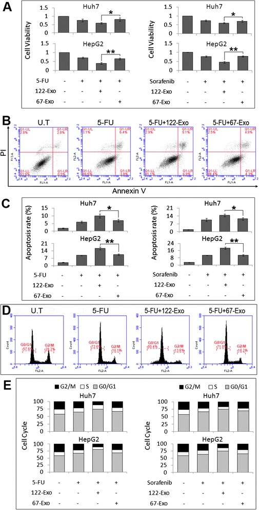
“Adipose tissue-derived mesenchymal stem cells (AMSCs) expressing miR-122 can increase HCC drug sensitivity by delivering exosomal miR-122 to HCC cells, leading to G0/G1 cell cycle arrest and apoptosis [122].”
From Emerging role of exosomes in cancer therapy: progress and challenges (2025) · cites this paper as [122]
Self-check
Percentages, sums and reported differences re-computed from the paper’s own numbers.
The self-check needs the paper’s full text.
Citations
Cites
No references could be resolved yet.
Research works printed but not linked: 42
- Bruix J, Sherman M. American Association for the Study of Liver Diseases. Management of hepatocellular carcinoma. Hepatology. 2005;42:1208–36. · DOI
- Zhang KZ, Zhang QB, Zhang QB, Sun HC, Ao JY, Chai ZT, et al. Arsenic trioxide induces differentiation of CD133+ hepatocellular carcinoma cells and prolongs posthepatectomy survival by targeting GLI1 expression in a mouse model. J Hematol Oncol. 2014;7:28. · DOI
- Smith AD, Roda D, Yap TA. Strategies for modern biomarker and drug development in oncology. J Hematol Oncol. 2014;7(1):70. · DOI
- Zhang H, Chen Z, Wang X, Huang Z, He Z, Chen Y. Long non-coding RNA: a new player in cancer. J Hematol Oncol. 2013;6:37. · DOI
- Wang WT, Chen YQ. Circulating miRNAs in cancer: from detection to therapy. J Hematol Oncol. 2014;7(1):86. · DOI
- Braoudaki M, Lambrou GI, Giannikou K, Milionis V, Stefanaki K, Birks DK, et al. Microrna expression signatures predict patient progression and disease outcome in pediatric embryonal central nervous system neoplasms. J Hematol Oncol. 2014;7(1):96. · DOI
- Fan S, Chen WX, Lv XB, Tang QL, Sun LJ, Liu BD, et al. miR-483-5p determines mitochondrial fission and cisplatin sensitivity in tongue squamous cell carcinoma by targeting FIS1. Cancer Lett. 2015;362:183–91. · DOI
- Sarkar FH, Li Y, Wang Z, Kong D, Ali S. Implication of microRNAs in drug resistance for designing novel cancer therapy. Drug Resist Update. 2010;13(3):57–66. · DOI
- Bandiera S, Pfeffer S, Baumert TF, Zeisel MB. miR-122-a key factor and therapeutic target in liver disease. J Hepatol. 2015;62:448–57. · DOI
- Tsai WC, Hsu SD, Hsu CS, Lai TC, Chen SJ, Shen R, et al. MicroRNA-122 plays a critical role in liver homeostasis and hepatocarcinogenesis. J Clin Invest. 2012;122:2884–97. · DOI
- Coulouarn C, Factor VM, Andersen JB, Durkin ME, Thorgeirsson SS. Loss of miR-122 expression in liver cancer correlates with suppression of the hepatic phenotype and gain of metastatic properties. Oncogene. 2009;28:3526–36. · DOI
- Xu Y, Xia F, Ma L, Shan J, Shen J, Yang Z, et al. MicroRNA-122 sensitizes HCC cancer cells to adriamycin and vincristine through modulating expression of MDR and inducing cell cycle arrest. Cancer Lett. 2011;310(2):160–9.
- Fornari F, Gramantieri L, Giovannini C, Veronese A, Ferracin M, Sabbioni S, et al. MiR-122/cyclin G1 interaction modulates p53 activity and affects doxorubicin sensitivity of human hepatocarcinoma cells. Cancer Res. 2009;69:5761–7. · DOI
- Bai S, Nasser MW, Wang B, Hsu SH, Datta J, Kutay H, et al. MicroRNA-122 inhibits tumorigenic properties of hepatocellular carcinoma cells and sensitizes these cells to sorafenib. J Biol Chem. 2009;284:32015–27. · DOI
- Hu G, Drescher KM, Chen XM. Exosomal miRNAs: biological properties and therapeutic potential. Front Genet. 2012;3:56.
- Katakowski M, Buller B, Zheng X, Lu Y, Rogers T, Osobamiro O, et al. Exosomes from marrow stromal cells expressing miR-146b inhibit glioma growth. Cancer Lett. 2013;335:201–4. · DOI · Paper page
- Yeo RW, Lai RC, Zhang B, Tan SS, Yin Y, Teh BJ, et al. Mesenchymal stem cell: an efficient mass producer of exosomes for drug delivery. Adv Drug Deliv Rev. 2013;65:336–41. · DOI · Paper page
- Lai RC, Arslan F, Lee MM, Sze NS, Choo A, Chen TS, et al. Exosome secreted by MSC reduces myocardial ischemia/reperfusion injury. Stem Cell Res. 2010;4:214–22. · DOI · Paper page
- Schäffler A, Büchler C. Concise review: adipose tissue-derived stromal cells—basic and clinical implications for novel cell-based therapies. Stem Cells. 2007;25:818–27. · DOI
- Kumar D, Gupta D, Shankar S, Srivastava RK. Biomolecular characterization of exosomes released from cancer stem cells: possible implications for biomarker and treatment of cancer. Oncotarget. 2015;6(5):3280–91. · DOI
- Zhang X, Yuan X, Shi H, Wu L, Qian H, Xu W. Exosomes in cancer: small particle, big player. J Hematol Oncol. 2015;8:83. · DOI
- O'Loughlin AJ, Woffindale CA, Wood MJ. Exosomes and the emerging field of exosome-based gene therapy. Curr Gene Ther. 2012;12:262–74. · DOI
- Xin H, Li Y, Buller B, Katakowski M, Zhang Y, Wang X, et al. Exosome-mediated transfer of miR-133b from multipotent mesenchymal stromal cells to neural cells contributes to neurite outgrowth. Stem Cells. 2012;30:1556–64. · DOI
- Yang C, Jiang F, Xu F, Jiang G. ADAM10 overexpression confers resistance to doxorubicin-induced apoptosis in hepatocellular carcinoma. Tumour Biol. 2012;33:1535–41. · DOI
- Wu J, Zhu AX. Targeting insulin-like growth factor axis in hepatocellular carcinoma. J Hematol Oncol. 2011;5:30. · DOI
- Tovar V, Alsinet C, Villanueva A, Hoshida Y, Chiang DY, Solé M, et al. IGF activation in a molecular subclass of hepatocellular carcinoma and pre-clinical efficacy of IGF-1R blockage. J Hepatol. 2010;52:550–9. · DOI
- Kimura SH, Ikawa M, Ito A, Okabe M, Nojima H. Cyclin G1 is involved in G2/M arrest in response to DNA damage and in growth control after damage recovery. Oncogene. 2001;20:3290–300. · DOI
- Wilhelm SM, Adnane L, Newell P, Villanueva A, Llovet JM, Lynch M. Preclinical overview of sorafenib, a multikinase inhibitor that targets both Raf and VEGF and PDGF receptor tyrosine kinase signaling. Mol Cancer Ther. 2008;7:3129–40. · DOI
- Liang Y, Zheng T, Song R, Wang J, Yin D, Wang L, et al. Hypoxia-mediated sorafenib resistance can be overcome by EF24 through Von Hippel-Lindau tumor suppressor-dependent HIF-1α inhibition in hepatocellular carcinoma. Hepatology. 2013;57:1847–57. · DOI
- Sun Z, Wang S, Zhao RC. The roles of mesenchymal stem cells in tumor inflammatory microenvironment. J Hematol Oncol. 2014;7:14. · DOI · Paper page
- Roccaro AM, Sacco A, Maiso P, Azab AK, Tai YT, Reagan M, et al. BM mesenchymal stromal cell-derived exosomes facilitate multiple myeloma progression. J Clin Invest. 2013;123:1542–55. · DOI
- Bruno S, Collino F, Deregibus MC, Grange C, Tetta C, Camussi G. Microvesicles derived from human bone marrow mesenchymal stem cells inhibit tumor growth. Stem Cells Dev. 2013;22:758–71. · DOI
- Wu S, Ju G, Du T, et al. Microvesicles derived from human umbilical cord Wharton’s jelly mesenchymal stem cells attenuate bladder tumor cell growth in vitro and in vivo. PloS One. 2013;8, e61366.
- Akyurekli C, Le Y, Richardson RB, Fergusson D, Tay J, Allan DS. A systematic review of preclinical studies on the therapeutic potential of mesenchymal stromal cell-derived microvesicles. Stem Cell Rev. 2015;11:150–60. · DOI
- Zhu W, Huang L, Li Y, Zhang X, Gu J, Yan Y, et al. Exosomes derived from human bone marrow mesenchymal stem cells promote tumor growth in vivo. Cancer Lett. 2012;315:28–37. · DOI · Paper page
- Zhang W, Liu S, Liu K, Ji B, Wang Y, Liu Y. Knockout of ADAM10 enhances sorafenib antitumor activity of hepatocellular carcinoma in vitro and in vivo. Oncol Rep. 2014;32(5):1913–22. · DOI
- Ou DL, Lee BS, Chang YC, Lin LI, Liou JY, Hsu C, et al. Potentiating the efficacy of molecular targeted therapy for hepatocellular carcinoma by inhibiting the insulin-like growth factor pathway. PLoS One. 2013;8, e66589.
- Tian Y, Li S, Song J, Ji T, Zhu M, Anderson GJ, et al. A doxorubicin delivery platform using engineered natural membrane vesicle exosomes for targeted tumor therapy. Biomaterials. 2014;35:2383–90. · DOI
- Liu Y, Yan X, Sun Z, Chen B, Han Q, Li J, et al. Flk-1+ adipose-derived mesenchymal stem cells differentiate into skeletal muscle satellite cells and ameliorate muscular dystrophy in mdx mice. Stem Cells Dev. 2007;16:695–706. · DOI
- Dudakovic A, Camilleri E, Riester SM, Lewallen EA, Kvasha S, Chen X, et al. High-resolution molecular validation of self-renewal and spontaneous differentiation in clinical-grade adipose-tissue derived human mesenchymal stem cells. J Cell Biochem. 2014;115(10):1816–28. · DOI
- Ullah M, Stich S, Notter M, Eucker J, Sittinger M, Ringe J. Transdifferentiation of mesenchymal stem cells-derived adipogenic-differentiated cells into osteogenic- or chondrogenic-differentiated cells proceeds via dedifferentiation and have a correlation with cell cycle arresting and driving genes. Differentiation. 2013;85(3):78–90. · DOI
- Ismail N, Wang Y, Dakhlallah D, Moldovan L, Agarwal K, Batte K, et al. Macrophage microvesicles induce macrophage differentiation and miR-223 transfer. Blood. 2013;121(6):984–95. · DOI
Cited by
Sorted by citations
Mesenchymal stem cell secretome: toward cell-free therapeutic strategies in regenerative medicine
Vizoso, F., Eiro, N., Cid, S., Schneider, J., & Perez-Fernandez, R. (2017) · International Journal of Molecular Sciences · doi:10.3390/ijms18091852 · Cited by 1,363
Exosome engineering: Current progress in cargo loading and targeted delivery
Fu, S., Wang, Y., Xia, X., & Zheng, J. C. (2020) · NanoImpact · doi:10.1016/j.impact.2020.100261 · Cited by 444
Engineered Extracellular Vesicles for Cancer Therapy
Zhang, X., Zhang, H., Gu, J., Zhang, J., Shi, H., Qian, H., Wang, D., Xu, W., Pan, J., & Santos, H. A. (2021) · Advanced Materials · doi:10.1002/adma.202005709 · Cited by 428
Lin, Z., Wu, Y., Xu, Y., Li, G., Li, Z., & Liu, T. (2022) · Molecular Cancer · doi:10.1186/s12943-022-01650-5 · Cited by 297
Emerging role of exosomes in cancer therapy: progress and challenges
Li, J., Wang, J., & Chen, Z. (2025) · Molecular Cancer · doi:10.1186/s12943-024-02215-4 · Cited by 297
Self-citationHong, P., Yang, H., Wu, Y., Li, K., & Tang, Z. (2019) · Stem Cell Research & Therapy · doi:10.1186/s13287-019-1358-y · Cited by 269
Exosome-Based Drug Delivery: Translation from Bench to Clinic
Koh, H. B., Kim, H. J., Kang, S.-W., & Yoo, T.-H. (2023) · Pharmaceutics · doi:10.3390/pharmaceutics15082042 · Cited by 259
Liu, Q., Li, D., Pan, X., & Liang, Y. (2023) · Journal of Nanobiotechnology · doi:10.1186/s12951-023-02081-0 · Cited by 201
Exosomes as bio-inspired nanocarriers for RNA delivery: preparation and applications
Amiri, A., Bagherifar, R., Ansari Dezfouli, E., Kiaie, S. H., Jafari, R., & Ramezani, R. (2022) · Journal of Translational Medicine · doi:10.1186/s12967-022-03325-7 · Cited by 186
Ding, Y., Cao, F., Sun, H., Wang, Y., Liu, S., Wu, Y., Cui, Q., Mei, W., & Li, F. (2019) · Cancer Letters · doi:10.1016/j.canlet.2018.10.039 · Cited by 177
Psaraki, A., Ntari, L., Karakostas, C., Korrou‐Karava, D., & Roubelakis, M. G. (2022) · Hepatology · doi:10.1002/hep.32129 · Cited by 145
Recent Advances in Exosome-Based Drug Delivery for Cancer Therapy
Kim, H., Jang, H., Cho, H., Choi, J., Hwang, K. Y., Choi, Y., Kim, S. H., & Yang, Y. (2021) · Cancers · doi:10.3390/cancers13174435 · Cited by 152
Sutaria, D. S., Badawi, M., Phelps, M. A., & Schmittgen, T. D. (2017) · Pharmaceutical Research · doi:10.1007/s11095-017-2123-5 · Cited by 140
Liquid biopsy in the clinical management of hepatocellular carcinoma
von Felden, J., Garcia-Lezana, T., Schulze, K., Losic, B., & Villanueva, A. (2020) · Gut · doi:10.1136/gutjnl-2019-320282 · Cited by 141
Recent discoveries concerning the tumor - mesenchymal stem cell interactions
Lazennec, G., & Lam, P. Y. (2016) · Biochimica et Biophysica Acta (BBA) - Reviews on Cancer · doi:10.1016/j.bbcan.2016.10.004 · Cited by 117
Chen, Y., Tian, Z., He, L., Liu, C., Wang, N., Rong, L., & Liu, B. (2021) · Stem Cell Research & Therapy · doi:10.1186/s13287-021-02282-0 · Cited by 106
MicroRNA-122 in human cancers: from mechanistic to clinical perspectives
Faramin Lashkarian, M., Hashemipour, N., Niaraki, N., Soghala, S., Moradi, A., Sarhangi, S., Hatami, M., Aghaei-Zarch, F., Khosravifar, M., Mohammadzadeh, A., et al. (2023) · Cancer Cell International · doi:10.1186/s12935-023-02868-z · Cited by 105
Extracellular vesicles in cancer nanomedicine
Tarasov, V. V., Svistunov, A. A., Chubarev, V. N., Dostdar, S. A., Sokolov, A. V., Brzecka, A., Sukocheva, O., Neganova, M. E., Klochkov, S. G., Somasundaram, S. G., et al. (2021) · Seminars in Cancer Biology · doi:10.1016/j.semcancer.2019.08.017 · Cited by 97
NAFLD and HBV interplay - related mechanisms underlying liver disease progression
Tourkochristou, E., Assimakopoulos, S. F., Thomopoulos, K., Marangos, M., & Triantos, C. (2022) · Frontiers in Immunology · doi:10.3389/fimmu.2022.965548 · Cited by 70
Amadeo, F., Trivino Cepeda, K., Littlewood, J., Wilm, B., Taylor, A., & Murray, P. (2021) · Emerging Topics in Life Sciences · doi:10.1042/etls20210013 · Cited by 20
MiR-128-3p – a gray eminence of the human central nervous system
Kiel, K., Król, S. K., Bronisz, A., & Godlewski, J. (2024) · Molecular Therapy Nucleic Acids · doi:10.1016/j.omtn.2024.102141 · Cited by 15
Extracellular Vesicles and Their Interplay with Biological Membranes
Yeo Xian Ping, J., Raj Neupane, Y., & Pastorin, G. (2022) · Physiology · doi:10.5772/intechopen.101297 · Cited by 9
Metadata
- Authors
- Lou, G., Song, X., Yang, F., Wu, S., Wang, J., Chen, Z., & Liu, Y.
- Venue
- Journal of Hematology & Oncology
- Published
- 2015-10-29
- Volume
- 8
- Issue
- 1
- ISSN
- 1756-8722
- PMC
- PMC4627430
- Cited by
- 651
- References
- 0
- Language
- en
- Funders
- National Natural Science Fund (81000730, 81201782)
- International Science and Technology Cooperation Project (re-innovation industrialization) (2012C14028)
- Medical and Health science and technology project in Zhejiang province (2014KYB085)
About these checks
Registry, retraction, Cites and statistics results are computed, not judged, with one exception: a printed reference that no index lists is read once by a model before it counts as a citation error. Claims are extracted by a model and kept only when their quote appears verbatim in the text; each cited source is then checked, with a model’s help, for whether it says what the claim relies on. Those verdicts can be wrong — read the claim’s quote and the cited source before relying on one.
Statistics and arithmetic are recomputed, not judged.
Confidence bands are shown only for fields calibrated on our annotated test set. None is calibrated yet, so no band is shown.
What counts as a claim ›How we measure confidence ›How we check this ›
Are you an author, or do you see a problem with this page? Request a review
