

The DOI matches its Crossref record; 86 of the 86 references it cites were found in a registry or an index.
Modulation of astrocyte reactivity improves functional deficits in mouse models of Alzheimer’s disease
Open accessAbstract
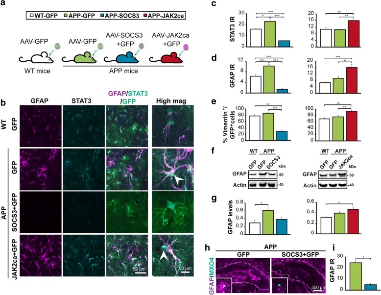
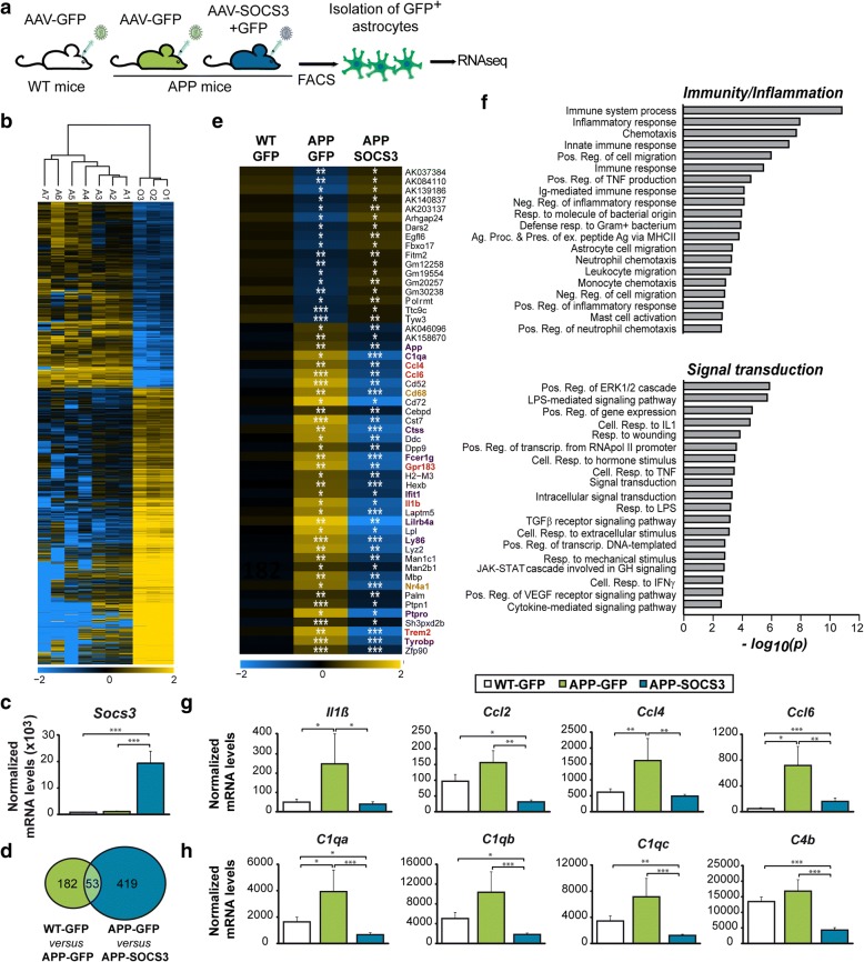
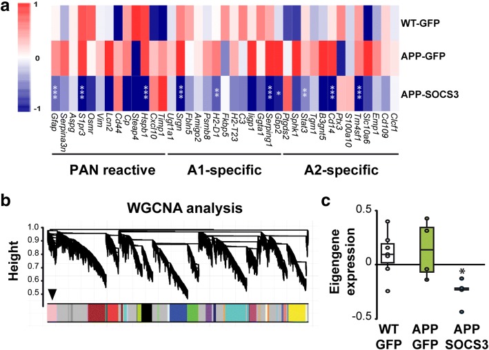
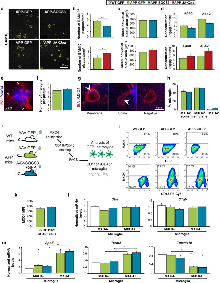
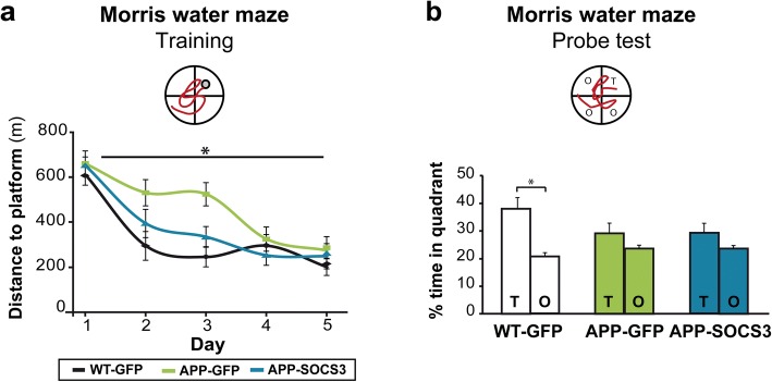
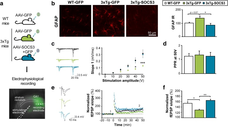
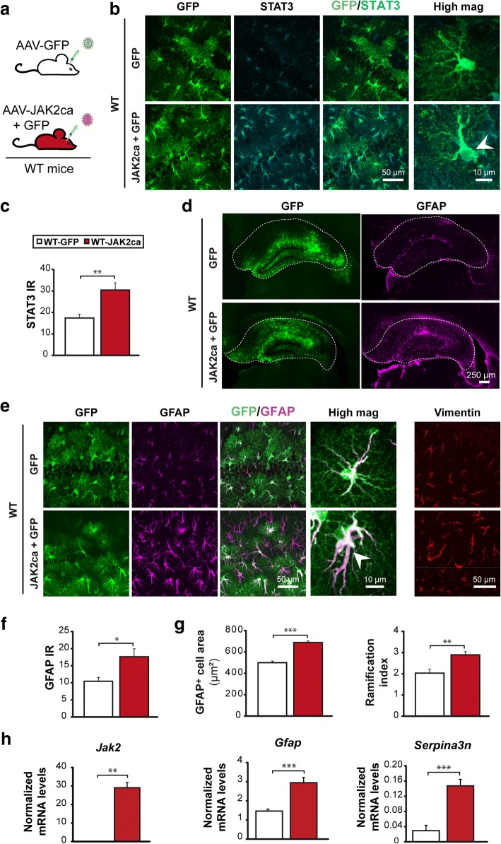
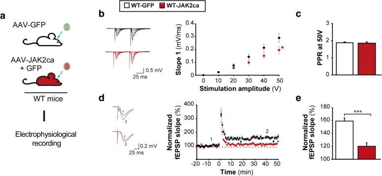
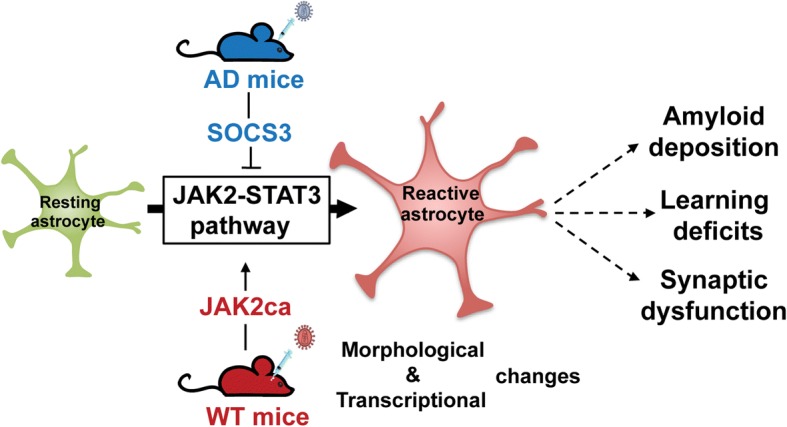
Astrocyte reactivity and neuroinflammation are hallmarks of CNS pathological conditions such as Alzheimer's disease. However, the specific role of reactive astrocytes is still debated. This controversy may stem from the fact that most strategies used to modulate astrocyte reactivity and explore its contribution to disease outcomes have only limited specificity. Moreover, reactive astrocytes are now emerging as heterogeneous cells and all types of astrocyte reactivity may not be controlled efficiently by such strategies.Here, we used cell type-specific approaches in vivo and identified the JAK2-STAT3 pathway, as necessary and sufficient for the induction and maintenance of astrocyte reactivity. Modulation of this cascade by viral gene transfer in mouse astrocytes efficiently controlled several morphological and molecular features of reactivity. Inhibition of this pathway in mouse models of Alzheimer's disease improved three key pathological hallmarks by reducing amyloid deposition, improving spatial learning and restoring synaptic deficits.In conclusion, the JAK2-STAT3 cascade operates as a master regulator of astrocyte reactivity in vivo. Its inhibition offers new therapeutic opportunities for Alzheimer's disease.
Overview
The claims and statistics of this paper haven’t been checked yet.
Run the check — free, no account needed. It reads the open-access text and checks the sources it cites.
Integrity signals
Registry
Cites
Statistics
Figures
Verification
Claims the paper makes
The claims and statistics of this paper haven’t been checked yet.
Run the check — free, no account needed. It reads the open-access text and checks the sources it cites.
Claims other papers make about this one
Each is checked against this paper’s own text: does it say what the citing paper says it does?
The source backs part of the claim, not all of it — or backs it for a different population, condition or comparison.
In the citing paper
“Importantly, only a subset, often a mix of A1 and A2 or pan-reactive transcripts, are upregulated in astrocytes from brains of study participants with HD74 or AD75,76, or from several mouse models of acute injuries and chronic diseases of the CNS42,69,76,77.”
From Reactive astrocyte nomenclature, definitions, and future directions (2021) · cites this paper as [42]
Experimental — accuracy not yet measuredThe source backs part of the claim, not all of it — or backs it for a different population, condition or comparison.
In the citing paper
“Finally, interventional strategies such as classical pharmacology56,98, genetic manipulation42,56, and biomaterials99 are available tools to modify pathological signaling in reactive astrocytes for therapeutic purposes.”
From Reactive astrocyte nomenclature, definitions, and future directions (2021) · cites this paper as [42]
Experimental — accuracy not yet measuredThe cited source says what the claim attributes to it.
In the citing paper
“For example, the manipulation of reactive astrocytes has resulted in improved outcomes38,42,43, worse35 outcomes, and no change44 in mouse models of AD and MS40,45,46.”
From Reactive astrocyte nomenclature, definitions, and future directions (2021) · cites this paper as [42]
The cited source says what the claim attributes to it.
In the citing paper
“On the other hand, blocking astrocyte reactivity has demonstrated positive effects on pathology (Ceyzeriat et al., 2018; Reichenbach et al., 2019).”
From Inhibition of the mitochondrial pyruvate carrier in astrocytes reduces amyloid and tau accumulation in the 3xTgAD mouse model of Alzheimer's disease (2024) · cites this paper as Ceyzeriat et al., 2018
Self-check
Percentages, sums and reported differences re-computed from the paper’s own numbers.
The self-check needs the paper’s full text.
Citations
Cites
Moderated estimation of fold change and dispersion for RNA-seq data with DESeq2
Love, M. I., Huber, W., & Anders, S. (2014) · Genome Biology · doi:10.1186/s13059-014-0550-8 · Cited by 95,106
STAR: ultrafast universal RNA-seq aligner
Dobin, A., Davis, C. A., Schlesinger, F., Drenkow, J., Zaleski, C., Jha, S., Batut, P., Chaisson, M., & Gingeras, T. R. (2013) · Bioinformatics · doi:10.1093/bioinformatics/bts635 · Cited by 51,709
Systematic and integrative analysis of large gene lists using DAVID bioinformatics resources
Huang, D. W., Sherman, B. T., & Lempicki, R. A. (2009) · Nature Protocols · doi:10.1038/nprot.2008.211 · Cited by 35,890
WGCNA: an R package for weighted correlation network analysis
Langfelder, P., & Horvath, S. (2008) · BMC Bioinformatics · doi:10.1186/1471-2105-9-559 · Cited by 27,252
Amyloid oligomers exacerbate tau pathology in a mouse model of tauopathy
Selenica, M.-L. B., Brownlow, M., Jimenez, J. P., Lee, D. C., Pena, G., Dickey, C. A., Gordon, M. N., & Morgan, D. (2013) · Neurodegenerative Diseases · doi:10.1159/000337230 · Cited by 10,359
Neurotoxic reactive astrocytes are induced by activated microglia
Liddelow, S. A., Guttenplan, K. A., Clarke, L. E., Bennett, F. C., Bohlen, C. J., Schirmer, L., Bennett, M. L., Münch, A. E., Chung, W.-S., Peterson, T. C., et al. (2017) · Nature · doi:10.1038/nature21029 · Cited by 7,971
Alzheimer's disease: the amyloid cascade hypothesis
Hardy, J. A., & Higgins, G. A. (1992) · Science · doi:10.1126/science.1566067 · Cited by 7,484
Place navigation impaired in rats with hippocampal lesions
Morris, R. G. M., Garrud, P., Rawlins, J. N. P., & O'Keefe, J. (1982) · Nature · doi:10.1038/297681a0 · Cited by 6,340
Neuroinflammation in Alzheimer's disease
Heneka, M. T., Carson, M. J., Khoury, J. E., Landreth, G. E., Brosseron, F., Feinstein, D. L., Jacobs, A. H., Wyss-Coray, T., Vitorica, J., Ransohoff, R. M., et al. (2015) · The Lancet Neurology · doi:10.1016/s1474-4422(15)70016-5 · Cited by 6,226
Iliff, J. J., Wang, M., Liao, Y., Plogg, B. A., Peng, W., Gundersen, G. A., Benveniste, H., Vates, G. E., Deane, R., Goldman, S. A., et al. (2012) · Science Translational Medicine · doi:10.1126/scitranslmed.3003748 · Cited by 5,824
A Unique Microglia Type Associated with Restricting Development of Alzheimer’s Disease
Keren-Shaul, H., Spinrad, A., Weiner, A., Matcovitch-Natan, O., Dvir-Szternfeld, R., Ulland, T. K., David, E., Baruch, K., Lara-Astaiso, D., Toth, B., et al. (2017) · Cell · doi:10.1016/j.cell.2017.05.018 · Cited by 5,493
Alzheimer's Disease Is a Synaptic Failure
Selkoe, D. J. (2002) · Science · doi:10.1126/science.1074069 · Cited by 4,420
Triple-Transgenic Model of Alzheimer’s Disease with Plaques and Tangles
Oddo, S., Caccamo, A., Shepherd, J. D., Murphy, M. P., Golde, T. E., Kayed, R., Metherate, R., Mattson, M. P., Akbari, Y., & LaFerla, F. M. (2003) · Neuron · doi:10.1016/s0896-6273(03)00434-3 · Cited by 4,126
Cahoy, J. D., Emery, B., Kaushal, A., Foo, L. C., Zamanian, J. L., Christopherson, K. S., Xing, Y., Lubischer, J. L., Krieg, P. A., Krupenko, S. A., et al. (2008) · The Journal of Neuroscience · doi:10.1523/jneurosci.4178-07.2008 · Cited by 3,129
Genomic analysis of reactive astrogliosis
Zamanian, J. L., Xu, L., Foo, L. C., Nouri, N., Zhou, L., Giffard, R. G., & Barres, B. A. (2012) · The Journal of Neuroscience · doi:10.1523/jneurosci.6221-11.2012 · Cited by 2,404
Reactive Astrocytes: Production, Function, and Therapeutic Potential
Liddelow, S. A., & Barres, B. A. (2017) · Immunity · doi:10.1016/j.immuni.2017.06.006 · Cited by 2,336
Astrocyte scar formation aids central nervous system axon regeneration
Anderson, M. A., Burda, J. E., Ren, Y., Ao, Y., O’Shea, T. M., Kawaguchi, R., Coppola, G., Khakh, B. S., Deming, T. J., & Sofroniew, M. V. (2016) · Nature · doi:10.1038/nature17623 · Cited by 1,851
Jankowsky, J. L., Fadale, D. J., Anderson, J., Xu, G. M., Gonzales, V., Jenkins, N. A., Copeland, N. G., Lee, M. K., Younkin, L. H., Wagner, S. L., et al. (2004) · Human Molecular Genetics · doi:10.1093/hmg/ddh019 · Cited by 1,575
Rothhammer, V., Mascanfroni, I. D., Bunse, L., Takenaka, M. C., Kenison, J. E., Mayo, L., Chao, C.-C., Patel, B., Yan, R., Blain, M., et al. (2016) · Nature Medicine · doi:10.1038/nm.4106 · Cited by 1,559
Reactive Gliosis and the Multicellular Response to CNS Damage and Disease
Burda, J. E., & Sofroniew, M. V. (2014) · Neuron · doi:10.1016/j.neuron.2013.12.034 · Cited by 1,500
Gliotransmitters Travel in Time and Space
Araque, A., Carmignoto, G., Haydon, P. G., Oliet, S. H. R., Robitaille, R., & Volterra, A. (2014) · Neuron · doi:10.1016/j.neuron.2014.02.007 · Cited by 1,308
Self-citationNormal aging induces A1-like astrocyte reactivity
Clarke, L. E., Liddelow, S. A., Chakraborty, C., Münch, A. E., Heiman, M., & Barres, B. A. (2018) · Proceedings of the National Academy of Sciences · doi:10.1073/pnas.1800165115 · Cited by 1,237
ApoE4 markedly exacerbates tau-mediated neurodegeneration in a mouse model of tauopathy
Initiative, A. D. N., Shi, Y., Yamada, K., Liddelow, S. A., Smith, S. T., Zhao, L., Luo, W., Tsai, R. M., Spina, S., Grinberg, L. T., et al. (2017) · Nature · doi:10.1038/nature24016 · Cited by 1,220
Block of A1 astrocyte conversion by microglia is neuroprotective in models of Parkinson’s disease
Yun, S. P., Kam, T.-I., Panicker, N., Kim, S., Oh, Y., Park, J.-S., Kwon, S.-H., Park, Y. J., Karuppagounder, S. S., Park, H., et al. (2018) · Nature Medicine · doi:10.1038/s41591-018-0051-5 · Cited by 1,123
The Complement System: An Unexpected Role in Synaptic Pruning During Development and Disease
Stephan, A. H., Barres, B. A., & Stevens, B. (2012) · Annual Review of Neuroscience · doi:10.1146/annurev-neuro-061010-113810 · Cited by 1,113
Microglial control of astrocytes in response to microbial metabolites
Rothhammer, V., Borucki, D. M., Tjon, E. C., Takenaka, M. C., Chao, C.-C., Ardura-Fabregat, A., de Lima, K. A., Gutiérrez-Vázquez, C., Hewson, P., Staszewski, O., et al. (2018) · Nature · doi:10.1038/s41586-018-0119-x · Cited by 1,051
Okada, S., Nakamura, M., Katoh, H., Miyao, T., Shimazaki, T., Ishii, K., Yamane, J., Yoshimura, A., Iwamoto, Y., Toyama, Y., et al. (2006) · Nature Medicine · doi:10.1038/nm1425 · Cited by 991
STAT3 is a Critical Regulator of Astrogliosis and Scar Formation after Spinal Cord Injury
Herrmann, J. E., Imura, T., Song, B., Qi, J., Ao, Y., Nguyen, T. K., Korsak, R. A., Takeda, K., Akira, S., & Sofroniew, M. V. (2008) · The Journal of Neuroscience · doi:10.1523/jneurosci.1709-08.2008 · Cited by 958
Glia-Derived d-Serine Controls NMDA Receptor Activity and Synaptic Memory
Panatier, A., Theodosis, D. T., Mothet, J.-P., Touquet, B., Pollegioni, L., Poulain, D. A., & Oliet, S. H. R. (2006) · Cell · doi:10.1016/j.cell.2006.02.051 · Cited by 934
Self-citationHol, E. M., & Pekny, M. (2015) · Current Opinion in Cell Biology · doi:10.1016/j.ceb.2015.02.004 · Cited by 917
Adult mouse astrocytes degrade amyloid-β in vitro and in situ
Wyss-Coray, T., Loike, J. D., Brionne, T. C., Lu, E., Anankov, R., Yan, F., Silverstein, S. C., & Husemann, J. (2003) · Nature Medicine · doi:10.1038/nm838 · Cited by 909
Astrocytes: a central element in neurological diseases
Pekny, M., Pekna, M., Messing, A., Steinhäuser, C., Lee, J., Parpura, V., Hol, E. M., Sofroniew, M. V., & Verkhratsky, A. (2015) · Acta Neuropathologica · doi:10.1007/s00401-015-1513-1 · Cited by 793
Cell type- and brain region-resolved mouse brain proteome
Sharma, K., Schmitt, S., Bergner, C. G., Tyanova, S., Kannaiyan, N., Manrique-Hoyos, N., Kongi, K., Cantuti, L., Hanisch, U.-K., Philips, M.-A., et al. (2015) · Nature Neuroscience · doi:10.1038/nn.4160 · Cited by 785
GABA from reactive astrocytes impairs memory in mouse models of Alzheimer's disease
Jo, S., Yarishkin, O., Hwang, Y. J., Chun, Y. E., Park, M., Woo, D. H., Bae, J. Y., Kim, T., Lee, J., Chun, H., et al. (2014) · Nature Medicine · doi:10.1038/nm.3639 · Cited by 783
Neuronal morphometry directly from bitmap images
Ferreira, T. A., Blackman, A. V., Oyrer, J., Jayabal, S., Chung, A. J., Watt, A. J., Sjöström, P. J., & van Meyel, D. J. (2014) · Nature Methods · doi:10.1038/nmeth.3125 · Cited by 725
Sites of Neocortical Reorganization Critical for Remote Spatial Memory
Maviel, T., Durkin, T., Menzaghi, F., & Bontempi, B. (2004) · Science · doi:10.1126/science.1098180 · Cited by 711
The role of microglia in amyloid clearance from the AD brain
Lee, C. Y. D., & Landreth, G. E. (2010) · Journal of Neural Transmission · doi:10.1007/s00702-010-0433-4 · Cited by 701
Lian, H., Yang, L., Cole, A., Sun, L., Chiang, A. C.-A., Fowler, S. W., Shim, D. J., Rodriguez-Rivera, J., Taglialatela, G., Jankowsky, J. L., et al. (2015) · Neuron · doi:10.1016/j.neuron.2014.11.018 · Cited by 697
Astrocytes Are Endogenous Regulators of Basal Transmission at Central Synapses
Panatier, A., Vallée, J., Haber, M., Murai, K. K., Lacaille, J.-C., & Robitaille, R. (2011) · Cell · doi:10.1016/j.cell.2011.07.022 · Cited by 650
Self-citationAstrocytic Activation Generates De Novo Neuronal Potentiation and Memory Enhancement
Adamsky, A., Kol, A., Kreisel, T., Doron, A., Ozeri-Engelhard, N., Melcer, T., Refaeli, R., Horn, H., Regev, L., Groysman, M., et al. (2018) · Cell · doi:10.1016/j.cell.2018.05.002 · Cited by 631
Lian, H., Litvinchuk, A., Chiang, A. C.-A., Aithmitti, N., Jankowsky, J. L., & Zheng, H. (2016) · The Journal of Neuroscience · doi:10.1523/jneurosci.2117-15.2016 · Cited by 597
Apolipoprotein E promotes astrocyte colocalization and degradation of deposited amyloid-β peptides
Koistinaho, M., Lin, S., Wu, X., Esterman, M., Koger, D., Hanson, J., Higgs, R., Liu, F., Malkani, S., Bales, K. R., et al. (2004) · Nature Medicine · doi:10.1038/nm1058 · Cited by 571
Heterogeneity of reactive astrocytes
Anderson, M. A., Ao, Y., & Sofroniew, M. V. (2013) · Neuroscience Letters · doi:10.1016/j.neulet.2013.12.030 · Cited by 478
Deficient glutamate tranport is associated with neurodegeneration in Alzheimer's disease
Masliah, E., Hansen, L., Alford, M., Deteresa, R., & Mallory, M. (1996) · Annals of Neurology · doi:10.1002/ana.410400512 · Cited by 476
Elusive roles for reactive astrocytes in neurodegenerative diseases
Ben Haim, L., Carrillo-de Sauvage, M.-A., Ceyzériat, K., & Escartin, C. (2015) · Frontiers in Cellular Neuroscience · doi:10.3389/fncel.2015.00278 · Cited by 453
Self-citationSTAT3 labels a subpopulation of reactive astrocytes required for brain metastasis
Priego, N., Zhu, L., Monteiro, C., Mulders, M., Wasilewski, D., Bindeman, W., Doglio, L., Martínez, L., Martínez-Saez, E., Ramón y Cajal, S., et al. (2018) · Nature Medicine · doi:10.1038/s41591-018-0044-4 · Cited by 452
McManus, M. J., Murphy, M. P., & Franklin, J. L. (2011) · The Journal of Neuroscience · doi:10.1523/jneurosci.0552-11.2011 · Cited by 432
GFAP promoter elements required for region‐specific and astrocyte‐specific expression
Lee, Y., Messing, A., Su, M., & Brenner, M. (2008) · Glia · doi:10.1002/glia.20622 · Cited by 424
Reactive astrocytes function as phagocytes after brain ischemia via ABCA1-mediated pathway
Morizawa, Y. M., Hirayama, Y., Ohno, N., Shibata, S., Shigetomi, E., Sui, Y., Nabekura, J., Sato, K., Okajima, F., Takebayashi, H., et al. (2017) · Nature Communications · doi:10.1038/s41467-017-00037-1 · Cited by 424
Isolation of glia from Alzheimer’s mice reveals inflammation and dysfunction
Orre, M., Kamphuis, W., Osborn, L. M., Jansen, A. H. P., Kooijman, L., Bossers, K., & Hol, E. M. (2014) · Neurobiology of Aging · doi:10.1016/j.neurobiolaging.2014.06.004 · Cited by 394
Ben Haim, L., Ceyzériat, K., Carrillo-de Sauvage, M. A., Aubry, F., Auregan, G., Guillermier, M., Ruiz, M., Petit, F., Houitte, D., Faivre, E., et al. (2015) · The Journal of Neuroscience · doi:10.1523/jneurosci.3516-14.2015 · Cited by 335
Self-citationDelekate, A., Füchtemeier, M., Schumacher, T., Ulbrich, C., Foddis, M., & Petzold, G. C. (2014) · Nature Communications · doi:10.1038/ncomms6422 · Cited by 332
Attenuating astrocyte activation accelerates plaque pathogenesis in APP/PS1 mice
Kraft, A. W., Hu, X., Yoon, H., Yan, P., Xiao, Q., Wang, Y., Gil, S. C., Brown, J., Wilhelmsson, U., Restivo, J. L., et al. (2013) · The FASEB Journal · doi:10.1096/fj.12-208660 · Cited by 324
Targeting Astrocytes Ameliorates Neurologic Changes in a Mouse Model of Alzheimer's Disease
Furman, J. L., Sama, D. M., Gant, J. C., Beckett, T. L., Murphy, M. P., Bachstetter, A. D., Van Eldik, L. J., & Norris, C. M. (2012) · The Journal of Neuroscience · doi:10.1523/jneurosci.2323-12.2012 · Cited by 313
Astrogliosis: An integral player in the pathogenesis of Alzheimer's disease
Osborn, L. M., Kamphuis, W., Wadman, W. J., & Hol, E. M. (2016) · Progress in Neurobiology · doi:10.1016/j.pneurobio.2016.01.001 · Cited by 312
Kershaw, N. J., Murphy, J. M., Liau, N. P. D., Varghese, L. N., Laktyushin, A., Whitlock, E. L., Lucet, I. S., Nicola, N. A., & Babon, J. J. (2013) · Nature Structural & Molecular Biology · doi:10.1038/nsmb.2519 · Cited by 301
Allaman, I., Gavillet, M., Bélanger, M., Laroche, T., Viertl, D., Lashuel, H. A., & Magistretti, P. J. (2010) · Journal of Neuroscience · doi:10.1523/jneurosci.5098-09.2010 · Cited by 291
Babon, J. J., Kershaw, N. J., Murphy, J. M., Varghese, L. N., Laktyushin, A., Young, S. N., Lucet, I. S., Norton, R. S., & Nicola, N. A. (2012) · Immunity · doi:10.1016/j.immuni.2011.12.015 · Cited by 279
Ding, Y., Qiao, A., Wang, Z., Goodwin, J. S., Lee, E.-S., Block, M. L., Allsbrook, M., McDonald, M. P., & Fan, G.-H. (2008) · The Journal of Neuroscience · doi:10.1523/jneurosci.3153-08.2008 · Cited by 269
Astroglial CB1 Receptors Determine Synaptic D-Serine Availability to Enable Recognition Memory
Robin, L. M., Oliveira da Cruz, J. F., Langlais, V. C., Martin-Fernandez, M., Metna-Laurent, M., Busquets-Garcia, A., Bellocchio, L., Soria-Gomez, E., Papouin, T., Varilh, M., et al. (2018) · Neuron · doi:10.1016/j.neuron.2018.04.034 · Cited by 262
Self-citationPapouin, T., Dunphy, J. M., Tolman, M., Dineley, K. T., & Haydon, P. G. (2017) · Neuron · doi:10.1016/j.neuron.2017.04.021 · Cited by 243
Sekar, S., McDonald, J., Cuyugan, L., Aldrich, J., Kurdoglu, A., Adkins, J., Serrano, G., Beach, T. G., Craig, D. W., Valla, J., et al. (2015) · Neurobiology of Aging · doi:10.1016/j.neurobiolaging.2014.09.027 · Cited by 199
The complex STATes of astrocyte reactivity: How are they controlled by the JAK-STAT3 pathway?
Ceyzériat, K., Abjean, L., Carrillo-de Sauvage, M.-A., Ben Haim, L., & Escartin, C. (2016) · Neuroscience · doi:10.1016/j.neuroscience.2016.05.043 · Cited by 185
Self-citationViral gene transfer of APPsα rescues synaptic failure in an Alzheimer’s disease mouse model
Fol, R. R., Braudeau, J., Ludewig, S., Abel, T., Weyer, S. W., Roederer, J.-P., Brod, F., Audrain, M., Bemelmans, A., Buchholz, C. J., et al. (2015) · Acta Neuropathologica · doi:10.1007/s00401-015-1498-9 · Cited by 182
Self-citationReactive astrocytes in Alzheimer’s disease: A double-edged sword
Chun, H., & Lee, C. J. (2018) · Neuroscience Research · doi:10.1016/j.neures.2017.11.012 · Cited by 176
Early Activation of STAT3 Regulates Reactive Astrogliosis Induced by Diverse Forms of Neurotoxicity
O'Callaghan, J. P., Kelly, K. A., VanGilder, R. L., Sofroniew, M. V., & Miller, D. B. (2014) · PLoS ONE · doi:10.1371/journal.pone.0102003 · Cited by 156
Kamphuis, W., Kooijman, L., Orre, M., Stassen, O., Pekny, M., & Hol, E. M. (2015) · Glia · doi:10.1002/glia.22800 · Cited by 155
CXCR3 promotes plaque formation and behavioral deficits in an Alzheimer’s disease model
Krauthausen, M., Kummer, M. P., Zimmermann, J., Reyes-Irisarri, E., Terwel, D., Bulic, B., Heneka, M. T., & Müller, M. (2014) · Journal of Clinical Investigation · doi:10.1172/jci66771 · Cited by 152
Wan, J., Fu, A. K. Y., Ip, F. C. F., Ng, H., Hugon, J., Page, G., Wang, J. H.-C., Lai, K., Wu, Z., & Ip, N. Y. (2010) · Journal of Neuroscience · doi:10.1523/jneurosci.0519-10.2010 · Cited by 150
Signaling Pathways in Reactive Astrocytes, a Genetic Perspective
Kang, W., & Hébert, J. M. (2011) · Molecular Neurobiology · doi:10.1007/s12035-011-8163-7 · Cited by 148
Marchetti, C., & Marie, H. (2011) · Reviews in the Neurosciences · doi:10.1515/rns.2011.035 · Cited by 141
Mertens, C., & Darnell, J. E., Jr. (2007) · Cell · doi:10.1016/j.cell.2007.10.033 · Cited by 136
A neuroprotective astrocyte state is induced by neuronal signal EphB1 but fails in ALS models
Tyzack, G. E., Hall, C. E., Sibley, C. R., Cymes, T., Forostyak, S., Carlino, G., Meyer, I. F., Schiavo, G., Zhang, S.-C., Gibbons, G. M., et al. (2017) · Nature Communications · doi:10.1038/s41467-017-01283-z · Cited by 128
Sompol, P., Furman, J. L., Pleiss, M. M., Kraner, S. D., Artiushin, I. A., Batten, S. R., Quintero, J. E., Simmerman, L. A., Beckett, T. L., Lovell, M. A., et al. (2017) · The Journal of Neuroscience · doi:10.1523/jneurosci.0877-17.2017 · Cited by 123
Notch1–STAT3–ETB R signaling axis controls reactive astrocyte proliferation after brain injury
LeComte, M. D., Shimada, I. S., Sherwin, C., & Spees, J. L. (2015) · Proceedings of the National Academy of Sciences · doi:10.1073/pnas.1501029112 · Cited by 109
Single-Cell Detection of Secreted Aβ and sAPPα from Human IPSC-Derived Neurons and Astrocytes
Liao, M.-C., Muratore, C., Gierahn, T. M., Sullivan, S. E., Srikanth, P., Jager, P. L. D., Love, J. C., & Young‐Pearse, T. L. (2016) · Journal of Neuroscience · doi:10.1523/jneurosci.2735-15.2016 · Cited by 99
Escartin, C., Brouillet, E., Gubellini, P., Trioulier, Y., Jacquard, C., Smadja, C., Knott, G. W., Goff, L. K., Déglon, N., Hantraye, P., et al. (2006) · Journal of Neuroscience · doi:10.1523/jneurosci.0302-06.2006 · Cited by 95
Self-citationNeutralization of TNFSF10 ameliorates functional outcome in a murine model of Alzheimer’s disease
Cantarella, G., Benedetto, G. D., Puzzo, D., Privitera, L., Loreto, C., Saccone, S., Giunta, S., Palmeri, A., & Bernardini, R. (2014) · Brain · doi:10.1093/brain/awu318 · Cited by 87
Fernandez, A. M., Jiménez, S., Mecha, M., Dávila, D., Guaza, C., Vitórica, J., & Torres‐Alemán, I. (2011) · Molecular Psychiatry · doi:10.1038/mp.2011.128 · Cited by 86
Discordant Growth of Monozygotic Twins Starts at the Blastocyst Stage: A Case Study
Noli, L., Capalbo, A., Ogilvie, C. M., Khalaf, Y., & Ilić, D. (2015) · Stem Cell Reports · doi:10.1016/j.stemcr.2015.10.006 · Cited by 76
Dual pathways mediate β-amyloid stimulated glutathione release from astrocytes
Ye, B., Shen, H., Zhang, J., Zhu, Y.-G., Ransom, B. R., Chen, X., & Ye, Z. (2015) · Glia · doi:10.1002/glia.22886 · Cited by 62
Sancheti, H., Patil, I., Kanamori, K., Brinton, R. D., Zhang, W. Y., Lin, A., & Cadenas, E. (2014) · Journal of Cerebral Blood Flow & Metabolism · doi:10.1038/jcbfm.2014.137 · Cited by 55
Haan, S., Wüller, S., Kaczor, J., Rolvering, C., Nocker, T., Behrmann, I., & Haan, C. (2009) · Oncogene · doi:10.1038/onc.2009.155 · Cited by 51
Thal, D. R., Härtig, W., & Schober, R. (1999) · PubMed · Cited by 26
Oeckl, P., Lattke, M., Wirth, T., Baumann, B., & Ferger, B. (2012) · Neurobiology of Disease · doi:10.1016/j.nbd.2012.06.010 · Cited by 24
Kim, D.-H., Kim, S., Cho, S., Shin, K., & Kim, S. (2010) · Molecular Immunology · doi:10.1016/j.molimm.2010.11.005 · Cited by 16
Cited by
Sorted by citations
Reactive astrocyte nomenclature, definitions, and future directions
Escartin, C., Galea, E., Lakatos, A., O’Callaghan, J. P., Petzold, G. C., Serrano-Pozo, A., Steinhäuser, C., Volterra, A., Carmignoto, G., Agarwal, A., et al. (2021) · Nature Neuroscience · doi:10.1038/s41593-020-00783-4 · Cited by 2,513
Self-citationAstrocyte Reactivity: Subtypes, States, and Functions in CNS Innate Immunity
Sofroniew, M. V. (2020) · Trends in Immunology · doi:10.1016/j.it.2020.07.004 · Cited by 721
Functional roles of reactive astrocytes in neuroinflammation and neurodegeneration
Patani, R., Hardingham, G. E., & Liddelow, S. A. (2023) · Nature Reviews Neurology · doi:10.1038/s41582-023-00822-1 · Cited by 531
Astrocyte contribution to dysfunction, risk and progression in neurodegenerative disorders
Brandebura, A. N., Paumier, A., Onur, T. S., & Allen, N. J. (2023) · Nature Reviews Neuroscience · doi:10.1038/s41583-022-00641-1 · Cited by 427
Astrocyte morphology: Diversity, plasticity, and role in neurological diseases
Zhou, B., Zuo, Y., & Jiang, R. (2019) · CNS Neuroscience & Therapeutics · doi:10.1111/cns.13123 · Cited by 417
Metadata
- Authors
- Ceyzériat, K., Ben Haim, L., Denizot, A., Pommier, D., Matos, M., Guillemaud, O., Palomares, M.-A., Abjean, L., Petit, F., Gipchtein, P., Gaillard, M.-C., Guillermier, M., Bernier, S., Gaudin, M., Aurégan, G., Joséphine, C., Déchamps, N., Veran, J., Langlais, V., Cambon, K., Bemelmans, A. P., Baijer, J., Bonvento, G., Dhenain, M., Deleuze, J.-F., Oliet, S. H. R., Brouillet, E., Hantraye, P., Carrillo-de Sauvage, M.-A., Olaso, R., Panatier, A., & Escartin, C.
- Affiliations
- Université Paris-Sud (FR)
- Université de Bordeaux (FR)
- Commissariat à l'Énergie Atomique et aux Énergies Alternatives (FR)
- Corresponding author
- Escartin, Carole
- ORCID
- Venue
- Acta Neuropathologica Communications
- Published
- 2018-10-16
- Volume
- 6
- Issue
- 1
- Pages
- 104-104
- ISSN
- 2051-5960 (ISSN-L)
- PMC
- PMC6190663
- OpenAlex
- W2897601491
- Cited by
- 256
- References
- 86
- Citations by year
- 2026: 18 · 2025: 38 · 2024: 50 · 2023: 38 · 2022: 30 · 2021: 40 · 2020: 31 · 2019: 11
- FWCI
- 9.22
- Type
- journal article
- Language
- en
- Open access
- gold
- doi.org
- Licence
- Locations
- Acta Neuropathologica Communications (publishedVersion, cc-by)
- Europe PMC (PubMed Central) (publishedVersion, cc-by)
- PubMed Central (publishedVersion, cc-by)
- PubMed (publishedVersion)
- HAL (submittedVersion, other-oa)
- DOAJ (DOAJ: Directory of Open Access Journals) (submittedVersion)
- Funders
- Fédération pour la Recherche sur le Cerveau
- Agence Nationale de la Recherche (2010-JCJC-1402-1; 2011-BSV4-021-03; ANR-16-TERC-0016-01, 2011-INBS-0011)
- Fondation Vaincre Alzheimer (FR-15015)
- Association France Alzheimer and Fondation de France
- Topics
- Life Sciences › Neuroscience › Neurology › Neuroinflammation and Neurodegeneration Mechanisms
- Life Sciences › Neuroscience › Cellular and Molecular Neuroscience › Alzheimer's disease research and treatments
- Life Sciences › Neuroscience › Cellular and Molecular Neuroscience › Neuroscience and Neuropharmacology Research
- Keywords
- astrocyte activation, Alzheimer's disease, JAK2/STAT3 signaling, neuroinflammation, viral gene delivery, amyloid deposition, spatial learning deficits, synaptic dysfunction, therapeutic target
- MeSH terms
- Suppressor of Cytokine Signaling 3 Protein, Alzheimer Disease, Animals, Apolipoproteins E, Astrocytes, Disease Models, Animal, Glial Fibrillary Acidic Protein, Hippocampus, Male, Mice, Transgenic, Mutation, Amyloid beta-Peptides, Aspartic Acid Endopeptidases, Amyloid beta-Protein Precursor, Maze Learning, Excitatory Postsynaptic Potentials, Extracellular Signal-Regulated MAP Kinases, STAT1 Transcription Factor, Mice, Janus Kinase 2, Presenilin-1, Amyloid Precursor Protein Secretases
About these checks
Registry, retraction, Cites and statistics results are computed, not judged, with one exception: a printed reference that no index lists is read once by a model before it counts as a citation error. Claims are extracted by a model and kept only when their quote appears verbatim in the text; each cited source is then checked, with a model’s help, for whether it says what the claim relies on. Those verdicts can be wrong — read the claim’s quote and the cited source before relying on one.
Statistics and arithmetic are recomputed, not judged.
Confidence bands are shown only for fields calibrated on our annotated test set. None is calibrated yet, so no band is shown.
What counts as a claim ›How we measure confidence ›How we check this ›
Are you an author, or do you see a problem with this page? Request a review
