

Functional brain networks develop from a local to distributed organization
Open accessThe DOI matches its Crossref record; 103 of the 104 references it cites were found in a registry or an index.
Abstract
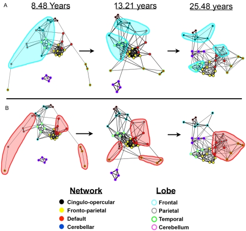
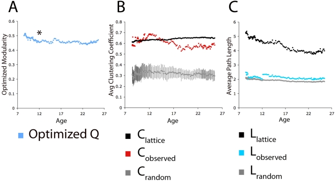
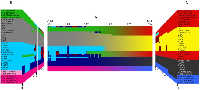
The mature human brain is organized into a collection of specialized functional networks that flexibly interact to support various cognitive functions. Studies of development often attempt to identify the organizing principles that guide the maturation of these functional networks. In this report, we combine resting state functional connectivity MRI (rs-fcMRI), graph analysis, community detection, and spring-embedding visualization techniques to analyze four separate networks defined in earlier studies. As we have previously reported, we find, across development, a trend toward 'segregation' (a general decrease in correlation strength) between regions close in anatomical space and 'integration' (an increased correlation strength) between selected regions distant in space. The generalization of these earlier trends across multiple networks suggests that this is a general developmental principle for changes in functional connectivity that would extend to large-scale graph theoretic analyses of large-scale brain networks. Communities in children are predominantly arranged by anatomical proximity, while communities in adults predominantly reflect functional relationships, as defined from adult fMRI studies. In sum, over development, the organization of multiple functional networks shifts from a local anatomical emphasis in children to a more "distributed" architecture in young adults. We argue that this "local to distributed" developmental characterization has important implications for understanding the development of neural systems underlying cognition. Further, graph metrics (e.g., clustering coefficients and average path lengths) are similar in child and adult graphs, with both showing "small-world"-like properties, while community detection by modularity optimization reveals stable communities within the graphs that are clearly different between young children and young adults. These observations suggest that early school age children and adults both have relatively efficient systems that may solve similar information processing problems in divergent ways.
Overview
The claims and statistics of this paper haven’t been checked yet.
Free: it reads the open-access text and checks the first 5 claims that cite a source we can check. Create a free account to check every claim.
Integrity signals
Registry
Cites
Statistics
Figures

Verification
Claims the paper makes
The claims and statistics of this paper haven’t been checked yet.
Free: it reads the open-access text and checks the first 5 claims that cite a source we can check. Create a free account to check every claim.
Claims other papers make about this one
Each is checked against this paper’s own text: does it say what the citing paper says it does?
The source backs part of the claim, not all of it — or backs it for a different population, condition or comparison.
In the citing paper
“Human neuroimaging studies have shown corresponding patterns of regional brain changes across development [12] with functional connectivity studies [13,14] suggesting refinement of local subcortical circuits precedes refinement of distal cortical ones.”
From Development of the emotional brain (2019) · cites this paper as [13]
Experimental — accuracy not yet measuredThe source backs part of the claim, not all of it — or backs it for a different population, condition or comparison.
In the citing paper
“A plausible temporal mechanism for the shifts in cognitive capacity from childhood to adulthood is a fine-tuning of circuits from subcortico-subcortical to cortico-subcortical to cortico-cortical consistent with postmortem animal studies of regional synaptic changes (Rakic et al., 1994) and functional connectivity studies (Fair et al., 2009) of refinement of local circuits preceding refinement of”
From Beyond simple models of adolescence to an integrated circuit-based account: A commentary (2016) · cites this paper as Fair et al., 2009
Experimental — accuracy not yet measuredThe source backs part of the claim, not all of it — or backs it for a different population, condition or comparison.
In the citing paper
“Evidence has converged on an executive attention network, with the anterior cingulate being the central structure, as the major network responsible for resolving cognitive conflicts (21, 22).”
From Multimodal imaging of the self-regulating developing brain (2012) · cites this paper as [21]
Experimental — accuracy not yet measuredThe source backs part of the claim, not all of it — or backs it for a different population, condition or comparison.
In the citing paper
“Prior work in fMRI using this modularity algorithm required that the researcher set an arbitrary threshold, or cutoff point, for what constitutes similarity between any two given individuals. Researchers would then arrive at thresholds by looking for consistency in community detection results at multiple thresholds (e.g., [2], [21], [22]).”
From Organizing Heterogeneous Samples Using Community Detection of GIMME-Derived Resting State Functional Networks (2014) · cites this paper as [22]
Experimental — accuracy not yet measuredThe source backs part of the claim, not all of it — or backs it for a different population, condition or comparison.
In the citing paper
“The functional network studies have consistently shown no statistical difference in global topological properties between children and adults”
From Developmental Changes in Organization of Structural Brain Networks (2013) · cites this paper as Fair et al. 2009
Experimental — accuracy not yet measured
5 more claims about this paper
Create a free account to see every claim other papers make about this one.
Self-check
Percentages, sums and reported differences re-computed from the paper’s own numbers.
The self-check needs the paper’s full text.
Citations
Cites
Controlling the False Discovery Rate: A Practical and Powerful Approach to Multiple Testing
Benjamini, Y., & Hochberg, Y. (1995) · Journal of the Royal Statistical Society Series B: Statistical Methodology · doi:10.1111/j.2517-6161.1995.tb02031.x · Cited by 109,482 · cited 1×
“To account for multiple comparisons the Benjamini and Hochberg False Discovery Rate [97] was applied.”
MethodCollective dynamics of ‘small-world’ networks
Watts, D. J., & Strogatz, S. H. (1998) · Nature · doi:10.1038/30918 · Cited by 43,060 · cited 1×
“long-range processes. It is this hybrid graph topology (i.e., high clustering coefficients and short path lengths) that matches the observed “small world” networks in many complex systems”
BackgroundModularity and community structure in networks
Newman, M. E. J. (2006) · Proceedings of the National Academy of Sciences · doi:10.1073/pnas.0601602103 · Cited by 12,256 · cited 5×
“As stated by Newman: ‘‘A good division of a graph into communities is not merely one in which there are few edges between communities; it is one in which there are fewer than expected edges between communities.”
BackgroundMethodInfluentialA default mode of brain function
Raichle, M. E., MacLeod, A. M., Snyder, A. Z., Powers, W. J., Gusnard, D. A., & Shulman, G. L. (2001) · Proceedings of the National Academy of Sciences · doi:10.1073/pnas.98.2.676 · Cited by 12,190 · cited 2×
“While the distinct function of the default mode network is often linked to internally directed mental activity [39], this notion continues to be debated [25,32,41–44].”
Functional connectivity in the motor cortex of resting human brain using echo‐planar mri
Biswal, B., Zerrin Yetkin, F., Haughton, V. M., & Hyde, J. S. (1995) · Magnetic Resonance in Medicine · doi:10.1002/mrm.1910340409 · Cited by 9,812
The human brain is intrinsically organized into dynamic, anticorrelated functional networks
Fox, M. D., Snyder, A. Z., Vincent, J. L., Corbetta, M., Van Essen, D. C., & Raichle, M. E. (2005) · Proceedings of the National Academy of Sciences · doi:10.1073/pnas.0504136102 · Cited by 8,553 · cited 3×
“Resting-state fcMRI analyses have repeatedly shown that these regions, along with associated medial temporal regions, are correlated at rest in adults [15,16,32,40].”
Distributed hierarchical processing in the primate cerebral cortex
Felleman, D. J., & Van Essen, D. C. (1991) · Cerebral Cortex · doi:10.1093/cercor/1.1.1 · Cited by 7,783 · cited 1×
“Increased myelination is thought to proceed from primary sensory and motor regions to association areas [68– 71], roughly following the hierarchical organization introduced by Felleman and Van Essen [72] (Note that while the most frequently referenced neuroanatomical changes that occur throughout development have been…”
BackgroundDissociable intrinsic connectivity networks for salience processing and executive control
Seeley, W. W., Menon, V., Schatzberg, A. F., Keller, J., Glover, G. H., Kenna, H., Reiss, A. L., & Greicius, M. D. (2007) · The Journal of Neuroscience · doi:10.1523/jneurosci.5587-06.2007 · Cited by 7,426 · cited 3×
“In addition, several previous findings using this technique [21,22,32] have now been replicated using continuous resting blocks [27,33,34] and other continuous resting data [89].”
BackgroundMethodFunctional connectivity in the resting brain: a network analysis of the default mode hypothesis
Greicius, M. D., Krasnow, B., Reiss, A. L., & Menon, V. (2003) · Proceedings of the National Academy of Sciences · doi:10.1073/pnas.0135058100 · Cited by 6,358 · cited 1×
“Resting-state fcMRI analyses have repeatedly shown that these regions, along with associated medial temporal regions, are correlated at rest in adults [15,16,32,40].”
BackgroundThe brainweb: phase synchronization and large-scale integration
Varela, F., Lachaux, J.-P., Rodriguez, E., & Martinerie, J. (2001) · Nature Reviews Neuroscience · doi:10.1038/35067550 · Cited by 4,761 · cited 1×
“In adults, spontaneous correlated activity has been suggested to be important for gating information flow [54], building internal representations [43,44,54], and maintaining mature network relationships [43,44,54].”
Background
Cited by
Items 801–900 of 1,003
Imaging Developmental and Interventional Plasticity Following Perinatal Stroke
Craig, B. T., Hilderley, A. J., Kirton, A., & Carlson, H. L. (2020) · Canadian Journal of Neurological Sciences / Journal Canadien des Sciences Neurologiques · doi:10.1017/cjn.2020.166 · Cited by 25
“Throughout childhood, networks strengthen and follow a local-to-global pattern of development.(164) Primary sensory networks appear first and are adult-like by around 8 years of age.”
BackgroundSilveira, S., Boney, S., Tapert, S. F., & Mishra, J. (2021) · Developmental Cognitive Neuroscience · doi:10.1016/j.dcn.2021.100962 · Cited by 20
Sex Differences in Brain and Behavioral Development
Beltz, A. M., Blakemore, J. E., & Berenbaum, S. A. (2013) · Elsevier eBooks · doi:10.1016/b978-0-12-397267-5.00064-9 · Cited by 27
Pediatric applications of functional magnetic resonance imaging
Altman, N. R., & Bernal, B. (2015) · Pediatric Radiology · doi:10.1007/s00247-015-3365-1 · Cited by 20
London, K., & Howman-Giles, R. B. (2014) · NeuroImage · doi:10.1016/j.neuroimage.2014.11.047 · Cited by 20
“Functional MRI studies in children have suggested that there is a shift in resting state connectivity from being dominated with constrained local networks towards more spatially distant networks as children age (Fair et al., 2009).”
Functional hierarchy of the human neocortex across the lifespan
Taylor, H. P., Huynh, K. M., Thung, K., Lin, G., Lyu, W., Lin, W., Ahmad, S., & Yap, P. (2026) · Nature · doi:10.1038/s41586-026-10219-x · Cited by 44
da Rocha, A. F., Rocha, F., Burattini, M. N., & Massad, E. (2010) · Brain Research · doi:10.1016/j.brainres.2010.06.046 · Cited by 19
“In addition, Fair et al. (2009) showed that brain processing changes from a more local to a more distributed one from early school-age children to adults, while maintaining " small-world " -like properties.”
Lei, H. C., Li, L., Chen, M., Xu, J., Jiang, M., Wang, Y., Jiang, L., Chen, X., Qiu, J., Tan, S., et al. (2021) · Neuroscience Bulletin · doi:10.1007/s12264-021-00650-7 · Cited by 20
Fu, C., Aisikaer, A., Chen, Z., Yu, Q. B., Yin, J., & Yang, W. (2021) · Frontiers in Neurology · doi:10.3389/fneur.2021.668856 · Cited by 21
Wang, J., Wang, Z., Zhang, H., Feng, S., Lü, Y., Wang, S., Wang, H., Sun, Y. E., & Chen, Y. (2021) · Cerebral Cortex · doi:10.1093/cercor/bhab166 · Cited by 24
Altered resting-state networks in adolescent non-suicidal self-injury—a graph theory analysis
Mürner-Lavanchy, I. M., Koenig, J., Reichl, C., Brunner, R. M., & Kaess, M. (2022) · Social Cognitive and Affective Neuroscience · doi:10.1093/scan/nsac007 · Cited by 20
“First, the neurobiological and neurophysiological processes underlying graph metrics are still unknown, and thesemetricsmay vary depending onmethodological decisions such as the parcellation scheme (Farahani et al., 2019).”
BackgroundAlterations in Structural and Functional Connectivity in ADHD: Implications for Theories of ADHD
González-Madruga, K., Staginnus, M., & Fairchild, G. F. (2022) · Current topics in behavioral neurosciences · doi:10.1007/7854_2022_345 · Cited by 23
Impacts of adrenarcheal DHEA levels on spontaneous cortical activity during development
Penhale, S. H., Picci, G., Ott, L. R., Taylor, B. K., Frenzel, M. R., Eastman, J. A., Wang, Y., Calhoun, V. D., Stephen, J. M., & Wilson, T. W. (2022) · Developmental Cognitive Neuroscience · doi:10.1016/j.dcn.2022.101153 · Cited by 21
Eng, C. M., Pocsai, M., Fulton, V. E., Moron, S., Thiessen, E. D., & Fisher, A. V. (2022) · Developmental Science · doi:10.1111/desc.13328 · Cited by 28
“The study of temporally correlated neural activity at rest shows correlations between brain areas more distant in space (i.e., long-range connections) strengthen with development (Fair et al., 2009).”
Cao, X., Wang, Z., Chen, Y., & Zhu, J. (2024) · Development and Psychopathology · doi:10.1017/s0954579424000725 · Cited by 23
“…maturation throughout adolescence and into young adulthood through the formation of optimally sculpted neural representations, such as greater functional segregation, significant reorganization, and strengthening of connections in network components (Fair et al., 2009; Uddin et al., 2011).”
BackgroundWang, P., Lin, H., Liu, G.-C., Yang, Y., Ko, C., & Yen, C. (2016) · Psychiatry Research Neuroimaging · doi:10.1016/j.pscychresns.2016.07.009 · Cited by 21
“Impairment of long-range connections can be seen as a failure of the integration process within a distributed functional network of the human brain (Fair et al., 2008; Fair et al., 2009; Fair et al., 2007), a deviation from the normal neurodevelopmental trajectory.”
BackgroundTask switching costs in preschool children and adults
Peng, A., Kirkham, N. Z., & Mareschal, D. (2018) · Journal of Experimental Child Psychology · doi:10.1016/j.jecp.2018.01.019 · Cited by 23
“This is at odds with the fact that cognitive development occurs on a protracted timescale (e.g., Fair et al., 2009; Giedd et al., 1999).”
Topology of the Structural Social Brain Network in Typical Adults
Li, L., Bachevalier, J., Hu, X. P., Klin, A., Preuss, T. M., Shultz, S., & JONES, W. R. (2018) · Brain Connectivity · doi:10.1089/brain.2018.0592 · Cited by 18
“These studies, in and of themselves, also tell us something quite profound about network topology and how that topology emerges at the intersection of structure/function and of brain/behavior (Fair et al., 2009; Hagmann et al., 2008).”
BackgroundKovacs-Balint, Z., Feczko, E., Pincus, M., Earl, E., Miranda-Domínguez, Ó., Howell, B., Morin, E. L., Maltbie, E. A., Li, L., Steele, J., et al. (2018) · Cerebral Cortex · doi:10.1093/cercor/bhy222 · Cited by 20
Self-citationDevelopment of human electrophysiological brain networks
Briley, P. M., Liddle, E. B., Groom, M. J., Smith, H. J. F., Morris, P. G., Colclough, G. L., Brookes, M. J., & Liddle, P. F. (2018) · Journal of Neurophysiology · doi:10.1152/jn.00293.2018 · Cited by 20
Miranda-Domínguez, Ó., Ragothaman, A., Hermosillo, R. J. M., Feczko, E., Morris, R., Carlson‐Kuhta, P., Nutt, J. G., Mancini, M., Fair, D. A., & Horak, F. B. (2020) · Neuroscience · doi:10.1016/j.neuroscience.2020.06.036 · Cited by 22
Self-citationExploring functional brain activity in neonates: A resting-state fMRI study
Huang, Z., Wang, Q., Zhou, S., Tang, C., Yi, F., & Nie, J. (2020) · Developmental Cognitive Neuroscience · doi:10.1016/j.dcn.2020.100850 · Cited by 21
“Besides the popular functional networks analysis, it is of great significance to probe into the local brain activity since the segregated functions exist in early brain development (Fair et al., 2009; Hagmann et al., 2010; Huang et al., 2015).”
BackgroundConnectivity Matrices and Brain Graphs
(2016) · Elsevier eBooks · doi:10.1016/b978-0-12-407908-3.00003-0 · Cited by 36
Siless, V., Davidow, J. Y., Nielsen, J. A., Fan, Q., Hedden, T., Hollinshead, M. O., Beam, E., Bustamante, C. M. V., Garrad, M. C., Santillana, R. M., et al. (2020) · NeuroImage · doi:10.1016/j.neuroimage.2020.116703 · Cited by 20
A Ferret Model of Encephalopathy of Prematurity
Wood, T. R., Moralejo, D. H., Corry, K. A., Snyder, J. M., Traudt, C. M., Curtis, C., Nance, E. A., Parikh, P., & Juul, S. E. (2018) · Developmental Neuroscience · doi:10.1159/000498968 · Cited by 19
Faghiri, A., Stephen, J. M., Wang, Y., Wilson, T. W., & Calhoun, V. D. (2019) · Brain Connectivity · doi:10.1089/brain.2018.0641 · Cited by 26
“In one influential study, Fair et al. (2009) used graph theory to demonstrate this ‘‘local to distributed’’ developmental trajectory.”
MethodSex differences in brain and behavioral development
Beltz, A. M., Kelly, D. P., & Berenbaum, S. A. (2020) · Elsevier eBooks · doi:10.1016/b978-0-12-814411-4.00027-5 · Cited by 30
Sanders, A. F. P., Baum, G. L., Harms, M. P., Kandala, S., Bookheimer, S. Y., Dapretto, M., Somerville, L. H., Thomas, K. M., Essen, D. C. V., Yacoub, E. S., et al. (2022) · Developmental Cognitive Neuroscience · doi:10.1016/j.dcn.2022.101145 · Cited by 19
“…these caveats, the nonlinear changesin VIS1, PMM, and ORA CT may reflect more complex age-related periods of large-scale brain network fine tuning, such as a succession from a less organized brain early in life to a more mature reorganization (Dosenbach et al., 2010; Fair et al., 2009).”
BackgroundXiao, X., Hammond, C., Salmeron, B. J., Wang, D., Gu, H., Zhai, T., Nguyen, H., Lu, H., Ross, T. J., & Yang, Y. (2023) · Biological Psychiatry · doi:10.1016/j.biopsych.2023.08.028 · Cited by 22
Feng, X., Piper, R. J., Prentice, F., Clayden, J. D., & Baldeweg, T. (2024) · Seizure · doi:10.1016/j.seizure.2024.02.021 · Cited by 23
Disconnected neuromagnetic networks in children born very preterm
Ye, A. X., AuCoin-Power, M., Taylor, M. J., & Doesburg, S. M. (2015) · NeuroImage Clinical · doi:10.1016/j.nicl.2015.08.016 · Cited by 22
Surface-based regional homogeneity in bipolar disorder: A resting-state fMRI study
Zhang, B., Wang, F., Dong, H.-M., Jiang, X., Wei, S., Chang, M., Yin, Z., Yang, N., Zuo, X., Tang, Y., et al. (2019) · Psychiatry Research · doi:10.1016/j.psychres.2019.05.045 · Cited by 18
Resting-state abnormalities of posterior cingulate in autism spectrum disorder
Leung, M.-K., & Lau, W. K. (2020) · Progress in molecular biology and translational science · doi:10.1016/bs.pmbts.2020.04.010 · Cited by 23
Functional connectivity patterns predict naturalistic viewing versus rest across development
Sanchez–Alonso, S., Rosenberg, M. D., & Aslin, R. Ν. (2021) · NeuroImage · doi:10.1016/j.neuroimage.2020.117630 · Cited by 22
“A number of studies have examined the developmental trajectory of the intrinsic functional organization of the brain at „rest‟ during early and late childhood (Fair et al., 2009; Gao et al., 2015; Smyser et al., 2010; Supekar et al., 2009).”
BackgroundGotlieb, R. J. M., Yang, X., & Immordino‐Yang, M. H. (2021) · Social Cognitive and Affective Neuroscience · doi:10.1093/scan/nsab108 · Cited by 23
Wang, J. X., Bartolotti, J., Amaral, L. A. N., & Booth, J. R. (2013) · PLoS ONE · doi:10.1371/journal.pone.0059204 · Cited by 20
Developmental Resting State Functional Connectivity for Clinicians
Hulvershorn, L. A., Cullen, K. R., Francis, M., & Schreiner, M. W. (2014) · Current Behavioral Neuroscience Reports · doi:10.1007/s40473-014-0020-3 · Cited by 20
Group-to-Individual Generalizability and Individual-Level Inferences in Cognitive Neuroscience
Mattoni, M., Fisher, A. J., Gates, K., Chein, J., & Olino, T. (2025) · Neuroscience and Biobehavioral Reviews · doi:10.1016/j.neubiorev.2025.106024 · Cited by 17
Xiao, L., Cai, B., Qu, G., Zhang, G., Stephen, J. M., Wilson, T. W., Calhoun, V. D., & Wang, Y.-P. (2022) · IEEE Transactions on Biomedical Engineering · doi:10.1109/tbme.2022.3160447 · Cited by 21
“Recently, brain developmental fMRI studies have shown that the human brain undergoes important changes in functional connectome across the lifespan [11]–[13].”
BackgroundViering, T., Hoekstra, P. J., Philipsen, A., Naaijen, J., Dietrich, A., Hartman, C. A., Buitelaar, J. K., Hildebrandt, A., Gießing, C., & Thiel, C. M. (2021) · Scientific Reports · doi:10.1038/s41598-021-94426-8 · Cited by 19
Network controllability mediates the relationship between rigid structure and flexible dynamics
Gu, S., Fotiadis, P., Parkes, L., Xia, C. H., Gur, R. C., Gur, R. E., Roalf, D. R., Satterthwaite, T. D., & Bassett, D. S. (2022) · Network Neuroscience · doi:10.1162/netn_a_00225 · Cited by 21
“…this developmental period that potentially contributes to the maturity of brain modules, in support of the emergence of functional roles of cognitive systems (Gu, Satterthwaite, et al., 2015) associated with network segregation (Baker et al., 2015; Baum et al., 2017; Fair et al., 2009, 2007).”
BackgroundLiu, X., TYLER, L. K., Cam‐CAN, Davis, S. W., Rowe, J. B., & Tsvetanov, K. A. (2023) · Neurobiology of Aging · doi:10.1016/j.neurobiolaging.2023.06.001 · Cited by 20
Translating Developmental Neuroscience to Understand Risk for Psychiatric Disorders
Meyer, H. C., & Lee, F. S. (2023) · American Journal of Psychiatry · doi:10.1176/appi.ajp.19010091 · Cited by 19
Domschke, K., Akhrif, A., Romanos, M., Bajer, C., Mainusch, M., Winkelmann, J., Zimmer, C., & Neufang, S. (2015) · Cerebral Cortex · doi:10.1093/cercor/bhv259 · Cited by 20
Sato, J. R., Biazoli, C. E., Salum, G. A., Gadelha, A., Crossley, N. A., Vieira, G., Zugman, A., Picon, F. A., Pan, P. M., Hoexter, M. Q., et al. (2016) · Developmental Cognitive Neuroscience · doi:10.1016/j.dcn.2016.05.002 · Cited by 21
Effect of rs1344706 in the ZNF804A gene on the brain network
Chen, X., Zhang, Z., Zhang, Q.-M., Zhao, W., Zhai, J., Chen, M., Du, B., Deng, X., Ji, F., Wang, C., et al. (2017) · NeuroImage Clinical · doi:10.1016/j.nicl.2017.12.017 · Cited by 17
Anckaerts, C., van Gastel, J., Leysen, V., Hinz, R., Azmi, A., Simoens, P., Shah, D., Kara, F., Langbeen, A., Bols, P. E. J., et al. (2018) · Neurobiology of Aging · doi:10.1016/j.neurobiolaging.2018.10.012 · Cited by 19
Liu, D., Chen, L., Duan, S., Yin, X., Yang, W. C., Shi, Y., Zhang, J., & Wang, J. (2018) · Frontiers in Neuroscience · doi:10.3389/fnins.2018.00875 · Cited by 18
Structural brain network development in children following prenatal methamphetamine exposure
Roos, A., Fouché, J., Toit, S. D., du Plessis, S., Stein, D. J., & Donald, K. A. (2020) · The Journal of Comparative Neurology · doi:10.1002/cne.24858 · Cited by 17
“In contrast, children with PME in our study, failed to show the expected greater frontal changes over time (Fair et al., 2009), consistent with existing understanding that frontal regions are targeted”
BackgroundResultInfluentialMeta-analysis of the neural correlates of vigilant attention in children and adolescents
Morandini, H. A. E., Silk, T. J., Griffiths, K. R., Rao, P., Hood, S. D., & Zepf, F. D. (2020) · Cortex · doi:10.1016/j.cortex.2020.08.008 · Cited by 17
Chaddock‐Heyman, L., Weng, T. B., Loui, P., Kienzler, C., Weisshappel, R., Drollette, E. S., Raine, L. B., Westfall, D. R., Kao, S., Pindus, D. M., et al. (2021) · Psychophysiology · doi:10.1111/psyp.13890 · Cited by 19
Vuontela, V., Jiang, P., Tokariev, M., Savolainen, P., Ma, Y., Aronen, E. T., Fontell, T., Liiri, T., Ahlström, M., Salonen, O. L. M., et al. (2012) · Brain and Cognition · doi:10.1016/j.bandc.2012.11.003 · Cited by 20
Chen, C., Zhou, J., Liu, C., Witt, K., Zhang, Y., Jing, B., Li, C., Wang, X., & Li, L. (2015) · Journal of Neuropsychiatry · doi:10.1176/appi.neuropsych.13030044 · Cited by 19
Liu, L., Li, H., Zhang, M., Wang, Z., Wei, N., Liu, L., Meng, X., & Ding, G. (2016) · Developmental Science · doi:10.1111/desc.12440 · Cited by 18
Characterization of Autism Spectrum Disorder across the Age Span by Intrinsic Network Patterns
Morgan, B. R., Ibrahim, G. M., Vogan, V. M., Leung, R. C., Lee, W., & Taylor, M. J. (2019) · Brain Topography · doi:10.1007/s10548-019-00697-w · Cited by 21
Structured dynamics of neural activity across developing neocortexPreprint
Ackman, J. B., Zeng, H., & Crair, M. C. (2014) · bioRxiv (Cold Spring Harbor Laboratory) · doi:10.1101/012237 · Cited by 18
“The increasing strength of intra-and inter-hemispheric connections with age is consistent with greater long-range connection strength between disparate brain regions during brain development 48,49 .”
BackgroundInfluentialBreukelaar, I. A., Griffiths, K. R., Harris, A. W. F., Foster, S., Williams, L. M., & Korgaonkar, M. S. (2020) · Cortex · doi:10.1016/j.cortex.2020.08.014 · Cited by 17
Does prefrontal connectivity during task switching help or hinder children’s performance?
Schwarze, S. A., Laube, C., Khosravani, N., Lindenberger, U., Bunge, S. A., & Fandakova, Y. (2023) · Developmental Cognitive Neuroscience · doi:10.1016/j.dcn.2023.101217 · Cited by 18
Geography education improves spatial ability: evidence from fMRI and behavioral experiments
Dong, W., Ying, Q., Yang, T., Zhu, L., Liu, Y., & Wan, X. (2023) · Cartography and Geographic Information Science · doi:10.1080/15230406.2023.2171493 · Cited by 16
“From a graphical perspective, this finding reflects strong regional specialization and integrated spatial ability (Fair et al., 2009).”
BackgroundKavčič, A., Demšar, J., Georgiev, D., Bon, J., & Šalamon, A. S. (2023) · Clinical Neurophysiology · doi:10.1016/j.clinph.2023.03.357 · Cited by 18
He, Y., Zeng, D., Li, Q., Chu, L., Dong, X., Liang, X., Sun, L., Liao, X., Zhao, T., Chen, X., et al. (2025) · PLoS Biology · doi:10.1371/journal.pbio.3002710 · Cited by 23
“Multiscale brain structural re-organization from childhood to adolescence displayed a more distributed and segregated pattern [71, 72].”
Stochastic geometric network models for groups of functional and structural connectomes
Friedman, E. J., Landsberg, A. S., Owen, J. P., Li, Y., & Mukherjee, P. (2014) · NeuroImage · doi:10.1016/j.neuroimage.2014.07.039 · Cited by 19
Modularity-based graph partitioning using conditional expected models
Chang, Y.-T., Leahy, R. M., & Pantazis, D. (2012) · Physical Review E · doi:10.1103/physreve.85.016109 · Cited by 17
Structural connectivity and weight loss in children with obesity: a study of the “connectobese”
Augustijn, M. J. C. M., Biase, M. A. D., Zalesky, A., Acker, L. V., Guchtenaere, A. D., D’Hondt, E., Lenoir, M., Deconinck, F. J. A., & Caeyenberghs, K. (2019) · International Journal of Obesity · doi:10.1038/s41366-019-0380-6 · Cited by 20
Effective connectivity in the default mode network after paediatric traumatic brain injury
Vaughn, K. A., DeMaster, D., Kook, J. H., Vannucci, M., & Ewing‐Cobbs, L. (2021) · European Journal of Neuroscience · doi:10.1111/ejn.15546 · Cited by 17
“With age, functional connectivity within these developing networks expands to include distant brain regions (Fair et al., 2007, 2009; Supekar et al., 2009), leading to brain networks that become more efficient and widespread.”
BackgroundFischer, A. S., Holt-Gosselin, B., Hagan, K. E., Fleming, S. L., Nimarko, A. F., Gotlib, I. H., & Singh, M. K. (2022) · Biological Psychiatry Cognitive Neuroscience and Neuroimaging · doi:10.1016/j.bpsc.2022.02.009 · Cited by 18
“Region of interest spheres created were centered on peak coordinates from the literature from regions in which youth at high versus low familial risk for mood disorders differed in their blood oxygen level–dependent response (14,15,17,18,61,64,65): bilateral amygdala seeds (central coordinates: 622, 25, 117), bilatera…”
BackgroundBey, K., Montag, C., Reuter, M., Weber, B., & Markett, S. A. (2015) · NeuroImage · doi:10.1016/j.neuroimage.2015.07.041 · Cited by 19
“A dual regression approach was then applied to create subject-level spatial maps and time series corresponding to each of the components obtained from the group-ICA (Beckmann et al., 2009; Filippini et al., 2009).”
MethodIdle Behaviors of the Hippocampus Reflect Endogenous Cortisol Levels in Youth
Thomason, M. E., Tocco, M. A., Quednau, K., Bedway, A. R., & Carré, J. M. (2013) · Journal of the American Academy of Child & Adolescent Psychiatry · doi:10.1016/j.jaac.2013.04.004 · Cited by 18
Keulers, E. H. H., Goulas, A., Jolles, J., & Stiers, P. (2012) · NeuroImage · doi:10.1016/j.neuroimage.2011.12.079 · Cited by 16
“…have shown that network efficiency does not change with age, but that in childhood brain networks are organized more locally , based on anatomical proximity, whereas adult brain networks comprise regions spread over different brain lobes (Fair et al., 2009; Power et al., 2010; Vogel et al., 2010).”
InfluentialBrain Modulyzer: Interactive Visual Analysis of Functional Brain Connectivity
Murugesan, S., Bouchard, K., Brown, J. A., Hamann, B., Seeley, W. W., Trujillo, A. J., & Weber, G. H. (2016) · IEEE Transactions on Computational Biology and Bioinformatics · doi:10.1109/tcbb.2016.2564970 · Cited by 21
“We provide a brief review of visualization methods in three domains: visual analysis in functional brain networks, visualizations in hierarchical modular structures, and visualization of communities in functional brain networks.”
MethodChorlian, D. B., Rangaswamy, M., Manz, N., Meyers, J. L., Kang, S. J., Kamarajan, C., Pandey, A. K., Wang, J., Wetherill, L. F., Edenberg, H. J., et al. (2016) · International Journal of Psychophysiology · doi:10.1016/j.ijpsycho.2016.11.007 · Cited by 17
Integrating brain, cognition and culture
Posner, M. I., & Rothbart, M. K. (2017) · Journal of Cultural Cognitive Science · doi:10.1007/s41809-017-0001-7 · Cited by 22
Scholtens, L. H., Barrett, L. F., & van den Heuvel, M. P. (2018) · Brain Connectivity · doi:10.1089/brain.2018.0622 · Cited by 16
“Indeed, in humans, studies have shown clear developmental change of large-scale patterns of anatomical and functional connectivity (Betzel et al., 2014; Dosenbach et al., 2010; Fair et al., 2008, 2009; Hagmann et al., 2010; van den Heuvel et al., 2015b).”
BackgroundAmemiya, K., Morita, T., Saito, D. N., Ban, M., Shimada, K., Okamoto, Y., Kosaka, H., Okazawa, H., Asada, M., & Naito, E. (2019) · Brain Structure and Function · doi:10.1007/s00429-018-01821-5 · Cited by 16
“Resting-state functional connectivity studies have revealed slow maturation of cerebrocerebellar functional networks in general (Fair et al. 2009).”
BackgroundLewis, C. P., Klimes‐Dougan, B., Croarkin, P. E., & Cullen, K. R. (2025) · Neuropsychopharmacology · doi:10.1038/s41386-025-02168-2 · Cited by 34
Harlalka, V., Bapi, R. S., Vinod, P. K. U., & Roy, D. (2018) · Brain Connectivity · doi:10.1089/brain.2018.0616 · Cited by 14
“Furthermore, it is also shown that functional brain network develops from local to distributed organization with development (Fair et al., 2009).”
Ha, M., Park, S. H., Park, I., Kim, T., Lee, J., Kim, M., & Kwon, J. S. (2023) · Schizophrenia · doi:10.1038/s41537-023-00375-8 · Cited by 21
“In addition, as the neural circuit matures, the cerebellum develops as a solid hub and establishes functional links with various cortical intrinsic networks over time, communicating on a large scale and contributing to cognitive processing as a hub entity 77,78 .”
BackgroundBrain signal variability and executive functions across the life span
Goodman, Z. T., Nomi, J. S., Kornfeld, S., Bolt, T., Saumure, R. A., Romero, C., Bainter, S. A., & Uddin, L. Q. (2023) · Network Neuroscience · doi:10.1162/netn_a_00347 · Cited by 26
“In contrast, brain networks become increasingly integrated and localized throughout adolescence (Fair et al., 2009).”
BackgroundMillevert, C., Vidas-Guscic, N., Vanherp, L., Jonckers, E., Verhoye, M., Staelens, S. G., Bertoglio, D., & Weckhuysen, S. (2023) · Journal of Neuroscience · doi:10.1523/jneurosci.1043-23.2023 · Cited by 20
Deficits in adults with autism spectrum disorders when processing multiple objects in dynamic scenes
O’Hearn, K. M., Lakusta, L., Schroer, E., Minshew, N. J., & Luna, B. (2011) · Autism Research · doi:10.1002/aur.179 · Cited by 23
“Other work indicates that network function has a long developmental trajectory [Fair et al., 2009; Luna et al., 2001] and might be affected by ASD [Just, Cherkassky, Keller, Kana, & Minshew, 2007; Minshew & Williams, 2007].”
BackgroundShaw, D. J., Mareček, R., Grosbras, M., Leonard, G. T., Pike, G. B., & Paus, T. (2016) · Social Cognitive and Affective Neuroscience · doi:10.1093/scan/nsv138 · Cited by 17
“In this light, we consider their increased integration in response to ambiguous expressions during adolescence to index an important event behind the development of face processing (Fair et al., 2007; 2009).”
BackgroundRichards, T. L., Berninger, V. W., Yagle, K. J., Abbott, R. D., & Peterson, D. J. (2017) · PubMed · Cited by 20
A Novel Synchronization-Based Approach for Functional Connectivity Analysis
Lombardi, A., Tangaro, S., Bellotti, R., Bertolino, A., Blasi, G., Pergola, G., Taurisano, P., & Guaragnella, C. (2017) · Complexity · doi:10.1155/2017/7190758 · Cited by 16
“Indeed, lack of highly specialized modules may not allow a rapid execution of complex cognitive task [45, 46].”
BackgroundToich, J. T. F., Taylor, P. A., Holmes, M., Gohel, S., Cotton, M. F., Dobbels, E., Laughton, B., Little, F., van der Kouwe, A. J. W., Biswal, B. B., et al. (2018) · Frontiers in Human Neuroscience · doi:10.3389/fnhum.2017.00635 · Cited by 22
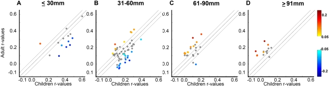
“Studies in typically developing healthy children find that longdistance connections between functionally related regions tend to be relatively weak and strengthen with age, while short-distance relationships are stronger and weaken with development (Fair et al., 2009; Power et al., 2010).”
Backgroundde Lacy, N., Kodish, I. M., Rachakonda, S., & Calhoun, V. D. (2018) · Human Brain Mapping · doi:10.1002/hbm.24187 · Cited by 16
“Perhaps the best-described phenomenon is increasing long- versus short-range connectivity (Dosenbach et al., 2010; Fair et al., 2009; Supekar, Musen, & Menon, 2009), particularly between task-positive lateral/medial frontal, striatal and parieto-temporal regions associated with cognitive control.”
BackgroundFunctional connectivity between cognitive control regions is sensitive to familial risk for ADHD
Mulder, M., van Belle, J., van Engeland, H., & Durston, S. (2010) · Human Brain Mapping · doi:10.1002/hbm.21141 · Cited by 16
“Studies reporting a shift from a local to more distributed architecture and from more subcortical to cortico-cortical organization with age have typically included younger and wider ranges [7– 31 yrs, Fair et al., 2009; 7–22 yrs, Supekar et al., 2009].”
BackgroundMeng, L., & Xiang, J. (2016) · Brain and Development · doi:10.1016/j.braindev.2016.05.004 · Cited by 16
Langen, C. D., Muetzel, R. L., Blanken, L. M. E., van der Lugt, A., Tiemeier, H. W., Verhulst, F. C., Niessen, W. J., & White, T. J. H. (2018) · Brain and Behavior · doi:10.1002/brb3.1031 · Cited by 18
Wolf, K., Weber, E. M. G., van den Bosch, J. J. F., Volz, S., Nöth, U., Deichmann, R., Naumer, M. J., Pfeiffer, T., & Fiebach, C. J. (2018) · Frontiers in Psychology · doi:10.3389/fpsyg.2018.01106 · Cited by 20
Chen, H. J., Wang, Y. F., Wen, J., Xu, Q., Lu, G., & Zhang, L. J. (2019) · Human Brain Mapping · doi:10.1002/hbm.24804 · Cited by 15
“With graph theory analysis, researchers found that the network properties change as the age increases (Achard & Bullmore, 2007; Fair et al., 2009).”
MethodPreserved interhemispheric functional connectivity in a case of corpus callosum agenesis
Khanna, P. C., Poliakov, A. V., Ishak, G. E., Poliachik, S. L., Friedman, S. D., Saneto, R. P., Novotny, E. J., Ojemann, J. G., & Shaw, D. (2011) · Neuroradiology · doi:10.1007/s00234-011-0883-x · Cited by 16
“As recent studies have suggested that functional networks develop in a “local-to-distributed” pattern [12], understanding how these patterns change across development [13] and in a range of disease states that affect hemispheric integration represents future directions to explore.”
BackgroundThe Development of Emotion Regulation: A Neuropsychological Perspective
Lewis, M. D., Todd, R. M., & Xu, X. (2010) · Wiley eBooks · doi:10.1002/9780470880166.hlsd002003 · Cited by 15
Kennedy, M. F., Mohamed, A. Z., Schwenn, P., Beaudequin, D. A., Shan, Z., Hermens, D. F., & Lagopoulos, J. (2022) · Brain Imaging and Behavior · doi:10.1007/s11682-022-00673-2 · Cited by 25
Liu, J., Chen, H., Cornea, E. A., Gilmore, J. H., & Gao, W. (2023) · Cerebral Cortex · doi:10.1093/cercor/bhad288 · Cited by 22
He, X., Li, Y., Xiao, X., Li, Y., Fang, J., & Zhou, R. (2025) · Cognitive Neurodynamics · doi:10.1007/s11571-024-10203-z · Cited by 16
“Based on the graph-theoretical method, researchers have applied the connectivity in complex brain networks to conduct functional connectivity analysis to determine the connections between distinct brain areas (Fair et al. 2009; Hutchison et al. 2013; Wang et al. 2023).”
Luo, Y., Schulz, K. P., Alvarez, T. L., Halperin, J. M., & Li, X. (2018) · Cortex · doi:10.1016/j.cortex.2018.09.013 · Cited by 14
Pluck, G. C., Villagomez-Pacheco, D., Karolys, M. I., Montaño-Córdova, M. E., & Almeida‐Meza, P. (2019) · Trends in Neuroscience and Education · doi:10.1016/j.tine.2019.100121 · Cited by 16
Dégeilh, F., Beauchamp, M. H., Leblanc, É., Daneault, V., & Bernier, A. (2019) · Developmental Neuroscience · doi:10.1159/000507616 · Cited by 16
“Thus, rsFC reflects functional brain organization and when measured during childhood can provide insight into fundamental organizing principles underlying brain development [35,36].”
Peng, Y., Zhao, J., Lü, X., Dong, J., Zhang, S., Zhang, J., Liu, H., Zheng, X., Wang, X., Lan, Y., et al. (2022) · Frontiers in Neurology · doi:10.3389/fneur.2022.821286 · Cited by 19
“Peak coordinates of ROIs were selected according to pieces of literature, in which 10-mm-radius and 4-mm-radius spheres around peak x, y, and z coordinates were delineated for cortical areas and subcortical structures, respectively (17, 26).”
MethodCrippa, A., Maurits, N. M., Lorist, M. M., & Roerdink, J. B. T. M. (2010) · Computers & Graphics · doi:10.1016/j.cag.2010.12.008 · Cited by 13
“For interpretation and diagnosis it is essential that local differences can be visualized in the original network representation [5,6].”
Background
Metadata
- Authors
- Fair, D. A., Cohen, A. L., Power, J. D., Dosenbach, N. U. F., Church, J. A., Miezin, F. M., Schlaggar, B. L., & Petersen, S. E.
- Affiliations
- Oregon Health & Science University (US)
- Washington University in St. Louis (US)
- Corresponding author
- Fair, Damien A.
- Petersen, Steven E.
- ORCID
- Venue
- PLoS Computational Biology
- Publisher
- Public Library of Science (PLoS)
- Published
- 2009-05-01
- Volume
- 5
- Issue
- 5
- Pages
- e1000381
- ISSN
- 1553-734X (ISSN-L)
- 1553-7358
- PMC
- PMC2671306
- OpenAlex
- W2159549823
- Cited by
- 1,530
- References
- 104
- Citations by year
- 2026: 69 · 2025: 78 · 2024: 89 · 2023: 66 · 2022: 87 · 2021: 78 · 2020: 86 · 2019: 103 · 2018: 109 · 2017: 95 · 2016: 102 · 2015: 107 · 2014: 106 · 2013: 113 · 2012: 84
- FWCI
- 39.01
- Type
- journal article
- Language
- en
- Open access
- gold
- doi.org
- Locations
- PLoS Computational Biology (publishedVersion, cc-by)
- PubMed Central (publishedVersion, cc-by)
- PubMed (publishedVersion)
- Digital Commons@Becker (Washington University School of Medicine) (submittedVersion)
- RePEc: Research Papers in Economics (submittedVersion)
- DOAJ (DOAJ: Directory of Open Access Journals) (submittedVersion, cc-by-sa)
- Figshare (submittedVersion, cc-by)
- Funders
- National Science Foundation (0548890)
- Dana Foundation
- Wellcome Trust
- National Institutes of Health
- National Institute of Neurological Disorders and Stroke (R01NS046424, K02NS053425, F30NS062489, NS46424, NS 41255, NS062489, R21NS041255, NS053425)
- Topics
- Life Sciences › Neuroscience › Cognitive Neuroscience › Functional Brain Connectivity Studies
- Health Sciences › Medicine › Radiology, Nuclear Medicine and Imaging › Advanced Neuroimaging Techniques and Applications
- Physical Sciences › Physics and Astronomy › Statistical and Nonlinear Physics › Complex Network Analysis Techniques
- Keywords
- functional brain networks, brain development, resting-state functional connectivity, integration and segregation, community detection, small-world networks, graph theory, children and young adults, modularity optimization, cognitive development
- MeSH terms
- Adolescent, Adult, Brain, Child, Humans, Magnetic Resonance Imaging, Models, Neurological, Nerve Net, Neural Pathways
About these checks
Registry, retraction, Cites and statistics results are computed, not judged, with one exception: a printed reference that no index lists is read once by a model before it counts as a citation error. Claims are extracted by a model and kept only when their quote appears verbatim in the text; each cited source is then checked, with a model’s help, for whether it says what the claim relies on. Those verdicts can be wrong — read the claim’s quote and the cited source before relying on one.
Statistics and arithmetic are recomputed, not judged.
Confidence bands are shown only for fields calibrated on our annotated test set. None is calibrated yet, so no band is shown.
What counts as a claim ›How we measure confidence ›How we check this ›
Are you an author, or do you see a problem with this page? Request a review
Data sources
Bibliographic data: Semantic Scholar, OpenAlex, Crossref.
Contains information from the Semantic Scholar Academic Graph Datasets, made available under the ODC Attribution License (ODC-BY 1.0).
Semantic Scholar’s data is described in: Kinney, R., et al. (2023). The Semantic Scholar Open Data Platform. arXiv:2301.10140.
