

Functional brain networks develop from a local to distributed organization
Open accessThe DOI matches its Crossref record; 103 of the 104 references it cites were found in a registry or an index.
Abstract
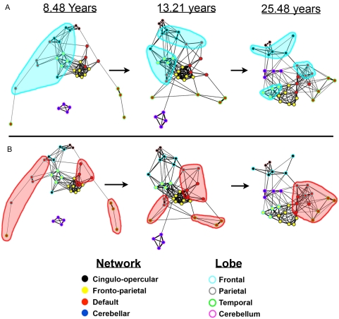
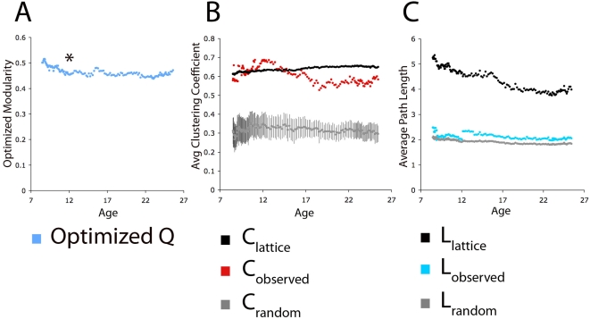
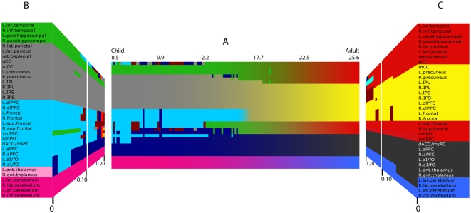
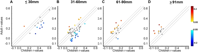
The mature human brain is organized into a collection of specialized functional networks that flexibly interact to support various cognitive functions. Studies of development often attempt to identify the organizing principles that guide the maturation of these functional networks. In this report, we combine resting state functional connectivity MRI (rs-fcMRI), graph analysis, community detection, and spring-embedding visualization techniques to analyze four separate networks defined in earlier studies. As we have previously reported, we find, across development, a trend toward 'segregation' (a general decrease in correlation strength) between regions close in anatomical space and 'integration' (an increased correlation strength) between selected regions distant in space. The generalization of these earlier trends across multiple networks suggests that this is a general developmental principle for changes in functional connectivity that would extend to large-scale graph theoretic analyses of large-scale brain networks. Communities in children are predominantly arranged by anatomical proximity, while communities in adults predominantly reflect functional relationships, as defined from adult fMRI studies. In sum, over development, the organization of multiple functional networks shifts from a local anatomical emphasis in children to a more "distributed" architecture in young adults. We argue that this "local to distributed" developmental characterization has important implications for understanding the development of neural systems underlying cognition. Further, graph metrics (e.g., clustering coefficients and average path lengths) are similar in child and adult graphs, with both showing "small-world"-like properties, while community detection by modularity optimization reveals stable communities within the graphs that are clearly different between young children and young adults. These observations suggest that early school age children and adults both have relatively efficient systems that may solve similar information processing problems in divergent ways.
Overview
The claims and statistics of this paper haven’t been checked yet.
Free: it reads the open-access text and checks the first 5 claims that cite a source we can check. Create a free account to check every claim.
Integrity signals
Registry
Cites
Statistics
Figures

Verification
Claims the paper makes
The claims and statistics of this paper haven’t been checked yet.
Free: it reads the open-access text and checks the first 5 claims that cite a source we can check. Create a free account to check every claim.
Claims other papers make about this one
Each is checked against this paper’s own text: does it say what the citing paper says it does?
The source backs part of the claim, not all of it — or backs it for a different population, condition or comparison.
In the citing paper
“Human neuroimaging studies have shown corresponding patterns of regional brain changes across development [12] with functional connectivity studies [13,14] suggesting refinement of local subcortical circuits precedes refinement of distal cortical ones.”
From Development of the emotional brain (2019) · cites this paper as [13]
Experimental — accuracy not yet measuredThe source backs part of the claim, not all of it — or backs it for a different population, condition or comparison.
In the citing paper
“A plausible temporal mechanism for the shifts in cognitive capacity from childhood to adulthood is a fine-tuning of circuits from subcortico-subcortical to cortico-subcortical to cortico-cortical consistent with postmortem animal studies of regional synaptic changes (Rakic et al., 1994) and functional connectivity studies (Fair et al., 2009) of refinement of local circuits preceding refinement of”
From Beyond simple models of adolescence to an integrated circuit-based account: A commentary (2016) · cites this paper as Fair et al., 2009
Experimental — accuracy not yet measuredThe source backs part of the claim, not all of it — or backs it for a different population, condition or comparison.
In the citing paper
“Evidence has converged on an executive attention network, with the anterior cingulate being the central structure, as the major network responsible for resolving cognitive conflicts (21, 22).”
From Multimodal imaging of the self-regulating developing brain (2012) · cites this paper as [21]
Experimental — accuracy not yet measuredThe source backs part of the claim, not all of it — or backs it for a different population, condition or comparison.
In the citing paper
“Prior work in fMRI using this modularity algorithm required that the researcher set an arbitrary threshold, or cutoff point, for what constitutes similarity between any two given individuals. Researchers would then arrive at thresholds by looking for consistency in community detection results at multiple thresholds (e.g., [2], [21], [22]).”
From Organizing Heterogeneous Samples Using Community Detection of GIMME-Derived Resting State Functional Networks (2014) · cites this paper as [22]
Experimental — accuracy not yet measuredThe source backs part of the claim, not all of it — or backs it for a different population, condition or comparison.
In the citing paper
“The functional network studies have consistently shown no statistical difference in global topological properties between children and adults”
From Developmental Changes in Organization of Structural Brain Networks (2013) · cites this paper as Fair et al. 2009
Experimental — accuracy not yet measured
5 more claims about this paper
Create a free account to see every claim other papers make about this one.
Self-check
Percentages, sums and reported differences re-computed from the paper’s own numbers.
The self-check needs the paper’s full text.
Citations
Cites
Controlling the False Discovery Rate: A Practical and Powerful Approach to Multiple Testing
Benjamini, Y., & Hochberg, Y. (1995) · Journal of the Royal Statistical Society Series B: Statistical Methodology · doi:10.1111/j.2517-6161.1995.tb02031.x · Cited by 109,482 · cited 1×
“To account for multiple comparisons the Benjamini and Hochberg False Discovery Rate [97] was applied.”
MethodCollective dynamics of ‘small-world’ networks
Watts, D. J., & Strogatz, S. H. (1998) · Nature · doi:10.1038/30918 · Cited by 43,060 · cited 1×
“long-range processes. It is this hybrid graph topology (i.e., high clustering coefficients and short path lengths) that matches the observed “small world” networks in many complex systems”
BackgroundModularity and community structure in networks
Newman, M. E. J. (2006) · Proceedings of the National Academy of Sciences · doi:10.1073/pnas.0601602103 · Cited by 12,256 · cited 5×
“As stated by Newman: ‘‘A good division of a graph into communities is not merely one in which there are few edges between communities; it is one in which there are fewer than expected edges between communities.”
BackgroundMethodInfluentialA default mode of brain function
Raichle, M. E., MacLeod, A. M., Snyder, A. Z., Powers, W. J., Gusnard, D. A., & Shulman, G. L. (2001) · Proceedings of the National Academy of Sciences · doi:10.1073/pnas.98.2.676 · Cited by 12,190 · cited 2×
“While the distinct function of the default mode network is often linked to internally directed mental activity [39], this notion continues to be debated [25,32,41–44].”
Functional connectivity in the motor cortex of resting human brain using echo‐planar mri
Biswal, B., Zerrin Yetkin, F., Haughton, V. M., & Hyde, J. S. (1995) · Magnetic Resonance in Medicine · doi:10.1002/mrm.1910340409 · Cited by 9,812
The human brain is intrinsically organized into dynamic, anticorrelated functional networks
Fox, M. D., Snyder, A. Z., Vincent, J. L., Corbetta, M., Van Essen, D. C., & Raichle, M. E. (2005) · Proceedings of the National Academy of Sciences · doi:10.1073/pnas.0504136102 · Cited by 8,553 · cited 3×
“Resting-state fcMRI analyses have repeatedly shown that these regions, along with associated medial temporal regions, are correlated at rest in adults [15,16,32,40].”
Distributed hierarchical processing in the primate cerebral cortex
Felleman, D. J., & Van Essen, D. C. (1991) · Cerebral Cortex · doi:10.1093/cercor/1.1.1 · Cited by 7,783 · cited 1×
“Increased myelination is thought to proceed from primary sensory and motor regions to association areas [68– 71], roughly following the hierarchical organization introduced by Felleman and Van Essen [72] (Note that while the most frequently referenced neuroanatomical changes that occur throughout development have been…”
BackgroundDissociable intrinsic connectivity networks for salience processing and executive control
Seeley, W. W., Menon, V., Schatzberg, A. F., Keller, J., Glover, G. H., Kenna, H., Reiss, A. L., & Greicius, M. D. (2007) · The Journal of Neuroscience · doi:10.1523/jneurosci.5587-06.2007 · Cited by 7,426 · cited 3×
“In addition, several previous findings using this technique [21,22,32] have now been replicated using continuous resting blocks [27,33,34] and other continuous resting data [89].”
BackgroundMethodFunctional connectivity in the resting brain: a network analysis of the default mode hypothesis
Greicius, M. D., Krasnow, B., Reiss, A. L., & Menon, V. (2003) · Proceedings of the National Academy of Sciences · doi:10.1073/pnas.0135058100 · Cited by 6,358 · cited 1×
“Resting-state fcMRI analyses have repeatedly shown that these regions, along with associated medial temporal regions, are correlated at rest in adults [15,16,32,40].”
BackgroundThe brainweb: phase synchronization and large-scale integration
Varela, F., Lachaux, J.-P., Rodriguez, E., & Martinerie, J. (2001) · Nature Reviews Neuroscience · doi:10.1038/35067550 · Cited by 4,761 · cited 1×
“In adults, spontaneous correlated activity has been suggested to be important for gating information flow [54], building internal representations [43,44,54], and maintaining mature network relationships [43,44,54].”
Background
Cited by
Items 401–500 of 1,003
Medaglia, J. D., Satterthwaite, T. D., Kelkar, A., Ciric, R., Moore, T. M., Ruparel, K., Gur, R. C., Gur, R. E., & Bassett, D. S. (2018) · NeuroImage · doi:10.1016/j.neuroimage.2017.10.048 · Cited by 64
“This characteristic of brain state dynamics contextualizes several major developmental studies of functional brain connectivity (Hwang et al., 2013; Fair et al., 2009; Cao et al., 2015; Betzel et al., 2014; Gu et al., 2015; Hutchison and Morton, 2015).”
Yin, D., Song, F., Xu, D., Sun, L., Men, W., Zang, L., Yan, X., & Fan, M. (2013) · Human Brain Mapping · doi:10.1002/hbm.22406 · Cited by 73
“…criteria were as follows: (1) first-onset stroke, (2) pure motor deficits, (3) right-handedness, (4) sufficient cognitive abilities (Mini-Mental State Examination [Folstein et al., 1975], MMSE > 24), (5) examination time greater than 3 months from stroke onset, and (6) aged 45 to 80 years old.”
BackgroundAssociated Microscale Spine Density and Macroscale Connectivity Disruptions in Schizophrenia
van den Heuvel, M. P., Scholtens, L. H., de Reus, M. A., & Kahn, R. S. (2015) · Biological Psychiatry · doi:10.1016/j.biopsych.2015.10.005 · Cited by 74
Attention to learning of school subjects
Posner, M. I., & Rothbart, M. K. (2014) · Trends in Neuroscience and Education · doi:10.1016/j.tine.2014.02.003 · Cited by 71
Fishman, I., Datko, M., Cabrera, Y., Carper, R. A., & Müller, R. (2015) · Annals of Neurology · doi:10.1002/ana.24533 · Cited by 76
Functional Brain Parcellations of the Infant Brain and the Associated Developmental Trends
Shi, F., Salzwedel, A., Lin, W., Gilmore, J. H., & Gao, W. (2017) · Cerebral Cortex · doi:10.1093/cercor/bhx062 · Cited by 77
“Globally, we expect to see a general “local to distributed” pattern featuring decrease in short-range connections but increase in long-range integrations (Fair et al. 2009; Gao et al. 2011).”
BackgroundAnderson, D. R., & Davidson, M. C. (2019) · Computers in Human Behavior · doi:10.1016/j.chb.2019.05.008 · Cited by 79
Development of Microstructural and Morphological Cortical Profiles in the Neonatal Brain
Fenchel, D., Dimitrova, R., Seidlitz, J., Robinson, E. C., Batalle, D., Hutter, J., Christiaens, D., Pietsch, M., Brandon, J., Hughes, E. J., et al. (2020) · Cerebral Cortex · doi:10.1093/cercor/bhaa150 · Cited by 71
“Increases in network segregation in the developing brain has been previously demonstrated in neonates (Cao et al., 2017; Zhao et al., 2019) and in children and adolescents (Baum et al., 2017; He et al., 2018), and may show more distributed integrative patterns in adulthood (Fair et al., 2009).”
BackgroundThe Age of Reason: Functional Brain Network Development during Childhood
Tooley, U. A., Park, A. T., Leonard, J., Boroshok, A. L., McDermott, C. L., Tisdall, M. D., Bassett, D. S., & Mackey, A. P. (2022) · Journal of Neuroscience · doi:10.1523/jneurosci.0511-22.2022 · Cited by 90
Bernard, J. A., Orr, J. M., & Mittal, V. A. (2015) · NeuroImage · doi:10.1016/j.neuroimage.2015.09.022 · Cited by 73
Bray, S. (2009) · Frontiers in Human Neuroscience · doi:10.3389/neuro.09.032.2009 · Cited by 71
“In fact, some evidence suggests that brain networks come to rely less on spatial proximity and more on functional importance, becoming less “local” and more “distributed” with development (Fair et al., 2009).”
InfluentialAge-related functional brain changes in young children
Long, X., Benischek, A., Dewey, D., & Lebel, C. (2017) · NeuroImage · doi:10.1016/j.neuroimage.2017.04.059 · Cited by 71
Buss, A. T., & Spencer, J. P. (2014) · Monographs of the Society for Research in Child Development · doi:10.1002/mono.12096 · Cited by 87
Richmond, S., Johnson, K. A., Seal, M. L., Allen, N. B., & Whittle, S. (2016) · Neuroscience & Biobehavioral Reviews · doi:10.1016/j.neubiorev.2016.08.024 · Cited by 70
Chhatwal, J. P., & Sperling, R. A. (2012) · Journal of Alzheimer's Disease · doi:10.3233/jad-2012-120730 · Cited by 70
“This, in turn, suggests that the networks seen in task and task-free MRI represent a part of the underlying organization of the brain [15,17,41].”
Lunsford‐Avery, J. R., Damme, K. S. F., Engelhard, M., Kollins, S. H., & Mittal, V. A. (2020) · Scientific Reports · doi:10.1038/s41598-019-57024-3 · Cited by 72
Festa, F., Medori, S., & Macrì, M. (2023) · Biomedicines · doi:10.3390/biomedicines11061765 · Cited by 93
Ibrahim, G. M., Morgan, B. R., Lee, W., Smith, M. L., Donner, E. J., Wang, F., Beers, C. A., Federico, P., Taylor, M. J., Doesburg, S. M., et al. (2014) · Human Brain Mapping · doi:10.1002/hbm.22580 · Cited by 74
Frontoparietal connectivity in substance-naïve youth with and without a family history of alcoholism
Wetherill, R. R., Bava, S., Thompson, W. K., Boucquey, V., Pulido, C., Yang, T. T., & Tapert, S. F. (2012) · Brain Research · doi:10.1016/j.brainres.2011.11.013 · Cited by 69
“Each participant's anatomical image was segmented with FreeSurfer, to which time series data were aligned.”
Development of hippocampal functional connectivity during childhood
Blankenship, S. L., Redcay, E., Dougherty, L. R., & Riggins, T. (2016) · Human Brain Mapping · doi:10.1002/hbm.23353 · Cited by 82
“…suggest more diffuse or less segregated patterns of connectivity during childhood—an established developmental pattern [e.g., Durston et al., 2006; Fair et al., 2009; Supekar et al., 2009], which has been previously undocumented in the hippocampal network due to the limited research on this…”
InfluentialWang, S., Zhan, Y., Zhang, Y., Lyu, L., Lyu, H., Wang, G., Wu, R., Zhao, J., & Guo, W. (2017) · Progress in Neuro-Psychopharmacology and Biological Psychiatry · doi:10.1016/j.pnpbp.2017.08.012 · Cited by 69
“Several resting-state fMRI studies demonstrated that pruning, coupled with maturational processes of axonal myelination, could explain an apparent shift from short-range FC in normal children to long-range FC during normal adolescence (Fair et al., 2009, Paus et al., 2008).”
Rosch, K. S., Mostofsky, S. H., & Nebel, M. B. (2018) · Journal of Neurodevelopmental Disorders · doi:10.1186/s11689-018-9254-9 · Cited by 69
Sturman, D. A., & Moghaddam, B. (2011) · The Journal of Neuroscience · doi:10.1523/jneurosci.4210-10.2011 · Cited by 69
Modular structure of functional networks in olfactory memory
Meunier, D., Fonlupt, P., Saive, A.-L., Plailly, J., Ravel, N., & Royet, J. (2014) · NeuroImage · doi:10.1016/j.neuroimage.2014.03.041 · Cited by 76
A pediatric structural MRI analysis of healthy brain development from newborns to young adults
Levman, J. E. D., MacDonald, P., Lim, A., Forgeron, C., & Takahashi, E. (2017) · Human Brain Mapping · doi:10.1002/hbm.23799 · Cited by 74
“It is particularly challenging to assess imaging features in a pediatric population because of the structural changes between children and adults [Bunge et al., 2002; Casey et al., 1997; Fair et al., 2009; Gogtay et al., 2004; Reiss et al., 1996; Supekar et al., 2009; Thomas et al., 2001].”
BackgroundWallois, F., Routier, L., Heberle, C., Mahmoudzadeh, M., Bourel‐Ponchel, E., & Moghimi, S. (2020) · Neurophysiologie Clinique · doi:10.1016/j.neucli.2020.10.006 · Cited by 77
“[49] Fair DA, Cohen AL, Power JD, Dosenbach NUF, Church JA,”
BackgroundAchterberg, J., Akarca, D., Strouse, D., Duncan, J., & Astle, D. E. (2023) · Nature Machine Intelligence · doi:10.1038/s42256-023-00748-9 · Cited by 115
“Many stereotypical features of networks are downstream consequences of resolving challenges and trade-offs they face, across their lifetime (Fair et al., 2009; Kaiser, 2017) and evolution (Bosman & Aboitiz, 2015; Heuvel et al.”
Honnorat, N., Eavani, H., Satterthwaite, T. D., Gur, R. C., Gur, R. C., & Davatzikos, C. A. (2014) · NeuroImage · doi:10.1016/j.neuroimage.2014.11.008 · Cited by 72
“(ii) Clinical studies have also established that the strength of the correlation between neighboring brain regions decreases with brain development from childhood to adulthood, as the segregation and integration of the brain function improves (Fair et al., 2007, 2009).”
BackgroundWithin brain area tractography suggests local modularity using high resolution connectomics
Taylor, P. N., Wang, Y., & Kaiser, M. (2017) · Scientific Reports · doi:10.1038/srep39859 · Cited by 75
“Previous studies have highlighted the existence of modules at the macroscopic scale 61,62 .”
BackgroundDavis, E. P., Korja, R., Karlsson, L., Glynn, L. M., Sandman, C. A., Vegetabile, B., Kataja, E.-L., Nolvi, S., Sinervä, E., Pelto, J., et al. (2019) · eBioMedicine · doi:10.1016/j.ebiom.2019.07.025 · Cited by 68
“Thus, in infants and young children, brain networks typically consist of short connections among neighboring regions [49,50].”
BackgroundAge-related differences in resting-state functional connectivity from childhood to adolescence
Sanders, A. F. P., Harms, M. P., Kandala, S., Marek, S. A., Somerville, L. H., Bookheimer, S. Y., Dapretto, M., Thomas, K. M., Essen, D. C. V., Yacoub, E. S., et al. (2023) · Cerebral Cortex · doi:10.1093/cercor/bhad011 · Cited by 75
“Research characterising these changes has pointed to a shift from a local to a more distributed organisation, where functional connectivity increases between broadly distributed brain regions throughout childhood, adolescence, and into adulthood (Fair et al. 2009; Supekar et al. 2009; Menon 2013; Marek et al. 2015).”
BackgroundDimitriadis, S. I., Laskaris, N., & Micheloyannis, S. (2015) · Cognitive Neurodynamics · doi:10.1007/s11571-015-9330-8 · Cited by 67
Wilson, T. W., Franzen, J. D., Heinrichs‐Graham, E., White, M. L., Knott, N. L., & Wetzel, M. W. (2013) · Human Brain Mapping · doi:10.1002/hbm.21459 · Cited by 68
Alaerts, K., Nayar, K., Kelly, C., Raithel, J., Milham, M. P., & Di Martino, A. (2015) · Social Cognitive and Affective Neuroscience · doi:10.1093/scan/nsv029 · Cited by 68
“This is consistent with prior reports of typical developmental shifts in iFC strength from short- to long-range connections (Fair et al., 2007, 2009; Kelly et al., 2009; Supekar et al., 2009).”
ResultDietary Omega-3 Fatty Acids Modulate Large-Scale Systems Organization in the Rhesus Macaque Brain
Grayson, D. S., Kroenke, C. D., Neuringer, M., & Fair, D. A. (2014) · The Journal of Neuroscience · doi:10.1523/jneurosci.3038-13.2014 · Cited by 68
Self-citationRohr, C. S., Vinette, S. A., Parsons, K., Cho, I. Y. K., Dimond, D., Benischek, A., Lebel, C. A., Dewey, D., & Bray, S. (2016) · Cerebral Cortex · doi:10.1093/cercor/bhw236 · Cited by 86
Collin, G., Seidman, L. J., Keshavan, M. S., Stone, W. S., Qi, Z., Zhang, T., Tang, Y., Li, H., Anteraper, S. A., Niznikiewicz, M. A., et al. (2018) · Molecular Psychiatry · doi:10.1038/s41380-018-0288-x · Cited by 69
“nization of the brain’s functional modules takes place between late childhood and early adulthood [11, 14, 15].”
BackgroundLong-term alterations in brain and behavior after postnatal Zika virus infection in infant macaques
Raper, J., Kovacs-Balint, Z., Mavigner, M., Gumber, S., Burke, M. W., Habib, J., Mattingly, C., Fair, D., Earl, E., Feczko, E., et al. (2020) · Nature Communications · doi:10.1038/s41467-020-16320-7 · Cited by 67
Self-citationHo, T. C., Sacchet, M. D., Connolly, C. G., Margulies, D. S., Tymofiyeva, O., Paulus, M. P., Simmons, A. N., Gotlib, I. H., & Yang, T. T. (2017) · Neuropsychopharmacology · doi:10.1038/npp.2017.103 · Cited by 76
“Brain maturation during adolescence includes not only structural changes such as increased myelination and changes in gray matter density, but also functional changes that occur at the level of network connectivity and organization (Fair et al, 2007a, 2009; Power et al, 2010).”
BackgroundA longitudinal study of structural brain network changes with normal aging
Wu, K., Taki, Y., Sato, K., Qi, H., Kawashima, R., & Fukuda, H. (2013) · Frontiers in Human Neuroscience · doi:10.3389/fnhum.2013.00113 · Cited by 70
Contribution of Neuroimaging Studies to Understanding Development of Human Cognitive Brain Functions
Morita, T., Asada, M., & Naito, E. (2016) · Frontiers in Human Neuroscience · doi:10.3389/fnhum.2016.00464 · Cited by 87
CIFASD, T., Wozniak, J. R., Mueller, B. A., Mattson, S. N., Coles, C. D., Kable, J. A., Jones, K. L., Boys, C. J., Lim, K. O., Riley, E. P., et al. (2016) · Brain Imaging and Behavior · doi:10.1007/s11682-016-9624-4 · Cited by 80
“Age was also considered as a potential confounding factor to the network metrics because some studies have shown age-related resting-state FC patterns (Fair et al., 2009).”
BackgroundMapping the structural and functional network architecture of the medial temporal lobe using 7T MRI
Shah, P., Bassett, D. S., Wisse, L. E. M., Detre, J. A., Stein, J. M., Yushkevich, P. A., Shinohara, R. T., Pluta, J. B., Valenciano, E., Daffner, M., et al. (2017) · Human Brain Mapping · doi:10.1002/hbm.23887 · Cited by 68
“…an entire field of research mapping network changes across variables such as intelligence, gender, and age, as well as in various neurological and psychiatric diseases (Fair et al., 2009; Khambhati, Davis, Lucas, Litt, & Bassett, 2016; Li et al., 2009; Lynall et al., 2010; Supekar et al., 2008).”
BackgroundDevelopmental change in EEG theta activity in the medial prefrontal cortex during response control
Liu, Z., Woltering, S., & Lewis, M. D. (2013) · NeuroImage · doi:10.1016/j.neuroimage.2013.08.054 · Cited by 77
“This improved neural communication, reflected by stronger theta phase-locking and oscillatory power, may be supported by more efficient functional and anatomical connections among relevant brain regions during brain maturation (Fair et al., 2007, 2009; Hwang et al., 2010, in press; Uddin, 2011).”
BackgroundHomotopic connectivity in drug‐naïve, first‐episode, early‐onset schizophrenia
Li, H., Xu, Y. J., Zhang, K.-R., Hoptman, M. J., & Zuo, X. (2014) · Journal of Child Psychology and Psychiatry · doi:10.1111/jcpp.12307 · Cited by 70
Kousaie, S., Chai, X. J., Sander, K., & Klein, D. (2017) · Brain and Cognition · doi:10.1016/j.bandc.2017.06.003 · Cited by 86
“We performed the resting-state connectivity analysis from a single seed region in the ventromedial prefrontal cortex (vmPFC; −3 +39 −2) defined as a 6 mm sphere (Fair et al., 2009); the vmPFC seed was chosen given that it is an anchor region in the DMN (Raichle et al., 2001).”
InfluentialThe neural coding of feedback learning across child and adolescent development
Peters, S., Braams, B. R., Raijmakers, M. E. J., Koolschijn, P. C. M. P., & Crone, E. A. (2014) · Journal of Cognitive Neuroscience · doi:10.1162/jocn_a_00594 · Cited by 65
Turesky, T. K., Vanderauwera, J., & Gaab, N. (2020) · Developmental Cognitive Neuroscience · doi:10.1016/j.dcn.2020.100893 · Cited by 71
“…has been of major concern in developmental literature in children over five years of age, such that findings previously attributed to developmental changes (Fair et al., 2007, 2009; Dosenbach et al., 2010) were mitigated upon reanalysis with stricter motion criteria (Satterthwaite et al., 2012).”
BackgroundTrujillo, L. T., Stanfield-Wiswell, C., & Vela, R. D. (2017) · Frontiers in Neuroscience · doi:10.3389/fnins.2017.00425 · Cited by 92
“These different scales of neural activity reflect the functional segregation (specialized information processing within regional groups of brain regions) and integration (the combination of that specialized information across distributed brain regions) of the brain networks (Tononi et al., 1994, 1996, 1998a,b; Bullmor…”
BackgroundAberrant brain structural–functional connectivity coupling in euthymic bipolar disorder
Zhang, R., Shao, R., Xu, G., Lu, W., Zheng, W., Miao, Q., Chen, K., Gao, Y., Bi, Y., Guan, L., et al. (2019) · Human Brain Mapping · doi:10.1002/hbm.24608 · Cited by 69
“There is evidence that during brain maturation in late childhood and adolescence, functional modularity transforms from being organized largely on anatomical proximity to being congregated functionally, highlighting its critical role in functional specialization and coordination (Fair et al., 2009; Power, Fair, Schlag…”
BackgroundSeguin, C., Sina Mansour, L., Sporns, O., Zalesky, A., & Calamante, F. (2022) · NeuroImage · doi:10.1016/j.neuroimage.2022.119323 · Cited by 77
Corticospinal Tract Anatomy and Functional Connectivity of Primary Motor Cortex in Autism
Carper, R. A., Solders, S. K., Treiber, J. M., Fishman, I., & Müller, R. (2015) · Journal of the American Academy of Child & Adolescent Psychiatry · doi:10.1016/j.jaac.2015.07.007 · Cited by 68
Mennes, M., Vega Potler, N., Kelly, C., Di Martino, A., Castellanos, F. X., & Milham, M. P. (2012) · Frontiers in Psychiatry · doi:10.3389/fpsyt.2011.00083 · Cited by 63
Late Development of Cue Integration Is Linked to Sensory Fusion in Cortex
Dekker, T. M., Ban, H., van der Velde, B., Sereno, M. I., Welchman, A. E., & Nardini, M. (2015) · Current Biology · doi:10.1016/j.cub.2015.09.043 · Cited by 71
Brain network characterization of high-risk preterm-born school-age children
Fischi-Gómez, E., Muñoz‐Moreno, E., Vasung, L., Griffa, A., Borradori-Tolsa, C., Monnier, M., Lazeyras, F., Thiran, J., & Hüppi, P. S. (2016) · NeuroImage Clinical · doi:10.1016/j.nicl.2016.02.001 · Cited by 66
“…and early adulthood, the connectome reorganizes into amore integrated topology, with increased segregation of anatomically clustered regions and increased integration between cortical regions that are spatially distributed, but functionally connected, into a subnetwork (Fair et al., 2009).”
BackgroundKoziol, L. F., Budding, D. E., & Chidekel, D. (2010) · The Cerebellum · doi:10.1007/s12311-010-0192-7 · Cited by 68
Lohse, C., Bassett, D. S., Lim, K. O., & Carlson, J. M. (2014) · PLoS Computational Biology · doi:10.1371/journal.pcbi.1003712 · Cited by 62
“However, converse evidence from normal human development indicates that some distributed processing based on long distance connections is necessary for healthy cognitive function [77].”
BackgroundIntra- and Inter-Frequency Brain Network Structure in Health and Schizophrenia
Siebenhühner, F., Weiss, S. A., Coppola, R., Weinberger, D. R., & Bassett, D. S. (2013) · PLoS ONE · doi:10.1371/journal.pone.0072351 · Cited by 62
“In intra-frequency networks, these alterations are consistent with an abnormality in the distributed nature of healthy putative communication patterns present in adulthood [90], leading to a more local, more segregated, and less integrated information processing structure.”
BackgroundJiang, X., Zhang, T., Zhang, S., Kendrick, K. M., & Liu, T. (2021) · Psychoradiology · doi:10.1093/psyrad/kkab002 · Cited by 75
Strength of default mode resting‐state connectivity relates to white matter integrity in children
Gordon, E. M., Lee, P. S., Maisog, J. M., Foss‐Feig, J., Billington, M. E., VanMeter, J., & Vaidya, C. J. (2011) · Developmental Science · doi:10.1111/j.1467-7687.2010.01020.x · Cited by 62
Mapping Small-World Properties through Development in the Human Brain: Disruption in Schizophrenia
Tomasi, D. G., & Volkow, N. D. (2014) · PLoS ONE · doi:10.1371/journal.pone.0096176 · Cited by 68
“It has been proposed that connectivity decreases from childhood to adulthood reflect less specialized or efficient functional networks in children as brain networks develop from a local to a distributed organization with decreased short-range connections [48,49].”
BackgroundViviani, R., Messina, I., & Walter, M. (2011) · PLoS ONE · doi:10.1371/journal.pone.0027050 · Cited by 64
Trait Rumination, Depression, and Executive Functions in Early Adolescence
Wagner, C. A., Alloy, L. B., & Abramson, L. Y. (2014) · Journal of Youth and Adolescence · doi:10.1007/s10964-014-0133-8 · Cited by 81
“…with attenuated recruitment of task irrelevant regions (Brown et al. 2005; Durston et al. 2006; Luna et al. 2010), as well as increases in coordinated activity between neural networks (Rubia et al. 2007), and increases in functional connectivity between distant regions (Fair et al. 2007, 2009).”
Sleep confers a benefit for retention of statistical language learning in 6.5 month old infants
Simon, K. N. S., Werchan, D. M., Goldstein, M. R., Sweeney, L., Bootzin, R. R., Nadel, L., & Gómez, R. L. (2016) · Brain and Language · doi:10.1016/j.bandl.2016.05.002 · Cited by 75
“…et al., 2010) that shows the same posterior to anterior topographical trend in morphological growth and synaptogenesis (Huttenlocher, 1990; Huttenlocher & Dabholkar, 1997), brain metabolism (Chugani, Phelps, & Mazziotta, 1987), and increased long range neural connectivity (Fair et al., 2009).”
A Network Neuroscience Approach to Typical and Atypical Brain Development
Morgan, S. E., White, S. R., Bullmore, E. T., & Vértes, P. E. (2018) · Biological Psychiatry Cognitive Neuroscience and Neuroimaging · doi:10.1016/j.bpsc.2018.03.003 · Cited by 78
“Consistent with structural results, several developmental changes focus on hub regions [85, 86, 81] and inter-modular integration also increases with development [77, 87].”
InfluentialMagnetoencephalographic signatures of conscious processing before birth
Moser, J., Schleger, F., Weiss, M., Sippel, K., Semeia, L., & Preissl, H. (2021) · Developmental Cognitive Neuroscience · doi:10.1016/j.dcn.2021.100964 · Cited by 61
Lawrence, K. E., Hernandez, L. M., Bookheimer, S. Y., & Dapretto, M. (2019) · Autism Research · doi:10.1002/aur.1971 · Cited by 61
Decoding the processing of lying using functional connectivity MRI
Jiang, W., Liu, H., Zeng, L., Liao, J., Shen, H., Luo, A., Hu, D., & Wang, W. (2015) · Behavioral and Brain Functions · doi:10.1186/s12993-014-0046-4 · Cited by 61
“Longrange functional connections tend to increase over time, indicating that long-range functional connections are related to intelligence development [30,78].”
GadElkarim, J., Ajilore, O., Schonfeld, D., Zhan, L., Thompson, P. M., Feusner, J. D., Kumar, M. A., Altshuler, L. L., & Leow, A. (2013) · Human Brain Mapping · doi:10.1002/hbm.22324 · Cited by 68
“In neuroimaging, studying the community structure of the brain has lately shed light on alterations related to aging (Fair et al., 2009; Meunier et al., 2009) and schizophrenia (Alexander-Bloch et al., 2012; Bassett et al., 2008; liu et al., 2008; Lynall et al., 2010; van den Heuvel et al., 2010).”
BackgroundDhamala, E., Ooi, L. Q. R., Chen, J., Kong, R., Anderson, K. M., Chin, R., Yeo, B. T. T., & Holmes, A. J. (2022) · NeuroImage · doi:10.1016/j.neuroimage.2022.119485 · Cited by 63
Bathelt, J., O’Reilly, H., Clayden, J. D., Cross, J. H., & de Haan, M. D. H. (2013) · NeuroImage · doi:10.1016/j.neuroimage.2013.06.003 · Cited by 67
McAnulty, G. B., Duffy, F. H., Kosta, S., Weisenfeld, N., Warfield, S. K., Butler, S., Alidoost, M., Bernstein, J. H., Robertson, R. L., Zurakowski, D., et al. (2013) · BMC Pediatrics · doi:10.1186/1471-2431-13-25 · Cited by 80
“Presumably such connectivity is more conducive to better differentiated mental control function and visual motor integration [16,77,80].”
BackgroundThe development of brain network architecture
Wierenga, L. M., van den Heuvel, M. P., van Dijk, S., Rijks, Y., de Reus, M. A., & Durston, S. (2016) · Human Brain Mapping · doi:10.1002/hbm.23062 · Cited by 61
“Studies of age-related change in functional connectivity support this observation as they show age-related increases in functional connectivity between distant regions, with smaller changes, or even decreases, in short range connectivity [Dosenbach et al., 2010; Fair et al., 2009].”
Abellaneda‐Pérez, K., Vaqué‐Alcázar, L., Dídac Vidal-piñeiro, A., Jannati, A., Solana, E., Bargalló, N., Santarnecchi, E., Pascual‐Leone, Á., & Bartrés‐Faz, D. (2018) · NeuroImage · doi:10.1016/j.neuroimage.2018.11.036 · Cited by 63
“…that in the elderly, resting-state connectivity shows decreases in long-range functional connectivity (Andrews-Hanna et al., 2007; Cao et al., 2014; Ferreira & Busatto, 2013; Sala-Llonch et al., 2014; Tomasi & Volkow, 2012; Vidal-Piñeiro et al., 2014), but increases in the strength of…”
InfluentialAttention Deficit Hyperactivity Disorder
Matthews, M., Nigg, J. T., & Fair, D. A. (2013) · Current Topics in Behavioral Neurosciences · doi:10.1007/7854_2013_249 · Cited by 61
“With regard to development, cortico–cortical interactions measured with rs-fcMRI have been investigated from birth through adulthood (Supekar et al. 2009; Fair et al. 2007, 2008, 2009, 2010a; Kelly et al. 2009; Fransson et al. 2007).”
MethodSelf-citationGesture in the developing brain
Dick, A. S., Goldin‐Meadow, S., Solodkin, A., & Small, S. L. (2012) · Developmental Science · doi:10.1111/j.1467-7687.2011.01100.x · Cited by 60
Cerebellar Coordination of Neuronal Communication in Cerebral Cortex
McAfee, S. S., Liu, Y., Sillitoe, R. V., & Heck, D. H. (2022) · Frontiers in Systems Neuroscience · doi:10.3389/fnsys.2021.781527 · Cited by 80
Beuriat, P.-A., Cristofori, I., Gordon, B., & Grafman, J. H. (2022) · Behavioral and Brain Functions · doi:10.1186/s12993-022-00193-5 · Cited by 80
Graph-based network analysis in schizophrenia
Micheloyannis, S. (2012) · World Journal of Psychiatry · doi:10.5498/wjp.v2.i1.1 · Cited by 62
Estimating Dynamic Functional Brain Connectivity With a Sparse Hidden Markov Model
Zhang, G., Cai, B., Zhang, A., Stephen, J. M., Wilson, T. W., Calhoun, V. D., & Wang, Y. (2019) · IEEE Transactions on Medical Imaging · doi:10.1109/tmi.2019.2929959 · Cited by 72
“As the brain matures, the whole brain FNC will change into a more interconnected network from a relatively local distributed network [56].”
BackgroundDevelopment of the brain functional connectome follows puberty-dependent nonlinear trajectories
Gracia-Tabuenca, Z., Moreno, M. B., Barrios, F. A., & Alcauter, S. (2021) · NeuroImage · doi:10.1016/j.neuroimage.2021.117769 · Cited by 72
Liu, J., Qin, W., Yuan, K., Li, J., Wang, W., Li, Q., Wang, Y., Sun, J., von Deneen, K. M., Liu, Y., et al. (2011) · PLoS ONE · doi:10.1371/journal.pone.0023098 · Cited by 59
“The resting brain is organized into segregated systems of brain areas, and these subsystems interact in a flexible manner to support various cognitive functions [20,22].”
BackgroundLees, B., Garcia, A. M., Debenham, J., Kirkland, A. E., Bryant, B. E., Mewton, L. R., & Squeglia, L. M. (2021) · Neuropharmacology · doi:10.1016/j.neuropharm.2021.108500 · Cited by 66
Saggar, M., Shine, J. M., Liégeois, R., Dosenbach, N. U. F., & Fair, D. A. (2022) · Nature Communications · doi:10.1038/s41467-022-32381-2 · Cited by 73
Self-citationReorganization of Brain Networks in Aging and Age-related Diseases.
Sun, J., Tong, S., & Yang, G. (2012) · Europe PMC (PubMed Central) · Cited by 66
Camacho, M. C., Quiñones‐Camacho, L. E., & Perlman, S. B. (2020) · NeuroImage · doi:10.1016/j.neuroimage.2020.116688 · Cited by 67
Local-to-remote cortical connectivity in early- and adulthood-onset schizophrenia
Jiang, L., Xu, Y., Zhu, X.-T., Yang, Z., Hj, L., & Zuo, X. (2015) · Translational Psychiatry · doi:10.1038/tp.2015.59 · Cited by 57
Emotional Processing and Brain Activity in Youth at High Risk for Alcoholism
Cservenka, A., Fair, D. A., & Nagel, B. J. (2014) · Alcoholism: Clinical and Experimental Research · doi:10.1111/acer.12435 · Cited by 57
“The ROIs of interest were defined by Talairach coordinates previously published in the literature (Fair et al., 2008; Fair et al., 2009) (fronto-parietal: L DLPFC: x = -43, y = 22, z =34, R DLPFC: x = 43, y =”
MethodInfluentialSelf-citationDecreased spectral entropy modulation in patients with schizophrenia during a P300 task
Bachiller, A., Díez, Á., Suazo, V., Domínguez, C., Ayuso, M., Hornero, R., Poza, J., & Molina, V. (2014) · European Archives of Psychiatry and Clinical Neuroscience · doi:10.1007/s00406-014-0488-6 · Cited by 60
Schizophrenia symptoms and brain network efficiency: A resting-state fMRI study
Su, T.-W., Hsu, T., Lin, Y.-C., & Lin, C. (2015) · Psychiatry Research Neuroimaging · doi:10.1016/j.pscychresns.2015.09.013 · Cited by 65
Ghisleni, C., Bollmann, S., Poil, S.-S., Brandeis, D., Martin, E., Michels, L., O'Gorman, R. L., & Klaver, P. (2015) · Journal of Neuroscience · doi:10.1523/jneurosci.4375-14.2015 · Cited by 57
Combined effects of peer presence, social cues, and rewards on cognitive control in adolescents
Breiner, K., Li, A., Cohen, A. O., Steinberg, L. D., Bonnie, R. J., Scott, E. S., Taylor‐Thompson, K., Rudolph, M. D., Chein, J. M., Richeson, J. A., et al. (2018) · Developmental Psychobiology · doi:10.1002/dev.21599 · Cited by 68
“, 2004; Sowell, 2004; Sowell, Thompson, Colin, Jernigan, & Toga, 1999) and functional connectivity studies (Dosenbach et al., 2011; Fair et al., 2009) within and between prefrontal circuits.”
Self-citationChen, B., Linke, A. C., Olson, L., Ibarra, C., Reynolds, S., Müller, R., Kinnear, M., & Fishman, I. (2020) · Journal of Child Psychology and Psychiatry · doi:10.1111/jcpp.13268 · Cited by 62
“These progressive and regressive processes contribute to the increasing cortical differentiation and specialization of neural pathways, through continuous activity-dependent interactions between brain regions (Fair et al., 2009; Johnson, 2000, 2003), characterizing a typical maturational course for brain circuits.”
BackgroundLund, M. J., Alnæs, D., de Lange, A. G., Andreassen, O. A., Westlye, L. T., & Kaufmann, T. (2021) · NeuroImage Clinical · doi:10.1016/j.nicl.2021.102921 · Cited by 61
“This has been observed for instance in a reduction in short-length connections for the SM and anterior cingulate cortex in young children (Kelly et al., 2008; Supekar et al., 2009), and for segregation of frontal regions and the DMN in late childhood (Fair et al., 2009).”
BackgroundPark, B.-Y., Paquola, C., Bethlehem, R. A. I., Benkarim, O., Neuroscience in Psychiatry Network (NSPN) Consortium, Mišić, B., Smallwood, J., Bullmore, E. T., Bernhardt, B. C., Bullmore, E., et al. (2022) · Proceedings of the National Academy of Sciences · doi:10.1073/pnas.2116673119 · Cited by 54
Dong, H.-M., Zhang, X.-H., Labache, L., Zhang, S., Ooi, L. Q. R., Yeo, B. T. T., Margulies, D. S., Holmes, A. J., & Zuo, X. (2024) · Nature Neuroscience · doi:10.1038/s41593-024-01736-x · Cited by 76
“The increased emphasis on such transmodal integration is consistent with the developmental principle that, over time, local connections are replaced by remote distributed interactions 13 .”
Developmental changes in functional brain networks from birth through adolescence
Gozdas, E., Holland, S. K., & Altaye, M. (2018) · Human Brain Mapping · doi:10.1002/hbm.24457 · Cited by 70
Age‐related changes in the ease of dynamical transitions in human brain activity
Ezaki, T., Sakaki, M., Watanabe, T., & Masuda, N. (2018) · Human Brain Mapping · doi:10.1002/hbm.24033 · Cited by 55
“For this reason, we have not analyzed various other brain systems whose documented numbers of ROIs is large (Fair et al., 2009; Power et al., 2011).”
InfluentialKocagöncü, E., Quinn, A. J., Firouzian, A., Cooper, E., Greve, A., Gunn, R. N., Green, G. G. R., Woolrich, M. W., Henson, R. N., Lovestone, S., et al. (2020) · Neurobiology of Aging · doi:10.1016/j.neurobiolaging.2020.03.009 · Cited by 57
Graph network analysis of immediate motor-learning induced changes in resting state BOLD
Sami, S. A. K., & Miall, R. C. (2013) · Frontiers in Human Neuroscience · doi:10.3389/fnhum.2013.00166 · Cited by 58
“Recent studies investigating resting-state BOLD data have found that modularity shows meaningful decompositions of the network into related functional sub-networks across a wide range of populations and experimental conditions (Fair et al., 2009; Meunier et al., 2010).”
Background
Metadata
- Authors
- Fair, D. A., Cohen, A. L., Power, J. D., Dosenbach, N. U. F., Church, J. A., Miezin, F. M., Schlaggar, B. L., & Petersen, S. E.
- Affiliations
- Oregon Health & Science University (US)
- Washington University in St. Louis (US)
- Corresponding author
- Fair, Damien A.
- Petersen, Steven E.
- ORCID
- Venue
- PLoS Computational Biology
- Publisher
- Public Library of Science (PLoS)
- Published
- 2009-05-01
- Volume
- 5
- Issue
- 5
- Pages
- e1000381
- ISSN
- 1553-734X (ISSN-L)
- 1553-7358
- PMC
- PMC2671306
- OpenAlex
- W2159549823
- Cited by
- 1,530
- References
- 104
- Citations by year
- 2026: 69 · 2025: 78 · 2024: 89 · 2023: 66 · 2022: 87 · 2021: 78 · 2020: 86 · 2019: 103 · 2018: 109 · 2017: 95 · 2016: 102 · 2015: 107 · 2014: 106 · 2013: 113 · 2012: 84
- FWCI
- 39.01
- Type
- journal article
- Language
- en
- Open access
- gold
- doi.org
- Locations
- PLoS Computational Biology (publishedVersion, cc-by)
- PubMed Central (publishedVersion, cc-by)
- PubMed (publishedVersion)
- Digital Commons@Becker (Washington University School of Medicine) (submittedVersion)
- RePEc: Research Papers in Economics (submittedVersion)
- DOAJ (DOAJ: Directory of Open Access Journals) (submittedVersion, cc-by-sa)
- Figshare (submittedVersion, cc-by)
- Funders
- National Science Foundation (0548890)
- Dana Foundation
- Wellcome Trust
- National Institutes of Health
- National Institute of Neurological Disorders and Stroke (R01NS046424, K02NS053425, F30NS062489, NS46424, NS 41255, NS062489, R21NS041255, NS053425)
- Topics
- Life Sciences › Neuroscience › Cognitive Neuroscience › Functional Brain Connectivity Studies
- Health Sciences › Medicine › Radiology, Nuclear Medicine and Imaging › Advanced Neuroimaging Techniques and Applications
- Physical Sciences › Physics and Astronomy › Statistical and Nonlinear Physics › Complex Network Analysis Techniques
- Keywords
- functional brain networks, brain development, resting-state functional connectivity, integration and segregation, community detection, small-world networks, graph theory, children and young adults, modularity optimization, cognitive development
- MeSH terms
- Adolescent, Adult, Brain, Child, Humans, Magnetic Resonance Imaging, Models, Neurological, Nerve Net, Neural Pathways
About these checks
Registry, retraction, Cites and statistics results are computed, not judged, with one exception: a printed reference that no index lists is read once by a model before it counts as a citation error. Claims are extracted by a model and kept only when their quote appears verbatim in the text; each cited source is then checked, with a model’s help, for whether it says what the claim relies on. Those verdicts can be wrong — read the claim’s quote and the cited source before relying on one.
Statistics and arithmetic are recomputed, not judged.
Confidence bands are shown only for fields calibrated on our annotated test set. None is calibrated yet, so no band is shown.
What counts as a claim ›How we measure confidence ›How we check this ›
Are you an author, or do you see a problem with this page? Request a review
Data sources
Bibliographic data: Semantic Scholar, OpenAlex, Crossref.
Contains information from the Semantic Scholar Academic Graph Datasets, made available under the ODC Attribution License (ODC-BY 1.0).
Semantic Scholar’s data is described in: Kinney, R., et al. (2023). The Semantic Scholar Open Data Platform. arXiv:2301.10140.
