

The DOI matches its Crossref record; 63 of the 63 references it cites were found in a registry or an index; 23 of the 77 sources its claims cite were checked for support.
Reduced Expression of Brain-Enriched microRNAs in Glioblastomas Permits Targeted Regulation of a Cell Death Gene
Open accessAbstract
Glioblastoma is a highly aggressive malignant tumor involving glial cells in the human brain. We used high-throughput sequencing to comprehensively profile the small RNAs expressed in glioblastoma and non-tumor brain tissues. MicroRNAs (miRNAs) made up the large majority of small RNAs, and we identified over 400 different cellular pre-miRNAs. No known viral miRNAs were detected in any of the samples analyzed. Cluster analysis revealed several miRNAs that were significantly down-regulated in glioblastomas, including miR-128, miR-124, miR-7, miR-139, miR-95, and miR-873. Post-transcriptional editing was observed for several miRNAs, including the miR-376 family, miR-411, miR-381, and miR-379. Using the deep sequencing information, we designed a lentiviral vector expressing a cell suicide gene, the herpes simplex virus thymidine kinase (HSV-TK) gene, under the regulation of a miRNA, miR-128, that was found to be enriched in non-tumor brain tissue yet down-regulated in glioblastomas, Glioblastoma cells transduced with this vector were selectively killed when cultured in the presence of ganciclovir. Using an in vitro model to recapitulate expression of brain-enriched miRNAs, we demonstrated that neuronally differentiated SH-SY5Y cells transduced with the miRNA-regulated HSV-TK vector are protected from killing by expression of endogenous miR-128. Together, these results provide an in-depth analysis of miRNA dysregulation in glioblastoma and demonstrate the potential utility of these data in the design of miRNA-regulated therapies for the treatment of brain cancers.
Overview
AI snapshot
Written from the paper’s full text.
TL;DR
Deep sequencing of six glioblastomas and three non-tumor brain samples identified dysregulated microRNAs, including down-regulation of brain-enriched miR-128, and detected no known viral miRNAs. Engineering miR-128 target sites into HSV-TK permitted selective ganciclovir killing of glioblastoma cells while endogenous miR-128 in neuronally differentiated SH-SY5Y cells protected them.
Key findings
- Small RNA sequencing identified 484 mature human miRNAs/miRNA*s represented at least 10 times among 16,305,416 mapped reads; no viral miRNAs were detected in any of the nine samples.
- 21 miRNAs met the FDR-adjusted p<0.1 criterion, 17 of which were down-regulated in glioblastoma, including miR-124, miR-95, miR-132, miR-139-5p, miR-7, miR-128, miR-487b, and miR-873; up-regulated miRNAs included miR-10b and miR-93. Using the standard FDR p<0.05 cutoff identified only nine miRNAs.
- A-to-I editing in miRNA seed sequences occurred in both tumor and non-tumor brain, with no significant difference between sample types; examples included miR-376b (92% non-tumor brain, 91% glioblastoma), miR-376c (63%, 73%), and miR-376a (62%, 69%).
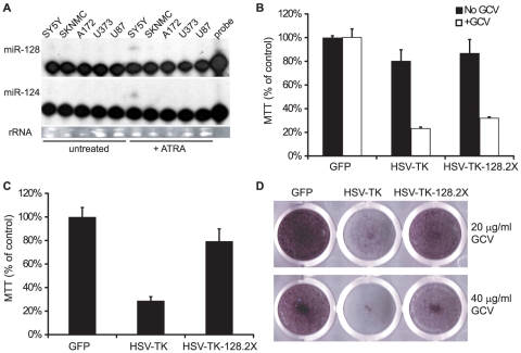
- Glioblastoma cells transduced with HSV-TK-128.2X were selectively killed by ganciclovir, and ectopic or endogenous miR-128 rescued cells; at 200 µg/mL ganciclovir, ∼70% of HSV-TK-expressing cells were killed.
Methods
- Small RNA libraries from six adult glioblastomas and three non-tumor brain tissues were sequenced on an Illumina GAII analyzer; 18–25 nt small RNAs were aligned to miRBase v16.0 using miRNAkey with SeqEM, filtered at RPKM >1, and analyzed for differential expression by t test and FDR adjustment.
- Primer extension validated expression of miR-21, miR-128, miR-124, and miR-132 in brain tissue samples and glioblastoma cell lines.
- HSV-TK vectors with two miR-128 target sites in the 3′UTR were transfected or lentivirally transduced into glioblastoma and neuroblastoma cell lines; cells were treated with ganciclovir, and viability was measured by crystal violet staining and MTT assays, with SH-SY5Y neuronal differentiation induced by all-trans retinoic acid.
Limitations
- Stated: high-throughput sequencing has limits; the standard FDR-adjusted p<0.05 cutoff identified only nine dysregulated miRNAs, so the authors expanded to p<0.1 to include 21, noting that additional miRNAs are likely dysregulated.
- Evident from the text: the sequencing analysis used a small sample set of six glioblastomas and three non-tumor brain tissues.
- Evident from the text: the therapeutic experiments were in vitro only, using cell lines and differentiated SH-SY5Y cells, with no in vivo tumor model or clinical validation reported.
Model-written summary. Check it against the paper before relying on it.
Integrity signals
Registry
Cites
Statistics
Figures
Verification
Claims the paper makes
67 Own result · 15 Not checked yet · 15 Own interpretation · 9 Partly supported · 9 No full text of the sources · 5 Own method · 2 Supported so far · 1 Not supported · 1 No basis given · accuracy not yet measured
The cited sources support only part of this claim — read each source’s verdict below.
In the paper
“In contrast to microarrays and RT-PCR arrays, high-throughput sequencing permits the detection of not only the dominant mature miRNA species (i.e. 5p versus 3p) but also potentially important sequence variations that may occur, such as 5′ nucleotide additions, which affect seed-based targeting, and RNA editing, which alters the targeting capacity of the mature miRNA [24]–[27].”
§ Reduced Expression of Brain-Enriched microRNAs in Glioblastomas Permits Targeted Regulation of a Cell Death Gene > Introduction
What the sources say
- Partly supportedcites [24] K Nishikura, 2010, Functions and regulation of RNA editing by ADAR deaminases., Annu Rev Biochem sourceExperimental — accuracy not yet measured
The section “Redirection of Silencing Target Genes by Edited miRNAs” reports that editing a mature miRNA’s seed sequence can redirect it to different targets. The copy also identifies edited mature miRNAs, but it does not establish the claimed advantage of high-throughput sequencing over microarrays or RT-PCR arrays, detection of the dominant 5p versus 3p species, or detection and targeting effects of 5′ nucleotide additions. (LLM, conf=0.85)
- Not checked — no full text of the sourcecites [25] Y Kawahara, 2007, Redirection of silencing targets by adenosine-to-inosine editing of miRNAs., Science source
- Not checked — no full text of the sourcecites [26] BP Lewis, 2005, Conserved seed pairing, often flanked by adenosines, indicates that thousands of human genes are microRNA targets., Cell source
- Partly supportedcites [27] H Seitz, 2008, Argonaute loading improves the 5′ precision of both MicroRNAs and their miRNA strands in flies., Curr Biol sourceExperimental — accuracy not yet measured
The Results and Discussion report that high-throughput sequencing identified miRNA and miRNA∗ reads and detected nontemplated additions at the 5′ end. The Computational Methods describe identifying reads from both strands. Our copy does not address RNA editing or compare sequencing with microarrays and RT-PCR arrays, so those parts of the citing sentence are not supported by this source. (LLM, conf=0.86)
plaindirect- Asserted: passes — The authors put it forward as true.
- Checkable: passes — It can be checked against evidence.
- Load-bearing: passes — The paper’s conclusions depend on it.
- Based: passes — The paper gives a reason why it holds.
- Partly supported
The cited sources support only part of this claim — read each source’s verdict below.
In the paper
“Congruent with previous studies [42], [43], ATRA treatment of SH-SY5Y cells induced expression of the brain-enriched miRNAs miR-128 and miR-124 (Fig. 5A) as well as miR-132 and miR-7 (not shown).”
§ Reduced Expression of Brain-Enriched microRNAs in Glioblastomas Permits Targeted Regulation of a Cell Death Gene > Results > Endogenous miR-128 protects neuronal cells from HSV-TK induced cell death
What the sources say
- Partly supportedcites [42] S Meseguer, MicroRNAs-10a and -10b Contribute to Retinoic Acid-induced Differentiation of Neuroblastoma Cells and Target the Alternative Splicing Regulatory Factor SFRS1 (SF2/ASF)., J Biol Chem sourceExperimental — accuracy not yet measured
The Results (page unavailable) report that retinoic acid treatment of SH-SY5Y cells up-regulated miR-132. Our copy does not report treatment-induced expression of miR-128, miR-124, or miR-7; it mentions miR-124a only as a brain-specific miRNA, not as a treatment result. (LLM, conf=0.85)
- Partly supportedcites [43] MT Le, 2009, MicroRNA-125b promotes neuronal differentiation in human cells by repressing multiple targets., Mol Cell Biol sourceExperimental — accuracy not yet measured
The section “Profiling miRNA expression in SH-SY5Y cells during differentiation” reports increased miR-124a and miR-7, but does not identify miR-128 or miR-132 among the six miRNAs consistently upregulated. It measured cells after RA treatment and after subsequent BDNF treatment, so the passage also does not establish that ATRA alone induced all four miRNAs. Page numbers are unavailable in our copy. (LLM, conf=0.88)
plaindirectn = 2- Asserted: passes — The authors put it forward as true.
- Checkable: passes — It can be checked against evidence.
- Load-bearing: passes — The paper’s conclusions depend on it.
- Based: passes — The paper gives a reason why it holds.
- Partly supported
A result the paper reports from its own data — it cites no other source for it, so there is none to check. The quote shows it is in the paper.
In the paper
“The most abundant miRNAs in both glioblastomas and non-tumor brain tissues were members of the let-7 family, which accounted for ∼23% of the total reads (Table S2, S3).”
§ Reduced Expression of Brain-Enriched microRNAs in Glioblastomas Permits Targeted Regulation of a Cell Death Gene > Results > Small RNA deep sequencing reveals differences in human miRNA expression patterns
plainn = 3- Asserted: passes — The authors put it forward as true.
- Checkable: passes — It can be checked against evidence.
- Load-bearing: unclear — It is unclear whether the paper’s conclusions depend on it.
- Based: passes — The paper gives a reason why it holds.
A result the paper reports from its own data — it cites no other source for it, so there is none to check. The quote shows it is in the paper.
In the paper
“Post-transcriptional editing was observed for several miRNAs, including the miR-376 family, miR-411, miR-381, and miR-379.”
§ Reduced Expression of Brain-Enriched microRNAs in Glioblastomas Permits Targeted Regulation of a Cell Death Gene > Abstract
plain- Asserted: passes — The authors put it forward as true.
- Checkable: passes — It can be checked against evidence.
- Load-bearing: unclear — It is unclear whether the paper’s conclusions depend on it.
- Based: passes — The paper gives a reason why it holds.
A result the paper reports from its own data — it cites no other source for it, so there is none to check. The quote shows it is in the paper.
In the paper
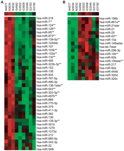
“we therefore used a FDR cut-off of p<0.1. 21 aberrantly expressed miRNAs met the criteria (Fig. 1, Table S5, Table S6, Fig. S2).”
§ Reduced Expression of Brain-Enriched microRNAs in Glioblastomas Permits Targeted Regulation of a Cell Death Gene > Results > Small RNA deep sequencing reveals differences in human miRNA expression patterns
plain- Asserted: passes — The authors put it forward as true.
- Checkable: passes — It can be checked against evidence.
- Load-bearing: passes — The paper’s conclusions depend on it.
- Based: passes — The paper gives a reason why it holds.
A result the paper reports from its own data — it cites no other source for it, so there is none to check. The quote shows it is in the paper.
In the paper
“Our analysis identified at least 20 miRNAs and miRNA star strands (miRNA*) that are significantly differentially expressed.”
§ Reduced Expression of Brain-Enriched microRNAs in Glioblastomas Permits Targeted Regulation of a Cell Death Gene > Introduction
plainn = 6- Asserted: passes — The authors put it forward as true.
- Checkable: passes — It can be checked against evidence.
- Load-bearing: passes — The paper’s conclusions depend on it.
- Based: passes — The paper gives a reason why it holds.
A result the paper reports from its own data — it cites no other source for it, so there is none to check. The quote shows it is in the paper.
In the paper
“miR-21, miR-9, miR-26a, miR-181a, and miR-125b were also abundantly detected and, together with let-7 (a-i) family members, made up >65% of all identified miRNAs (Table S2, S3).”
§ Reduced Expression of Brain-Enriched microRNAs in Glioblastomas Permits Targeted Regulation of a Cell Death Gene > Results > Small RNA deep sequencing reveals differences in human miRNA expression patterns
plainn = 6- Asserted: passes — The authors put it forward as true.
- Checkable: passes — It can be checked against evidence.
- Load-bearing: unclear — It is unclear whether the paper’s conclusions depend on it.
- Based: passes — The paper gives a reason why it holds.
A result the paper reports from its own data — it cites no other source for it, so there is none to check. The quote shows it is in the paper.
In the paper
“From this set of 449, 68 miRNAs and miRNA*s were differentially expressed between non-tumor brain tissues and glioblastomas (p<0.05, Student's t test; Table S6).”
§ Reduced Expression of Brain-Enriched microRNAs in Glioblastomas Permits Targeted Regulation of a Cell Death Gene > Results > Small RNA deep sequencing reveals differences in human miRNA expression patterns
plaincorrelational- Asserted: passes — The authors put it forward as true.
- Checkable: passes — It can be checked against evidence.
- Load-bearing: passes — The paper’s conclusions depend on it.
- Based: passes — The paper gives a reason why it holds.
A result the paper reports from its own data — it cites no other source for it, so there is none to check. The quote shows it is in the paper.
In the paper
“When we applied the standard FDR-adjusted p-value <0.05 cut-off, only nine miRNAs were identified that met the criteria (Table S5).”
§ Reduced Expression of Brain-Enriched microRNAs in Glioblastomas Permits Targeted Regulation of a Cell Death Gene > Results > Small RNA deep sequencing reveals differences in human miRNA expression patterns
plain- Asserted: passes — The authors put it forward as true.
- Checkable: passes — It can be checked against evidence.
- Load-bearing: passes — The paper’s conclusions depend on it.
- Based: passes — The paper gives a reason why it holds.
A result the paper reports from its own data — it cites no other source for it, so there is none to check. The quote shows it is in the paper.
In the paper
“MicroRNAs (miRNAs) made up the large majority of small RNAs, and we identified over 400 different cellular pre-miRNAs.”
§ Reduced Expression of Brain-Enriched microRNAs in Glioblastomas Permits Targeted Regulation of a Cell Death Gene > Abstract
plain- Asserted: passes — The authors put it forward as true.
- Checkable: passes — It can be checked against evidence.
- Load-bearing: unclear — It is unclear whether the paper’s conclusions depend on it.
- Based: passes — The paper gives a reason why it holds.
- Probably not a substantial claim. 43
- It may state two separate claims as one. 32
- The quote only partly states this claim. 2
At least one cited source doesn’t support this claim or points to a different paper, and none confirmed it — see the verdicts below.
In the paper
“In fact, individual miRNAs are predicted to target as many as 200 different mRNAs [12].”
§ Reduced Expression of Brain-Enriched microRNAs in Glioblastomas Permits Targeted Regulation of a Cell Death Gene > Introduction
What the sources say
- Disputedcites [12] RC Friedman, 2009, Most mammalian mRNAs are conserved targets of microRNAs., Genome Res sourceExperimental — accuracy not yet measured
The Results and Discussion section estimates 438 ± 60 or 497 ± 49 conserved targets per miRNA family, rather than an upper figure of 200 mRNAs. The widespread scope of conserved targeting section also estimates 292 ± 18 targets per broadly conserved miRNA even when considering only 7–8mer sites. (LLM, conf=0.84)
Checked by a reasoning model — may be wrong.
plain- Asserted: passes — The authors put it forward as true.
- Checkable: passes — It can be checked against evidence.
- Load-bearing: fails — The paper’s conclusions may not depend on it.
- Based: passes — The paper gives a reason why it holds.
Probably not a substantial claim. The paper’s conclusions may not depend on it.
- Disputed
The cited sources support only part of this claim — read each source’s verdict below.
In the paper
“miR-128, miR-124, and miR-137 are all enriched in the brain and have been shown to regulate neuronal differentiation, maturation, and/or survival [15], [20]–[23].”
§ Reduced Expression of Brain-Enriched microRNAs in Glioblastomas Permits Targeted Regulation of a Cell Death Gene > Introduction
What the sources say
- Partly supportedcites [15] J Silber, 2008, miR-124 and miR-137 inhibit proliferation of glioblastoma multiforme cells and induce differentiation of brain tumor stem cells., BMC Med sourceExperimental — accuracy not yet measured
The Results section reports that miR-124 and miR-137 promote neuronal differentiation in adult neural stem cells. Its expression table also compares miR-128a with non-neoplastic brain tissue, but the paper does not show that miR-128 regulates neuronal differentiation, maturation, or survival, or establish that all three are enriched in the brain relative to other tissues. Page numbers are unavailable in our copy. (LLM, conf=0.87)
- Partly supportedcites [20] LF Sempere, 2004, Expression profiling of mammalian microRNAs uncovers a subset of brain-expressed microRNAs with possible roles in murine and human neuronal differentiation., Genome Biol sourceExperimental — accuracy not yet measured
The Results and discussion section lists miR-128, miR-124a/b, and miR-137 among miRNAs expressed in the brain. It also reports that miR-128 and miR-124a/b increase during neuronal differentiation in cultured cells. The paper suggests a possible role in neuronal differentiation, but does not show that these miRNAs regulate differentiation, maturation, or survival; it does not report miR-137 increasing in that experiment. Page numbers are unavailable in our copy. (LLM, conf=0.90)
- Not checked — no full text of the sourcecites [21] J Godlewski, 2010, MicroRNAs and glioblastoma; the stem cell connection., Cell Death Differ source
- Supportedcites [22] C Evangelisti, 2009, MiR-128 up-regulation inhibits Reelin and DCX expression and reduces neuroblastoma cell motility and invasiveness., FASEB J source
The Abstract identifies miR-128 as brain-enriched. The Discussion links miR-128 to differentiation and reports that its forced expression produces effects associated with retinoic-acid-induced differentiation, including reduced migration. In this multi-citation sentence, this paper supports the miR-128 portion of the claim. (LLM, conf=0.86)
- Partly supportedcites [23] C Conaco, 2006, Reciprocal actions of REST and a microRNA promote neuronal identity., Proc Natl Acad Sci U S A sourceExperimental — accuracy not yet measured
The Results report that miR-124a is highly enriched in the brain, and the Results and Discussion link it to suppression of nonneuronal transcripts and maintenance of neuronal identity. The passages found for miR-128 and miR-137 do not address their brain enrichment or roles in neuronal differentiation, maturation, or survival, so this source supports only the miR-124 portion of the claim. Page numbers are unavailable in our copy. (LLM, conf=0.88)
plaindirect- Asserted: passes — The authors put it forward as true.
- Checkable: passes — It can be checked against evidence.
- Load-bearing: passes — The paper’s conclusions depend on it.
- Based: passes — The paper gives a reason why it holds.
It may state two separate claims as one.
- Partly supported
The sources with a verdict support this claim; the rest weren’t checked yet, have no full text we could read, or their verdicts aren’t rated yet.
In the paper
“the pro-oncogenic miRNA miR-10b is upregulated in glioblastomas and has recently been shown to be a significant contributor to tumor growth in vivo [9], [17].”
§ Reduced Expression of Brain-Enriched microRNAs in Glioblastomas Permits Targeted Regulation of a Cell Death Gene > Introduction
What the sources say
- Not checked — no full text of the sourcecites [9] SA Ciafre, 2005, Extensive modulation of a set of microRNAs in primary glioblastoma., Biochem Biophys Res Commun source
- Supportedcites [17] G Gabriely, 2011, Human Glioma Growth Is Controlled by MicroRNA-10b., Cancer Res source
The Introduction says miR-10b is strongly upregulated in glioma. The Abstract reports that inhibiting miR-10b significantly reduced tumor growth in a mouse model of human glioma, supporting its contribution to growth in vivo. (LLM, conf=0.94)
plaindirect- Asserted: passes — The authors put it forward as true.
- Checkable: passes — It can be checked against evidence.
- Load-bearing: fails — The paper’s conclusions may not depend on it.
- Based: passes — The paper gives a reason why it holds.
Probably not a substantial claim. The paper’s conclusions may not depend on it. It may state two separate claims as one.
The sources with a verdict support this claim; the rest weren’t checked yet, have no full text we could read, or their verdicts aren’t rated yet.
In the paper
“Previous miRNA profiling studies, based on miRNA microarrays and PCR arrays, have revealed a number of miRNAs to be altered in gliomas [9], [14]–[16].”
§ Reduced Expression of Brain-Enriched microRNAs in Glioblastomas Permits Targeted Regulation of a Cell Death Gene > Introduction
What the sources say
- Not checked — no full text of the sourcecites [9] SA Ciafre, 2005, Extensive modulation of a set of microRNAs in primary glioblastoma., Biochem Biophys Res Commun source
- Not checked — no full text of the sourcecites [14] J Godlewski, 2008, Targeting of the Bmi-1 oncogene/stem cell renewal factor by microRNA-128 inhibits glioma proliferation and self-renewal., Cancer Res source
- Supportedcites [15] J Silber, 2008, miR-124 and miR-137 inhibit proliferation of glioblastoma multiforme cells and induce differentiation of brain tumor stem cells., BMC Med source
The Results section reports that this study used quantitative RT-PCR to profile miRNAs in gliomas and found altered expression, including changes consistent with earlier studies. The Discussion identifies microarrays as the technology used in previous GBM profiling studies, supporting the claim's description of both approaches. (LLM, conf=0.91)
- Supportedcites [16] JT Huse, 2009, The PTEN-regulating microRNA miR-26a is amplified in high-grade glioma and facilitates gliomagenesis in vivo., Genes Dev source
In “miR-26a is overexpressed in high-grade glioma and directly targets PTEN,” the authors report using microarray analysis to identify 29 miRNAs elevated in glioblastoma samples relative to controls. This paper is therefore an example of the miRNA-profiling studies cited in the sentence; the sentence does not require every cited study to have used both microarrays and PCR arrays. (LLM, conf=0.90)
plaincorrelational- Asserted: passes — The authors put it forward as true.
- Checkable: passes — It can be checked against evidence.
- Load-bearing: fails — The paper’s conclusions may not depend on it.
- Based: passes — The paper gives a reason why it holds.
Probably not a substantial claim. The paper’s conclusions may not depend on it.
The cited sources support only part of this claim — read each source’s verdict below.
In the paper
“Furthermore, several of these miRNA variants (miR-376 family members, miR-411, miR-379, and miR-320a) have previously been described in deep sequencing studies performed on human brain [36], [37], and therefore, are not specific to glioblastoma.”
§ Reduced Expression of Brain-Enriched microRNAs in Glioblastomas Permits Targeted Regulation of a Cell Death Gene > Results > miRNA editing and miRNA sequence variants in the brain
What the sources say
- Partly supportedcites [36] Y Kawahara, 2008, Frequency and fate of microRNA editing in human brain., Nucleic Acids Res sourceExperimental — accuracy not yet measured
In Results and Discussion (‘Expression of edited mature miRNAs’; page not provided), the paper identifies edited miR-379-5p and miR-411-5p in human brain and discusses edited miR-376 family members. This supports those variants occurring outside glioblastoma, but our copy does not identify a miR-320a variant. The paper describes its own approach as targeted cloning, not deep sequencing; its discussion of a deep-sequencing survey concerns a separate study. (LLM, conf=0.88)
- Partly supportedcites [37] E Marti, A myriad of miRNA variants in control and Huntington's disease brain regions detected by massively parallel sequencing., Nucleic Acids Res sourceExperimental — accuracy not yet measured
The Results section ('miRNA variability is ubiquitous in control and HD brain samples'; page not available) reports variants of miR-376a/b/c, miR-411, and miR-379 in human brain samples, supporting the point that those variants are not specific to glioblastoma. It discusses miR-320b and miR-320c, however, rather than the claimed miR-320a; searching our copy for miR-320a did not establish that part of the list. (LLM, conf=0.86)
plain- Asserted: passes — The authors put it forward as true.
- Checkable: passes — It can be checked against evidence.
- Load-bearing: unclear — It is unclear whether the paper’s conclusions depend on it.
- Based: passes — The paper gives a reason why it holds.
It may state two separate claims as one.
- Partly supported
The cited sources support only part of this claim — read each source’s verdict below.
In the paper
“Similarly, CREB-induced miR-132 and miR-212, which share seed sequence homology, promote dendritic outgrowth from newborn neurons [55] and can target methyl CpG binding protein 2 (MeCP2), a regulator of neuroplasticity [56], [57].”
§ Reduced Expression of Brain-Enriched microRNAs in Glioblastomas Permits Targeted Regulation of a Cell Death Gene > Discussion
What the sources say
- Partly supportedcites [55] ST Magill, microRNA-132 regulates dendritic growth and arborization of newborn neurons in the adult hippocampus., Proc Natl Acad Sci U S A sourceExperimental — accuracy not yet measured
The Results state that miR-132 and miR-212 have identical seed sequences, and the Discussion concludes that miR-132 mediates dendritic outgrowth in newborn hippocampal neurons. But the Results identify miR-132, not miR-212, as active in those neurons, so the paper does not support attributing that effect to both. The MeCP2 statement has separate citations [56] and [57], rather than this paper's citation [55]. (LLM, conf=0.88)
- Not checked — no full text of the sourcecites [56] ME Klein, 2007, Homeostatic regulation of MeCP2 expression by a CREB-induced microRNA., Nat Neurosci source
- Supportedcites [57] HI Im, MeCP2 controls BDNF expression and cocaine intake through homeostatic interactions with microRNA-212., Nat Neurosci source
The Results section (‘miR-212 inhibits striatal MeCP2 expression’) reports that miR-212 reduces MeCP2 expression and notes that miR-132 regulates a MeCP2 transcript. The Discussion (‘MeCP2 acts through homeostatic interactions with miR-212’) likewise describes interactions between MeCP2 and both miRNAs. The dendritic-outgrowth clause is attached to a different citation, [55], and is not judged here. (LLM, conf=0.91)
plaindirect- Asserted: passes — The authors put it forward as true.
- Checkable: passes — It can be checked against evidence.
- Load-bearing: fails — The paper’s conclusions may not depend on it.
- Based: passes — The paper gives a reason why it holds.
Probably not a substantial claim. The paper’s conclusions may not depend on it. It may state two separate claims as one.
- Partly supported
The cited sources support only part of this claim — read each source’s verdict below.
In the paper
“Brain-enriched miR-124 and miR-7, for example, show poor expression in neural progenitors, but are highly expressed in neurons and have been implicated in neural differentiation [20], [23], [54].”
§ Reduced Expression of Brain-Enriched microRNAs in Glioblastomas Permits Targeted Regulation of a Cell Death Gene > Discussion
What the sources say
- Partly supportedcites [20] LF Sempere, 2004, Expression profiling of mammalian microRNAs uncovers a subset of brain-expressed microRNAs with possible roles in murine and human neuronal differentiation., Genome Biol sourceExperimental — accuracy not yet measured
In Results and discussion (‘Diverse miRNA expression patterns in mammalian organs’), the paper lists both miR-7 and miR-124 among brain-expressed miRNAs. In ‘A subset of brain-expressed miRNAs is induced in retinoic acid-treated EC cells’, it reports that miR-124 increases during neuronal differentiation, but miR-7 is not among the induced miRNAs. The paper does not directly compare expression in neural progenitors and neurons, so it does not establish that full contrast for both miRNAs. (LLM, conf=0.87)
- Partly supportedcites [23] C Conaco, 2006, Reciprocal actions of REST and a microRNA promote neuronal identity., Proc Natl Acad Sci U S A sourceExperimental — accuracy not yet measured
The Results (page not available) report that miR-124a was barely detectable in dividing cortical progenitors, appeared in mature cortical neurons, and was associated with neuronal differentiation. The passages found do not establish the corresponding expression pattern or differentiation role for miR-7; a search for miR-7 found no passage addressing it. (LLM, conf=0.88)
- Partly supportedcites [54] X Li, 2005, A microRNA mediates EGF receptor signaling and promotes photoreceptor differentiation in the Drosophila eye., Cell sourceExperimental — accuracy not yet measured
The Results report miR-7 in differentiating Drosophila photoreceptor neurons and show that miR-7 stimulates photoreceptor differentiation. The paper does not establish the sentence’s claims about miR-124, brain enrichment, or neuronal expression beyond this photoreceptor setting. (LLM, conf=0.86)
plaincorrelational- Asserted: passes — The authors put it forward as true.
- Checkable: passes — It can be checked against evidence.
- Load-bearing: passes — The paper’s conclusions depend on it.
- Based: passes — The paper gives a reason why it holds.
It may state two separate claims as one.
- Partly supported
The cited sources support only part of this claim — read each source’s verdict below.
In the paper
“HSV-TK-based lentiviral vectors are currently in phase I/II clinical trials for glioblastoma treatment [39], [40].”
§ Reduced Expression of Brain-Enriched microRNAs in Glioblastomas Permits Targeted Regulation of a Cell Death Gene > Results > miRNAs as potential therapeutic tools to regulate cell suicide gene expression
What the sources say
- Partly supportedcites [39] KJ Pulkkanen, 2005, Gene therapy for malignant glioma: current clinical status., Mol Ther sourceExperimental — accuracy not yet measured
The review describes HSV-tk clinical trials for malignant glioma, including phase I adenoviral studies and a phase IIb adenoviral study (Abstract; page unavailable). It does not identify an HSV-tk lentiviral phase I/II trial; its discussion of lentiviral vectors says they had mainly been used in preclinical malignant-glioma studies (Abstract; page unavailable). (LLM, conf=0.87)
- Not checked — no full text of the sourcecites [40] NG Rainov, 2003, Gene therapy for human malignant brain tumors., Cancer J source
plain- Asserted: passes — The authors put it forward as true.
- Checkable: passes — It can be checked against evidence.
- Load-bearing: fails — The paper’s conclusions may not depend on it.
- Based: passes — The paper gives a reason why it holds.
Probably not a substantial claim. The paper’s conclusions may not depend on it.
- Partly supported
The cited sources support only part of this claim — read each source’s verdict below.
In the paper
“which could have dramatic consequences on mRNA targeting [25], [27].”
§ Reduced Expression of Brain-Enriched microRNAs in Glioblastomas Permits Targeted Regulation of a Cell Death Gene > Discussion
What the sources say
- Not checked — no full text of the sourcecites [25] Y Kawahara, 2007, Redirection of silencing targets by adenosine-to-inosine editing of miRNAs., Science source
- Partly supportedcites [27] H Seitz, 2008, Argonaute loading improves the 5′ precision of both MicroRNAs and their miRNA strands in flies., Curr Biol sourceExperimental — accuracy not yet measured
The Results and Discussion (page not provided) describe variation in miRNA 5′ ends and suggest that different seed sequences may affect binding to target RNAs. This supports the possibility of targeting consequences from 5′-end polymorphisms. The passages do not address A-to-I editing or its consequences for targeting. (LLM, conf=0.84)
hedgedconditional- Asserted: passes — The authors put it forward as true.
- Checkable: passes — It can be checked against evidence.
- Load-bearing: fails — The paper’s conclusions may not depend on it.
- Based: passes — The paper gives a reason why it holds.
Probably not a substantial claim. The paper’s conclusions may not depend on it.
The cited sources support only part of this claim — read each source’s verdict below.
In the paper
“When cultured in the presence of all-trans retinoic acid (ATRA), SH-SY5Y cells undergo morphological changes, differentiate into neuron-like cells, express neuronal markers, and up-regulate a number of brain-specific miRNAs [42], [43].”
§ Reduced Expression of Brain-Enriched microRNAs in Glioblastomas Permits Targeted Regulation of a Cell Death Gene > Results > Endogenous miR-128 protects neuronal cells from HSV-TK induced cell death
What the sources say
- Partly supportedcites [42] S Meseguer, MicroRNAs-10a and -10b Contribute to Retinoic Acid-induced Differentiation of Neuroblastoma Cells and Target the Alternative Splicing Regulatory Factor SFRS1 (SF2/ASF)., J Biol Chem sourceExperimental — accuracy not yet measured
The Results report that retinoic acid treatment of SH-SY5Y cells up-regulated 26 miRNAs. The Figure 2 caption describes RA-induced morphological differentiation and neurite outgrowth, and the neuronal-differentiation section reports increased neuronal markers. However, the paper identifies only miR-132 among its listed up-regulated miRNAs as brain-specific; it does not establish that a number of brain-specific miRNAs were up-regulated. Page numbers were unavailable in our copy. (LLM, conf=0.86)
- Partly supportedcites [43] MT Le, 2009, MicroRNA-125b promotes neuronal differentiation in human cells by repressing multiple targets., Mol Cell Biol sourceExperimental — accuracy not yet measured
In “Profiling miRNA expression in SH-SY5Y cells during differentiation” (page unavailable), the paper reports morphological changes, neuronal-marker expression, and six miRNAs upregulated during differentiation. The claim omits a key condition: the paper’s differentiation protocol used five days of retinoic acid followed by seven days of BDNF, rather than ATRA alone. It also does not establish that all six upregulated miRNAs are brain-specific. (LLM, conf=0.86)
plaindirectn = 2- Asserted: passes — The authors put it forward as true.
- Checkable: passes — It can be checked against evidence.
- Load-bearing: passes — The paper’s conclusions depend on it.
- Based: passes — The paper gives a reason why it holds.
It may state two separate claims as one.
- Partly supported
A result the paper reports from its own data — it cites no other source for it, so there is none to check. The quote shows it is in the paper.
In the paper
“we identified over 400 different cellular pre-miRNAs”
§ Reduced Expression of Brain-Enriched microRNAs in Glioblastomas Permits Targeted Regulation of a Cell Death Gene > Abstract
plain- Asserted: passes — The authors put it forward as true.
- Checkable: passes — It can be checked against evidence.
- Load-bearing: fails — The paper’s conclusions may not depend on it.
- Based: passes — The paper gives a reason why it holds.
Probably not a substantial claim. The paper’s conclusions may not depend on it.
A result the paper reports from its own data — it cites no other source for it, so there is none to check. The quote shows it is in the paper.
In the paper
“No known viral miRNAs were detected in any of the samples analyzed.”
- Also stated in: § Small RNA deep sequencing reveals differences in human miRNA expression patterns · plain
“No viral miRNAs were detected in any of the samples analyzed.”
§ Reduced Expression of Brain-Enriched microRNAs in Glioblastomas Permits Targeted Regulation of a Cell Death Gene > Abstract
plainn = 3- Asserted: passes — The authors put it forward as true.
- Checkable: passes — It can be checked against evidence.
- Load-bearing: fails — The paper’s conclusions may not depend on it.
- Based: passes — The paper gives a reason why it holds.
Probably not a substantial claim. The paper’s conclusions may not depend on it.
- Also stated in: § Small RNA deep sequencing reveals differences in human miRNA expression patterns · plain
A result the paper reports from its own data — it cites no other source for it, so there is none to check. The quote shows it is in the paper.
In the paper
“Between 1.8 and 4.9 million reads were obtained per sample (Table S1)”
§ Reduced Expression of Brain-Enriched microRNAs in Glioblastomas Permits Targeted Regulation of a Cell Death Gene > Results > Small RNA deep sequencing reveals differences in human miRNA expression patterns
plain- Asserted: passes — The authors put it forward as true.
- Checkable: passes — It can be checked against evidence.
- Load-bearing: fails — The paper’s conclusions may not depend on it.
- Based: passes — The paper gives a reason why it holds.
Probably not a substantial claim. The paper’s conclusions may not depend on it.
A result the paper reports from its own data — it cites no other source for it, so there is none to check. The quote shows it is in the paper.
In the paper
“Among the 16,305,416 mapped reads, 484 mature human miRNAs/miRNA*s were represented at least 10 times (Table S2).”
§ Reduced Expression of Brain-Enriched microRNAs in Glioblastomas Permits Targeted Regulation of a Cell Death Gene > Results > Small RNA deep sequencing reveals differences in human miRNA expression patterns
plainn = 6- Asserted: passes — The authors put it forward as true.
- Checkable: passes — It can be checked against evidence.
- Load-bearing: fails — The paper’s conclusions may not depend on it.
- Based: passes — The paper gives a reason why it holds.
Probably not a substantial claim. The paper’s conclusions may not depend on it.
A result the paper reports from its own data — it cites no other source for it, so there is none to check. The quote shows it is in the paper.
In the paper
“the sequence length distributions showed peaks at ∼22–23 nt for each sample, corresponding to the average length of a mature miRNA (Fig. S1).”
§ Reduced Expression of Brain-Enriched microRNAs in Glioblastomas Permits Targeted Regulation of a Cell Death Gene > Results > Small RNA deep sequencing reveals differences in human miRNA expression patterns
plain- Asserted: passes — The authors put it forward as true.
- Checkable: passes — It can be checked against evidence.
- Load-bearing: fails — The paper’s conclusions may not depend on it.
- Based: passes — The paper gives a reason why it holds.
Probably not a substantial claim. The paper’s conclusions may not depend on it.
A result the paper reports from its own data — it cites no other source for it, so there is none to check. The quote shows it is in the paper.
In the paper
“under the regulation of a miRNA, miR-128, that was found to be enriched in non-tumor brain tissue yet down-regulated in glioblastomas”
§ Reduced Expression of Brain-Enriched microRNAs in Glioblastomas Permits Targeted Regulation of a Cell Death Gene > Abstract
plaincorrelational- Asserted: passes — The authors put it forward as true.
- Checkable: passes — It can be checked against evidence.
- Load-bearing: passes — The paper’s conclusions depend on it.
- Based: passes — The paper gives a reason why it holds.
It may state two separate claims as one.
A result the paper reports from its own data — it cites no other source for it, so there is none to check. The quote shows it is in the paper.
In the paper
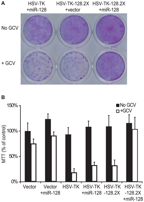
“Both the ectopic expression of miR-128 in glioblastoma cells and the natural, endogenous level of miR-128 expression seen in neuronal cells indeed limited cell death when compared to control glioblastoma cells.”
§ Reduced Expression of Brain-Enriched microRNAs in Glioblastomas Permits Targeted Regulation of a Cell Death Gene > Introduction
plaindirect- Asserted: passes — The authors put it forward as true.
- Checkable: passes — It can be checked against evidence.
- Load-bearing: passes — The paper’s conclusions depend on it.
- Based: passes — The paper gives a reason why it holds.
It may state two separate claims as one.
A claim is checked only against the cited paper’s full text, never its abstract, and none is available for its sources.
In the paper
“We also observed high read counts for miR-21, amongst other miRNAs, in all glioblastoma samples compared to non-tumor samples, congruent with previous studies showing that this miRNA is highly up-regulated in glioblastoma (Table S4) [5], [9], [14], [15]; however, there was variability in the values for miR-21 across the samples and thus, the miRNA did not meet the final FDR selection criteria.”
§ Reduced Expression of Brain-Enriched microRNAs in Glioblastomas Permits Targeted Regulation of a Cell Death Gene > Results > Small RNA deep sequencing reveals differences in human miRNA expression patterns
What the sources say
- Not checked — no full text of the sourcecites [5] JA Chan, 2005, MicroRNA-21 is an antiapoptotic factor in human glioblastoma cells., Cancer Res source
- Not checked — no full text of the sourcecites [9] SA Ciafre, 2005, Extensive modulation of a set of microRNAs in primary glioblastoma., Biochem Biophys Res Commun source
- Not checked — no full text of the sourcecites [14] J Godlewski, 2008, Targeting of the Bmi-1 oncogene/stem cell renewal factor by microRNA-128 inhibits glioma proliferation and self-renewal., Cancer Res source
plaincorrelational- Asserted: passes — The authors put it forward as true.
- Checkable: passes — It can be checked against evidence.
- Load-bearing: fails — The paper’s conclusions may not depend on it.
- Based: passes — The paper gives a reason why it holds.
Probably not a substantial claim. The paper’s conclusions may not depend on it. It may state two separate claims as one.
A result the paper reports from its own data — it cites no other source for it, so there is none to check. The quote shows it is in the paper.
In the paper
“These 5′ nucleotide variations were observed in all nine sequencing libraries, and with the exception of miR-124-3p, there was no correlation of 5′ end variability and miRNA expression differences in tumors versus non-tumor samples (Table S4, S7, and S8).”
§ Reduced Expression of Brain-Enriched microRNAs in Glioblastomas Permits Targeted Regulation of a Cell Death Gene > Results > miRNA editing and miRNA sequence variants in the brain
plain- Asserted: passes — The authors put it forward as true.
- Checkable: passes — It can be checked against evidence.
- Load-bearing: unclear — It is unclear whether the paper’s conclusions depend on it.
- Based: passes — The paper gives a reason why it holds.
It may state two separate claims as one.
A result the paper reports from its own data — it cites no other source for it, so there is none to check. The quote shows it is in the paper.
In the paper
“A to G substitutions for each of the miRNAs were found in both the glioblastoma and non-tumor brain tissue libraries, and there were no significant differences in the averaged abundance of edited miRNAs between the two sample types (Table 1).”
§ Reduced Expression of Brain-Enriched microRNAs in Glioblastomas Permits Targeted Regulation of a Cell Death Gene > Results > miRNA editing and miRNA sequence variants in the brain
plain- Asserted: passes — The authors put it forward as true.
- Checkable: passes — It can be checked against evidence.
- Load-bearing: unclear — It is unclear whether the paper’s conclusions depend on it.
- Based: passes — The paper gives a reason why it holds.
It may state two separate claims as one.
- Rests on an assumption: In deep sequencing libraries, A to I editing is marked by the substitution of A with G.
A result the paper reports from its own data — it cites no other source for it, so there is none to check. The quote shows it is in the paper.
In the paper
“We therefore expanded our cut-off to p<0.1 to include 21 significantly dysregulated miRNAs, 17 of which were down-regulated in glioblastoma.”
§ Reduced Expression of Brain-Enriched microRNAs in Glioblastomas Permits Targeted Regulation of a Cell Death Gene > Discussion
plain- Asserted: passes — The authors put it forward as true.
- Checkable: passes — It can be checked against evidence.
- Load-bearing: passes — The paper’s conclusions depend on it.
- Based: passes — The paper gives a reason why it holds.
It may state two separate claims as one.
A result the paper reports from its own data — it cites no other source for it, so there is none to check. The quote shows it is in the paper.
In the paper
“Interestingly, the dominant form of miR-324-3p was consistent with the miRNA sequence reported in miRBase; however, in non-tumor brain, we observed a significant number of reads mapping to a miR-324-3p isoform that was two nucleotides longer at the 5′ end (Table S7).”
§ Reduced Expression of Brain-Enriched microRNAs in Glioblastomas Permits Targeted Regulation of a Cell Death Gene > Results > miRNA editing and miRNA sequence variants in the brain
plain- Asserted: passes — The authors put it forward as true.
- Checkable: passes — It can be checked against evidence.
- Load-bearing: fails — The paper’s conclusions may not depend on it.
- Based: passes — The paper gives a reason why it holds.
Probably not a substantial claim. The paper’s conclusions may not depend on it.
A result the paper reports from its own data — it cites no other source for it, so there is none to check. The quote shows it is in the paper.
In the paper
“Several of the miRNAs down-regulated in glioblastoma (including miR-124-3p and miR-323-3p) exhibited 5′ truncations or additions while one up-regulated miRNA, miR-10b-5p, exhibited 5′ variations (Table S8).”
§ Reduced Expression of Brain-Enriched microRNAs in Glioblastomas Permits Targeted Regulation of a Cell Death Gene > Results > miRNA editing and miRNA sequence variants in the brain
plaincorrelational- Asserted: passes — The authors put it forward as true.
- Checkable: passes — It can be checked against evidence.
- Load-bearing: unclear — It is unclear whether the paper’s conclusions depend on it.
- Based: passes — The paper gives a reason why it holds.
It may state two separate claims as one.
None of the sources cited for this claim have been checked yet.
In the paper
“Congruent with previous studies, A to G changes occurred in >60% of the reads for members of the miR-376 family (miR-376a, b, and c) at nucleotide position six of the mature miRNA sequence (Table 1, Fig. S3A) [36].”
§ Reduced Expression of Brain-Enriched microRNAs in Glioblastomas Permits Targeted Regulation of a Cell Death Gene > Results > miRNA editing and miRNA sequence variants in the brain
What the sources say
- Not checked yetcites [36] Y Kawahara, 2008, Frequency and fate of microRNA editing in human brain., Nucleic Acids Res source
Checked before — its earlier verdict is held back until a new check runs.
plain- Asserted: passes — The authors put it forward as true.
- Checkable: passes — It can be checked against evidence.
- Load-bearing: fails — The paper’s conclusions may not depend on it.
- Based: passes — The paper gives a reason why it holds.
Probably not a substantial claim. The paper’s conclusions may not depend on it.
- Rests on an assumption: In deep sequencing libraries, A to I editing is marked by the substitution of A with G.
- Not checked yet
A result the paper reports from its own data — it cites no other source for it, so there is none to check. The quote shows it is in the paper.
In the paper
“The 3p miRNAs for miR-330 and miR-204 were preferentially detected in non-tumor brain tissue, while the 5p miRNAs were preferentially detected in glioblastomas (Table S7; Fig. S3B).”
§ Reduced Expression of Brain-Enriched microRNAs in Glioblastomas Permits Targeted Regulation of a Cell Death Gene > Results > Variations in 5p and 3p strand dominance
plaincorrelationaln = 9- Asserted: passes — The authors put it forward as true.
- Checkable: passes — It can be checked against evidence.
- Load-bearing: unclear — It is unclear whether the paper’s conclusions depend on it.
- Based: passes — The paper gives a reason why it holds.
It may state two separate claims as one.
A result the paper reports from its own data — it cites no other source for it, so there is none to check. The quote shows it is in the paper.
In the paper
“the data presented here provide a comprehensive analysis of the miRNA signature in glioblastoma”
§ Reduced Expression of Brain-Enriched microRNAs in Glioblastomas Permits Targeted Regulation of a Cell Death Gene > Discussion
plain- Asserted: passes — The authors put it forward as true.
- Checkable: fails — It may be too vague to check against evidence.
- Load-bearing: passes — The paper’s conclusions depend on it.
- Based: passes — The paper gives a reason why it holds.
Probably not a substantial claim. It may be too vague to check against evidence.
A result the paper reports from its own data — it cites no other source for it, so there is none to check. The quote shows it is in the paper.
In the paper
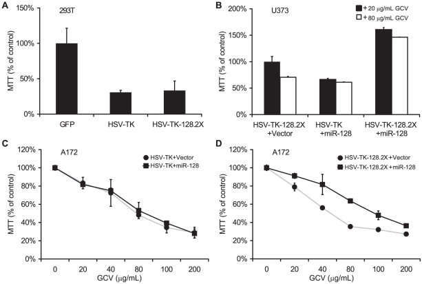
“At the highest dose, 200 µg/mL GCV, ∼70% of HSV-TK-expressing cells were killed (Fig. 4C & D).”
§ Reduced Expression of Brain-Enriched microRNAs in Glioblastomas Permits Targeted Regulation of a Cell Death Gene > Results > Endogenous miR-128 protects neuronal cells from HSV-TK induced cell death
plaindirect- Asserted: passes — The authors put it forward as true.
- Checkable: passes — It can be checked against evidence.
- Load-bearing: unclear — It is unclear whether the paper’s conclusions depend on it.
- Based: passes — The paper gives a reason why it holds.
The quote only partly states this claim.
A result the paper reports from its own data — it cites no other source for it, so there is none to check. The quote shows it is in the paper.
In the paper
“we report a number of miRNA sequence variants that are expressed in the brain.”
§ Reduced Expression of Brain-Enriched microRNAs in Glioblastomas Permits Targeted Regulation of a Cell Death Gene > Discussion
plain- Asserted: passes — The authors put it forward as true.
- Checkable: passes — It can be checked against evidence.
- Load-bearing: fails — The paper’s conclusions may not depend on it.
- Based: passes — The paper gives a reason why it holds.
Probably not a substantial claim. The paper’s conclusions may not depend on it.
A result the paper reports from its own data — it cites no other source for it, so there is none to check. The quote shows it is in the paper.
In the paper
“A to G substitutions also occurred in the seed region of miR-320 family members; however, these miRNAs were unusual in that several positions (two, three, and four) exhibited nucleotide substitutions (Fig. S3A and data not shown).”
§ Reduced Expression of Brain-Enriched microRNAs in Glioblastomas Permits Targeted Regulation of a Cell Death Gene > Results > miRNA editing and miRNA sequence variants in the brain
plain- Asserted: passes — The authors put it forward as true.
- Checkable: passes — It can be checked against evidence.
- Load-bearing: fails — The paper’s conclusions may not depend on it.
- Based: passes — The paper gives a reason why it holds.
Probably not a substantial claim. The paper’s conclusions may not depend on it.
- Rests on an assumption: In deep sequencing libraries, A to I editing is marked by the substitution of A with G.
A result the paper reports from its own data — it cites no other source for it, so there is none to check. The quote shows it is in the paper.
In the paper
“Two major isoforms were observed for both miR-30a-3p and miR-330-3p.”
§ Reduced Expression of Brain-Enriched microRNAs in Glioblastomas Permits Targeted Regulation of a Cell Death Gene > Results > miRNA editing and miRNA sequence variants in the brain
plain- Asserted: passes — The authors put it forward as true.
- Checkable: passes — It can be checked against evidence.
- Load-bearing: fails — The paper’s conclusions may not depend on it.
- Based: passes — The paper gives a reason why it holds.
Probably not a substantial claim. The paper’s conclusions may not depend on it.
A result the paper reports from its own data — it cites no other source for it, so there is none to check. The quote shows it is in the paper.
In the paper
“miR-212, miR-218, and miR-379, amongst others, also exhibited lower read counts in glioblastoma compared to non-tumor brain tissue (Table S4), but did not meet the final FDR selection criteria.”
§ Reduced Expression of Brain-Enriched microRNAs in Glioblastomas Permits Targeted Regulation of a Cell Death Gene > Results > Small RNA deep sequencing reveals differences in human miRNA expression patterns
plaincorrelational- Asserted: passes — The authors put it forward as true.
- Checkable: passes — It can be checked against evidence.
- Load-bearing: unclear — It is unclear whether the paper’s conclusions depend on it.
- Based: passes — The paper gives a reason why it holds.
It may state two separate claims as one.
A result the paper reports from its own data — it cites no other source for it, so there is none to check. The quote shows it is in the paper.
In the paper
“We also noted A to G substitutions at nucleotide position four for miR-381 and position five for miR-411, miR-200b, and miR-379 (Table 1, Fig. S3A).”
§ Reduced Expression of Brain-Enriched microRNAs in Glioblastomas Permits Targeted Regulation of a Cell Death Gene > Results > miRNA editing and miRNA sequence variants in the brain
plain- Asserted: passes — The authors put it forward as true.
- Checkable: passes — It can be checked against evidence.
- Load-bearing: fails — The paper’s conclusions may not depend on it.
- Based: passes — The paper gives a reason why it holds.
Probably not a substantial claim. The paper’s conclusions may not depend on it. It may state two separate claims as one.
- Rests on an assumption: In deep sequencing libraries, A to I editing is marked by the substitution of A with G.
A result the paper reports from its own data — it cites no other source for it, so there is none to check. The quote shows it is in the paper.
In the paper
“however, in the majority of the glioblastoma libraries, the miRNAs expressed from the 3p arms were under-represented while miR-30a-5p and miR-330-5p were enriched (Fig. S3B).”
§ Reduced Expression of Brain-Enriched microRNAs in Glioblastomas Permits Targeted Regulation of a Cell Death Gene > Results > miRNA editing and miRNA sequence variants in the brain
plaincorrelational- Asserted: passes — The authors put it forward as true.
- Checkable: passes — It can be checked against evidence.
- Load-bearing: unclear — It is unclear whether the paper’s conclusions depend on it.
- Based: passes — The paper gives a reason why it holds.
It may state two separate claims as one.
A result the paper reports from its own data — it cites no other source for it, so there is none to check. The quote shows it is in the paper.
In the paper
“Similarly, miR-30a-5p was enriched in glioblastomas while in non-tumor brain tissue, miR-30a-5p and miR-30a-3p were expressed at similar levels (Table S7; Fig. S3B).”
§ Reduced Expression of Brain-Enriched microRNAs in Glioblastomas Permits Targeted Regulation of a Cell Death Gene > Results > Variations in 5p and 3p strand dominance
plaincorrelational- Asserted: passes — The authors put it forward as true.
- Checkable: passes — It can be checked against evidence.
- Load-bearing: unclear — It is unclear whether the paper’s conclusions depend on it.
- Based: passes — The paper gives a reason why it holds.
It may state two separate claims as one.
A result the paper reports from its own data — it cites no other source for it, so there is none to check. The quote shows it is in the paper.
In the paper
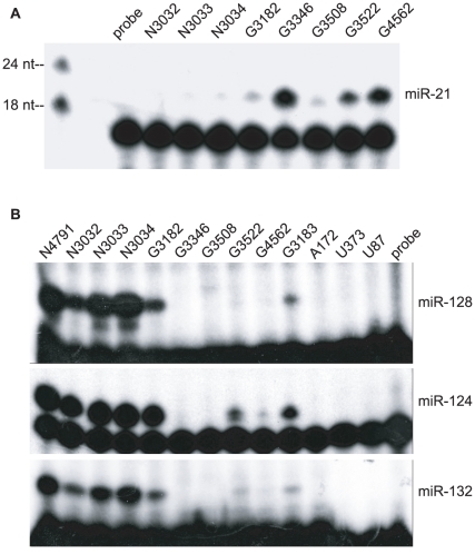
“Primer extension analysis confirmed enhanced expression of miR-21 (Fig. 2A) and decreased expression of miR-128, miR-124, and miR-132 (Fig. 2B) in glioblastomas compared to non-tumor brain tissue samples.”
§ Reduced Expression of Brain-Enriched microRNAs in Glioblastomas Permits Targeted Regulation of a Cell Death Gene > Results > Validation of miRNA expression patterns by primer extension
plaincorrelational- Asserted: passes — The authors put it forward as true.
- Checkable: passes — It can be checked against evidence.
- Load-bearing: passes — The paper’s conclusions depend on it.
- Based: passes — The paper gives a reason why it holds.
It may state two separate claims as one.
A result the paper reports from its own data — it cites no other source for it, so there is none to check. The quote shows it is in the paper.
In the paper
“we also observed down-regulation of miR-124, miR-128, miR-132, and miR-7”
§ Reduced Expression of Brain-Enriched microRNAs in Glioblastomas Permits Targeted Regulation of a Cell Death Gene > Discussion
plain- Asserted: passes — The authors put it forward as true.
- Checkable: passes — It can be checked against evidence.
- Load-bearing: passes — The paper’s conclusions depend on it.
- Based: passes — The paper gives a reason why it holds.
It may state two separate claims as one.
A result the paper reports from its own data — it cites no other source for it, so there is none to check. The quote shows it is in the paper.
In the paper
“Our sequencing data reveal that miR-128 has an invariant sequence and does not exhibit post-transcriptional editing or significant 5′ end variation.”
§ Reduced Expression of Brain-Enriched microRNAs in Glioblastomas Permits Targeted Regulation of a Cell Death Gene > Results > miRNAs as potential therapeutic tools to regulate cell suicide gene expression
plain- Asserted: passes — The authors put it forward as true.
- Checkable: passes — It can be checked against evidence.
- Load-bearing: unclear — It is unclear whether the paper’s conclusions depend on it.
- Based: passes — The paper gives a reason why it holds.
It may state two separate claims as one.
None of the sources cited for this claim have been checked yet.
In the paper
“Glioblastoma is associated with a number of mutations at the genomic level including mutations in the PTEN tumor suppressor and amplification of the gene encoding the epidermal growth factor receptor (EGFR) [3], [4].”
§ Reduced Expression of Brain-Enriched microRNAs in Glioblastomas Permits Targeted Regulation of a Cell Death Gene > Introduction
What the sources say
- Not checked — no full text of the sourcecites [3] PY Wen, 2008, Malignant gliomas in adults., N Engl J Med source
- Not checked yetcites [4] H Ohgaki, 2009, Genetic alterations and signaling pathways in the evolution of gliomas., Cancer Sci source
Checked before — its earlier verdict is held back until a new check runs.
plaincorrelational- Asserted: passes — The authors put it forward as true.
- Checkable: passes — It can be checked against evidence.
- Load-bearing: fails — The paper’s conclusions may not depend on it.
- Based: passes — The paper gives a reason why it holds.
Probably not a substantial claim. The paper’s conclusions may not depend on it. It may state two separate claims as one.
A conclusion the paper draws from its own results — it cites no other source for it, so there is none to check. The quote shows it is in the paper.
In the paper
“In addition, we identified variation in the 5′ ends of several miRNAs expressed in glioblastomas as well as editing in the seed region, which should modulate the mRNA targets bound by these miRNAs.”
§ Reduced Expression of Brain-Enriched microRNAs in Glioblastomas Permits Targeted Regulation of a Cell Death Gene > Introduction
hedgedconditionaln = 6- Asserted: passes — The authors put it forward as true.
- Checkable: passes — It can be checked against evidence.
- Load-bearing: passes — The paper’s conclusions depend on it.
- Based: passes — The paper gives a reason why it holds.
It may state two separate claims as one.
None of the sources cited for this claim have been checked yet.
In the paper
“Similar approaches have been used to selectively prevent transgene expression in hematopoetic cells and other cell types [28]–[31].”
§ Reduced Expression of Brain-Enriched microRNAs in Glioblastomas Permits Targeted Regulation of a Cell Death Gene > Introduction
What the sources say
- Not checked yetcites [28] EJ Kelly, 2009, MicroRNAs and the regulation of vector tropism., Mol Ther source
Checked before — its earlier verdict is held back until a new check runs.
- Not checked — no full text of the sourcecites [29] BD Brown, 2009, Exploiting and antagonizing microRNA regulation for therapeutic and experimental applications., Nat Rev Genet source
- Not checked — no full text of the sourcecites [30] BD Brown, 2006, Endogenous microRNA regulation suppresses transgene expression in hematopoietic lineages and enables stable gene transfer., Nat Med source
- Not checked yetcites [31] C Wu, 2009, Combinatorial control of suicide gene expression by tissue-specific promoter and microRNA regulation for cancer therapy., Mol Ther source
Checked before — its earlier verdict is held back until a new check runs.
plaindirect- Asserted: passes — The authors put it forward as true.
- Checkable: passes — It can be checked against evidence.
- Load-bearing: fails — The paper’s conclusions may not depend on it.
- Based: passes — The paper gives a reason why it holds.
Probably not a substantial claim. The paper’s conclusions may not depend on it.
- Not checked yet
In the paper
“Of note, miR-7 can directly repress expression of EGFR, which is often amplified at the genetic level and/or over-expressed at the protein level in gliomas [19].”
§ Reduced Expression of Brain-Enriched microRNAs in Glioblastomas Permits Targeted Regulation of a Cell Death Gene > Introduction
What the sources say
- Not checked — no full text of the sourcecites [19] B Kefas, 2008, microRNA-7 inhibits the epidermal growth factor receptor and the Akt pathway and is down-regulated in glioblastoma., Cancer Res source
plaindirect- Asserted: passes — The authors put it forward as true.
- Checkable: passes — It can be checked against evidence.
- Load-bearing: fails — The paper’s conclusions may not depend on it.
- Based: passes — The paper gives a reason why it holds.
Probably not a substantial claim. The paper’s conclusions may not depend on it.
In the paper
“More recently, dysregulated microRNA (miRNA) expression, such as over-expression of the anti-apoptotic miRNA miR-21, has been linked to tumor pathogenesis [5].”
§ Reduced Expression of Brain-Enriched microRNAs in Glioblastomas Permits Targeted Regulation of a Cell Death Gene > Introduction
What the sources say
- Not checked — no full text of the sourcecites [5] JA Chan, 2005, MicroRNA-21 is an antiapoptotic factor in human glioblastoma cells., Cancer Res source
plaincorrelational- Asserted: passes — The authors put it forward as true.
- Checkable: passes — It can be checked against evidence.
- Load-bearing: fails — The paper’s conclusions may not depend on it.
- Based: passes — The paper gives a reason why it holds.
Probably not a substantial claim. The paper’s conclusions may not depend on it.
In the paper
“Moreover, single pre-miRNA precursors occasionally give rise to not only the dominant, miRNA strand, but also a less highly expressed passenger or star strand, which may also be able to regulate mRNA expression [13].”
§ Reduced Expression of Brain-Enriched microRNAs in Glioblastomas Permits Targeted Regulation of a Cell Death Gene > Introduction
What the sources say
- Not checked yetcites [13] JS Yang, 2011, Widespread regulatory activity of vertebrate microRNA* species., RNA source
hedgedconditional- Asserted: passes — The authors put it forward as true.
- Checkable: passes — It can be checked against evidence.
- Load-bearing: fails — The paper’s conclusions may not depend on it.
- Based: passes — The paper gives a reason why it holds.
Probably not a substantial claim. The paper’s conclusions may not depend on it.
What the paper did — it cites no source for this, so there is none to check. The quote shows it is in the paper.
In the paper
“To validate results from our deep sequencing experiments, we selected four differentially expressed miRNAs for further analysis: miR-21, miR-128, miR-124, and miR-132.”
§ Reduced Expression of Brain-Enriched microRNAs in Glioblastomas Permits Targeted Regulation of a Cell Death Gene > Results > Validation of miRNA expression patterns by primer extension
plain- Asserted: passes — The authors put it forward as true.
- Checkable: passes — It can be checked against evidence.
- Load-bearing: fails — The paper’s conclusions may not depend on it.
- Based: passes — The paper gives a reason why it holds.
Probably not a substantial claim. The paper’s conclusions may not depend on it.
A conclusion the paper draws from its own results — it cites no other source for it, so there is none to check. The quote shows it is in the paper.
In the paper
“These data suggest that 5′ end polymorphisms can reflect fluctuations in strand dominance and presumably, reflect variations in Drosha processing of the primary miRNA transcript.”
§ Reduced Expression of Brain-Enriched microRNAs in Glioblastomas Permits Targeted Regulation of a Cell Death Gene > Results > miRNA editing and miRNA sequence variants in the brain
hedgedcorrelational- Asserted: passes — The authors put it forward as true.
- Checkable: passes — It can be checked against evidence.
- Load-bearing: fails — The paper’s conclusions may not depend on it.
- Based: passes — The paper gives a reason why it holds.
Probably not a substantial claim. The paper’s conclusions may not depend on it. It may state two separate claims as one.
A conclusion the paper draws from its own results — it cites no other source for it, so there is none to check. The quote shows it is in the paper.
In the paper
“Such changes may indicate post-transcriptional processing differences—for instance, the absence or presence of a factor in glial versus non-glial cells that perturbs miRNA strand selection.”
§ Reduced Expression of Brain-Enriched microRNAs in Glioblastomas Permits Targeted Regulation of a Cell Death Gene > Results > Variations in 5p and 3p strand dominance
hedgedconditionaln = 9- Asserted: passes — The authors put it forward as true.
- Checkable: passes — It can be checked against evidence.
- Load-bearing: fails — The paper’s conclusions may not depend on it.
- Based: passes — The paper gives a reason why it holds.
Probably not a substantial claim. The paper’s conclusions may not depend on it.
A conclusion the paper draws from its own results — it cites no other source for it, so there is none to check. The quote shows it is in the paper.
In the paper
“demonstrating that high-throughput sequencing can be an effective method for profiling miRNA expression.”
§ Reduced Expression of Brain-Enriched microRNAs in Glioblastomas Permits Targeted Regulation of a Cell Death Gene > Discussion
boosted- Asserted: passes — The authors put it forward as true.
- Checkable: passes — It can be checked against evidence.
- Load-bearing: fails — The paper’s conclusions may not depend on it.
- Based: passes — The paper gives a reason why it holds.
Probably not a substantial claim. The paper’s conclusions may not depend on it.
The paper states this without a source or data behind it here, so there is nothing to check it against. The quote shows it is in the paper.
In the paper
“Compared to microarray and PCR-based profiling platforms, which detect a pre-determined set of miRNAs, high-throughput sequencing provides a more complete picture of the miRNAs expressed in a given sample and is not restricted in the number of individual miRNAs that can be detected.”
§ Reduced Expression of Brain-Enriched microRNAs in Glioblastomas Permits Targeted Regulation of a Cell Death Gene > Discussion
plaindirect- Asserted: passes — The authors put it forward as true.
- Checkable: passes — It can be checked against evidence.
- Load-bearing: fails — The paper’s conclusions may not depend on it.
- Based: passes — The paper gives a reason why it holds.
Probably not a substantial claim. The paper’s conclusions may not depend on it. It may state two separate claims as one.
A conclusion the paper draws from its own results — it cites no other source for it, so there is none to check. The quote shows it is in the paper.
In the paper
“an approach that could be readily extended to cancers of other organs.”
§ Reduced Expression of Brain-Enriched microRNAs in Glioblastomas Permits Targeted Regulation of a Cell Death Gene > Discussion
hedged- Asserted: passes — The authors put it forward as true.
- Checkable: fails — It may be too vague to check against evidence.
- Load-bearing: fails — The paper’s conclusions may not depend on it.
- Based: fails — The paper may give no reason why it holds.
Probably not a substantial claim. It may be too vague to check against evidence. The paper’s conclusions may not depend on it. The paper may give no reason why it holds.
What the paper did — it cites no source for this, so there is none to check. The quote shows it is in the paper.
In the paper
“None of the samples were post-mortem.”
§ Reduced Expression of Brain-Enriched microRNAs in Glioblastomas Permits Targeted Regulation of a Cell Death Gene > Materials and Methods > Cell lines and tissue samples
plain- Asserted: passes — The authors put it forward as true.
- Checkable: passes — It can be checked against evidence.
- Load-bearing: fails — The paper’s conclusions may not depend on it.
- Based: passes — The paper gives a reason why it holds.
Probably not a substantial claim. The paper’s conclusions may not depend on it.
None of the sources cited for this claim have been checked yet.
In the paper
“One post-transcriptional miRNA modification is enzymatic editing of the dsRNA primary miRNA transcript by ADAR (adenosine deaminase acting on RNA) which results in the conversion of an adenosine (A) to an inosine (I) [24].”
§ Reduced Expression of Brain-Enriched microRNAs in Glioblastomas Permits Targeted Regulation of a Cell Death Gene > Results > miRNA editing and miRNA sequence variants in the brain
What the sources say
- Not checked yetcites [24] K Nishikura, 2010, Functions and regulation of RNA editing by ADAR deaminases., Annu Rev Biochem source
Checked before — its earlier verdict is held back until a new check runs.
plaindirect- Asserted: passes — The authors put it forward as true.
- Checkable: passes — It can be checked against evidence.
- Load-bearing: fails — The paper’s conclusions may not depend on it.
- Based: passes — The paper gives a reason why it holds.
Probably not a substantial claim. The paper’s conclusions may not depend on it.
- Not checked yet
What the paper did — it cites no source for this, so there is none to check. The quote shows it is in the paper.
In the paper
“De-identified tissues were used for all studies and the data were analyzed anonymously.”
§ Reduced Expression of Brain-Enriched microRNAs in Glioblastomas Permits Targeted Regulation of a Cell Death Gene > Materials and Methods > Cell lines and tissue samples
plain- Asserted: passes — The authors put it forward as true.
- Checkable: passes — It can be checked against evidence.
- Load-bearing: fails — The paper’s conclusions may not depend on it.
- Based: passes — The paper gives a reason why it holds.
Probably not a substantial claim. The paper’s conclusions may not depend on it. It may state two separate claims as one.
None of the sources cited for this claim have been checked yet.
In the paper
“A to I conversions can influence loading of miRNAs into RISC [34] and also alter the targeting capacity of a miRNA, especially if the editing occurs within the miRNA seed region [24], [25].”
§ Reduced Expression of Brain-Enriched microRNAs in Glioblastomas Permits Targeted Regulation of a Cell Death Gene > Results > miRNA editing and miRNA sequence variants in the brain
What the sources say
- Not checked yetcites [34] DS Schwarz, 2003, Asymmetry in the assembly of the RNAi enzyme complex., Cell source
- Not checked yetcites [24] K Nishikura, 2010, Functions and regulation of RNA editing by ADAR deaminases., Annu Rev Biochem source
Checked before — its earlier verdict is held back until a new check runs.
- Not checked — no full text of the sourcecites [25] Y Kawahara, 2007, Redirection of silencing targets by adenosine-to-inosine editing of miRNAs., Science source
plainconditional- Asserted: passes — The authors put it forward as true.
- Checkable: passes — It can be checked against evidence.
- Load-bearing: fails — The paper’s conclusions may not depend on it.
- Based: passes — The paper gives a reason why it holds.
Probably not a substantial claim. The paper’s conclusions may not depend on it. It may state two separate claims as one.
In the paper
“many of which (miR-128, miR-124, miR-7, miR-132, miR-139) are consistently dysregulated in not only gliomas but also other brain cancers including medulloblastomas and neuroblastomas [33].”
§ Reduced Expression of Brain-Enriched microRNAs in Glioblastomas Permits Targeted Regulation of a Cell Death Gene > Discussion
What the sources say
- Not checked — no full text of the sourcecites [33] JC Pang, 2009, Oncogenic role of microRNAs in brain tumors., Acta Neuropathol source
plaincorrelational- Asserted: passes — The authors put it forward as true.
- Checkable: passes — It can be checked against evidence.
- Load-bearing: fails — The paper’s conclusions may not depend on it.
- Based: passes — The paper gives a reason why it holds.
Probably not a substantial claim. The paper’s conclusions may not depend on it.
What the paper did — it cites no source for this, so there is none to check. The quote shows it is in the paper.
In the paper
“449 mature miRNAs and miRNAs* were identified using a filter of RPKM score >1 to normalize read counts between different libraries and by considering only those miRNAs occurring in at least two out of three non-tumor brain libraries or 50% of the glioblastoma libraries (Table S4).”
§ Reduced Expression of Brain-Enriched microRNAs in Glioblastomas Permits Targeted Regulation of a Cell Death Gene > Results > Small RNA deep sequencing reveals differences in human miRNA expression patterns
plainn = 3- Asserted: passes — The authors put it forward as true.
- Checkable: passes — It can be checked against evidence.
- Load-bearing: passes — The paper’s conclusions depend on it.
- Based: passes — The paper gives a reason why it holds.
Probably not a substantial claim. It may state two separate claims as one.
None of the sources cited for this claim have been checked yet.
In the paper
“we used the miRNAkey pipeline, implementing the SeqEM option to account for reads mapping to multiple miRNAs [32].”
§ Reduced Expression of Brain-Enriched microRNAs in Glioblastomas Permits Targeted Regulation of a Cell Death Gene > Results > Small RNA deep sequencing reveals differences in human miRNA expression patterns
What the sources say
- Not checked yetcites [32] R Ronen, 2010, miRNAkey: a software for microRNA deep sequencing analysis., Bioinformatics source
Checked before — its earlier verdict is held back until a new check runs.
plain- Asserted: passes — The authors put it forward as true.
- Checkable: passes — It can be checked against evidence.
- Load-bearing: fails — The paper’s conclusions may not depend on it.
- Based: passes — The paper gives a reason why it holds.
Probably not a substantial claim. The paper’s conclusions may not depend on it.
- Not checked yet
A claim is checked only against the cited paper’s full text, never its abstract, and none is available for its sources.
In the paper
“It is hypothesized that glioblastomas, like other cancers such as leukemia [51], arise and are maintained by a population of progenitor cells with stem-like characteristics including the ability to self-renew for unlimited proliferation [21], [52], [53].”
§ Reduced Expression of Brain-Enriched microRNAs in Glioblastomas Permits Targeted Regulation of a Cell Death Gene > Discussion
What the sources say
- Not checked — no full text of the sourcecites [51] BJ Huntly, 2005, Leukaemia stem cells and the evolution of cancer-stem-cell research., Nat Rev Cancer source
- Not checked — no full text of the sourcecites [21] J Godlewski, 2010, MicroRNAs and glioblastoma; the stem cell connection., Cell Death Differ source
- Not checked — no full text of the sourcecites [52] SK Singh, 2004, Cancer stem cells in nervous system tumors., Oncogene source
hedgeddirect- Asserted: passes — The authors put it forward as true.
- Checkable: passes — It can be checked against evidence.
- Load-bearing: passes — The paper’s conclusions depend on it.
- Based: passes — The paper gives a reason why it holds.
Probably not a substantial claim.
In the paper
“Finally, miR-128, which is also abundantly expressed in neurons, has been shown to target the Bmi-1 stem cell renewal factor [14].”
§ Reduced Expression of Brain-Enriched microRNAs in Glioblastomas Permits Targeted Regulation of a Cell Death Gene > Discussion
What the sources say
- Not checked — no full text of the sourcecites [14] J Godlewski, 2008, Targeting of the Bmi-1 oncogene/stem cell renewal factor by microRNA-128 inhibits glioma proliferation and self-renewal., Cancer Res source
plaindirect- Asserted: passes — The authors put it forward as true.
- Checkable: passes — It can be checked against evidence.
- Load-bearing: passes — The paper’s conclusions depend on it.
- Based: passes — The paper gives a reason why it holds.
It may state two separate claims as one.
None of the sources cited for this claim have been checked yet.
In the paper
“Additionally, single nucleotide changes have been shown to affect miRNA expression, processing, and/or function [35].”
§ Reduced Expression of Brain-Enriched microRNAs in Glioblastomas Permits Targeted Regulation of a Cell Death Gene > Results > miRNA editing and miRNA sequence variants in the brain
What the sources say
- Not checked yetcites [35] Y Kawahara, 2007, RNA editing of the microRNA-151 precursor blocks cleavage by the Dicer-TRBP complex., EMBO Rep source
Checked before — its earlier verdict is held back until a new check runs.
plaindirect- Asserted: passes — The authors put it forward as true.
- Checkable: passes — It can be checked against evidence.
- Load-bearing: fails — The paper’s conclusions may not depend on it.
- Based: passes — The paper gives a reason why it holds.
Probably not a substantial claim. The paper’s conclusions may not depend on it.
- Not checked yet
None of the sources cited for this claim have been checked yet.
In the paper
“The use of tissue-specific miRNAs to regulate transgene expression for therapeutic applications has been investigated in multiple cell types, including lymphoid and myeloid cells, muscle cells, liver cells, and brain cells [28]–[31].”
§ Reduced Expression of Brain-Enriched microRNAs in Glioblastomas Permits Targeted Regulation of a Cell Death Gene > Discussion
What the sources say
- Not checked yetcites [28] EJ Kelly, 2009, MicroRNAs and the regulation of vector tropism., Mol Ther source
Checked before — its earlier verdict is held back until a new check runs.
- Not checked — no full text of the sourcecites [29] BD Brown, 2009, Exploiting and antagonizing microRNA regulation for therapeutic and experimental applications., Nat Rev Genet source
- Not checked — no full text of the sourcecites [30] BD Brown, 2006, Endogenous microRNA regulation suppresses transgene expression in hematopoietic lineages and enables stable gene transfer., Nat Med source
- Not checked yetcites [31] C Wu, 2009, Combinatorial control of suicide gene expression by tissue-specific promoter and microRNA regulation for cancer therapy., Mol Ther source
Checked before — its earlier verdict is held back until a new check runs.
plain- Asserted: passes — The authors put it forward as true.
- Checkable: passes — It can be checked against evidence.
- Load-bearing: fails — The paper’s conclusions may not depend on it.
- Based: passes — The paper gives a reason why it holds.
Probably not a substantial claim. The paper’s conclusions may not depend on it.
- Not checked yet
None of the sources cited for this claim have been checked yet.
In the paper
“This vector showed the same therapeutic efficacy in glioblastoma U87 cells as a control HSV-TK vector lacking artificial miRNA target sites, but when introduced into human astrocytes, which express the three selected miRNAs, showed restricted HSV-TK expression, leading to an increase in cell viability.”
§ Reduced Expression of Brain-Enriched microRNAs in Glioblastomas Permits Targeted Regulation of a Cell Death Gene > Discussion
What the sources say
- Not checked yetcites [31] C Wu, 2009, Combinatorial control of suicide gene expression by tissue-specific promoter and microRNA regulation for cancer therapy., Mol Ther source
Checked before — its earlier verdict is held back until a new check runs.
plaindirect- Asserted: passes — The authors put it forward as true.
- Checkable: passes — It can be checked against evidence.
- Load-bearing: unclear — It is unclear whether the paper’s conclusions depend on it.
- Based: passes — The paper gives a reason why it holds.
It may state two separate claims as one.
- Not checked yet
In the paper
“A single A to I conversion in the seed of miR-376a-5p, for example, redirects the edited miRNA to a new set of mRNA targets [25].”
§ Reduced Expression of Brain-Enriched microRNAs in Glioblastomas Permits Targeted Regulation of a Cell Death Gene > Results > miRNA editing and miRNA sequence variants in the brain
What the sources say
- Not checked — no full text of the sourcecites [25] Y Kawahara, 2007, Redirection of silencing targets by adenosine-to-inosine editing of miRNAs., Science source
plaindirect- Asserted: passes — The authors put it forward as true.
- Checkable: passes — It can be checked against evidence.
- Load-bearing: fails — The paper’s conclusions may not depend on it.
- Based: passes — The paper gives a reason why it holds.
Probably not a substantial claim. The paper’s conclusions may not depend on it.
In the paper
“reduced expression of miR-128 is not specific to just glioblastomas as this miRNA is also down-regulated in other brain cancers [33].”
§ Reduced Expression of Brain-Enriched microRNAs in Glioblastomas Permits Targeted Regulation of a Cell Death Gene > Discussion
What the sources say
- Not checked — no full text of the sourcecites [33] JC Pang, 2009, Oncogenic role of microRNAs in brain tumors., Acta Neuropathol source
plain- Asserted: passes — The authors put it forward as true.
- Checkable: passes — It can be checked against evidence.
- Load-bearing: fails — The paper’s conclusions may not depend on it.
- Based: passes — The paper gives a reason why it holds.
Probably not a substantial claim. The paper’s conclusions may not depend on it.
What the paper did — it cites no source for this, so there is none to check. The quote shows it is in the paper.
In the paper
“Given the much broader dynamic range of deep sequencing compared to finite platforms such as miRNA microarrays or real-time PCR arrays, we therefore used a FDR cut-off of p<0.1.”
§ Reduced Expression of Brain-Enriched microRNAs in Glioblastomas Permits Targeted Regulation of a Cell Death Gene > Results > Small RNA deep sequencing reveals differences in human miRNA expression patterns
plaindirect- Asserted: passes — The authors put it forward as true.
- Checkable: passes — It can be checked against evidence.
- Load-bearing: passes — The paper’s conclusions depend on it.
- Based: passes — The paper gives a reason why it holds.
Probably not a substantial claim. It may state two separate claims as one.
None of the sources cited for this claim have been checked yet.
In the paper
“Additionally, miR-128 is highly enriched in the brain as it is expressed by neurons and is induced during neuronal differentiation of embryonal carcinoma (EC) cells [20], [21], thus making it an ideal candidate to limit cell suicide gene expression.”
§ Reduced Expression of Brain-Enriched microRNAs in Glioblastomas Permits Targeted Regulation of a Cell Death Gene > Results > miRNAs as potential therapeutic tools to regulate cell suicide gene expression
What the sources say
- Not checked yetcites [20] LF Sempere, 2004, Expression profiling of mammalian microRNAs uncovers a subset of brain-expressed microRNAs with possible roles in murine and human neuronal differentiation., Genome Biol source
- Not checked — no full text of the sourcecites [21] J Godlewski, 2010, MicroRNAs and glioblastoma; the stem cell connection., Cell Death Differ source
plain- Asserted: passes — The authors put it forward as true.
- Checkable: passes — It can be checked against evidence.
- Load-bearing: passes — The paper’s conclusions depend on it.
- Based: passes — The paper gives a reason why it holds.
It may state two separate claims as one.
A conclusion the paper draws from its own results — it cites no other source for it, so there is none to check. The quote shows it is in the paper.
In the paper
“clearly additional miRNAs identified here are dysregulated in glioblastoma (Fig. 1, Tables S4 and S6).”
§ Reduced Expression of Brain-Enriched microRNAs in Glioblastomas Permits Targeted Regulation of a Cell Death Gene > Discussion
boostedcorrelational- Asserted: passes — The authors put it forward as true.
- Checkable: passes — It can be checked against evidence.
- Load-bearing: passes — The paper’s conclusions depend on it.
- Based: passes — The paper gives a reason why it holds.
The quote only partly states this claim.
The paper relies on this without support.
In deep sequencing libraries, A to I editing is marked by the substitution of A with G.
“In deep sequencing libraries, this conversion is marked by the substitution of A with G.”
The paper relies on this without support.
Lentiviral vectors can enter tissue surrounding the tumor and kill otherwise healthy cells, which could have dire consequences in the brain.
“one major drawback in glioblastoma treatment is that such vectors can also enter the tissue surrounding the tumor, killing otherwise healthy cells, which could have dire consequences in the brain.”
The paper relies on this without support.
miRNAs post-transcriptionally down-regulate cellular gene expression by binding to complementary sequences generally located in the 3′ UTRs of target mRNAs.
“MiRNAs are ∼22 nt non-coding RNAs that post-transcriptionally down-regulate cellular gene expression by binding to complementary sequences generally located in the 3′ untranslated regions (UTRs) of target mRNAs.”
The paper relies on this without support.
Target specificity is predominantly determined by nucleotides 2-8 of the mature miRNA, the miRNA seed, which generally exhibits full sequence complementarity to target mRNAs.
“Target specificity is predominantly determined by nucleotides 2-8 of the mature miRNA, the miRNA “seed,” which generally exhibits full sequence complementarity to target mRNAs.”
The paper relies on this without support.
miRNA effects on target mRNAs are usually modest.
“While miRNA effects on target mRNAs are usually modest”
Self-check
Percentages, sums and reported differences re-computed from the paper’s own numbers.
Each contradiction between two passages listed here was confirmed by an independent check.
Rated minor: it would not touch a result — a value no result rests on, a p-value that stays on the same side of the significance level, or a conflict that is clerical, resolved elsewhere in the paper, or outside its own findings.
In the paper
§ Reduced Expression of Brain-Enriched microRNAs in Glioblastomas Permits Targeted Regulation of a Cell Death Gene > Results > Small RNA deep sequencing reveals differences in human miRNA expression patterns
“Small RNA populations corresponding to 18–25 nt in length were sequenced using an Illumina GAII analyzer.”
§ Reduced Expression of Brain-Enriched microRNAs in Glioblastomas Permits Targeted Regulation of a Cell Death Gene > Materials and Methods > Deep Sequencing and Bioinformatics
“Small RNAs (18–24 nt) were extracted and ligated to adapter sequences for Illumina sequencing.”
Rated minor: it would not touch a result — a value no result rests on, a p-value that stays on the same side of the significance level, or a conflict that is clerical, resolved elsewhere in the paper, or outside its own findings.
In the paper
§ Reduced Expression of Brain-Enriched microRNAs in Glioblastomas Permits Targeted Regulation of a Cell Death Gene > Results > miRNA editing and miRNA sequence variants in the brain
“miR-324, together with four other miRNAs that show isoform differences in tumor versus non-tumor samples (miR-106b, miR-335, and miR-411)”
§ Reduced Expression of Brain-Enriched microRNAs in Glioblastomas Permits Targeted Regulation of a Cell Death Gene > Results > miRNA editing and miRNA sequence variants in the brain
“miR-106b, miR-335, and miR-411”
Rated minor: it would not touch a result — a value no result rests on, a p-value that stays on the same side of the significance level, or a conflict that is clerical, resolved elsewhere in the paper, or outside its own findings.
In the paper
§ Reduced Expression of Brain-Enriched microRNAs in Glioblastomas Permits Targeted Regulation of a Cell Death Gene > Materials and Methods > Primer Extension to detect mature miRNAs
“Primer extensions were performed with 5 µg of total RNA”
§ Reduced Expression of Brain-Enriched microRNAs in Glioblastomas Permits Targeted Regulation of a Cell Death Gene > Results > Validation of miRNA expression patterns by primer extension
“6 µg total RNA were used for each reaction.”
Rated minor: it would not touch a result — a value no result rests on, a p-value that stays on the same side of the significance level, or a conflict that is clerical, resolved elsewhere in the paper, or outside its own findings.
In the paper
§ Reduced Expression of Brain-Enriched microRNAs in Glioblastomas Permits Targeted Regulation of a Cell Death Gene > Materials and Methods > Crystal Violet Staining and MTT assays
“SH-SY5Y cells were seeded in 24-well plates at 25,000 cells per well 48 hrs post-transduction and cultured for 10 days in media containing GCV and either 10 µM ATRA or 10 µM di-methyl sulfoxide (DMSO) vehicle control.”
§ Reduced Expression of Brain-Enriched microRNAs in Glioblastomas Permits Targeted Regulation of a Cell Death Gene > Results > Endogenous miR-128 protects neuronal cells from HSV-TK induced cell death
“Cells were cultured in the presence of 10 µM ATRA to induce neuronal differentiation and 40 µg/ml GCV for two weeks (n = 2).”
Citations
Cites
Cluster analysis and display of genome-wide expression patterns
Eisen, M. B., Spellman, P. T., Brown, P. O., & Botstein, D. (1998) · Proceedings of the National Academy of Sciences · doi:10.1073/pnas.95.25.14863 · Cited by 16,184
Lewis, B. P., Burge, C. B., & Bartel, D. P. (2005) · Cell · doi:10.1016/j.cell.2004.12.035 · Cited by 11,855
MicroRNA expression profiles classify human cancers
Lü, J., Getz, G. A., Miska, E. A., Alvarez-Saavedra, E., Lamb, J., Peck, D., Sweet-Cordero, A., Ebert, B. L., Mak, R. H., Ferrando, A. A., et al. (2005) · Nature · doi:10.1038/nature03702 · Cited by 9,637
Most mammalian mRNAs are conserved targets of microRNAs
Friedman, R. C., Farh, K. K.-H., Burge, C. B., & Bartel, D. P. (2009) · Genome Research · doi:10.1101/gr.082701.108 · Cited by 8,462
Identification of human brain tumour initiating cells
Singh, S. K., Hawkins, C. E., Clarke, I. D., Squire, J. A., Bayani, J., Hide, T., Henkelman, R. M., Cusimano, M. D., & Dirks, P. B. (2004) · Nature · doi:10.1038/nature03128 · Cited by 7,564
A microRNA expression signature of human solid tumors defines cancer gene targets
Volinia, S., Calin, G. A., Liu, C.-G., Ambs, S., Cimmino, A., Petrocca, F., Visone, R., Iorio, M., Roldo, C., Ferracin, M., et al. (2006) · Proceedings of the National Academy of Sciences · doi:10.1073/pnas.0510565103 · Cited by 5,697
miRBase: tools for microRNA genomics
Griffiths‐Jones, S., Saini, H. K., van Dongen, S., & Enright, A. J. (2007) · Nucleic Acids Research · doi:10.1093/nar/gkm952 · Cited by 4,432
MicroRNA Gene Expression Deregulation in Human Breast Cancer
Iorio, M. V., Ferracin, M., Liu, C.-G., Veronese, A., Spizzo, R., Sabbioni, S., Magri, E., Pedriali, M., Fabbri, M., Campiglio, M., et al. (2005) · Cancer Research · doi:10.1158/0008-5472.can-05-1783 · Cited by 4,053
Wen, P. Y., & Kesari, S. (2008) · New England Journal of Medicine · doi:10.1056/nejmra0708126 · Cited by 3,511
Asymmetry in the Assembly of the RNAi Enzyme Complex
Schwarz, D. S., Hutvágner, G., Du, T., Xu, Z., Aronin, N., & Zamore, P. D. (2003) · Cell · doi:10.1016/s0092-8674(03)00759-1 · Cited by 2,922
Cited by
Sorted by citations
Møller, H. G., Rasmussen, A. P., Andersen, H. H., Johnsen, K. B., Henriksen, M., & Duroux, M. C. (2012) · Molecular Neurobiology · doi:10.1007/s12035-012-8349-7 · Cited by 273
The microRNAs within the DLK1-DIO3 genomic region: involvement in disease pathogenesis
Benetatos, L., Hatzimichael, E. C., Londin, E. R., Vartholomatos, G., Loher, P., Rigoutsos, I., & Briasoulis, E. C. (2012) · Cellular and Molecular Life Sciences · doi:10.1007/s00018-012-1080-8 · Cited by 279
Glioblastoma Therapy: Past, Present and Future
Obrador, E., Moreno-Murciano, P., Oriol-Caballo, M., López-Blanch, R., Pineda, B., Gutiérrez-Arroyo, J., Loras, A., Gonzalez-Bonet, L., Martinez-Cadenas, C., Estrela, J., et al. (2024) · International Journal of Molecular Sciences · doi:10.3390/ijms25052529 · Cited by 271
Wang, K., Liu, F., Liu, C.-Y., An, T., Zhang, J., Zhou, L.-Y., Wang, M.-H., Dong, Y.-H., Li, N., Gao, J.-N., et al. (2016) · Cell Death and Differentiation · doi:10.1038/cdd.2016.28 · Cited by 216
Adlakha, Y. K., & Saini, N. (2014) · Molecular Cancer · doi:10.1186/1476-4598-13-33 · Cited by 213
Metadata
- Authors
- Skalsky, R. L., & Cullen, B. R.
- Affiliations
- Duke University (US)
- Corresponding author
- Cullen, Bryan R.
- Venue
- PLoS ONE
- Publisher
- Public Library of Science (PLoS)
- Published
- 2011-09-02
- Volume
- 6
- Issue
- 9
- Pages
- e24248
- ISSN
- 1932-6203 (ISSN-L)
- PMC
- PMC3166303
- OpenAlex
- W2020186279
- Cited by
- 170
- References
- 63
- Citations by year
- 2026: 1 · 2025: 2 · 2024: 3 · 2023: 4 · 2022: 8 · 2021: 10 · 2020: 10 · 2019: 10 · 2018: 15 · 2017: 13 · 2016: 19 · 2015: 21 · 2014: 18 · 2013: 23 · 2012: 13
- FWCI
- 5.77
- Type
- journal article
- Language
- en
- Open access
- gold
- doi.org
- Locations
- PLoS ONE (publishedVersion, cc-by)
- Europe PMC (PubMed Central) (publishedVersion, cc-by)
- PubMed Central (publishedVersion, cc-by)
- PubMed (publishedVersion)
- DOAJ (DOAJ: Directory of Open Access Journals) (submittedVersion, cc-by-sa)
- Figshare (submittedVersion, cc-by)
- Funders
- Pediatric Brain Tumor Foundation
- National Institutes of Health
- National Cancer Institute (T32CA009111, T342-CA009111)
- Topics
- Life Sciences › Biochemistry, Genetics and Molecular Biology › Cancer Research › MicroRNA in disease regulation
- Health Sciences › Medicine › Oncology › Glioma Diagnosis and Treatment
- Life Sciences › Biochemistry, Genetics and Molecular Biology › Molecular Biology › RNA Interference and Gene Delivery
- Keywords
- glioblastoma, microRNA, miR-128, miR-124, miR-7, miR-139, miR-381, miR-379, HSV thymidine kinase, ganciclovir, neuronal differentiation, SH-SY5Y cells, small RNA profiling
- MeSH terms
- Base Sequence, Brain, Genetic Vectors, Glioblastoma, Humans, Neurons, Organ Specificity, Thymidine Kinase, Genetic Variation, Reproducibility of Results, Genetic Therapy, Down-Regulation, Cell Death, RNA Editing, DNA Primers, Herpesvirus 1, Human, MicroRNAs, Genes, Transgenic, Suicide, Cell Line, Tumor, High-Throughput Nucleotide Sequencing, Transcriptome
About these checks
Registry, retraction, Cites and statistics results are computed, not judged, with one exception: a printed reference that no index lists is read once by a model before it counts as a citation error. Claims are extracted by a model and kept only when their quote appears verbatim in the text; each cited source is then checked, with a model’s help, for whether it says what the claim relies on. Those verdicts can be wrong — read the claim’s quote and the cited source before relying on one.
A language model drew the claims from the paper on 2026-11-10, under version 3 of the claim definition (model deepseek-v4.1-flash, prompt extract-v3).
A second model labelled each claim on 2026-11-10 (model jev-1.13-20260917, definition revision gold-guideline-v0.2).
Cited sources are checked with a language model’s help (gpt-6-sol).
Statistics and arithmetic are recomputed, not judged.
Confidence bands are shown only for fields calibrated on our annotated test set. None is calibrated yet, so no band is shown.
What counts as a claim ›How we measure confidence ›How we check this ›
Are you an author, or do you see a problem with this page? Request a review
