

The DOI matches its Crossref record; 114 of the 114 references it cites were found in a registry or an index; 53 of the 93 sources its claims cite were checked for support.
Gene Expression Analysis of Zebrafish Melanocytes, Iridophores, and Retinal Pigmented Epithelium Reveals Indicators of Biological Function and Developmental Origin
Open accessAbstract
In order to facilitate understanding of pigment cell biology, we developed a method to concomitantly purify melanocytes, iridophores, and retinal pigmented epithelium from zebrafish, and analyzed their transcriptomes. Comparing expression data from these cell types and whole embryos allowed us to reveal gene expression co-enrichment in melanocytes and retinal pigmented epithelium, as well as in melanocytes and iridophores. We found 214 genes co-enriched in melanocytes and retinal pigmented epithelium, indicating the shared functions of melanin-producing cells. We found 62 genes significantly co-enriched in melanocytes and iridophores, illustrative of their shared developmental origins from the neural crest. This is also the first analysis of the iridophore transcriptome. Gene expression analysis for iridophores revealed extensive enrichment of specific enzymes to coordinate production of their guanine-based reflective pigment. We speculate the coordinated upregulation of specific enzymes from several metabolic pathways recycles the rate-limiting substrate for purine synthesis, phosphoribosyl pyrophosphate, thus constituting a guanine cycle. The purification procedure and expression analysis described here, along with the accompanying transcriptome-wide expression data, provide the first mRNA sequencing data for multiple purified zebrafish pigment cell types, and will be a useful resource for further studies of pigment cell biology.
Overview
AI snapshot
Written from the paper’s full text.
TL;DR
The authors developed a density-gradient/FACS method to purify zebrafish melanocytes, iridophores, and retinal pigmented epithelium (RPE) and generated the first transcriptome-wide mRNA sequencing data for these three pigment cell types, including the first iridophore transcriptome. Comparative expression analysis identified shared and cell-type-specific gene sets, notably a guanine-synthesis cycle model in iridophores.
Key findings
- RNA-seq detected 8,472 genes expressed in melanocytes, 8,096 in iridophores, and 9,053 in RPE at >1 RPKM; melanocyte–RPE expression was most correlated (r = 0.90), while melanocyte–iridophore and RPE–iridophore correlations were modest (r = 0.52 and r = 0.49), and each pigment cell type correlated with whole embryos at <0.02.
- The authors found 214 genes co-enriched in melanocytes and RPE, 62 co-enriched in melanocytes and iridophores, and only one gene, alcama, co-enriched in RPE and iridophores; 28 genes were shared among all three pigment cell types at ≥4 RPKM, within 2-fold of each other, and ≥100-fold above whole embryos.
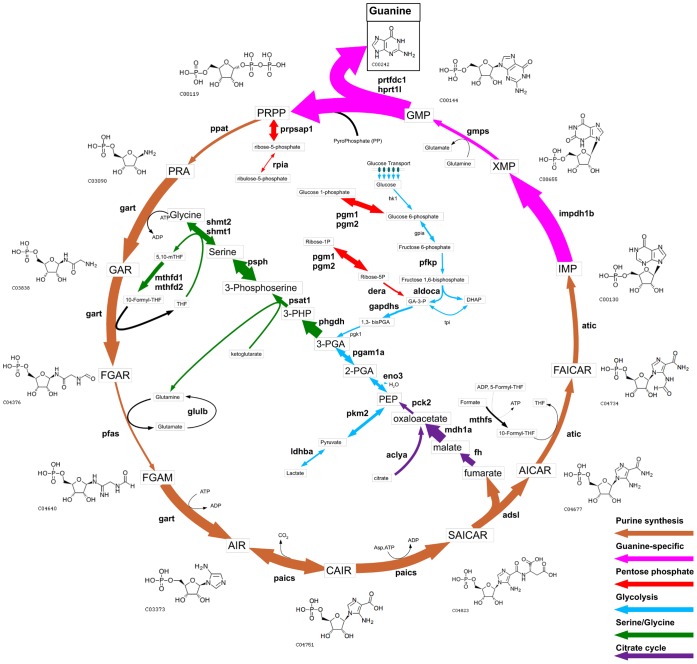
- Iridophores showed extensive enrichment of guanine-production machinery: 5 glucose transporters, 7/11 glycolysis steps, and 9/9 de novo purine synthesis enzymes were enriched; impdh1b was 71-fold higher than in melanocytes, prtfdc1 was 198-fold higher, and the adenine-specific adssl was not significantly different from melanocytes (0.7-fold).
- The iridophore-enriched list included 346 genes, with 30 genes expressed at least 30-fold above melanocytes and RPE and 100-fold above whole embryos; the authors propose a guanine cycle model in which PRPP is recycled during guanine pigment production.
Methods
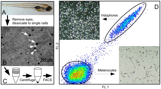
- Zebrafish homozygous for temperature-sensitive mitfavc7 were dissociated, pigment cells enriched by Percoll density gradient centrifugation, and sorted by FACS using iridophore autofluorescence; mRNA was extracted and Illumina cDNA libraries were sequenced for 11 melanocyte, 5 iridophore, and 5 RPE samples, plus whole 3 dpf embryos.
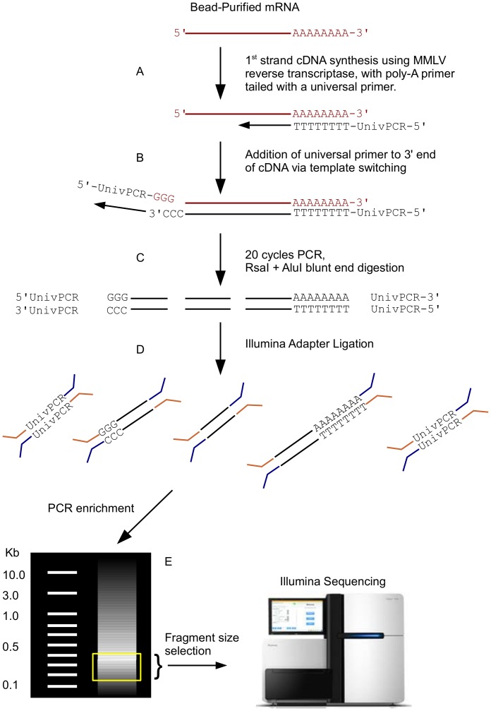
- Sequence tags were aligned with Novoalign to a custom non-redundant database of 25,102 zebrafish genes; expression was normalized as RPKM, statistical tests were performed in R, and selected expression changes were validated by qRT-PCR.
- Enrichment analyses used a 1 RPKM baseline and thresholds including ≥2-fold over the third cell type with p<0.05, ≥8-fold over whole embryos for co-enrichment/cell-specific genes, and stricter cutoffs for selected shared or iridophore-specific gene lists.
Limitations
- Stated: mitfa may be partially compromised by aberrant splicing at permissive temperatures, though melanocytes appeared indistinguishable from wild-type; RPE preparations lacked separate markers for eye-associated neural crest-derived melanocytes or choroidal melanocytes, and one RPE library was prepared later than 5 dpf.
- Stated: the authors did not have statistical power to confidently track developmental changes in gene expression within specific cell types, and xanthophores were not isolated by the procedure.
- Evident: the method relies on density and autofluorescence rather than lineage markers, so purity depends on those physical properties; the reduced-representation cDNA strategy could miss short cDNAs lacking restriction sites, although the authors included enzyme sites in amplification primers to mitigate this.
Model-written summary. Check it against the paper before relying on it.
Integrity signals
Registry
Cites
Statistics
Figures
Verification
Claims the paper makes
66 Own result · 35 Own interpretation · 18 Own method · 17 Partly supported · 16 Supported · 13 No full text of the sources · 5 Supported so far · 1 Not supported · 1 Couldn’t confirm · 1 Sources not found · 1 Not checked yet · 1 No basis given · accuracy not yet measured
The cited sources support only part of this claim — read each source’s verdict below.
In the paper
“Melanocytes, iridophores, and RPE develop normally at 25°C in mitfavc7.”
§ Gene Expression Analysis of Zebrafish Melanocytes, Iridophores, and Retinal Pigmented Epithelium Reveals Indicators of Biological Function and Developmental Origin > Materials and Methods > Zebrafish Strain and Sample Collection Time Points
What the sources say
- Partly supportedcites [41] SL Johnson, 2011, mitfa is required at multiple stages of melanocyte differentiation but not to establish the melanocyte stem cell, Developmental Biology sourceExperimental — accuracy not yet measured
The Results section (‘Identification of temperature-sensitive alleles of mitfa’; page unavailable) says mitfavc7 develops normal or nearly normal melanocyte patterns at 25°C, not unequivocally normal ones. The paper also says RPE has no requirement for mitfa, but it does not establish that iridophores develop normally in mitfavc7 at 25°C; its discussion of excess iridophores concerns temperature-sensitive alleles at restrictive temperatures. (LLM, conf=0.85)
plain- Asserted: passes — The authors put it forward as true.
- Checkable: passes — It can be checked against evidence.
- Load-bearing: passes — The paper’s conclusions depend on it.
- Based: passes — The paper gives a reason why it holds.
- Partly supported
Every source the paper cites for this claim was checked and supports it.
In the paper
“Zebrafish bearing mutations in the de novo purine synthesis enzymes gart and paics have iridophore defects, indicating purine synthesis is important for iridophore pigmentation [30].”
§ Gene Expression Analysis of Zebrafish Melanocytes, Iridophores, and Retinal Pigmented Epithelium Reveals Indicators of Biological Function and Developmental Origin > Introduction
What the sources say
- Supportedcites [30] A Ng, 2009, Zebrafish mutations in gart and paics identify crucial roles for de novo purine synthesis in vertebrate pigmentation and ocular development, Development source
The Introduction reports that zebrafish gart and paics mutants have pigmentation defects affecting iridophores. The paper’s opening summary states that iridophore pigmentation is almost completely absent in zygotic mutants, supporting the claim that de novo purine synthesis is important for it. (LLM, conf=0.94)
hedgeddirect- Asserted: passes — The authors put it forward as true.
- Checkable: passes — It can be checked against evidence.
- Load-bearing: passes — The paper’s conclusions depend on it.
- Based: passes — The paper gives a reason why it holds.
- Supported
Every source the paper cites for this claim was checked and supports it.
In the paper
“This mutant facilitated the collection of RPE, as mitfa is not required for RPE development in zebrafish [41].”
§ Gene Expression Analysis of Zebrafish Melanocytes, Iridophores, and Retinal Pigmented Epithelium Reveals Indicators of Biological Function and Developmental Origin > Materials and Methods > Zebrafish Strain and Sample Collection Time Points
What the sources say
- Supportedcites [41] SL Johnson, 2011, mitfa is required at multiple stages of melanocyte differentiation but not to establish the melanocyte stem cell, Developmental Biology source
The Results section states that the RPE has no requirement for mitfa. The Introduction also says that zebrafish mitfa null alleles spare the retinal pigment epithelium. No page number is available in our copy. (LLM, conf=0.94)
plain- Asserted: passes — The authors put it forward as true.
- Checkable: passes — It can be checked against evidence.
- Load-bearing: passes — The paper’s conclusions depend on it.
- Based: passes — The paper gives a reason why it holds.
- Supported
Every source the paper cites for this claim was checked and supports it.
In the paper
“For instance, ltk, known to be a marker of iridophores, is highly enriched in iridophores compared to melanocytes (red square with x in Figure S4) [27].”
§ Gene Expression Analysis of Zebrafish Melanocytes, Iridophores, and Retinal Pigmented Epithelium Reveals Indicators of Biological Function and Developmental Origin > Results and Discussion > Purification of Melanocytes, Iridophores, and RPE from Whole Embryos to Generate Cell-specific Gene Expression Data
What the sources say
- Supportedcites [27] SS Lopes, 2008, Leukocyte Tyrosine Kinase Functions in Pigment Cell Development, PLoS Genetics source
The Discussion reports persistent, robust ltk expression in the iridophore lineage and says cells adopting melanophore fate extinguish ltk expression. The Results section ‘shd expression is prominent in NCCs and iridophores’ further reports that, at late stages, ltk expression in neural-crest derivatives is restricted to iridophores. These passages support the stated comparison, though the paper also notes earlier expression in some neural-crest cells. (LLM, conf=0.91)
boostedcorrelational- Asserted: passes — The authors put it forward as true.
- Checkable: passes — It can be checked against evidence.
- Load-bearing: unclear — It is unclear whether the paper’s conclusions depend on it.
- Based: passes — The paper gives a reason why it holds.
- Supported
The cited sources support only part of this claim — read each source’s verdict below.
In the paper
“Known to be expressed in iridophores”
§ Gene Expression Analysis of Zebrafish Melanocytes, Iridophores, and Retinal Pigmented Epithelium Reveals Indicators of Biological Function and Developmental Origin > Results and Discussion > Iridophore Gene Enrichment
What the sources say
- Partly supportedcites [77] JA Lister, 2011, Embryonic expression of zebrafish MiT family genes tfe3b, tfeb, and tfec, Developmental Dynamics sourceExperimental — accuracy not yet measured
In Results and Discussion, under “tfec” (page not provided), the authors observe tfec-expressing cells in locations associated with iridophores. They call the pattern “strongly suggestive” of iridophore pigment cells, but do not establish expression in identified iridophores as a known fact. (LLM, conf=0.84)
- Not checked — no full text of the sourcecites [78] MC Chung, 2001, TFEC can function as a transcriptional activator of the nonmuscle myosin II heavy chain-A gene in transfected cells, Biochemistry source
- Not checked — no full text of the sourcecites [79] KC Mansky, 2002, The microphthalmia transcription factor and the related helix-loop-helix zipper factors TFE-3 and TFE-C collaborate to activate the tartrate-resistant acid phosphatase, Journal of Leukocyte Biology source
boostedcorrelational- Asserted: passes — The authors put it forward as true.
- Checkable: passes — It can be checked against evidence.
- Load-bearing: unclear — It is unclear whether the paper’s conclusions depend on it.
- Based: passes — The paper gives a reason why it holds.
- Partly supported
The cited sources support only part of this claim — read each source’s verdict below.
In the paper
“Although not required for RPE development in zebrafish, mitfa is expressed in the RPE at a relatively high level, consistent with the reported ability of mitfa to promote pigmented fate in zebrafish retinas [59].”
§ Gene Expression Analysis of Zebrafish Melanocytes, Iridophores, and Retinal Pigmented Epithelium Reveals Indicators of Biological Function and Developmental Origin > Results and Discussion > Melanocyte and RPE Co-Expression
What the sources say
- Partly supportedcites [59] BM Lane, 2012, Otx but not mitf transcription factors are required for zebrafish retinal pigment epithelium development, PLoS ONE sourceExperimental — accuracy not yet measured
The Introduction (page unavailable) states that mitfa is expressed in the developing zebrafish RPE, but does not describe its expression there as relatively high. The Results section on Mitfa and Mitfb (page unavailable) supports the surrounding statement that mitfa is not required for RPE development. (LLM, conf=0.87)
plain- Asserted: passes — The authors put it forward as true.
- Checkable: passes — It can be checked against evidence.
- Load-bearing: unclear — It is unclear whether the paper’s conclusions depend on it.
- Based: passes — The paper gives a reason why it holds.
- Partly supported
Every source the paper cites for this claim was checked and supports it.
In the paper
“We found a single receptor tyrosine kinase enriched in iridophores, ltk, which we have previously shown to be required for iridophore development [27].”
§ Gene Expression Analysis of Zebrafish Melanocytes, Iridophores, and Retinal Pigmented Epithelium Reveals Indicators of Biological Function and Developmental Origin > Results and Discussion > Iridophore Gene Enrichment
What the sources say
- Supportedcites [27] SS Lopes, 2008, Leukocyte Tyrosine Kinase Functions in Pigment Cell Development, PLoS Genetics source
The Discussion states that, in zebrafish, Ltk is crucial for specifying iridophores from neural crest cells. The Results also report that shd/ltk mutants usually lacked iridophores, supporting the claim that ltk is required for their development. (LLM, conf=0.95)
boosteddirect- Asserted: passes — The authors put it forward as true.
- Checkable: passes — It can be checked against evidence.
- Load-bearing: unclear — It is unclear whether the paper’s conclusions depend on it.
- Based: passes — The paper gives a reason why it holds.
- Supported
The sources with a verdict support this claim; the rest weren’t checked yet, have no full text we could read, or their verdicts aren’t rated yet.
In the paper
“Also included is the cell adhesion molecule pcdh10a, which is expressed by migrating zebrafish neural crest cells, and acts as a tumor suppressor in several cancers [61]-[63].”
§ Gene Expression Analysis of Zebrafish Melanocytes, Iridophores, and Retinal Pigmented Epithelium Reveals Indicators of Biological Function and Developmental Origin > Results and Discussion > Melanocyte and Iridophore Co-Expression
What the sources say
- Supportedcites [61] Zhong X, Zhu Y, Mao J, Zhang J, Zheng S (2012) Frequent epigenetic silencing of PCDH10 by methylation in human colorectal cancer. Journal of Cancer Research and Clinical Oncology doi 10.1007/s00432–012–1353–5. source
The citation in the printed sentence supports the tumor-suppressor statement, rather than the citing authors’ co-enrichment observation. The Discussion (p. 5) concludes that PCDH10 suppresses tumor growth and invasion in colorectal cancer and reports related findings in other cancers, including gastric cancer. (LLM, conf=0.84)
- Not checked — no full text of the sourcecites [62] KC Bertrand, 2011, PCDH10 is a candidate tumour suppressor gene in medulloblastoma, Child’s Nervous Syst source
- Not checked — no full text of the sourcecites [63] Z Li, 2011, Epigenetic inactivation of PCDH10 in human prostate cancer cell lines, Cell Biology International source
plaincorrelational- Asserted: passes — The authors put it forward as true.
- Checkable: passes — It can be checked against evidence.
- Load-bearing: unclear — It is unclear whether the paper’s conclusions depend on it.
- Based: passes — The paper gives a reason why it holds.
- Supported
Every source the paper cites for this claim was checked and supports it.
In the paper
“We found a single receptor tyrosine kinase enriched in iridophores, ltk, which we have previously shown to be required for iridophore development [27].”
§ Gene Expression Analysis of Zebrafish Melanocytes, Iridophores, and Retinal Pigmented Epithelium Reveals Indicators of Biological Function and Developmental Origin > Results and Discussion > Iridophore Gene Enrichment
What the sources say
- Supportedcites [27] SS Lopes, 2008, Leukocyte Tyrosine Kinase Functions in Pigment Cell Development, PLoS Genetics source
The citation is attached to the citing authors’ statement that they previously showed ltk is required for iridophore development. The source’s Discussion says Ltk is crucial for iridophore specification in zebrafish, and its Results report that shd/ltk mutants usually lacked iridophores. The description of ltk as the single enriched receptor tyrosine kinase is presented as the citing authors’ own finding. (LLM, conf=0.92)
plaincorrelational- Asserted: passes — The authors put it forward as true.
- Checkable: passes — It can be checked against evidence.
- Load-bearing: passes — The paper’s conclusions depend on it.
- Based: passes — The paper gives a reason why it holds.
- Supported
The cited sources support only part of this claim — read each source’s verdict below.
In the paper
“Many of the genes previously known to be expressed by iridophores are components of the purine synthesis pathway, as iridophore pigment largely consists of stacks of guanine plates [30], [71].”
§ Gene Expression Analysis of Zebrafish Melanocytes, Iridophores, and Retinal Pigmented Epithelium Reveals Indicators of Biological Function and Developmental Origin > Results and Discussion > Iridophore Gene Enrichment
What the sources say
- Partly supportedcites [30] A Ng, 2009, Zebrafish mutations in gart and paics identify crucial roles for de novo purine synthesis in vertebrate pigmentation and ocular development, Development sourceExperimental — accuracy not yet measured
The Introduction identifies gart and paics as purine-synthesis genes and describes guanine stacks in iridophores. The Results say these genes are highly expressed in pigment cells, but do not establish that many previously known iridophore-expressed genes belong to the pathway. (LLM, conf=0.82)
- Partly supportedcites [71] K Curran, 2010, Interplay between Foxd3 and Mitf regulates cell fate plasticity in the zebrafish neural crest, Developmental Biology sourceExperimental — accuracy not yet measured
The Results section (‘pnp4a is a novel iridoblast marker’; page not provided) identifies pnp4a as an iridophore marker. The Discussion (‘pnp4a as a novel iridophore marker’; page not provided) links its enzyme family to purine synthesis and iridescence to guanine crystals. This supports one example, but not the claim that many known iridophore-expressed genes are purine-synthesis components. (LLM, conf=0.86)
plaincorrelational- Asserted: passes — The authors put it forward as true.
- Checkable: passes — It can be checked against evidence.
- Load-bearing: passes — The paper’s conclusions depend on it.
- Based: passes — The paper gives a reason why it holds.
- Partly supported
- Probably not a substantial claim. 40
- It may state two separate claims as one. 22
- The quote only partly states this claim. 3
At least one cited source doesn’t support this claim or points to a different paper, and none confirmed it — see the verdicts below.
In the paper
“Previous descriptions of gene expression in the RPE of zebrafish, chicken, and human have elucidated many of the genes playing roles in RPE biology [36]–[38].”
§ Gene Expression Analysis of Zebrafish Melanocytes, Iridophores, and Retinal Pigmented Epithelium Reveals Indicators of Biological Function and Developmental Origin > Introduction
What the sources say
- Disputedcites [36] YF Leung, 2007, Gene expression profiling of zebrafish embryonic retinal pigment epithelium in vivo, Investigative Ophthalmology & Visual Science sourceExperimental — accuracy not yet measured
The Introduction says the study profiled the developing zebrafish retina, not its retinal pigment epithelium (RPE). The Materials and Methods describe removing the RPE before collecting the retinal tissue. For the zebrafish portion of this multi-citation sentence, the paper therefore describes gene expression in a different tissue. (LLM, conf=0.88)
Checked by a reasoning model — may be wrong.
- Not checked — no full text of the sourcecites [37] LJ Rizzolo, 2007, Analysis of the RPE transcriptome reveals dynamic changes during the development of the outer blood-retinal barrier, Mol Vis
- Not checked — no full text of the sourcecites [38] G Wistow, 2002, Expressed sequence tag analysis of human RPE/choroid for the NEIBank Project: over 6000 non-redundant transcripts, novel genes and splice variants, Mol Vis
plaindirect- Asserted: passes — The authors put it forward as true.
- Checkable: passes — It can be checked against evidence.
- Load-bearing: fails — The paper’s conclusions may not depend on it.
- Based: passes — The paper gives a reason why it holds.
Probably not a substantial claim. The paper’s conclusions may not depend on it.
- Disputed
Every source the paper cites for this claim was checked and supports it.
In the paper
“Zebrafish bearing mutations in the de novo purine synthesis enzymes gart and paics have iridophore defects, indicating purine synthesis is important for iridophore pigmentation [30].”
§ Gene Expression Analysis of Zebrafish Melanocytes, Iridophores, and Retinal Pigmented Epithelium Reveals Indicators of Biological Function and Developmental Origin > Introduction
What the sources say
- Supportedcites [30] A Ng, 2009, Zebrafish mutations in gart and paics identify crucial roles for de novo purine synthesis in vertebrate pigmentation and ocular development, Development source
The Introduction identifies gart and paics as de novo purine synthesis enzymes and reports pigmentation defects affecting iridophores in both mutants. The Discussion states that the results support a model in which the GTP branch of purine synthesis mediates embryonic pigmentation. (LLM, conf=0.95)
plaindirect- Asserted: passes — The authors put it forward as true.
- Checkable: passes — It can be checked against evidence.
- Load-bearing: passes — The paper’s conclusions depend on it.
- Based: passes — The paper gives a reason why it holds.
Probably not a substantial claim.
- Supported
Every source the paper cites for this claim was checked and supports it.
In the paper
“Many of these genes are responsible for producing melanin, and defects in melanin production are often associated with reduced visual function [39].”
§ Gene Expression Analysis of Zebrafish Melanocytes, Iridophores, and Retinal Pigmented Epithelium Reveals Indicators of Biological Function and Developmental Origin > Introduction
What the sources say
- Supportedcites [39] K Grønskov, 2007, Oculocutaneous albinism, Orphanet Journal of Rare Diseases source
The Definition section states that reduced melanin biosynthesis causes oculocutaneous albinism and that reduced melanin in the eyes results in reduced visual acuity. It also links the different types to mutations in different genes. Page numbers are not provided in our copy. (LLM, conf=0.94)
plaincorrelational- Asserted: passes — The authors put it forward as true.
- Checkable: passes — It can be checked against evidence.
- Load-bearing: fails — The paper’s conclusions may not depend on it.
- Based: passes — The paper gives a reason why it holds.
Probably not a substantial claim. The paper’s conclusions may not depend on it.
- Supported
The cited sources support only part of this claim — read each source’s verdict below.
In the paper
“Many of these genes are responsible for producing melanin, and defects in melanin production are often associated with reduced visual function [39].”
§ Gene Expression Analysis of Zebrafish Melanocytes, Iridophores, and Retinal Pigmented Epithelium Reveals Indicators of Biological Function and Developmental Origin > Introduction
What the sources say
- Partly supportedcites [39] K Grønskov, 2007, Oculocutaneous albinism, Orphanet Journal of Rare Diseases sourceExperimental — accuracy not yet measured
The Etiology section identifies multiple genes involved in melanin biosynthesis, including TYR and TYRP1, and the Definition section links reduced ocular melanin to reduced visual acuity. The paper discusses these genes in melanocytes; it does not establish that they are specifically RPE genes. (LLM, conf=0.83)
plaindirect- Asserted: passes — The authors put it forward as true.
- Checkable: passes — It can be checked against evidence.
- Load-bearing: fails — The paper’s conclusions may not depend on it.
- Based: passes — The paper gives a reason why it holds.
Probably not a substantial claim. The paper’s conclusions may not depend on it.
- Partly supported
Every source the paper cites for this claim was checked and supports it.
In the paper
“The RPE develops from the eye primordium, and is continuous with the layer of cells that forms the iris [31].”
§ Gene Expression Analysis of Zebrafish Melanocytes, Iridophores, and Retinal Pigmented Epithelium Reveals Indicators of Biological Function and Developmental Origin > Introduction
What the sources say
- Supportedcites [31] K Bharti, 2006, The other pigment cell: specification and development of the pigmented epithelium of the vertebrate eye, Pigment Cell Res source
The Introduction says RPE cells arise from the optic neuroepithelium during development of the eye from its primordium. “The development of the iris and the ciliary body” says the RPE extends into the back layer of the iris, supporting the described continuity. (LLM, conf=0.93)
plaindirect- Asserted: passes — The authors put it forward as true.
- Checkable: passes — It can be checked against evidence.
- Load-bearing: fails — The paper’s conclusions may not depend on it.
- Based: passes — The paper gives a reason why it holds.
Probably not a substantial claim. The paper’s conclusions may not depend on it. It may state two separate claims as one.
- Supported
The cited sources support only part of this claim — read each source’s verdict below.
In the paper
“We note the possibility that mitfa may be partially compromised by aberrant splicing products in this mutant at permissive temperatures, but following development at 25°C, melanocyte numbers, morphology, and pigmentation are indistinguishable from wild-type zebrafish [41].”
§ Gene Expression Analysis of Zebrafish Melanocytes, Iridophores, and Retinal Pigmented Epithelium Reveals Indicators of Biological Function and Developmental Origin > Materials and Methods > Zebrafish Strain and Sample Collection Time Points
What the sources say
- Partly supportedcites [41] SL Johnson, 2011, mitfa is required at multiple stages of melanocyte differentiation but not to establish the melanocyte stem cell, Developmental Biology sourceExperimental — accuracy not yet measured
The Results section, “Identification of temperature-sensitive alleles of mitfa” (page unavailable), says mitfavc7 developed a normal or nearly normal melanocyte pattern at 25°C. It also says the allele appeared somewhat compromised even at 23°C. The source therefore supports a near-normal pattern at 25°C, but not the stronger claim that melanocyte numbers, morphology, and pigmentation were all indistinguishable from wild type. (LLM, conf=0.88)
hedged- Asserted: passes — The authors put it forward as true.
- Checkable: passes — It can be checked against evidence.
- Load-bearing: passes — The paper’s conclusions depend on it.
- Based: passes — The paper gives a reason why it holds.
It may state two separate claims as one.
- Partly supported
Every source the paper cites for this claim was checked and supports it.
In the paper
“In contrast, several neural crest-derived pigment cells are found in zebrafish and other poikilotherms, including reflective iridophores [5].”
§ Gene Expression Analysis of Zebrafish Melanocytes, Iridophores, and Retinal Pigmented Epithelium Reveals Indicators of Biological Function and Developmental Origin > Introduction
What the sources say
- Supportedcites [5] DM Parichy, 2000, Mutational analysis of endothelin receptor b1 (rose) during neural crest and pigment pattern development in the zebrafish Danio rerio, Developmental Biology source
The Introduction describes three classes of pigment cells in fishes and amphibians, including silver iridophores, and says zebrafish neural crest cells differentiate into all three. The phenotype section also describes iridophores as highly reflective; page numbers are not provided in our copy. (LLM, conf=0.93)
plain- Asserted: passes — The authors put it forward as true.
- Checkable: passes — It can be checked against evidence.
- Load-bearing: passes — The paper’s conclusions depend on it.
- Based: passes — The paper gives a reason why it holds.
Probably not a substantial claim.
- Supported
Every source the paper cites for this claim was checked and supports it.
In the paper
“Several requirements for iridophore development from the neural crest are known [5], [26], [27].”
§ Gene Expression Analysis of Zebrafish Melanocytes, Iridophores, and Retinal Pigmented Epithelium Reveals Indicators of Biological Function and Developmental Origin > Introduction
What the sources say
- Supportedcites [5] DM Parichy, 2000, Mutational analysis of endothelin receptor b1 (rose) during neural crest and pigment pattern development in the zebrafish Danio rerio, Developmental Biology source
The section on pigment pattern metamorphosis reports a severe iridophore deficit in adult ednrb1 mutants, and the mutant-phenotype section describes an essential role for ednrb1 in adult pigment pattern development. The Introduction identifies iridophores as neural crest-derived pigment cells. Page numbers are unavailable in our copy. (LLM, conf=0.88)
- Supportedcites [26] K Curran, 2009, Foxd3 controls melanophore specification in the zebrafish neural crest by regulation of Mitf, Developmental Biology source
The Discussion (page not available) identifies Foxd3 as promoting iridophore specification and reports that Mitfa represses iridophore development. The Introduction identifies iridophores as neural crest-derived pigment cells. Together, these passages support the broad statement that requirements for their development are known. (LLM, conf=0.90)
- Supportedcites [27] SS Lopes, 2008, Leukocyte Tyrosine Kinase Functions in Pigment Cell Development, PLoS Genetics source
The Discussion (Figure 8; page not provided) identifies Ltk signalling and Sox10 function as requirements for iridoblast specification from neural crest cells. The Results also report that iridophore-lineage markers are absent in ltk mutants. (LLM, conf=0.90)
plain- Asserted: passes — The authors put it forward as true.
- Checkable: passes — It can be checked against evidence.
- Load-bearing: fails — The paper’s conclusions may not depend on it.
- Based: passes — The paper gives a reason why it holds.
Probably not a substantial claim. The paper’s conclusions may not depend on it.
- Supported
Every source the paper cites for this claim was checked and supports it.
In the paper
“consistent with the reported ability of mitfa to promote pigmented fate in zebrafish retinas [59].”
§ Gene Expression Analysis of Zebrafish Melanocytes, Iridophores, and Retinal Pigmented Epithelium Reveals Indicators of Biological Function and Developmental Origin > Results and Discussion > Melanocyte and RPE Co-Expression
What the sources say
- Supportedcites [59] BM Lane, 2012, Otx but not mitf transcription factors are required for zebrafish retinal pigment epithelium development, PLoS ONE source
The Results section, “Mitfa has the ability to promote pigmented cell fate in the developing retina,” reports patches of pigmented cells after mitfa was expressed in the developing zebrafish retina. The authors conclude that mitfa can induce pigmentation there, supporting the attributed claim. (LLM, conf=0.94)
plaindirect- Asserted: passes — The authors put it forward as true.
- Checkable: passes — It can be checked against evidence.
- Load-bearing: fails — The paper’s conclusions may not depend on it.
- Based: passes — The paper gives a reason why it holds.
Probably not a substantial claim. The paper’s conclusions may not depend on it.
- Supported
The cited sources support only part of this claim — read each source’s verdict below.
In the paper
“In addition, the formation of COPI transport vesicles is known to be mediated by the interaction of GAPDH with RAB2 [75].”
§ Gene Expression Analysis of Zebrafish Melanocytes, Iridophores, and Retinal Pigmented Epithelium Reveals Indicators of Biological Function and Developmental Origin > Results and Discussion > Iridophore Gene Enrichment
What the sources say
- Partly supportedcites [75] EJ Tisdale, 2004, Glyceraldehyde-3-phosphate dehydrogenase interacts with Rab2 and plays an essential role in endoplasmic reticulum to Golgi transport exclusive of its glycolytic activity, Journal of Biological Chemistry sourceExperimental — accuracy not yet measured
The Results show that activated Rab2 interacts directly with GAPDH, and the Abstract links Rab2 to vesicle formation from vesicular tubular clusters. But the Abstract says GAPDH’s precise function there is unknown; the cited paper does not establish that the interaction mediates formation of COPI vesicles. (LLM, conf=0.87)
boosteddirect- Asserted: passes — The authors put it forward as true.
- Checkable: passes — It can be checked against evidence.
- Load-bearing: fails — The paper’s conclusions may not depend on it.
- Based: passes — The paper gives a reason why it holds.
Probably not a substantial claim. The paper’s conclusions may not depend on it.
- Partly supported
The cited sources support only part of this claim — read each source’s verdict below.
In the paper
“Included in this enrichment list are several factors known to be important for iridophore development, including ltk, ednrb1, and pnp4a [5], [26], [27].”
§ Gene Expression Analysis of Zebrafish Melanocytes, Iridophores, and Retinal Pigmented Epithelium Reveals Indicators of Biological Function and Developmental Origin > Results and Discussion > Iridophore Gene Enrichment
What the sources say
- Supportedcites [5] DM Parichy, 2000, Mutational analysis of endothelin receptor b1 (rose) during neural crest and pigment pattern development in the zebrafish Danio rerio, Developmental Biology source
The cited paper supports the ednrb1 part of the multi-citation sentence. In the section on ednrb1 expression during pigment pattern metamorphosis, it reports a severe deficit of iridophores in adult ednrb1 mutants; the mutant-phenotype discussion calls the gene essential for adult pigment pattern development. The sentence does not specifically assign ltk or pnp4a to this paper. (LLM, conf=0.93)
- Could not verifycites [26] K Curran, 2009, Foxd3 controls melanophore specification in the zebrafish neural crest by regulation of Mitf, Developmental Biology sourceExperimental — accuracy not yet measured
The Discussion says Foxd3 appears to promote iridophore specification, but it does not address whether ltk, ednrb1, or pnp4a are important for iridophore development. Searches of our copy for those gene names and their expanded terms found no passage establishing the attributed claim. The indexed passages provide no page numbers. (LLM, conf=0.72)
- Supportedcites [27] SS Lopes, 2008, Leukocyte Tyrosine Kinase Functions in Pigment Cell Development, PLoS Genetics source
The Discussion states that Ltk is crucial for iridophore specification, supporting the ltk part of this multi-citation sentence. The Results also report that shd/ltk mutants usually lack iridophores. The sentence does not assign all three genes to this citation alone. (LLM, conf=0.92)
boosteddirect- Asserted: passes — The authors put it forward as true.
- Checkable: passes — It can be checked against evidence.
- Load-bearing: fails — The paper’s conclusions may not depend on it.
- Based: passes — The paper gives a reason why it holds.
Probably not a substantial claim. The paper’s conclusions may not depend on it.
- Supported
Every source the paper cites for this claim was checked and supports it.
In the paper
“Roles for GPNMB have been described for melanocytes, melanoma, and the pigmented iris in mammals, and it has been suggested to act both as a plasma membrane protein and a component of the melanosome [70].”
§ Gene Expression Analysis of Zebrafish Melanocytes, Iridophores, and Retinal Pigmented Epithelium Reveals Indicators of Biological Function and Developmental Origin > Results and Discussion > Iridophore Gene Enrichment
What the sources say
- Supportedcites [70] M Tomihari, 2009, Gpnmb is a melanosome-associated glycoprotein that contributes to melanocyte/keratinocyte adhesion in a RGD-dependent fashion, Experimental Dermatology source
The Introduction describes Gpnmb's links to iris pigment dispersion in mice, human melanoma, and melanocyte biology. The Discussion reports that Gpnmb resides mostly in melanosomes, with a smaller portion on the cell surface; the paper also describes a role in melanocyte–keratinocyte adhesion. (LLM, conf=0.92)
hedgedcorrelational- Asserted: passes — The authors put it forward as true.
- Checkable: passes — It can be checked against evidence.
- Load-bearing: fails — The paper’s conclusions may not depend on it.
- Based: passes — The paper gives a reason why it holds.
Probably not a substantial claim. The paper’s conclusions may not depend on it. It may state two separate claims as one.
- Supported
The cited sources support only part of this claim — read each source’s verdict below.
In the paper
“In humans, Rab27 mutations cause hypopigmentation associated with the immunodeficiency disorder Griscelli syndrome type II [74].”
§ Gene Expression Analysis of Zebrafish Melanocytes, Iridophores, and Retinal Pigmented Epithelium Reveals Indicators of Biological Function and Developmental Origin > Results and Discussion > Iridophore Gene Enrichment
What the sources say
- Partly supportedcites [74] P Bahadoran, 2001, Rab27a: A key to melanosome transport in human melanocytes, The Journal of Cell Biology sourceExperimental — accuracy not yet measured
The Abstract describes Griscelli syndrome as involving immune deficiency and partial albinism and says a Rab27a mutation can cause it. The Discussion also links absent Rab27a to pigmentary dilution in human patients. Our copy does not identify the condition specifically as Griscelli syndrome type II. (LLM, conf=0.83)
plaindirect- Asserted: passes — The authors put it forward as true.
- Checkable: passes — It can be checked against evidence.
- Load-bearing: fails — The paper’s conclusions may not depend on it.
- Based: passes — The paper gives a reason why it holds.
Probably not a substantial claim. The paper’s conclusions may not depend on it.
- Partly supported
The sources with a verdict support this claim; the rest weren’t checked yet, have no full text we could read, or their verdicts aren’t rated yet.
In the paper
“previously reported to be expressed in neural crest tissues [64]–[67].”
§ Gene Expression Analysis of Zebrafish Melanocytes, Iridophores, and Retinal Pigmented Epithelium Reveals Indicators of Biological Function and Developmental Origin > Results and Discussion > Melanocyte and Iridophore Co-Expression
What the sources say
- Not checked yetcites [64] Thisse C, Thisse B (2005) High Throughput Expression Analysis of ZF-Models Consortium Clones. ZFIN Direct Data Submission (http://zfin.org).
- Not checked — no full text of the sourcecites [65] A Tallafuss, 2006, Characterization of retinoid-X receptor genes rxra, rxrba, rxrbb and rxrg during zebrafish development, Gene Expression Patterns source
- Supportedcites [66] JS Waxman, 2007, Comparison of the expression patterns of newly identified zebrafish retinoic acid and retinoid X receptors, Developmental Dynamics source
In “rxr Expression Patterns Through 24 hpf,” the paper reports rxrga expression in the third stream of cranial neural crest at 24 hpf. It specifies that expression was not observed in migrating crest. (LLM, conf=0.93)
- Not checked — no full text of the sourcecites [67] C He, 2009, Tissue-specific and embryonic expression of the retinoid X receptors in Sebastiscus marmoratus. Comparative Biochemistry and Physiology, Part B, Biochemistry & Molecular Biology source
plain- Asserted: passes — The authors put it forward as true.
- Checkable: passes — It can be checked against evidence.
- Load-bearing: fails — The paper’s conclusions may not depend on it.
- Based: passes — The paper gives a reason why it holds.
Probably not a substantial claim. The paper’s conclusions may not depend on it.
The cited sources support only part of this claim — read each source’s verdict below.
In the paper
“Phosphorylation of GAPDH occurs through the Src/PI3K/AKT pathway, often downstream of receptor tyrosine kinase activation [76].”
§ Gene Expression Analysis of Zebrafish Melanocytes, Iridophores, and Retinal Pigmented Epithelium Reveals Indicators of Biological Function and Developmental Origin > Results and Discussion > Iridophore Gene Enrichment
What the sources say
- Partly supportedcites [76] Q Huang, 2011, AKT2 suppresses GAPDH mediated-apoptosis in ovarian cancer cells via phosphorylating GAPDH at threonine 237 and decreasing its nuclear translocation, Journal of Biological Chemistry sourceExperimental — accuracy not yet measured
The Results (page not provided) show that Akt2 phosphorylates GAPDH and that wortmannin or Akt2 siRNA inhibits the observed phosphorylation; the paper also places Akt downstream of PI3K. The cited passages do not establish a Src/PI3K/Akt route to GAPDH phosphorylation or show that it often follows receptor tyrosine kinase activation. (LLM, conf=0.88)
plaindirect- Asserted: passes — The authors put it forward as true.
- Checkable: passes — It can be checked against evidence.
- Load-bearing: fails — The paper’s conclusions may not depend on it.
- Based: passes — The paper gives a reason why it holds.
Probably not a substantial claim. The paper’s conclusions may not depend on it.
- Partly supported
The cited sources support only part of this claim — read each source’s verdict below.
In the paper
“which is expressed by migrating zebrafish neural crest cells, and acts as a tumor suppressor in several cancers [61]-[63].”
§ Gene Expression Analysis of Zebrafish Melanocytes, Iridophores, and Retinal Pigmented Epithelium Reveals Indicators of Biological Function and Developmental Origin > Results and Discussion > Melanocyte and Iridophore Co-Expression
What the sources say
- Partly supportedcites [61] Zhong X, Zhu Y, Mao J, Zhang J, Zheng S (2012) Frequent epigenetic silencing of PCDH10 by methylation in human colorectal cancer. Journal of Cancer Research and Clinical Oncology doi 10.1007/s00432–012–1353–5. sourceExperimental — accuracy not yet measured
The Discussion (p. 5) identifies human PCDH10 as a functional tumor-suppression gene in colorectal cancer and discusses findings in other tumors. The paper does not establish that zebrafish pcdh10a is expressed in migrating neural crest cells, or that the zebrafish gene acts as a tumor suppressor. (LLM, conf=0.88)
- Not checked — no full text of the sourcecites [62] KC Bertrand, 2011, PCDH10 is a candidate tumour suppressor gene in medulloblastoma, Child’s Nervous Syst source
- Not checked — no full text of the sourcecites [63] Z Li, 2011, Epigenetic inactivation of PCDH10 in human prostate cancer cell lines, Cell Biology International source
plaindirect- Asserted: passes — The authors put it forward as true.
- Checkable: passes — It can be checked against evidence.
- Load-bearing: fails — The paper’s conclusions may not depend on it.
- Based: passes — The paper gives a reason why it holds.
Probably not a substantial claim. The paper’s conclusions may not depend on it. It may state two separate claims as one.
- Partly supported
The cited sources support only part of this claim — read each source’s verdict below.
In the paper
“TFEC forms hetero and homodimers with other bHLH members and can function as a transcriptional activator or repressor [77]–[79].”
§ Gene Expression Analysis of Zebrafish Melanocytes, Iridophores, and Retinal Pigmented Epithelium Reveals Indicators of Biological Function and Developmental Origin > Results and Discussion > Iridophore Gene Enrichment
What the sources say
- Partly supportedcites [77] JA Lister, 2011, Embryonic expression of zebrafish MiT family genes tfe3b, tfeb, and tfec, Developmental Dynamics sourceExperimental — accuracy not yet measured
The Introduction (page not provided) says MiT proteins, including TFEC, can form homodimers and heterodimers with one another, but explicitly says they do not dimerize with other bHLH or bHLH-ZIP proteins outside that family. The paper says TFEC regulates gene expression and lists a study of its inhibition of TFE3-dependent activation, but it does not establish the claimed ability to function as both a transcriptional activator and a repressor. (LLM, conf=0.86)
- Not checked — no full text of the sourcecites [78] MC Chung, 2001, TFEC can function as a transcriptional activator of the nonmuscle myosin II heavy chain-A gene in transfected cells, Biochemistry source
- Not checked — no full text of the sourcecites [79] KC Mansky, 2002, The microphthalmia transcription factor and the related helix-loop-helix zipper factors TFE-3 and TFE-C collaborate to activate the tartrate-resistant acid phosphatase, Journal of Leukocyte Biology source
plaindirect- Asserted: passes — The authors put it forward as true.
- Checkable: passes — It can be checked against evidence.
- Load-bearing: fails — The paper’s conclusions may not depend on it.
- Based: passes — The paper gives a reason why it holds.
Probably not a substantial claim. The paper’s conclusions may not depend on it. It may state two separate claims as one.
- Partly supported
The cited sources support only part of this claim — read each source’s verdict below.
In the paper
“Because melanocyte and RPE gene expression have been previously characterized, we do not discuss them in detail here, but present the data as the supplemental tables listed below [36]–[38], [68], [69].”
§ Gene Expression Analysis of Zebrafish Melanocytes, Iridophores, and Retinal Pigmented Epithelium Reveals Indicators of Biological Function and Developmental Origin > Results and Discussion > Cell-Type Specific Gene Expression
What the sources say
- Could not verifycites [36] YF Leung, 2007, Gene expression profiling of zebrafish embryonic retinal pigment epithelium in vivo, Investigative Ophthalmology & Visual Science sourceExperimental — accuracy not yet measured
The Materials and Methods describe removing the RPE before profiling retinal RNA, and the Results compare retinal with whole-embryo gene expression. Searches for RPE and melanocyte expression found no passage in our copy characterizing gene expression in either cell type. No page numbers were available for these passages. (LLM, conf=0.75)
- Not checked — no full text of the sourcecites [37] LJ Rizzolo, 2007, Analysis of the RPE transcriptome reveals dynamic changes during the development of the outer blood-retinal barrier, Mol Vis
- Not checked — no full text of the sourcecites [38] G Wistow, 2002, Expressed sequence tag analysis of human RPE/choroid for the NEIBank Project: over 6000 non-redundant transcripts, novel genes and splice variants, Mol Vis
- Partly supportedcites [68] R Flockhart, 2012, BRAFV600E remodels the melanocyte transcriptome and induces BANCR to regulate melanoma cell migration, Genome Research sourceExperimental — accuracy not yet measured
The Results (page unavailable) characterize gene expression in normal human melanocytes, including BRAF-regulated transcripts. The cited paper does not address RPE gene expression, so it supports only the melanocyte part of the statement. (LLM, conf=0.86)
- Partly supportedcites [69] J An, 2011, A Comparative Transcriptomic Analysis of Uveal Melanoma and Normal Uveal Melanocyte, PLoS ONE sourceExperimental — accuracy not yet measured
The Results section (‘Gene profiling and the category of highly-expressed genes in two libraries’; page unavailable) reports gene-expression profiling of normal uveal melanocytes. The cited paper does not characterize RPE gene expression, so it supports only the melanocyte part of the claim. (LLM, conf=0.88)
plain- Asserted: passes — The authors put it forward as true.
- Checkable: passes — It can be checked against evidence.
- Load-bearing: fails — The paper’s conclusions may not depend on it.
- Based: passes — The paper gives a reason why it holds.
Probably not a substantial claim. The paper’s conclusions may not depend on it.
- Could not verify
The cited sources support only part of this claim — read each source’s verdict below.
In the paper
“Relatedly, endothelin receptor (ednrb1) signaling is required for iridophore development in zebrafish [5].”
§ Gene Expression Analysis of Zebrafish Melanocytes, Iridophores, and Retinal Pigmented Epithelium Reveals Indicators of Biological Function and Developmental Origin > Results and Discussion > Gene Expression Indicative of Cellular Function or Developmental Origin
What the sources say
- Partly supportedcites [5] DM Parichy, 2000, Mutational analysis of endothelin receptor b1 (rose) during neural crest and pigment pattern development in the zebrafish Danio rerio, Developmental Biology sourceExperimental — accuracy not yet measured
The Abstract states that zebrafish ednrb1 is required for a subset of adult iridophores. The section ‘ednrb1 Is Expressed during Pigment Pattern Metamorphosis’ reports a severe iridophore deficit in mutant adults but no gross pigment-pattern defect in mutant embryos. The claim omits the adult-stage and subset qualifications, making the requirement sound general. (LLM, conf=0.86)
plaindirect- Asserted: passes — The authors put it forward as true.
- Checkable: passes — It can be checked against evidence.
- Load-bearing: fails — The paper’s conclusions may not depend on it.
- Based: passes — The paper gives a reason why it holds.
Probably not a substantial claim. The paper’s conclusions may not depend on it.
- Partly supported
The cited sources that could be checked couldn’t confirm or dispute this claim.
In the paper
“Pigment cell enrichment of ribosomal components is not surprising, considering several mouse coat color mutants are ribosomal proteins, although they are not the ribosomal components enriched here [50]–[53].”
§ Gene Expression Analysis of Zebrafish Melanocytes, Iridophores, and Retinal Pigmented Epithelium Reveals Indicators of Biological Function and Developmental Origin > Results and Discussion > Gene Expression Indicative of a Pigment Cell Identity
What the sources say
- Not checked — no full text of the sourcecites [50] ER Oliver, 2004, Ribosomal protein L24 defect in belly spot and tail (Bst), a mouse Minute, Development (Cambridge, England) source
- Not checked — no full text of the sourcecites [51] N Draptchinskaia, 1999, The gene encoding ribosomal protein S19 is mutated in Diamond-Blackfan anaemia, Nature Genetics source
- Could not verifycites [52] WC Skarnes, 2011, A conditional knockout resource for the genome-wide study of mouse gene function, Nature sourceExperimental — accuracy not yet measured
The Abstract and Discussion describe a resource for generating mouse knockout alleles, not coat-color phenotypes or ribosomal proteins. Searches of our copy for coat-color mutants and ribosomal pigmentation found no passage addressing the attributed relationship; the claim about which components were enriched also cannot be checked against this source. (LLM, conf=0.74)
- Could not verifycites [53] MH Hrabe de Angelis, 2000, Genome-wide, large-scale production of mutant mice by ENU mutagenesis, Nature Genetics sourceExperimental — accuracy not yet measured
The indexed copy's bibliographic entry (section ‘1999 (7)’, no page) identifies the paper but provides no findings about coat color mutants or ribosomal proteins. Searches for those terms and for pigmentation returned only catalog material, so the claim cannot be checked against the available text. (LLM, conf=0.72)
plain- Asserted: passes — The authors put it forward as true.
- Checkable: passes — It can be checked against evidence.
- Load-bearing: fails — The paper’s conclusions may not depend on it.
- Based: passes — The paper gives a reason why it holds.
Probably not a substantial claim. The paper’s conclusions may not depend on it. It may state two separate claims as one.
Every source the paper cites for this claim was checked and supports it.
In the paper
“It is intriguing that alcama has been found to mediate endothelin 1 signaling in cartilage development [58].”
§ Gene Expression Analysis of Zebrafish Melanocytes, Iridophores, and Retinal Pigmented Epithelium Reveals Indicators of Biological Function and Developmental Origin > Results and Discussion > Gene Expression Indicative of Cellular Function or Developmental Origin
What the sources say
- Supportedcites [58] P Choudhry, 2011, Alcama mediates Edn1 signaling during zebrafish cartilage morphogenesis, Developmental Biology source
The Conclusion states that Alcama functions downstream of Edn1 during cartilage formation and supports a mechanism by which it mediates Edn1 signaling. The Introduction identifies this finding specifically in zebrafish, consistent with the citing sentence’s general paraphrase. (LLM, conf=0.94)
plaindirect- Asserted: passes — The authors put it forward as true.
- Checkable: passes — It can be checked against evidence.
- Load-bearing: fails — The paper’s conclusions may not depend on it.
- Based: passes — The paper gives a reason why it holds.
Probably not a substantial claim. The paper’s conclusions may not depend on it.
- Supported
Every source the paper cites for this claim was checked and supports it.
In the paper
“Another oncogene shown to regulate glycolysis and associated feeder pathways is Ets-1 [83].”
§ Gene Expression Analysis of Zebrafish Melanocytes, Iridophores, and Retinal Pigmented Epithelium Reveals Indicators of Biological Function and Developmental Origin > Results and Discussion > Iridophore Gene Enrichment
What the sources say
- Supportedcites [83] ML Verschoor, 2010, Ets-1 regulates energy metabolism in cancer cells, PLoS ONE source
The Abstract reports that Ets-1 over-expression up-regulates enzymes involved in glycolysis and associated feeder pathways in ovarian cancer cells. The Discussion calls Ets-1 an oncogenic transcription factor. The cited claim accurately summarizes those findings. (LLM, conf=0.92)
plaindirect- Asserted: passes — The authors put it forward as true.
- Checkable: passes — It can be checked against evidence.
- Load-bearing: fails — The paper’s conclusions may not depend on it.
- Based: passes — The paper gives a reason why it holds.
Probably not a substantial claim. The paper’s conclusions may not depend on it. It may state two separate claims as one.
- Supported
The cited sources support only part of this claim — read each source’s verdict below.
In the paper
“It is established that MYC upregulates glycolysis, DNA-synthesis, and nucleotide metabolism [80]–[82].”
§ Gene Expression Analysis of Zebrafish Melanocytes, Iridophores, and Retinal Pigmented Epithelium Reveals Indicators of Biological Function and Developmental Origin > Results and Discussion > Iridophore Gene Enrichment
What the sources say
- Partly supportedcites [80] SJ Yeung, 2008, Roles of p53, MYC and HIF-1 in regulating glycolysis–the seventh hallmark of cancer, Cellular and Molecular Life Sciences sourceExperimental — accuracy not yet measured
The section “Coordinated Regulation of Proteins that Control the Glycolytic Flux” (pp. 7–8) says increased c-MYC activity helps upregulate glycolytic-enzyme genes and glycolytic capacity. Our copy does not say that MYC upregulates DNA synthesis or nucleotide metabolism; searches for those terms found no supporting passage. (LLM, conf=0.87)
- Partly supportedcites [81] M Albajar, 2011, MYC in chronic myeloid leukemia: induction of aberrant DNA synthesis and association with poor response to imatinib, Molecular Cancer Research sourceExperimental — accuracy not yet measured
The Introduction (page not provided) describes MYC-induced DNA synthesis and mentions enhanced energetic metabolism. The section “Ectopic MYC increased DNA synthesis in K562 cells treated with imatinib” (page not provided) reports increased DNA synthesis after MYC induction. Neither passage establishes the more specific claims that MYC upregulates glycolysis or nucleotide metabolism. (LLM, conf=0.88)
- Partly supportedcites [82] Y Liu, 2008, Global regulation of nucleotide biosynthetic genes by c-Myc, PLoS ONE sourceExperimental — accuracy not yet measured
The Discussion states that MYC upregulates glycolytic genes, and the Results show that MYC induction upregulates genes for nucleotide biosynthesis. The Discussion links these effects to providing energy and nucleotides for DNA synthesis, but our copy does not establish that MYC upregulates DNA synthesis itself. Page numbers are unavailable in our copy. (LLM, conf=0.84)
boosteddirect- Asserted: passes — The authors put it forward as true.
- Checkable: passes — It can be checked against evidence.
- Load-bearing: fails — The paper’s conclusions may not depend on it.
- Based: passes — The paper gives a reason why it holds.
Probably not a substantial claim. The paper’s conclusions may not depend on it. It may state two separate claims as one.
- Partly supported
The sources with a verdict support this claim; the rest weren’t checked yet, have no full text we could read, or their verdicts aren’t rated yet.
In the paper
“Members of the Cart1/Alx3/Alx4 family of homeodomain proteins are known to regulate formation of skeletal elements in organisms ranging from sea urchin to mammals [86], [87].”
§ Gene Expression Analysis of Zebrafish Melanocytes, Iridophores, and Retinal Pigmented Epithelium Reveals Indicators of Biological Function and Developmental Origin > Results and Discussion > Iridophore Gene Enrichment
What the sources say
- Supportedcites [86] CA Ettensohn, 2003, Alx1, a member of the Cart1/Alx3/Alx4 subfamily of Paired-class homeodomain proteins, is an essential component of the gene network controlling skeletogenic fate specification in the sea urchin embryo, Development source
The Introduction identifies Alx1 as the first known invertebrate member of the Cart1/Alx3/Alx4 subfamily and reports that it is required for skeletogenic fate specification in sea urchins. It also states that these proteins function in vertebrate skeletogenesis; the section on evolution of biomineralization describes skeletal defects in mice and humans. Together, these passages support the claimed range from sea urchins to mammals. (LLM, conf=0.93)
- Not checked — no full text of the sourcecites [87] A Beverdam, 2001, Severe nasal clefting and abnormal embryonic apoptosis in Alx3/Alx4 double mutant mice, Development source
boosteddirect- Asserted: passes — The authors put it forward as true.
- Checkable: passes — It can be checked against evidence.
- Load-bearing: fails — The paper’s conclusions may not depend on it.
- Based: passes — The paper gives a reason why it holds.
Probably not a substantial claim. The paper’s conclusions may not depend on it.
- Supported
The sources with a verdict support this claim; the rest weren’t checked yet, have no full text we could read, or their verdicts aren’t rated yet.
In the paper
“In humans, mutation and haploinsufficiency of ALX4 are associated with skull ossification defects [88], [89].”
§ Gene Expression Analysis of Zebrafish Melanocytes, Iridophores, and Retinal Pigmented Epithelium Reveals Indicators of Biological Function and Developmental Origin > Results and Discussion > Iridophore Gene Enrichment
What the sources say
- Not checked — no full text of the sourcecites [88] LA Mavrogiannis, 2001, Haploinsufficiency of the human homeobox gene ALX4 causes skull ossification defects, Nature Genetics source
- Supportedcites [89] W Wuyts, 2000, The ALX4 homeobox gene is mutated in patients with ossification defects of the skull (foramina parietalia permagna, OMIM 168500), Journal of Medical Genetics source
The Discussion reports ALX4 mutations in two human families with foramina parietalia permagna, a skull ossification defect. It specifically says the inactivating frameshift mutation in one family indicates that ALX4 haploinsufficiency results in the condition. (LLM, conf=0.94)
plaincorrelational- Asserted: passes — The authors put it forward as true.
- Checkable: passes — It can be checked against evidence.
- Load-bearing: fails — The paper’s conclusions may not depend on it.
- Based: passes — The paper gives a reason why it holds.
Probably not a substantial claim. The paper’s conclusions may not depend on it.
Every source the paper cites for this claim was checked and supports it.
In the paper
“Known to have several roles in the embryo and an early specifier of posterior neural crest in Xenopus”
§ Gene Expression Analysis of Zebrafish Melanocytes, Iridophores, and Retinal Pigmented Epithelium Reveals Indicators of Biological Function and Developmental Origin > Results and Discussion > Iridophore Gene Enrichment
What the sources say
- Supportedcites [85] B Li, 2009, The posteriorizing gene Gbx2 is a direct target of Wnt signalling and the earliest factor in neural crest induction, Development source
The Discussion describes Gbx2 roles in midbrain–hindbrain boundary formation and neural plate posteriorization, as well as early neural crest induction. The Introduction states that Gbx2 promotes posterior neural fold fate (neural crest) and acts upstream of the earliest expressed neural crest genes. (LLM, conf=0.93)
plaindirect- Asserted: passes — The authors put it forward as true.
- Checkable: passes — It can be checked against evidence.
- Load-bearing: fails — The paper’s conclusions may not depend on it.
- Based: passes — The paper gives a reason why it holds.
Probably not a substantial claim. The paper’s conclusions may not depend on it. It may state two separate claims as one.
- Supported
A result the paper reports from its own data — it cites no other source for it, so there is none to check. The quote shows it is in the paper.
In the paper
“In summary, our efforts have generated a non-redundant zebrafish cDNA database that identifies most (∼99%) of annotated genes, and includes 7% (10/126) records of dubious utility, but would tend not to confound RNA-seq analysis such as reported here.”
§ Gene Expression Analysis of Zebrafish Melanocytes, Iridophores, and Retinal Pigmented Epithelium Reveals Indicators of Biological Function and Developmental Origin > Materials and Methods > Sequence Analysis
plain- Asserted: passes — The authors put it forward as true.
- Checkable: passes — It can be checked against evidence.
- Load-bearing: fails — The paper’s conclusions may not depend on it.
- Based: passes — The paper gives a reason why it holds.
Probably not a substantial claim. The paper’s conclusions may not depend on it.
In the paper
“Included in this group are several well-known regulators and markers of neural crest and pigment cell development, including the transcription factors sox10 and the expressed repetitive element crestin [60].”
§ Gene Expression Analysis of Zebrafish Melanocytes, Iridophores, and Retinal Pigmented Epithelium Reveals Indicators of Biological Function and Developmental Origin > Results and Discussion > Melanocyte and Iridophore Co-Expression
What the sources say
- Not checked — no full text of the sourcecites [60] AL Rubinstein, 2000, Genes dependent on zebrafish cyclops function identified by AFLP differential gene expression screen, Genesis source
boostedcorrelational- Asserted: passes — The authors put it forward as true.
- Checkable: passes — It can be checked against evidence.
- Load-bearing: passes — The paper’s conclusions depend on it.
- Based: passes — The paper gives a reason why it holds.
The quote only partly states this claim.
A result the paper reports from its own data — it cites no other source for it, so there is none to check. The quote shows it is in the paper.
In the paper
“By this general assessment, melanocytes, RPE, and iridophores are distinct from whole embryos, with average correlation values less than 0.02.”
§ Gene Expression Analysis of Zebrafish Melanocytes, Iridophores, and Retinal Pigmented Epithelium Reveals Indicators of Biological Function and Developmental Origin > Results and Discussion > Melanocyte, Iridophore, and RPE Gene Expression is Correlated Compared to Whole Embryos
plain- Asserted: passes — The authors put it forward as true.
- Checkable: passes — It can be checked against evidence.
- Load-bearing: passes — The paper’s conclusions depend on it.
- Based: passes — The paper gives a reason why it holds.
It may state two separate claims as one.
A result the paper reports from its own data — it cites no other source for it, so there is none to check. The quote shows it is in the paper.
In the paper
“When comparing iridophores to melanocytes and RPE, we find 5 facilitated glucose transporters, 7/11 steps of glycolysis, and 9/9 enzymes for de novo purine synthesis to be enriched.”
§ Gene Expression Analysis of Zebrafish Melanocytes, Iridophores, and Retinal Pigmented Epithelium Reveals Indicators of Biological Function and Developmental Origin > Results and Discussion > Iridophore Gene Enrichment
plaincorrelational- Asserted: passes — The authors put it forward as true.
- Checkable: passes — It can be checked against evidence.
- Load-bearing: passes — The paper’s conclusions depend on it.
- Based: passes — The paper gives a reason why it holds.
It may state two separate claims as one.
A result the paper reports from its own data — it cites no other source for it, so there is none to check. The quote shows it is in the paper.
In the paper
“Interestingly, there is one other bHLH in enriched in iridophores when compared to melanocytes and RPE, but it did not meet the 8-fold requirement over whole embryos.”
§ Gene Expression Analysis of Zebrafish Melanocytes, Iridophores, and Retinal Pigmented Epithelium Reveals Indicators of Biological Function and Developmental Origin > Results and Discussion > Iridophore Gene Enrichment
plaincorrelational- Asserted: passes — The authors put it forward as true.
- Checkable: passes — It can be checked against evidence.
- Load-bearing: unclear — It is unclear whether the paper’s conclusions depend on it.
- Based: passes — The paper gives a reason why it holds.
It may state two separate claims as one. The quote only partly states this claim.
What the paper did — it cites no source for this, so there is none to check. The quote shows it is in the paper.
In the paper
“This study was carried out in accordance with the Washington University Animal Use Committee guidelines under approved protocol #20110236.”
§ Gene Expression Analysis of Zebrafish Melanocytes, Iridophores, and Retinal Pigmented Epithelium Reveals Indicators of Biological Function and Developmental Origin > Materials and Methods > Zebrafish Strain and Sample Collection Time Points
plain- Asserted: passes — The authors put it forward as true.
- Checkable: passes — It can be checked against evidence.
- Load-bearing: fails — The paper’s conclusions may not depend on it.
- Based: passes — The paper gives a reason why it holds.
Probably not a substantial claim. The paper’s conclusions may not depend on it.
In the paper
“The RPE forms part of the blood-retina barrier, providing the eye with an immune-privileged status [33].”
§ Gene Expression Analysis of Zebrafish Melanocytes, Iridophores, and Retinal Pigmented Epithelium Reveals Indicators of Biological Function and Developmental Origin > Introduction
What the sources say
- Not checked — no full text of the sourcecites [33] EA Runkle, 2011, The blood-retinal barrier: structure and functional significance, Methods Mol Biol source
plaindirect- Asserted: passes — The authors put it forward as true.
- Checkable: passes — It can be checked against evidence.
- Load-bearing: fails — The paper’s conclusions may not depend on it.
- Based: passes — The paper gives a reason why it holds.
Probably not a substantial claim. The paper’s conclusions may not depend on it. It may state two separate claims as one.
What the paper did — it cites no source for this, so there is none to check. The quote shows it is in the paper.
In the paper
“All sequencing data used in this study are publicly available at NCBI’s GEO database under accession GSE46387 (http://www.ncbi.nlm.nih.gov/geo/query/acc.cgi?acc=GSE46387).”
§ Gene Expression Analysis of Zebrafish Melanocytes, Iridophores, and Retinal Pigmented Epithelium Reveals Indicators of Biological Function and Developmental Origin > Materials and Methods > Sequence Analysis
plain- Asserted: passes — The authors put it forward as true.
- Checkable: passes — It can be checked against evidence.
- Load-bearing: fails — The paper’s conclusions may not depend on it.
- Based: passes — The paper gives a reason why it holds.
Probably not a substantial claim. The paper’s conclusions may not depend on it.
A claim is checked only against the cited paper’s full text, never its abstract, and none is available for its sources.
In the paper
“Defects in the RPE contribute to diseases such as macular degeneration and retinitis pigmentosa, which result in vision problems [34], [35].”
§ Gene Expression Analysis of Zebrafish Melanocytes, Iridophores, and Retinal Pigmented Epithelium Reveals Indicators of Biological Function and Developmental Origin > Introduction
What the sources say
- Not checked — no full text of the sourcecites [34] S Dridi, 2012, ERK1/2 activation is a therapeutic target in age-related macular degeneration, Proc Natl Acad Sci USA source
- Not checked — no full text of the sourcecites [35] DA Thompson, 2003, Genetic defects in vitamin A metabolism of the retinal pigment epithelium, Developments in Ophthalmology source
plaindirect- Asserted: passes — The authors put it forward as true.
- Checkable: passes — It can be checked against evidence.
- Load-bearing: fails — The paper’s conclusions may not depend on it.
- Based: passes — The paper gives a reason why it holds.
Probably not a substantial claim. The paper’s conclusions may not depend on it.
The paper states this without a source or data behind it here, so there is nothing to check it against. The quote shows it is in the paper.
In the paper
“When held at 32°C, the neural crest-derived melanocytes do not develop, but RPE and iridophores develop normally.”
§ Gene Expression Analysis of Zebrafish Melanocytes, Iridophores, and Retinal Pigmented Epithelium Reveals Indicators of Biological Function and Developmental Origin > Materials and Methods > Zebrafish Strain and Sample Collection Time Points
plaindirect- Asserted: passes — The authors put it forward as true.
- Checkable: passes — It can be checked against evidence.
- Load-bearing: passes — The paper’s conclusions depend on it.
- Based: passes — The paper gives a reason why it holds.
It may state two separate claims as one.
A claim is checked only against the cited paper’s full text, never its abstract, and none is available for its sources.
In the paper
“This is not entirely surprising, as RPE65 is known to be present in keratinocytes, melanocytes, and melanoma, in addition to the RPE [46], [47].”
§ Gene Expression Analysis of Zebrafish Melanocytes, Iridophores, and Retinal Pigmented Epithelium Reveals Indicators of Biological Function and Developmental Origin > Results and Discussion > Purification of Melanocytes, Iridophores, and RPE from Whole Embryos to Generate Cell-specific Gene Expression Data
What the sources say
- Not checked — no full text of the sourcecites [46] G Hinterhuber, 2004, RPE65 of retinal pigment epithelium, a putative receptor molecule for plasma retinol-binding protein, is expressed in human keratinocytes. J Invest Dermatol, Feb source
- Not checked — no full text of the sourcecites [47] PM Amann, 2012, Vitamin A metabolism in benign and malignant melanocytic skin cells: importance of lecithin/retinol acyltransferase and RPE65, J Cell Physiol. Feb source
boostedcorrelational- Asserted: passes — The authors put it forward as true.
- Checkable: passes — It can be checked against evidence.
- Load-bearing: fails — The paper’s conclusions may not depend on it.
- Based: passes — The paper gives a reason why it holds.
Probably not a substantial claim. The paper’s conclusions may not depend on it.
What the paper did — it cites no source for this, so there is none to check. The quote shows it is in the paper.
In the paper
“In summary, our efforts have generated a non-redundant zebrafish cDNA database that identifies most (∼99%) of annotated genes, and includes 7% (10/126) records of dubious utility, but would tend not to confound RNA-seq analysis such as reported here.”
§ Gene Expression Analysis of Zebrafish Melanocytes, Iridophores, and Retinal Pigmented Epithelium Reveals Indicators of Biological Function and Developmental Origin > Materials and Methods > Sequence Analysis
plain- Asserted: passes — The authors put it forward as true.
- Checkable: passes — It can be checked against evidence.
- Load-bearing: fails — The paper’s conclusions may not depend on it.
- Based: passes — The paper gives a reason why it holds.
Probably not a substantial claim. The paper’s conclusions may not depend on it.
What the paper did — it cites no source for this, so there is none to check. The quote shows it is in the paper.
In the paper
“This purification procedure relies on the inherent densities of melanin and guanine-filled cells; hence it can be used without other complicated lineage markers.”
§ Gene Expression Analysis of Zebrafish Melanocytes, Iridophores, and Retinal Pigmented Epithelium Reveals Indicators of Biological Function and Developmental Origin > Introduction
plaindirect- Asserted: passes — The authors put it forward as true.
- Checkable: passes — It can be checked against evidence.
- Load-bearing: passes — The paper’s conclusions depend on it.
- Based: passes — The paper gives a reason why it holds.
The quote only partly states this claim.
What the paper did — it cites no source for this, so there is none to check. The quote shows it is in the paper.
In the paper
“The other pigment cells in zebrafish, the pteridine-containing xanthophores, do not pass through the density gradient and are not isolated by this method.”
§ Gene Expression Analysis of Zebrafish Melanocytes, Iridophores, and Retinal Pigmented Epithelium Reveals Indicators of Biological Function and Developmental Origin > Results and Discussion > Purification of Melanocytes, Iridophores, and RPE from Whole Embryos to Generate Cell-specific Gene Expression Data
plain- Asserted: passes — The authors put it forward as true.
- Checkable: passes — It can be checked against evidence.
- Load-bearing: unclear — It is unclear whether the paper’s conclusions depend on it.
- Based: passes — The paper gives a reason why it holds.
It may state two separate claims as one.
In the paper
“It provides trophic support and recycles wastes from the photoreceptors of the retina [32].”
§ Gene Expression Analysis of Zebrafish Melanocytes, Iridophores, and Retinal Pigmented Epithelium Reveals Indicators of Biological Function and Developmental Origin > Introduction
What the sources say
- Not checked — no full text of the sourcecites [32] O Strauss, 2005, The retinal pigment epithelium in visual function, Physiological Reviews source
plaindirect- Asserted: passes — The authors put it forward as true.
- Checkable: passes — It can be checked against evidence.
- Load-bearing: fails — The paper’s conclusions may not depend on it.
- Based: passes — The paper gives a reason why it holds.
Probably not a substantial claim. The paper’s conclusions may not depend on it. It may state two separate claims as one.
In the paper
“Rab GTPases regulate many aspects of membranous vesicle formation and traffic [73].”
§ Gene Expression Analysis of Zebrafish Melanocytes, Iridophores, and Retinal Pigmented Epithelium Reveals Indicators of Biological Function and Developmental Origin > Results and Discussion > Iridophore Gene Enrichment
What the sources say
- Not checked — no full text of the sourcecites [73] BL Grosshans, 2006, Rabs and their effectors: achieving specificity in membrane traffic, Proc Natl Acad Sci USA source
plaindirect- Asserted: passes — The authors put it forward as true.
- Checkable: passes — It can be checked against evidence.
- Load-bearing: fails — The paper’s conclusions may not depend on it.
- Based: passes — The paper gives a reason why it holds.
Probably not a substantial claim. The paper’s conclusions may not depend on it.
What the paper did — it cites no source for this, so there is none to check. The quote shows it is in the paper.
In the paper
“To independently validate these enrichment sets, we generated separate biological samples for each cell type and compared expression of selected genes from the enrichment sets via qRT-PCR.”
§ Gene Expression Analysis of Zebrafish Melanocytes, Iridophores, and Retinal Pigmented Epithelium Reveals Indicators of Biological Function and Developmental Origin > Results and Discussion > Cell-Type Specific Gene Expression
plain- Asserted: passes — The authors put it forward as true.
- Checkable: passes — It can be checked against evidence.
- Load-bearing: passes — The paper’s conclusions depend on it.
- Based: passes — The paper gives a reason why it holds.
Probably not a substantial claim.
In the paper
“ARFs are a large family of ras-related GTPases that regulate membrane trafficking and organelle structure (For review see [72]).”
§ Gene Expression Analysis of Zebrafish Melanocytes, Iridophores, and Retinal Pigmented Epithelium Reveals Indicators of Biological Function and Developmental Origin > Results and Discussion > Iridophore Gene Enrichment
What the sources say
- Not checked — no full text of the sourcecites [72] JG Donaldson, 2005, Localization and function of Arf family GTPases, Biochemical Society Trans source
plaindirect- Asserted: passes — The authors put it forward as true.
- Checkable: passes — It can be checked against evidence.
- Load-bearing: fails — The paper’s conclusions may not depend on it.
- Based: passes — The paper gives a reason why it holds.
Probably not a substantial claim. The paper’s conclusions may not depend on it. It may state two separate claims as one.
What the paper did — it cites no source for this, so there is none to check. The quote shows it is in the paper.
In the paper
“We observed good correlation of RNA-seq and qRT-PCR relative expression levels, indicating our RNA-seq data is a reliable indicator of the gene expression in these pigment cells (r2 = 0.75, Figure S4).”
§ Gene Expression Analysis of Zebrafish Melanocytes, Iridophores, and Retinal Pigmented Epithelium Reveals Indicators of Biological Function and Developmental Origin > Results and Discussion > Cell-Type Specific Gene Expression
hedgedcorrelational- Asserted: passes — The authors put it forward as true.
- Checkable: passes — It can be checked against evidence.
- Load-bearing: passes — The paper’s conclusions depend on it.
- Based: passes — The paper gives a reason why it holds.
It may state two separate claims as one.
In the paper
“However, in situ analysis reveals no enriched expression in xanthophores, suggesting that slc45a2 expression is not shared by all pigment cells [55].”
§ Gene Expression Analysis of Zebrafish Melanocytes, Iridophores, and Retinal Pigmented Epithelium Reveals Indicators of Biological Function and Developmental Origin > Results and Discussion > Gene Expression Indicative of a Pigment Cell Identity
What the sources say
- Not checked — no full text of the sourcecites [55] Dooley CM, Schwarz H, Mueller K, Mongera A, Konantz M, <etal>et al</etal>.. (2012) Slc45a2 and V-ATPase are regulators of melanosomal pH homeostasis in zebrafish, providing a mechanism for human pigment evolution and disease. Pigment Cell & Melanoma Research doi: 10.1111/pcmr.12053. source
hedged- Asserted: passes — The authors put it forward as true.
- Checkable: passes — It can be checked against evidence.
- Load-bearing: passes — The paper’s conclusions depend on it.
- Based: passes — The paper gives a reason why it holds.
It may state two separate claims as one.
A conclusion the paper draws from its own results — it cites no other source for it, so there is none to check. The quote shows it is in the paper.
In the paper
“The third highest expressed gene on this list, slc23l, may act as a guanine transporter.”
§ Gene Expression Analysis of Zebrafish Melanocytes, Iridophores, and Retinal Pigmented Epithelium Reveals Indicators of Biological Function and Developmental Origin > Results and Discussion > Iridophore Gene Enrichment
hedgedconditional- Asserted: passes — The authors put it forward as true.
- Checkable: passes — It can be checked against evidence.
- Load-bearing: fails — The paper’s conclusions may not depend on it.
- Based: passes — The paper gives a reason why it holds.
Probably not a substantial claim. The paper’s conclusions may not depend on it.
- Rests on an assumption: In mammals, the SLC23 gene family has roles in transporting nucleobases, such as guanine, as well as vitamin C.
A conclusion the paper draws from its own results — it cites no other source for it, so there is none to check. The quote shows it is in the paper.
In the paper
“Therefore, we suggest these lists of co-expressed genes are likely enriched for common metabolic functions, in the case of melanocytes and RPE, or developmental origins, in the case of melanocytes and iridophores.”
§ Gene Expression Analysis of Zebrafish Melanocytes, Iridophores, and Retinal Pigmented Epithelium Reveals Indicators of Biological Function and Developmental Origin > Results and Discussion > Gene Expression Indicative of Cellular Function or Developmental Origin
hedgedcorrelational- Asserted: passes — The authors put it forward as true.
- Checkable: passes — It can be checked against evidence.
- Load-bearing: passes — The paper’s conclusions depend on it.
- Based: passes — The paper gives a reason why it holds.
It may state two separate claims as one.
- Rests on an assumption: Genes co-enriched among pigment cell types indicate shared pigment cell functions.
In the paper
“Because correlation values can be artificially skewed by outlying data points [49], we calculated the average correlation from 24,102 overlapping windows of 1000 genes, sorted by increasing whole embryo expression (Figure S6).”
§ Gene Expression Analysis of Zebrafish Melanocytes, Iridophores, and Retinal Pigmented Epithelium Reveals Indicators of Biological Function and Developmental Origin > Results and Discussion > Melanocyte, Iridophore, and RPE Gene Expression is Correlated Compared to Whole Embryos
What the sources say
- Not checked — no full text of the sourcecites [49] A Fujita, 2009, Comparing Pearson, Spearman and Hoeffding’s D measure for gene expression association analysis, Journal of Bioinformatics and Computational Biology source
plain- Asserted: passes — The authors put it forward as true.
- Checkable: passes — It can be checked against evidence.
- Load-bearing: passes — The paper’s conclusions depend on it.
- Based: passes — The paper gives a reason why it holds.
Probably not a substantial claim.
A conclusion the paper draws from its own results — it cites no other source for it, so there is none to check. The quote shows it is in the paper.
In the paper
“Presumably, this low value (0.02) reflects the baseline correlation component from expression of housekeeping genes shared by all cells”
§ Gene Expression Analysis of Zebrafish Melanocytes, Iridophores, and Retinal Pigmented Epithelium Reveals Indicators of Biological Function and Developmental Origin > Results and Discussion > Melanocyte, Iridophore, and RPE Gene Expression is Correlated Compared to Whole Embryos
hedgeddirect- Asserted: passes — The authors put it forward as true.
- Checkable: passes — It can be checked against evidence.
- Load-bearing: fails — The paper’s conclusions may not depend on it.
- Based: passes — The paper gives a reason why it holds.
Probably not a substantial claim. The paper’s conclusions may not depend on it.
In the paper
“Upon inspection of the known pathway of guanine production, we observed synthesis of guanine from GMP likely results in the recycling of 5-Phosphoribosyl 1-Pyrophosphate (PRPP), the rate-limiting substrate in purine synthesis (KEGG Pathway: dre00230).”
§ Gene Expression Analysis of Zebrafish Melanocytes, Iridophores, and Retinal Pigmented Epithelium Reveals Indicators of Biological Function and Developmental Origin > Results and Discussion > Iridophore Gene Enrichment
What the sources say
- Not matchedcites KEGG Pathway: dre00230
Couldn’t be matched to a paper in Crossref, so it can’t be checked.
hedgedconditional- Asserted: passes — The authors put it forward as true.
- Checkable: passes — It can be checked against evidence.
- Load-bearing: fails — The paper’s conclusions may not depend on it.
- Based: passes — The paper gives a reason why it holds.
Probably not a substantial claim. The paper’s conclusions may not depend on it.
- Not matched
What the paper did — it cites no source for this, so there is none to check. The quote shows it is in the paper.
In the paper
“To contribute to the understanding of pigment cell biology, we have developed a method for rapidly and reliably purifying melanocytes, iridophores, and retinal pigmented epithelium from zebrafish embryos, followed by global gene expression analysis by mRNA sequencing.”
§ Gene Expression Analysis of Zebrafish Melanocytes, Iridophores, and Retinal Pigmented Epithelium Reveals Indicators of Biological Function and Developmental Origin > Results and Discussion > Conclusion
plain- Asserted: passes — The authors put it forward as true.
- Checkable: passes — It can be checked against evidence.
- Load-bearing: passes — The paper’s conclusions depend on it.
- Based: passes — The paper gives a reason why it holds.
It may state two separate claims as one.
A conclusion the paper draws from its own results — it cites no other source for it, so there is none to check. The quote shows it is in the paper.
In the paper
“which uniquely allowed us to identify co-expressed genes indicative of shared function or developmental origin, as well as those that are specifically enriched in single cell types.”
§ Gene Expression Analysis of Zebrafish Melanocytes, Iridophores, and Retinal Pigmented Epithelium Reveals Indicators of Biological Function and Developmental Origin > Results and Discussion > Conclusion
plaindirect- Asserted: passes — The authors put it forward as true.
- Checkable: passes — It can be checked against evidence.
- Load-bearing: passes — The paper’s conclusions depend on it.
- Based: passes — The paper gives a reason why it holds.
It may state two separate claims as one.
The paper relies on this without support.
This is the first analysis of the iridophore transcriptome.
“This is also the first analysis of the iridophore transcriptome.”
“Furthermore, this is the first characterization of the iridophore transcriptome.”
The paper relies on this without support.
The purification procedure and expression analysis provide the first mRNA sequencing data for multiple purified zebrafish pigment cell types.
“The purification procedure and expression analysis described here, along with the accompanying transcriptome-wide expression data, provide the first mRNA sequencing data for multiple purified zebrafish pigment cell types, and will be a useful resource for further studies of pigment cell biology.”
The paper relies on this without support.
It is unknown whether markers of neural crest identity persist in iridophores following development, and whether these markers are shared by other neural crest-derived pigment cells, such as melanocytes.
“However, it is unknown if markers of neural crest identity persist in iridophores following development, and whether these markers are shared by other neural crest-derived pigment cells, such as melanocytes.”
The paper relies on this without support.
It is unknown whether RPE and melanocytes use different pathways of melanin production, or if they are essentially identical.
“However, it is unknown whether RPE and melanocytes use different pathways of melanin production, or if they are essentially identical.”
The paper relies on this without support.
The percentage of melanocytes, iridophores, and RPE compared to all other cells in the fish is less than 1%.
“However, the percentage of melanocytes, iridophores, and RPE compared to all other cells in the fish is less than 1%, making cell-specific gene expression analysis from the whole organism difficult.”
The paper relies on this without support.
Genes co-enriched among pigment cell types indicate shared pigment cell functions.
“We reasoned that genes co-enriched among pigment cell types would indicate shared pigment cell functions.”
The paper relies on this without support.
RPE and iridophores do not share a pigment-type production or developmental origin.
“Given that the RPE and iridophores do not share a pigment-type production or developmental origin, we expected fewer genes to be co-enriched in these cells, but not expressed by neural-crest melanocytes.”
The paper relies on this without support.
Melanocytes and RPE share the function of producing melanin and melanosomes.
“Due to their shared function of producing melanin and melanosomes, we expected many genes to be shared between melanocytes and the RPE when compared to iridophores.”
The paper relies on this without support.
mitfa is not required for RPE development in zebrafish.
“Although not required for RPE development in zebrafish, mitfa is expressed in the RPE at a relatively high level, consistent with the reported ability of mitfa to promote pigmented fate in zebrafish retinas [59].”
The paper relies on this without support.
Melanocytes and iridophores share a common developmental origin from the neural crest.
“Because of the common developmental origin from the neural crest for melanocytes and iridophores, we speculated that neural crest-specific gene expression would be readily identifiable.”
The paper relies on this without support.
The iridophore transcriptome has not previously been explored.
“In contrast to melanocytes and RPE, the iridophore transcriptome has not previously been explored.”
The paper relies on this without support.
Iridophores produce a guanine-based pigment, whereas melanocytes and RPE produce melanin.
“Since iridophore produce a guanine-based pigment, rather than the melanin characteristic of melanocytes and RPE, we expected to find many genes to be specifically enriched in this cell type.”
The paper relies on this without support.
In mammals, the SLC23 gene family has roles in transporting nucleobases, such as guanine, as well as vitamin C.
“In mammals, the SLC23 gene family has roles in transporting nucleobases, such as guanine, as well as vitamin C.”
The paper relies on this without support.
Iridophores are relatively less-studied than melanocytes and RPE, and aside from requirements for foxd3, ednrb1, and ltk, not much is known about transcriptional regulation of iridophore identity.
“Iridophores are a relatively less-studied pigment cell than melanocytes and RPE, and aside from the requirements for foxd3, ednrb1, and ltk, not much is known regarding the transcriptional regulation of iridophore identity.”
The paper relies on this without support.
mycl1a is a paralog of the classic oncogenic protein MYC.
“This gene, mycl1a, is a paralog of the classic oncogenic protein MYC.”
Claims other papers make about this one
Each is checked against this paper’s own text: does it say what the citing paper says it does?
The cited source doesn’t support the claim as written.
In the citing paper
“This gene encodes purine nucleoside phosphorylase 4a, an enzyme converting guanosine mono-phosphate to guanine [48].”
From A systems biology approach uncovers the core gene regulatory network governing iridophore fate choice from the neural crest (2018) · cites this paper as [48]
Checked by a reasoning model — may be wrong.
Experimental — accuracy not yet measuredThe source backs part of the claim, not all of it — or backs it for a different population, condition or comparison.
In the citing paper
“Although Higdon et al showed that tfec expression was prominent in the RNA-seq profiles of iridophores, they also detected low levels of expression in purified melanocytes [40].”
From The MITF paralog tfec is required in neural crest development for fate specification of the iridophore lineage from a multipotent pigment cell progenitor (2021) · cites this paper as [40]
Experimental — accuracy not yet measuredThe source backs part of the claim, not all of it — or backs it for a different population, condition or comparison.
In the citing paper
“We note that the Mitfa concentration is not required to attain zero, but simply to drop to a lower steady-state value; given the expectation that in situ hybridisation techniques have a ‘detection threshold’, we consider that this final lower value would reflect expression levels undetectable by our detection methods in differentiated iridophores, although they would still be measurable by micro”
From A systems biology approach uncovers the core gene regulatory network governing iridophore fate choice from the neural crest (2018) · cites this paper as [48]
Experimental — accuracy not yet measuredThe source backs part of the claim, not all of it — or backs it for a different population, condition or comparison.
In the citing paper
“RNA-seq analysis of gene expression in purified iridophores and melanocytes has shown that it is expressed at high levels not only in differentiated iridophores, but also, albeit at lower levels, in melanocytes [48].”
From A systems biology approach uncovers the core gene regulatory network governing iridophore fate choice from the neural crest (2018) · cites this paper as [48]
Experimental — accuracy not yet measuredThe cited source says what the claim attributes to it.
In the citing paper
“Higdon et al. performed transcriptomic analysis to compare FACS-purified iridophores, melanocytes, and retinal pigment epithelium (RPE), and found tfec to be one of several transcription factor genes with enriched expression specifically in the iridophore lineage [40].”
From The MITF paralog tfec is required in neural crest development for fate specification of the iridophore lineage from a multipotent pigment cell progenitor (2021) · cites this paper as [40]
The check wasn’t confident either way, so it gives no verdict — never disputed.
In the citing paper
“Higdon and Johnson showed by RNA-seq that mpv17 is highly enriched in iridophores compared to other pigment cells such as melanocytes (Higdon et al., 2013).”
From A defect in the mitochondrial protein Mpv17 underlies the transparent casper zebrafish (2017) · cites this paper as Higdon et al., 2013
Experimental — accuracy not yet measuredIn the citing paper
“For D. rerio, the housekeeping gene actinb1 was selected for its relatively high and uniform expression (64) and was specifically calibrated for the MARS-Seq protocol.”
From Lysosome-related organelles orchestrate guanine crystal formation in pigment cells (2026) · cites this paper as [64]
Self-check
Percentages, sums and reported differences re-computed from the paper’s own numbers.
Each contradiction between two passages listed here was confirmed by an independent check.
Rated minor: it would not touch a result — a value no result rests on, a p-value that stays on the same side of the significance level, or a conflict that is clerical, resolved elsewhere in the paper, or outside its own findings.
In the paper
“includes 7% (10/126) records of dubious utility”
§ Gene Expression Analysis of Zebrafish Melanocytes, Iridophores, and Retinal Pigmented Epithelium Reveals Indicators of Biological Function and Developmental Origin > Materials and Methods > Sequence Analysis
Reported 7 · recomputed 7.936508
Rated minor: it would not touch a result — a value no result rests on, a p-value that stays on the same side of the significance level, or a conflict that is clerical, resolved elsewhere in the paper, or outside its own findings.
In the paper
§ Gene Expression Analysis of Zebrafish Melanocytes, Iridophores, and Retinal Pigmented Epithelium Reveals Indicators of Biological Function and Developmental Origin > Materials and Methods > mRNA Extraction, cDNA Synthesis, and Illumina Library Preparation
“Blunt-end enzymatic fragmentation of cDNA was used instead of sonication and gel extraction to minimize loss of sample material and eliminate the end-repair step of Illumina library preparation.”
§ Gene Expression Analysis of Zebrafish Melanocytes, Iridophores, and Retinal Pigmented Epithelium Reveals Indicators of Biological Function and Developmental Origin > Materials and Methods > mRNA Extraction, cDNA Synthesis, and Illumina Library Preparation
“PCR amplification of the library is followed by RsaI and AluI enzymatic digestion of cDNAs (C), followed by the standard Illumina library preparation steps of end-repair, a single adenine addition, Y-adapter ligation (D), PCR enrichment, and size selection”
Cited works that were retracted
Works this paper cites that were later retracted, from the retraction notices publishers deposit. Major when the notice came out before this paper was published.
A note, not an issue: for example, a cited work that was retracted only after this paper was published, which its authors could not have known.
In the paper
“Y Gu, 2011, TCRP1 promotes radioresistance of oral squamous cell carcinoma cells via Akt signal pathway, Molecular and Cellular Biochemistry, 357, 107, 10.1007/s11010-011-0880-8”
Cited work 10.1007/s11010-011-0880-8 · retracted 2024-04-24 · notice 10.1007/s11010-024-05015-y · this paper published 2013-07-09
Citations
Cites
1 retracted
Gu, Y., Fan, S., Liu, B., Zheng, G., Yu, Y., Ouyang, Y., & He, Z. (2011) · Molecular and Cellular Biochemistry · doi:10.1007/s11010-011-0880-8 · Cited by 26
RetractedMapping and quantifying mammalian transcriptomes by RNA-Seq
Mortazavi, A., Williams, B. A., McCue, K., Schaeffer, L., & Wold, B. (2008) · Nature Methods · doi:10.1038/nmeth.1226 · Cited by 13,696
Stages of embryonic development of the zebrafish
Kimmel, C. B., Ballard, W. W., Kimmel, S. R., Ullmann, B., & Schilling, T. F. (1995) · Developmental Dynamics · doi:10.1002/aja.1002030302 · Cited by 12,037
The Retinal Pigment Epithelium in Visual Function
Strauss, O. (2005) · Physiological Reviews · doi:10.1152/physrev.00021.2004 · Cited by 2,828
A conditional knockout resource for the genome-wide study of mouse gene function
Skarnes, W. C., Rosen, B., West, A. P., Koutsourakis, M., Bushell, W., Iyer, V., Mujica, A. O., Thomas, M., Harrow, J., Cox, T., et al. (2011) · Nature · doi:10.1038/nature10163 · Cited by 1,708
SLC24A5, a putative cation exchanger, affects pigmentation in zebrafish and humans
Lamason, R. L., Mohideen, M.-A. P. K., Mest, J. R., Wong, A. C., Norton, H. L., Aros, M. C., Jurynec, M. J., Mao, X., Humphreville, V. R., Humbert, J. E., et al. (2005) · Science · doi:10.1126/science.1116238 · Cited by 1,043
Rabs and their effectors: achieving specificity in membrane traffic
Grosshans, B. L., Ortiz, D., & Novick, P. (2006) · Proceedings of the National Academy of Sciences · doi:10.1073/pnas.0601617103 · Cited by 1,005
The gene encoding ribosomal protein S19 is mutated in Diamond-Blackfan anaemia
Draptchinskaia, N., Gustavsson, P., Andersson, B., Pettersson, M., Willig, T.-N., Dianzani, I., Ball, S., Tchernia, G., Klar, J., Matsson, H., et al. (1999) · Nature Genetics · doi:10.1038/5951 · Cited by 795
Zhu, Y., Machleder, E. M., Chenchik, A. A., Li, R., & Siebert, P. D. (2001) · BioTechniques · doi:10.2144/01304pf02 · Cited by 773
Mechanisms of hair graying: incomplete melanocyte stem cell maintenance in the niche
Nishimura, E. K., Granter, S. R., & Fisher, D. E. (2005) · Science · doi:10.1126/science.1099593 · Cited by 750
Cited by
Sorted by citations
Thyroid hormone regulates distinct paths to maturation in pigment cell lineages
Saunders, L. M., Mishra, A. K., Aman, A. J., Lewis, V. M., Toomey, M. B., Packer, J. S., Qiu, X., McFaline-Figueroa, J. L., Corbo, J. C., Trapnell, C., et al. (2019) · eLife · doi:10.7554/elife.45181 · Cited by 170
Fish pigmentation and the melanocortin system
Cal, L., Suarez-Bregua, P., Cerdá-Reverter, J. M., Braasch, I., & Rotllant, J. (2017) · Comparative Biochemistry and Physiology Part A: Molecular & Integrative Physiology · doi:10.1016/j.cbpa.2017.06.001 · Cited by 159
Guanine Crystallization in Aqueous Solutions Enables Control over Crystal Size and Polymorphism
Gur, D., Pierantoni, M., Dov, N. E., Hirsh, A., Feldman, Y., Weiner, S., & Addadi, L. (2016) · Crystal Growth & Design · doi:10.1021/acs.cgd.6b00566 · Cited by 89
A defect in the mitochondrial protein Mpv17 underlies the transparent casper zebrafish
D’Agati, G., Beltre, R., Sessa, A., Burger, A., Zhou, Y., Mosimann, C., & White, R. M. (2017) · Developmental Biology · doi:10.1016/j.ydbio.2017.07.017 · Cited by 87
TFAP2 paralogs regulate melanocyte differentiation in parallel with MITF
Seberg, H. E., Van Otterloo, E., Loftus, S. K., Liu, H., Bonde, G., Sompallae, R., Gildea, D. E., Santana, J. F., Manak, J. R., Pavan, W. J., et al. (2017) · PLOS Genetics · doi:10.1371/journal.pgen.1006636 · Cited by 85
Salis, P., Lorin, T., Lewis, V. M., Rey, C., Marcionetti, A., Escande, M., Roux, N., Besseau, L., Salamin, N., Sémon, M., et al. (2019) · Pigment Cell & Melanoma Research · doi:10.1111/pcmr.12766 · Cited by 83
An atlas of neural crest lineages along the posterior developing zebrafish at single-cell resolution
Howard, A. G. A., Baker, P. A., Ibarra‐García‐Padilla, R., Moore, J. A., Rivas, L. J., Tallman, J. J., Singleton, E. W., Westheimer, J. L., Corteguera, J. A., & Uribe, R. A. (2021) · eLife · doi:10.7554/elife.60005 · Cited by 76
Zebrafish pigment cells develop directly from persistent highly multipotent progenitors
Subkhankulova, T., Sosa, K. C., Урошлев, Л. А., Nikaido, M., Shriever, N. P., Kasianov, A. S., Yang, X., Rodrigues, F. S. L. M., Carney, T. J., Bavister, G., et al. (2023) · Nature Communications · doi:10.1038/s41467-023-36876-4 · Cited by 105
Revealing the Biochemical and Genetic Basis of Color Variation in a Polymorphic Lizard
McLean, C. A., Lutz, A., Rankin, K. J., Stuart-Fox, D. M., & Moussalli, A. (2017) · Molecular Biology and Evolution · doi:10.1093/molbev/msx136 · Cited by 69
Cellular diversity of the regenerating caudal fin
Hou, Y., Lee, H. J., Chen, Y., Ge, J., Osman, F. O. I., McAdow, A. R., Mokalled, M. H., Johnson, S. L., Zhao, G., & Wang, T. (2020) · Science Advances · doi:10.1126/sciadv.aba2084 · Cited by 72
Self-citationPetratou, K., Subkhankulova, T., Lister, J. A., Rocco, A., Schwetlick, H., & Kelsh, R. N. (2018) · PLOS Genetics · doi:10.1371/journal.pgen.1007402 · Cited by 69
Petratou, K., Spencer, S. A., Kelsh, R. N., & Lister, J. A. (2021) · PLOS ONE · doi:10.1371/journal.pone.0244794 · Cited by 55
Rodríguez, A., Mundy, N. I., Ibáñez, R., & Pröhl, H. (2020) · BMC Genomics · doi:10.1186/s12864-020-6719-5 · Cited by 52
Stuckert, A. M. M., Moore, E. C., Coyle, K. P., Davison, I., MacManes, M. D., Roberts, R. B., & Summers, K. (2019) · BMC Evolutionary Biology · doi:10.1186/s12862-019-1410-7 · Cited by 44
pnp4a Is the Causal Gene of the Medaka Iridophore Mutant guanineless
Kimura, T., Takehana, Y., & Naruse, K. (2017) · G3 Genes|Genomes|Genetics · doi:10.1534/g3.117.040675 · Cited by 46
Stuckert, A. M. M., Chouteau, M., McClure, M., LaPolice, T. M., Linderoth, T. P., Nielsen, R., Summers, K., & MacManes, M. D. (2021) · Molecular Ecology · doi:10.1111/mec.16024 · Cited by 41
RetractedMandal, B., Chen, H., Si, Z., Hou, X., Yang, H., Xu, X., Wang, J., & Wang, C. (2019) · Aquaculture · doi:10.1016/j.aquaculture.2019.734822 · Cited by 42
Mechanisms for Color Convergence in a Mimetic Radiation of Poison Frogs
Twomey, E. M., Kain, M. P., Claeys, M., Summers, K., Castroviejo‐Fisher, S., & Bocxlaer, I. V. (2020) · The American Naturalist · doi:10.1086/708157 · Cited by 38
Watanabe, M., Sawada, R., Aramaki, T., Skerrett, I. M., & Kondo, S. (2016) · Journal of Biological Chemistry · doi:10.1074/jbc.m115.673129 · Cited by 38
Yang, B.-T., Wen, B., Ji, Y., Wang, Q., Zhang, H.-R., Zhang, Y., Gao, J., & Chen, Z. (2020) · Journal of Proteomics · doi:10.1016/j.jprot.2020.104085 · Cited by 38
Jiang, H., Newman, M., & Lardelli, M. T. (2018) · PLoS ONE · doi:10.1371/journal.pone.0206155 · Cited by 38
Deane‐Coe, P. E., Chu, E. T., Slavney, A., Boyko, A. R., & Sams, A. J. (2018) · PLoS Genetics · doi:10.1371/journal.pgen.1007648 · Cited by 34
Liang, Y., Gerwin, J., Meyer, A., & Kratochwil, C. F. (2020) · Frontiers in Cell and Developmental Biology · doi:10.3389/fcell.2020.00062 · Cited by 35
Kuriyama, T., Murakami, A., Brandley, M., & Hasegawa, M. (2020) · Frontiers in Ecology and Evolution · doi:10.3389/fevo.2020.00232 · Cited by 34
Cechmanek, P. B., & McFarlane, S. (2017) · Developmental Dynamics · doi:10.1002/dvdy.24525 · Cited by 33
Martorano, L., Peron, M., Laquatra, C., Lidron, E., Facchinello, N., Meneghetti, G., Tiso, N., Rasola, A., Ghezzi, D., & Argenton, F. (2019) · Disease Models & Mechanisms · doi:10.1242/dmm.037226 · Cited by 34
Nakayama, T., Nakajima, K., Cox, A., Fisher, M. A., Howell, M., Fish, M. B., Yaoita, Y., & Grainger, R. M. (2016) · Developmental Biology · doi:10.1016/j.ydbio.2016.08.020 · Cited by 33
Ahi, E. P., & Sefc, K. M. (2017) · PeerJ · doi:10.7717/peerj.2843 · Cited by 32
Development of a novel zebrafish xenograft model in ache mutants using liver cancer cell lines
Avcı, M. E., Keşküş, A. G., Targen, S., Işılak, M. E., Öztürk, M., Çetin-Atalay, R., Adams, M. M., & Konu, Ö. (2018) · Scientific Reports · doi:10.1038/s41598-018-19817-w · Cited by 31
Epigenetic dynamics shaping melanophore and iridophore cell fate in zebrafish
Jang, H. J., Chen, Y., Ge, J., Wilkening, A. N., Hou, Y., Lee, H. J., Choi, Y. R., Lowdon, R. F., Xing, X., Li, D., et al. (2021) · Genome biology · doi:10.1186/s13059-021-02493-x · Cited by 31
Self-citationGene Expression Variations of Red—White Skin Coloration in Common Carp (Cyprinus carpio)
Li, X., Song, Y., Xiao, G.-B., Zhu, B.-H., Xu, G.-C., Sun, M., Xiao, J., Mahboob, S., Al‐Ghanim, K. A., Sun, X., et al. (2015) · International Journal of Molecular Sciences · doi:10.3390/ijms160921310 · Cited by 31
Uveal melanoma driver mutations in GNAQ /11 yield numerous changes in melanocyte biology
Perez, D. E., Henle, A. M., Amsterdam, A., Hagen, H. R., & Lees, J. A. (2018) · Pigment Cell & Melanoma Research · doi:10.1111/pcmr.12700 · Cited by 32
Red carotenoids and associated gene expression explain colour variation in frillneck lizards
McLean, C. A., Lutz, A., Rankin, K. J., Elliott, A., Moussalli, A., & Stuart-Fox, D. M. (2019) · Proceedings of the Royal Society B Biological Sciences · doi:10.1098/rspb.2019.1172 · Cited by 31
Molecular Plasticity in Animal Pigmentation: Emerging Processes Underlying Color Changes
Alvarado, S. G. (2020) · Integrative and Comparative Biology · doi:10.1093/icb/icaa142 · Cited by 27
Huang, Z., Ma, B., Guo, X., Wang, H., Ma, A., Sun, Z., & Wang, Q. (2021) · Aquaculture · doi:10.1016/j.aquaculture.2021.736979 · Cited by 25
Zhang, Y., Wang, Z., Guo, Y., Liu, L., Yu, J., Zhang, S., Liu, S., & Chu-wu, L. (2015) · International Journal of Molecular Sciences · doi:10.3390/ijms161126005 · Cited by 26
Genetic control over biogenic crystal morphogenesis in zebrafish
Deis, R., Lerer-Goldshtein, T., Baiko, O., Eyal, Z., Brenman-Begin, D., Goldsmith, M., Kaufmann, S., Heinig, U., Dong, Y., Lushchekina, S., et al. (2025) · Nature Chemical Biology · doi:10.1038/s41589-024-01722-1 · Cited by 24
Katkat, E., Demirci, Y., Heger, G., Karagulle, D., Papatheodorou, I., Brāzma, A., & Özhan, G. (2023) · Frontiers in Cell and Developmental Biology · doi:10.3389/fcell.2023.1297910 · Cited by 24
Pigmentation enhancement techniques during ornamental fish production
Lau, C. C., Nor, S. A. M., Tan, M. P., Sung, Y. Y., Wong, L. L., de Peer, Y. V., Sorgeloos, P., & Danish‐Daniel, M. (2023) · Reviews in Fish Biology and Fisheries · doi:10.1007/s11160-023-09777-4 · Cited by 25
Transcriptional and Behavioral Responses of Zebrafish Larvae to Microcystin-LR Exposure
Tzima, E., Serifi, I., Tsikari, I., Alzualde, A., LEONARDOS, I. D., & Papamarcaki, T. (2017) · International Journal of Molecular Sciences · doi:10.3390/ijms18020365 · Cited by 22
Ahi, E. P., Richter, F., & Sefc, K. M. (2017) · Scientific Reports · doi:10.1038/s41598-017-17778-0 · Cited by 22
Bian, F., Yang, X., Ou, Z., Luo, J., Tan, B., Yuan, M., Chen, T., & Yang, R. (2019) · Frontiers in Genetics · doi:10.3389/fgene.2019.00698 · Cited by 22
Ahi, E. P., & Sefc, K. M. (2017) · PeerJ · doi:10.7717/peerj.4080 · Cited by 21
The Estrogen-Responsive Transcriptome of Female Secondary Sexual Traits in the Gulf Pipefish
Anderson, A. P., Rose, E., Flanagan, S. P., & Jones, A. G. (2020) · Journal of Heredity · doi:10.1093/jhered/esaa008 · Cited by 19
Genetic mapping and molecular mechanism behind color variation in the Asian vine snake
Tang, C.-Y., Zhang, X., Xu, X. J., Sun, S., Peng, C., Song, M.-H., Yan, C., Sun, H., Liu, M., Xie, L., et al. (2023) · Genome biology · doi:10.1186/s13059-023-02887-z · Cited by 21
Phylogeography and support vector machine classification of colour variation in panther chameleons
Grbic, D., Saenko, S. V., Randriamoria, T. M., Debry, A., Raselimanana, A. P., & Milinkovitch, M. C. (2015) · Molecular Ecology · doi:10.1111/mec.13241 · Cited by 21
Transcriptomic Analysis of Skin Color in Anole Lizards
de Mello, P. L. H., Hime, P. M., & Glor, R. E. (2021) · Genome Biology and Evolution · doi:10.1093/gbe/evab110 · Cited by 20
Specialized molecular pathways drive the formation of light-scattering assemblies in leucophores
Barzilay, Y., Eyal, Z., Noy, Y., Varsano, N., Olender, T., Bera, S., Lerer‐Goldshtein, T., Kedmi, M., Porat, Z., Pinkas, I., et al. (2025) · Proceedings of the National Academy of Sciences · doi:10.1073/pnas.2424979122 · Cited by 17
Guanine crystal formation by bacteria
Paván, M. E., Movilla, F., Pavan, E. E., Salvo, F. D., López, N. I., & Pettinari, M. J. (2023) · BMC Biology · doi:10.1186/s12915-023-01572-8 · Cited by 18
Requirement of zebrafish pcdh10a and pcdh10b in melanocyte precursor migration
Williams, J. S., Hsu, J. Y., Rossi, C. C., & Artinger, K. (2018) · Developmental Biology · doi:10.1016/j.ydbio.2018.03.022 · Cited by 18
Jiang, B., Wang, L., Luo, M., Fu, J., Zhu, W., Liu, W., & Dong, Z. (2022) · Fish Physiology and Biochemistry · doi:10.1007/s10695-022-01073-5 · Cited by 16
Huang, D., Kapadia, E. H., Liang, Y., Shriver, L. P., Dai, S., Patti, G. J., Humbel, B. M., Laudet, V., & Parichy, D. M. (2025) · Proceedings of the National Academy of Sciences · doi:10.1073/pnas.2424180122 · Cited by 15
Wu, S., Huang, J., Li, Y., Liu, Z., Zhang, Q., Pan, Y., & Wang, X. (2021) · Aquaculture International · doi:10.1007/s10499-021-00666-w · Cited by 15
Cao, D., Gong, S., Yang, J., Li, W., Ge, Y., & Wei, Y. (2017) · Comparative Biochemistry and Physiology Part B Biochemistry and Molecular Biology · doi:10.1016/j.cbpb.2017.12.011 · Cited by 15
Expression profiling of the retina of pde6c, a zebrafish model of retinal degeneration
Zhang, L., Zhang, X., Zhang, G., Pang, C. P., Leung, Y. F., Zhang, M., & Zhong, W. (2017) · Scientific Data · doi:10.1038/sdata.2017.182 · Cited by 14
Colucci‐Guyon, E., Sousa-Batista, A. J., Oliveira, S. D., Blaud, M., Bellettini, I. C., Marteyn, B., Leblanc, K., Herbomel, P., & Duval, R. A. (2019) · Chemical Science · doi:10.1039/c8sc05593a · Cited by 13
Cleghorn, W. M., Burrell, A. L., Giarmarco, M. M., Brock, D. C., Wang, Y., Chambers, Z. S., Du, J., Kollman, J. M., & Brockerhoff, S. E. (2021) · Journal of Biological Chemistry · doi:10.1016/j.jbc.2021.101441 · Cited by 13
Nambi, K. S. N., Majeed, S. A., Taju, G., Sivasubbu, S., Raj, N. S., Madan, N., & Hameed, A. S. S. (2014) · Zebrafish · doi:10.1089/zeb.2014.1012 · Cited by 13
Rubio, A. O., Stuckert, A. M. M., Geralds, B., Nielsen, R., MacManes, M. D., & Summers, K. (2024) · Genome Biology and Evolution · doi:10.1093/gbe/evae123 · Cited by 12
PACAP system evolution and its role in melanophore function in teleost fish skin
Cardoso, J. C. R., Félix, R. C., Martins, R. S. T., Trindade, M., Fonseca, V. G., Fuentes, J., & Power, D. M. (2015) · Molecular and Cellular Endocrinology · doi:10.1016/j.mce.2015.04.020 · Cited by 12
Paul, S., Sudalaimani, D. K., Syamala, S. S., Balakrishnan, S., Vijayan, V., Arumugaswami, V., & Sivasubramaniam, S. (2022) · Molecular Biology Reports · doi:10.1007/s11033-022-07256-w · Cited by 12
Lin, S., Zhang, L., Wang, G., Huang, S., & Wang, Y. (2022) · Comparative Biochemistry and Physiology Part D Genomics and Proteomics · doi:10.1016/j.cbd.2022.100977 · Cited by 12
Pigment cell mechanism of postembryonic stripe pattern formation in the Japanese four‐lined snake
Murakami, A., Hasegawa, M., & Kuriyama, T. (2015) · Journal of Morphology · doi:10.1002/jmor.20489 · Cited by 11
Kuriyama, T., Esashi, J., & Hasegawa, M. (2016) · Zoology · doi:10.1016/j.zool.2016.11.003 · Cited by 11
Pigment Cell Development in Teleosts
Hashimoto, H., Goda, M., & Kelsh, R. N. (2021) · Springer eBooks · doi:10.1007/978-981-16-1490-3_7 · Cited by 11
Monteiro, J. P. C., Pröhl, H., Lyra, M. L., Brunetti, A. E., de Nardin, E. C., Condez, T. H., Haddad, C. F. B., & Rodríguez, A. (2024) · Molecular Ecology · doi:10.1111/mec.17458 · Cited by 9
Requirement of Zebrafish Adcy3a and Adcy5 in Melanosome Dispersion and Melanocyte Stripe Formation
Zhang, L., Wan, M., Tohti, R., Jin, D., & Zhong, T. P. (2022) · International Journal of Molecular Sciences · doi:10.3390/ijms232214182 · Cited by 12
Mao, L., Zhu, Y., Yan, J., Zhang, L., Zhu, S.-R., An, L., Meng, Q., Zhang, Z., & Wang, X. (2023) · Aquaculture and Fisheries · doi:10.1016/j.aaf.2022.11.008 · Cited by 9
Zebrafish pigment cells develop directly from persistent highly multipotent progenitorsPreprint
Nikaido, M., Subkhankulova, T., Урошлев, Л. А., Kasianov, A. S., Sosa, K. C., Bavister, G., Yang, X., Rodrigues, F. S. L. M., Carney, T. J., Schwetlick, H., et al. (2021) · bioRxiv (Cold Spring Harbor Laboratory) · doi:10.1101/2021.06.17.448805 · Cited by 9
Hozumi, S., Shirai, M., Wang, J., Aoki, S., & Kikuchi, Y. (2018) · Biochemical and Biophysical Research Communications · doi:10.1016/j.bbrc.2018.05.128 · Cited by 9
Rubio, A. O., Stuckert, A. M. M., LaPolice, T. M., Cole, T. J., & Summers, K. (2024) · Evolutionary Ecology · doi:10.1007/s10682-024-10297-1 · Cited by 9
Fukuzawa, T. (2015) · Cell and Tissue Research · doi:10.1007/s00441-015-2133-8 · Cited by 9
Bayala, E. X., Cisneros, I., Massardo, D., VanKuren, N. W., & Kronforst, M. R. (2023) · BMC Biology · doi:10.1186/s12915-023-01602-5 · Cited by 7
Zhang, K., Liu, Q., Wang, W., He, B., Hou, Y., Lin, Y., Ye, J., Ren, S., Qin, Y., Xiao, A., et al. (2023) · Aquaculture Reports · doi:10.1016/j.aqrep.2023.101756 · Cited by 6
Mgat4b-mediated selective N -glycosylation regulates melanocyte development and melanoma progression
Sharma, B., Aswin, K., Jain, T., Nasreen, A., Aggarwal, A., Subramaniam, Y. J., Rengaraju, J., Agrawal, S. J., Bhatt, M., Paul, B., et al. (2025) · Proceedings of the National Academy of Sciences · doi:10.1073/pnas.2423831122 · Cited by 4
Neuffer, S. J., Beltran‐Cardona, D., Jimenez‐Perez, K., Clancey, L. F., Brown, A. J., New, L., & Cooper, C. D. (2022) · Pigment Cell & Melanoma Research · doi:10.1111/pcmr.13055 · Cited by 4
Li, K., Zhao, N., Zhang, B., Jia, L., Liu, K., Wang, Q., He, X., & Bao, B. (2020) · Fish Physiology and Biochemistry · doi:10.1007/s10695-019-00758-8 · Cited by 5
Blandon, I. R., DiBona, E. R., Battenhouse, A. M., Vargas, S. M., Mace, C. E., & Seemann, F. (2024) · International Journal of Molecular Sciences · doi:10.3390/ijms25147775 · Cited by 4
Xue, C., Mi, H., Huang, D., Liang, H., Aboseif, A. M., Ren, M., & Zhang, L. (2024) · Aquaculture Reports · doi:10.1016/j.aqrep.2024.102472 · Cited by 4
Mantzana‐Oikonomaki, V., Tamayo‐Castillo, G., Vásquez-Chaves, V., Mora‐Machado, A., Zamora, W. J., Roberto, I., Pröhl, H., & Rodríguez, A. (2025) · Molecular Ecology · doi:10.1111/mec.70214 · Cited by 4
Complementarity of Seeing and Appearing
Brejcha, J., Pecháček, P., & Kleisner, K. (2018) · International series on intelligent systems, control and automation: science and engineering · doi:10.1007/978-3-319-97550-4_2 · Cited by 4
Subkhankulova, T., & Kelsh, R. N. (2019) · Methods in molecular biology · doi:10.1007/978-1-4939-9412-0_14 · Cited by 4
Collery, R. F., & Link, B. A. (2018) · bioRxiv (Cold Spring Harbor Laboratory) · doi:10.1101/365817 · Cited by 4
Genetic basis and expression of ventral colour in polymorphic common lizards
Recknagel, H., Leitão, H. G., & Elmer, K. R. (2024) · Molecular Ecology · doi:10.1111/mec.17278 · Cited by 3
Creation of colorless transparent tilapia using CRISPR/Cas9 mediated multi-gene mutation
Liang, G., Lu, B., Dai, S., Li, M., Yao, J., Liu, H., Liu, X., Liu, X., & Wang, D. (2025) · New Biotechnology · doi:10.1016/j.nbt.2025.07.009 · Cited by 4
Qi, M., Liu, W., Li, W., Chen, G., Shi, W., Yao, G., Bei, Y., Fan, Y., Meng, Q., Zhou, F., et al. (2024) · Aquaculture · doi:10.1016/j.aquaculture.2024.741344 · Cited by 3
Controlled pH alteration enables guanine accumulation and drives crystallization within iridosomes
Eyal, Z., Gorelick-Ashkenazi, A., Deis, R., Barzilay, Y., Broder, Y., Kellum, A. P., Varsano, N., Hartstein, M., Sorrentino, A., Kaplan-Ashiri, I., et al. (2024) · doi:10.1101/2024.07.20.604036 · Cited by 4
Ma, L., Zhu, Z., Zhang, S., Yang, R., Liu, C., Yu, Y., & Yang, X. (2023) · Biology · doi:10.3390/biology13010005 · Cited by 2
Gene regulatory network of melanocyte development
Lapedriza, A. (2015) · The University of Bath Online Publications Store (The University of Bath) · Cited by 2
Lysosome-related organelles orchestrate guanine crystal formation in pigment cells
Gorelick-Ashkenazi, A., Barzilay, Y., Lerer-Goldshtein, T., Olender, T., Eyal, Z., Glaser, M., Broder, Y., Mishol, N., Deis, R., Kedmi, M., et al. (2026) · Proceedings of the National Academy of Sciences · doi:10.1073/pnas.2524305123 · Cited by 1
de Souza, A. M., Gurgel, H. C. A., Borba, J. V., Rosemberg, D. B., & Luchiari, A. C. (2025) · Behavioural Processes · doi:10.1016/j.beproc.2025.105203 · Cited by 1
Fan, X., Yuan, S., Wen, J., Wang, S., Rong, W., & Feng, D. (2025) · BMJ Open Ophthalmology · doi:10.1136/bmjophth-2025-002272 · Cited by 1
Dual-receptor switch bifurcates Kitlga for Nile tilapia pigmentation
Yao, J., Li, T., Liang, G., Wen, Z., Liu, X., Fang, F., Liu, H., Wang, C., Liu, X., & Wang, D. (2026) · Water Biology and Security · doi:10.1016/j.watbs.2026.100631 · Cited by 2
Conserved and lineage-specific mechanisms drive chromatophore differentiation in reptiles
Helleboid, P., & Tzika, A. C. (2026) · Nature Communications · doi:10.1038/s41467-026-75281-5 · Cited by 1
Meena, P., Verma, R., & Awasthi, K. K. (2026) · Discover Life · doi:10.1007/s11084-026-09750-8 · Cited by 1
Comparative transcriptome analysis of Paphia undulata with different foot colors
Deng, S., Zhang, X., Li, Q., Zhang, J., Han, F., & Liu, X. (2018) · Marine Genomics · doi:10.1016/j.margen.2018.08.007 · Cited by 1
Mackay, E., Molero, S. I., Tirathdas, L., Peterson-Maduro, J., Zang, J., Neuhauss, S. C. F., Schulte‐Merker, S., & Wilson, S. W. (2020) · bioRxiv (Cold Spring Harbor Laboratory) · doi:10.1101/2020.09.02.279182 · Cited by 1
Pigment Genes and Cancer Genes in Fish ☆
Braasch, I., & Liedtke, D. (2016) · Elsevier eBooks · doi:10.1016/b978-0-12-809633-8.03239-8
Stuckert, A. M. M., Linderoth, T. P., MacManes, M. D., & Summers, K. (2019) · The Scholarship East Carolina University's Institutional Repository (East Carolina University) · doi:10.1101/706671
Iridophore Expression in Mutant oca2 Zebrafish (Danio Rerio)
McCarthy, J. (2019) · Research Exchange (Washington State University)
Metadata
- Authors
- Higdon, C. W., Mitra, R. D., & Johnson, S. L.
- Affiliations
- Washington University in St. Louis (US)
- Corresponding author
- Higdon, Charles W.
- Johnson, Stephen L.
- Venue
- PLoS ONE
- Publisher
- Public Library of Science (PLoS)
- Published
- 2013-07-09
- Volume
- 8
- Issue
- 7
- Pages
- e67801
- ISSN
- 1932-6203 (ISSN-L)
- PMC
- PMC3706446
- OpenAlex
- W2144853273
- Cited by
- 125
- References
- 114
- Citations by year
- 2026: 8 · 2025: 8 · 2024: 10 · 2023: 16 · 2022: 5 · 2021: 12 · 2020: 12 · 2019: 11 · 2018: 11 · 2017: 18 · 2016: 4 · 2015: 9 · 2014: 1
- FWCI
- 2.71
- Type
- journal article
- Language
- en
- Open access
- gold
- doi.org
- Locations
- PLoS ONE (publishedVersion, cc-by)
- Europe PMC (PubMed Central) (publishedVersion, cc-by)
- PubMed Central (publishedVersion, cc-by)
- PubMed (publishedVersion)
- Digital Commons@Becker (Washington University School of Medicine) (submittedVersion)
- DOAJ (DOAJ: Directory of Open Access Journals) (submittedVersion, cc-by-sa)
- Figshare (submittedVersion, cc-by)
- Funders
- W. M. Keck Foundation
- Alvin J. Siteman Cancer Center
- National Institutes of Health (RO1-GM56988)
- National Institute of General Medical Sciences (R01GM056988)
- National Institute of Neurological Disorders and Stroke (R01NS076993)
- Topics
- Life Sciences › Biochemistry, Genetics and Molecular Biology › Cell Biology › melanin and skin pigmentation
- Life Sciences › Biochemistry, Genetics and Molecular Biology › Cell Biology › Zebrafish Biomedical Research Applications
- Life Sciences › Biochemistry, Genetics and Molecular Biology › Molecular Biology › Developmental Biology and Gene Regulation
- Keywords
- zebrafish, melanocytes, iridophores, retinal pigment epithelium, transcriptome, cell purification, neural crest, purine biosynthesis, phosphoribosylpyrophosphate, melanogenesis, developmental origins
- MeSH terms
- Animals, Cell Differentiation, Embryo, Nonmammalian, Guanine, Melanocytes, Organ Specificity, Zebrafish, Gene Expression Regulation, Developmental, Gene Expression Profiling, Biosynthetic Pathways, Retinal Pigment Epithelium
About these checks
Registry, retraction, Cites and statistics results are computed, not judged, with one exception: a printed reference that no index lists is read once by a model before it counts as a citation error. Claims are extracted by a model and kept only when their quote appears verbatim in the text; each cited source is then checked, with a model’s help, for whether it says what the claim relies on. Those verdicts can be wrong — read the claim’s quote and the cited source before relying on one.
A language model drew the claims from the paper on 2026-11-02, under version 3 of the claim definition (model deepseek-v4.1-flash, prompt extract-v3).
A second model labelled each claim on 2026-11-02 (model jev-1.13-20260917, definition revision gold-guideline-v0.2).
Cited sources are checked with a language model’s help (gpt-6-sol).
Statistics and arithmetic are recomputed, not judged.
Confidence bands are shown only for fields calibrated on our annotated test set. None is calibrated yet, so no band is shown.
What counts as a claim ›How we measure confidence ›How we check this ›
Are you an author, or do you see a problem with this page? Request a review
