

The DOI matches its Crossref record; 336 of the 336 references it cites were found in a registry or an index.
Mitochondrial calcium cycling in neuronal function and neurodegeneration
Open accessAbstract
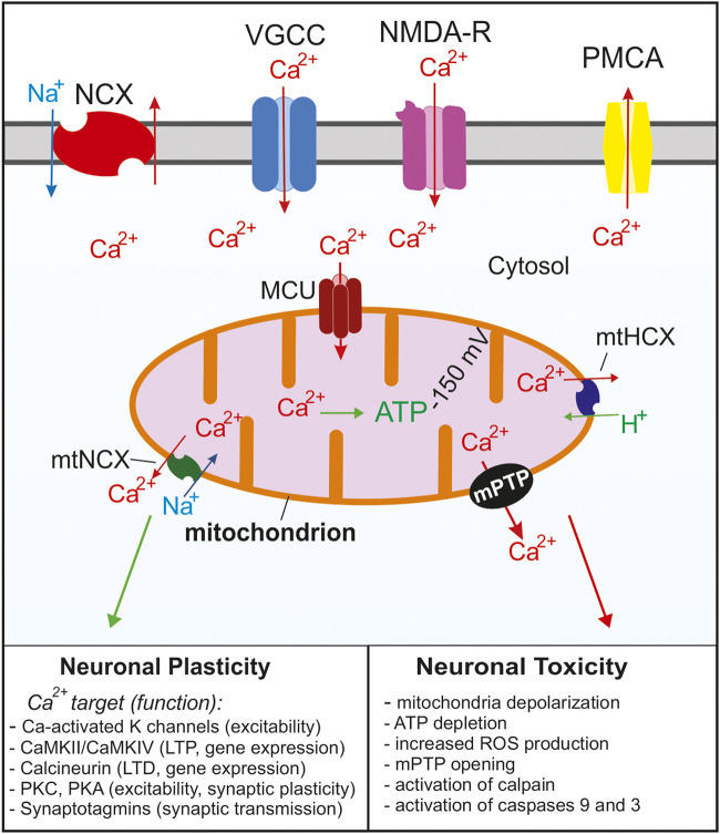
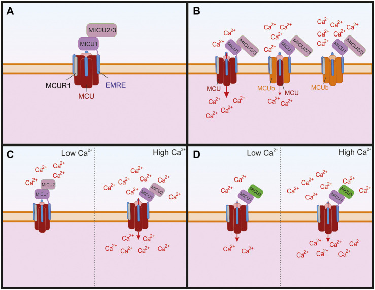
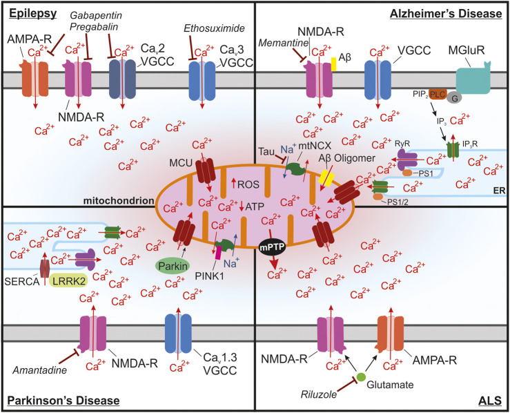
Mitochondria are essential for proper cellular function through their critical roles in ATP synthesis, reactive oxygen species production, calcium (Ca 2+ ) buffering, and apoptotic signaling. In neurons, Ca 2+ buffering is particularly important as it helps to shape Ca 2+ signals and to regulate numerous Ca 2+ -dependent functions including neuronal excitability, synaptic transmission, gene expression, and neuronal toxicity. Over the past decade, identification of the mitochondrial Ca 2+ uniporter (MCU) and other molecular components of mitochondrial Ca 2+ transport has provided insight into the roles that mitochondrial Ca 2+ regulation plays in neuronal function in health and disease. In this review, we discuss the many roles of mitochondrial Ca 2+ uptake and release mechanisms in normal neuronal function and highlight new insights into the Ca 2+ -dependent mechanisms that drive mitochondrial dysfunction in neurologic diseases including epilepsy, Alzheimer’s disease, Parkinson’s disease, and amyotrophic lateral sclerosis. We also consider how targeting Ca 2+ uptake and release mechanisms could facilitate the development of novel therapeutic strategies for neurological diseases.
Overview
The claims and statistics of this paper haven’t been checked yet.
Run the check — free, no account needed. It reads the open-access text and checks the sources it cites.
Integrity signals
Registry
Cites
Statistics
Figures
Verification
Claims the paper makes
The claims and statistics of this paper haven’t been checked yet.
Run the check — free, no account needed. It reads the open-access text and checks the sources it cites.
Claims other papers make about this one
Each is checked against this paper’s own text: does it say what the citing paper says it does?
The source backs part of the claim, not all of it — or backs it for a different population, condition or comparison.
In the citing paper
“excessive or prolonged activation of CaV channels leads to calcium overload in mitochondria, disrupting their normal function at multiple levels and significantly impacting cellular development and homeostasis101,102.”
From CYFIP1 governs the development of cortical axons by modulating calcium availability (2025) · cites this paper as [102]
Experimental — accuracy not yet measured
Self-check
Percentages, sums and reported differences re-computed from the paper’s own numbers.
The self-check needs the paper’s full text.
Citations
Cites
Staging of brain pathology related to sporadic Parkinson’s disease
Braak, H., Tredici, K. D., Rüb, U., de Vos, R. A. I., Jansen Steur, E. N. H., & Braak, E. (2003) · Neurobiology of Aging · doi:10.1016/s0197-4580(02)00065-9 · Cited by 10,777
Down-Regulation of Na+/K+ATPase Activity by Human Parvovirus B19 Capsid Protein VP1
Almilaji, A., Szteyn, K., Fein, E., Pakladok, T., Muñoz, C., Elvira, B., Towhid, S. T., Alesutan, I., Shumilina, E. V., Bock, C. –., et al. (2013) · Cellular Physiology and Biochemistry · doi:10.1159/000350083 · Cited by 8,700
Rosen, D. R., Siddique, T., Patterson, D., Figlewicz, D. A., Sapp, P., Hentati, A., Donaldson, D., Goto, J., O'Regan, J. P., Deng, H.-X., et al. (1993) · Nature · doi:10.1038/362059a0 · Cited by 7,062
The amyloid hypothesis of Alzheimer's disease at 25 years
Selkoe, D. J., & Hardy, J. (2016) · EMBO Molecular Medicine · doi:10.15252/emmm.201606210 · Cited by 6,164
Early Identification of Refractory Epilepsy
Kwan, P., & Brodie, M. J. (2000) · New England Journal of Medicine · doi:10.1056/nejm200002033420503 · Cited by 5,204 · cited 1×
“Unfortunately, 30% of patients have epilepsy that is poorly controlled by current pharmaceuticals, a condition referred to as refractory epilepsy (Kwan and Brodie, 2000).”
BackgroundShort-Term Synaptic Plasticity
Zucker, R. S., & Regehr, W. G. (2002) · Annual Review of Physiology · doi:10.1146/annurev.physiol.64.092501.114547 · Cited by 5,068
Querfurth, H. W., & LaFerla, F. M. (2010) · New England Journal of Medicine · doi:10.1056/nejmra0909142 · Cited by 4,569
Berg, A. T., Berkovic, S. F., Brodie, M. J., Buchhalter, J., Cross, J. H., Van Emde Boas, W., Engel, J., French, J., Glauser, T. A., Mathern, G. W., et al. (2010) · Epilepsia · doi:10.1111/j.1528-1167.2010.02522.x · Cited by 4,363
Motor Neuron Degeneration in Mice that Express a Human Cu,Zn Superoxide Dismutase Mutation
Gurney, M. E., Pu, H., Chiu, A. Y., Dal Canto, M. C., Polchow, C. Y., Alexander, D. D., Caliendo, J., Hentati, A., Kwon, Y. W., Deng, H.-X., et al. (1994) · Science · doi:10.1126/science.8209258 · Cited by 4,143
Interplay between Cortical Spreading Depolarization and Seizures
Kramer, D. R., Fujii, T., Ohiorhenuan, I. E., & Liu, C. Y. (2017) · Stereotactic and Functional Neurosurgery · doi:10.1159/000452841 · Cited by 3,721
An Energy Budget for Signaling in the Grey Matter of the Brain
Attwell, D., & Laughlin, S. B. (2001) · Journal of Cerebral Blood Flow & Metabolism · doi:10.1097/00004647-200110000-00001 · Cited by 3,543
Mitochondria: In Sickness and in Health
Nunnari, J., & Suomalainen, A. (2012) · Cell · doi:10.1016/j.cell.2012.02.035 · Cited by 3,479
Scheltens, P., Blennow, K., Breteler, M. M. B., de Strooper, B., Frisoni, G. B., Salloway, S., & Van der Flier, W. M. (2016) · The Lancet · doi:10.1016/s0140-6736(15)01124-1 · Cited by 3,125
Hyman, B. T., Phelps, C. H., Beach, T. G., Bigio, E. H., Cairns, N. J., Carrillo, M. C., Dickson, D. W., Duyckaerts, C., Frosch, M. P., Masliah, E., et al. (2012) · Alzheimer's & Dementia · doi:10.1016/j.jalz.2011.10.007 · Cited by 3,092
Regulation of cell death: the calcium–apoptosis link
Orrenius, S., Zhivotovsky, B., & Nicotera, P. (2003) · Nature Reviews Molecular Cell Biology · doi:10.1038/nrm1150 · Cited by 3,060
TRP channels as cellular sensors
Clapham, D. E. (2003) · Nature · doi:10.1038/nature02196 · Cited by 2,779
Calcium, ATP, and ROS: a mitochondrial love-hate triangle
Brookes, P. S., Yoon, Y., Robotham, J. L., Anders, M. W., & Sheu, S.-S. (2004) · American Journal of Physiology-Cell Physiology · doi:10.1152/ajpcell.00139.2004 · Cited by 2,575
Reactive astrocyte nomenclature, definitions, and future directions
Escartin, C., Galea, E., Lakatos, A., O’Callaghan, J. P., Petzold, G. C., Serrano-Pozo, A., Steinhäuser, C., Volterra, A., Carmignoto, G., Agarwal, A., et al. (2021) · Nature Neuroscience · doi:10.1038/s41593-020-00783-4 · Cited by 2,513 · cited 1×
“Even though this review focused primarily on neurons, it is important to point out that glial cells, such as astrocytes and microglia, also play crucial roles in epilepsy and neurodegenerative diseases as highlighted in many excellent reviews on this topic (Salter and Stevens, 2017; Patel et al., 2019; Santello et al.…”
Background2020 Alzheimer's disease facts and figures
(2020) · Alzheimer's & Dementia · doi:10.1002/alz.12068 · Cited by 2,353
Structure and Regulation of Voltage-Gated Ca 2+ Channels
Catterall, W. A. (2000) · Annual Review of Cell and Developmental Biology · doi:10.1146/annurev.cellbio.16.1.521 · Cited by 2,344
A forty-kilodalton protein of the inner membrane is the mitochondrial calcium uniporter
Stefani, D. D., Raffaello, A., Teardo, E., Szabó, I., & Rizzuto, R. (2011) · Nature · doi:10.1038/nature10230 · Cited by 1,957
Integrative genomics identifies MCU as an essential component of the mitochondrial calcium uniporter
Baughman, J. M., Perocchi, F., Girgis, H. S., Plovanich, M., Belcher-Timme, C. A., Sancak, Y., Bao, X. R., Strittmatter, L., Goldberger, O., Bogorad, R. L., et al. (2011) · Nature · doi:10.1038/nature10234 · Cited by 1,953
The Science of Stroke: Mechanisms in Search of Treatments
Moskowitz, M. A., Lo, E. H., & Iadecola, C. (2010) · Neuron · doi:10.1016/j.neuron.2010.07.002 · Cited by 1,930
Mitochondria as sensors and regulators of calcium signalling
Rizzuto, R., De Stefani, D., Raffaello, A., & Mammucari, C. (2012) · Nature Reviews Molecular Cell Biology · doi:10.1038/nrm3412 · Cited by 1,781
Microglia emerge as central players in brain disease
Salter, M. W., & Stevens, B. (2017) · Nature Medicine · doi:10.1038/nm.4397 · Cited by 1,767
Mitochondrial Respiratory-Chain Diseases
DiMauro, S., & Schon, E. A. (2003) · New England Journal of Medicine · doi:10.1056/nejmra022567 · Cited by 1,672
Mechanisms by which mitochondria transport calcium
Gunter, T. E., & Pfeiffer, D. R. (1990) · American Journal of Physiology-Cell Physiology · doi:10.1152/ajpcell.1990.258.5.c755 · Cited by 1,640
The Vanilloid Receptor: A Molecular Gateway to the Pain Pathway
Caterina, M. J., & Julius, D. J. (2001) · Annual Review of Neuroscience · doi:10.1146/annurev.neuro.24.1.487 · Cited by 1,571
Mitochondrial Transport of Cations: Channels, Exchangers, and Permeability Transition
Bernardi, P. (1999) · Physiological Reviews · doi:10.1152/physrev.1999.79.4.1127 · Cited by 1,546
The mitochondrial calcium uniporter is a highly selective ion channel
Kirichok, Y., Krapivinsky, G., & Clapham, D. E. (2004) · Nature · doi:10.1038/nature02246 · Cited by 1,409
Role of calcium ions in regulation of mammalian intramitochondrial metabolism
McCormack, J. G., Halestrap, A. P., & Denton, R. M. (1990) · Physiological Reviews · doi:10.1152/physrev.1990.70.2.391 · Cited by 1,398 · cited 1×
“8 μM (for comparison, isocitrate dehydrogenase has a Ca Kd of~40 μM) (McCormack et al., 1990), are the primary targets of matrix Ca.”
BackgroundStore-Operated Calcium Channels
Prakriya, M., & Lewis, R. S. (2015) · Physiological Reviews · doi:10.1152/physrev.00020.2014 · Cited by 1,274
Newmeyer, D. D., & Ferguson-Miller, S. M. (2003) · Cell · doi:10.1016/s0092-8674(03)00116-8 · Cited by 1,272
McShane, R., Areosa Sastre, A., & Minakaran, N. (2006) · Cochrane Database of Systematic Reviews · doi:10.1002/14651858.cd003154.pub5 · Cited by 1,155
Selective neuronal vulnerability in Parkinson disease
Surmeier, D. J., Obeso, J. A., & Halliday, G. M. (2017) · Nature Reviews Neuroscience · doi:10.1038/nrn.2016.178 · Cited by 1,116
Mitochondria in neuroplasticity and neurological disorders
Mattson, M. P., Gleichmann, M., & Cheng, A. (2008) · Neuron · doi:10.1016/j.neuron.2008.10.010 · Cited by 1,081
Mitochondrial reactive oxygen species in cell death signaling
Fleury, C., Mignotte, B., & Vayssière, J. (2002) · Biochimie · doi:10.1016/s0300-9084(02)01369-x · Cited by 1,069
The mitochondrion in apoptosis: how Pandora's box opens
Zamzami, N., & Kroemer, G. (2001) · Nature Reviews Molecular Cell Biology · doi:10.1038/35048073 · Cited by 1,055
Wei, A. D., Gutman, G. A., Aldrich, R. W., Chandy, K. G., Grissmer, S., & Wulff, H. (2005) · Pharmacological Reviews · doi:10.1124/pr.57.4.9 · Cited by 990
N-methyl-D-aspartic acid receptor structure and function
McBain, C. J., & Mayer, M. L. (1994) · Physiological Reviews · doi:10.1152/physrev.1994.74.3.723 · Cited by 976
Dimers of mitochondrial ATP synthase form the permeability transition pore
Giorgio, V., von Stockum, S., Antoniel, M., Fabbro, A., Fogolari, F., Forte, M. A., Glick, G. D., Petronilli, V., Zoratti, M., Szabó, I., et al. (2013) · Proceedings of the National Academy of Sciences · doi:10.1073/pnas.1217823110 · Cited by 973
Multiple Roles of Calcium Ions in the Regulation of Neurotransmitter Release
Neher, E., & Sakaba, T. (2008) · Neuron · doi:10.1016/j.neuron.2008.08.019 · Cited by 966
Du, H., Guo, L., Fang, F., Chen, D., Sosunov, A. A., McKhann, G. M., Yan, Y., Wang, C., Zhang, H., Molkentin, J. D., et al. (2008) · Nature Medicine · doi:10.1038/nm.1868 · Cited by 950
The scientific and clinical basis for the treatment of Parkinson disease (2009)
Olanow, C. W., Stern, M. B., & Sethi, K. D. (2009) · Neurology · doi:10.1212/wnl.0b013e3181a1d44c · Cited by 947
Neuronal calcium mishandling and the pathogenesis of Alzheimer's disease
Bezprozvanny, I., & Mattson, M. P. (2008) · Trends in Neurosciences · doi:10.1016/j.tins.2008.06.005 · Cited by 938
‘Rejuvenation’ protects neurons in mouse models of Parkinson’s disease
Chan, C. S., Guzmán, J. N., Ilijić, E., Mercer, J. N., Rick, C., Tkatch, T., Meredith, G. E., & Surmeier, D. J. (2007) · Nature · doi:10.1038/nature05865 · Cited by 936
Ca2+-activated K+ currents in neurones: types, physiological roles and modulation
Sah, P. (1996) · Trends in Neurosciences · doi:10.1016/s0166-2236(96)80026-9 · Cited by 892
Howland, D., Liu, J., She, Y., Goad, B., Maragakis, N. J., Kim, B. B., Erickson, J., Kulik, J., DeVito, L., Psaltis, G. C., et al. (2002) · Proceedings of the National Academy of Sciences · doi:10.1073/pnas.032539299 · Cited by 890
MICU1 encodes a mitochondrial EF hand protein required for Ca2+ uptake
Perocchi, F., Gohil, V. M., Girgis, H. S., Bao, X. R., McCombs, J. E., Palmer, A. E., & Mootha, V. K. (2010) · Nature · doi:10.1038/nature09358 · Cited by 888
Regulation of mitochondrial dehydrogenases by calcium ions
Denton, R. M. (2009) · Biochimica et Biophysica Acta (BBA) - Bioenergetics · doi:10.1016/j.bbabio.2009.01.005 · Cited by 885
VDAC, a multi-functional mitochondrial protein regulating cell life and death
Shoshan‐Barmatz, V., Pinto, V. D., Zweckstetter, M., Raviv, Z., Keinan, N., & Arbel, N. (2010) · Molecular Aspects of Medicine · doi:10.1016/j.mam.2010.03.002 · Cited by 824
An astrocytic basis of epilepsy
Tian, G.-F., Azmi, H., Takano, T., Xu, Q., Peng, W., Lin, J., Oberheim, N., Lou, N., Wang, X., Zielke, H. R., et al. (2005) · Nature Medicine · doi:10.1038/nm1277 · Cited by 806
The Ca2+-induced membrane transition in mitochondria
Haworth, R. A., & Hunter, D. R. (1979) · Archives of Biochemistry and Biophysics · doi:10.1016/0003-9861(79)90372-2 · Cited by 793
Neuronal calcium signaling: function and dysfunction
Brini, M., Calí, T., Ottolini, D., & Carafoli, E. (2014) · Cellular and Molecular Life Sciences · doi:10.1007/s00018-013-1550-7 · Cited by 790
Role of mitochondrial ROS in the brain: from physiology to neurodegeneration
Angelova, P. R., & Abramov, A. Y. (2018) · FEBS Letters · doi:10.1002/1873-3468.12964 · Cited by 789 · cited 2×
“Super oxide free radicals (O2 ) produced through OXPHOS are converted to H2O2 by enzymes such as superoxide dismutase and subsequently broken down into harmless byproducts (Beckhauser et al., 2016; Angelova and Abramov, 2018).”
BackgroundNCLX is an essential component of mitochondrial Na + /Ca 2+ exchange
Palty, R., Silverman, W. F., Hershfinkel, M., Caporale, T., Sensi, S. L., Parnis, J., Nolte, C., Fishman, D., Shoshan‐Barmatz, V., Herrmann, S., et al. (2009) · Proceedings of the National Academy of Sciences · doi:10.1073/pnas.0908099107 · Cited by 789
Reactive oxygen species and the free radical theory of aging
Liochev, S. I. (2013) · Free Radical Biology and Medicine · doi:10.1016/j.freeradbiomed.2013.02.011 · Cited by 768
Ichas, F., Jouaville, L. S., & Mazat, J. (1997) · Cell · doi:10.1016/s0092-8674(00)80301-3 · Cited by 748
Updated Energy Budgets for Neural Computation in the Neocortex and Cerebellum
Howarth, C., Gleeson, P., & Attwell, D. (2012) · Journal of Cerebral Blood Flow & Metabolism · doi:10.1038/jcbfm.2012.35 · Cited by 738
Astrocyte function from information processing to cognition and cognitive impairment
Santello, M., Toni, N., & Volterra, A. (2019) · Nature Neuroscience · doi:10.1038/s41593-018-0325-8 · Cited by 737
Hansson Petersen, C. A., Alikhani, N., Behbahani, H., Wiehager, B., Pavlov, P. F., Alafuzoff, I., Leinonen, V., Ito, A., Winblad, B., Glaser, E., et al. (2008) · Proceedings of the National Academy of Sciences · doi:10.1073/pnas.0806192105 · Cited by 693
Gandhi, S., Wood-Kaczmar, A., Yao, Z., Plun-Favreau, H., Deas, E., Klupsch, K., Downward, J., Latchman, D. S., Tabrizi, S. J., Wood, N. W., et al. (2009) · Molecular Cell · doi:10.1016/j.molcel.2009.02.013 · Cited by 682
Mallilankaraman, K. B., Doonan, P. J., Cárdenas, C., Chandramoorthy, H. C. K., Müller, M., Miller, R., Hoffman, N. E., Gandhirajan, R. K., Molgó, J., Birnbaum, M. J., et al. (2012) · Cell · doi:10.1016/j.cell.2012.10.011 · Cited by 668
EMRE Is an Essential Component of the Mitochondrial Calcium Uniporter Complex
Sancak, Y., Markhard, A. L., Kitami, T., Kovács-Bogdán, E., Kamer, K. J., Udeshi, N. D., Carr, S. A., Chaudhuri, D., Clapham, D. E., Li, A. A., et al. (2013) · Science · doi:10.1126/science.1242993 · Cited by 666
The kainic acid model of temporal lobe epilepsy
Lévesque, M., & Avoli, M. (2013) · Neuroscience & Biobehavioral Reviews · doi:10.1016/j.neubiorev.2013.10.011 · Cited by 650
Role of Mitochondrial Ca2+ in the Regulation of Cellular Energetics
Glancy, B. C., & Balaban, R. S. (2012) · Biochemistry · doi:10.1021/bi2018909 · Cited by 649
Pan, X., Liu, J., Nguyen, T. T. M., Liu, C., Sun, J., Teng, Y., Fergusson, M. M., Rovira, I. I., Allen, M. D., Springer, D., et al. (2013) · Nature Cell Biology · doi:10.1038/ncb2868 · Cited by 646
Mitochondria at the neuronal presynapse in health and disease
Devine, M. J., & Kittler, J. T. (2018) · Nature reviews. Neuroscience · doi:10.1038/nrn.2017.170 · Cited by 636
Amantadine in the Treatment of Parkinson's Disease
Schwab, R. S. (1969) · JAMA · doi:10.1001/jama.1969.03160070046011 · Cited by 633
Glutamate-induced neuron death requires mitochondrial calcium uptake
Stout, A. K., Raphael, H. M., Kanterewicz, B. I., Klann, E., & Reynolds, I. J. (1998) · Nature Neuroscience · doi:10.1038/1577 · Cited by 630
CALCIUM-ACTIVATED CHLORIDE CHANNELS
Hartzell, C., Putzier, I., & Arreola, J. (2004) · Annual Review of Physiology · doi:10.1146/annurev.physiol.67.032003.154341 · Cited by 627
Alavian, K. N., Beutner, G., Lazrove, E., Sacchetti, S., Park, H., Licznerski, P., Li, H., Nabili, P., Hockensmith, K., Graham, M. M., et al. (2014) · Proceedings of the National Academy of Sciences · doi:10.1073/pnas.1401591111 · Cited by 626
Hajnóczky, G., Csordás, G., Das, S., Garcı́a-Pérez, C., Saotome, M., Roy, S. S., & Yi, M. (2006) · Cell Calcium · doi:10.1016/j.ceca.2006.08.016 · Cited by 613
Mitochondrial permeability transition in Ca2+-dependent apoptosis and necrosis
Rasola, A., & Bernardi, P. (2011) · Cell Calcium · doi:10.1016/j.ceca.2011.04.007 · Cited by 590
Selective vulnerability in neurodegenerative diseases
Fu, H., Hardy, J., & Duff, K. E. (2018) · Nature Neuroscience · doi:10.1038/s41593-018-0221-2 · Cited by 585
The role of mitochondria in amyotrophic lateral sclerosis
Smith, E. F., Shaw, P. J., & Vos, K. J. D. (2017) · Neuroscience Letters · doi:10.1016/j.neulet.2017.06.052 · Cited by 579
Mitochondrial membrane potential and neuronal glutamate excitotoxicity: mortality and millivolts
Nicholls, D. G., & Ward, M. W. (2000) · Trends in Neurosciences · doi:10.1016/s0166-2236(99)01534-9 · Cited by 563
Arispe, N. J., Pollard, H. B., & Rojas, E. (1993) · Proceedings of the National Academy of Sciences · doi:10.1073/pnas.90.22.10573 · Cited by 561
Ueda, S., Masutani, H., Nakamura, H., Tanaka, T., Ueno, M., & Yodoi, J. (2002) · Antioxidants and Redox Signaling · doi:10.1089/15230860260196209 · Cited by 552
Mitochondrial Participation in the Intracellular Ca2+ Network
Babcock, D. F., Herrington, J. B., Goodwin, P. C., Park, Y. B., & Hille, B. (1997) · The Journal of Cell Biology · doi:10.1083/jcb.136.4.833 · Cited by 547
Stimulation by calcium ions of pyruvate dehydrogenase phosphate phosphatase
Denton, R. M., Randle, P. J., & Martin, B. R. (1972) · Biochemical Journal · doi:10.1042/bj1280161 · Cited by 542
Mechanisms of action of currently used antiseizure drugs
Sills, G. J., & Rogawski, M. A. (2020) · Neuropharmacology · doi:10.1016/j.neuropharm.2020.107966 · Cited by 529
Scharfman, H. E. (2007) · Current Neurology and Neuroscience Reports · doi:10.1007/s11910-007-0053-z · Cited by 526
Patrón, M., Checchetto, V., Raffaello, A., Teardo, E., Reane, D. V., Mantoan, M., Granatiero, V., Szabó, I., Stefani, D. D., & Rizzuto, R. (2014) · Molecular Cell · doi:10.1016/j.molcel.2014.01.013 · Cited by 525
Mitochondrial dysfunction and apoptosis in myopathic mice with collagen VI deficiency
Irwin, W., Bergamin, N., Sabatelli, P., Reggiani, C., Megighian, A., Merlini, L., Braghetta, P., Columbaro, M., Volpin, D., Bressan, G. M., et al. (2003) · Nature Genetics · doi:10.1038/ng1270 · Cited by 509
MICU1 Controls Both the Threshold and Cooperative Activation of the Mitochondrial Ca2+ Uniporter
Csordás, G., Golenár, T., Seifert, E. L., Kamer, K. J., Sancak, Y., Perocchi, F., Moffat, C., Weaver, D., de la Fuente, S., Bogorad, R. L., et al. (2013) · Cell Metabolism · doi:10.1016/j.cmet.2013.04.020 · Cited by 499
The role of excitotoxicity in the pathogenesis of amyotrophic lateral sclerosis
Bosch, L. V. D., Damme, P. V., Bogaert, E., & Robberecht, W. (2006) · Biochimica et Biophysica Acta (BBA) - Molecular Basis of Disease · doi:10.1016/j.bbadis.2006.05.002 · Cited by 491
Raffaello, A., Stefani, D. D., Sabbadin, D., Teardo, E., Merli, G., Picard, A., Checchetto, V., Moro, S., Szabó, I., & Rizzuto, R. (2013) · The EMBO Journal · doi:10.1038/emboj.2013.157 · Cited by 489
MCUR1 is an essential component of mitochondrial Ca2+ uptake that regulates cellular metabolism
Mallilankaraman, K. B., Cárdenas, C., Doonan, P. J., Chandramoorthy, H. C. K., Irrinki, K. M., Golenár, T., Csordás, G., Madireddi, P., Yang, J., Müller, M., et al. (2012) · Nature Cell Biology · doi:10.1038/ncb2622 · Cited by 487
Sudden unexpected death in epilepsy: Assessing the public health burden
Thurman, D. J., Hesdorffer, D. C., & French, J. A. (2014) · Epilepsia · doi:10.1111/epi.12666 · Cited by 486
Calcium signaling and neurodegenerative diseases
Борисович, Б. И. (2009) · Trends in Molecular Medicine · doi:10.1016/j.molmed.2009.01.001 · Cited by 480
Mitochondria buffer physiological calcium loads in cultured rat dorsal root ganglion neurons
Werth, J. L., & Thayer, S. (1994) · Journal of Neuroscience · doi:10.1523/jneurosci.14-01-00348.1994 · Cited by 479
Neuron–glia interactions in the pathophysiology of epilepsy
Patel, D. C., Tewari, B. P., Chaunsali, L., & Sontheimer, H. W. (2019) · Nature reviews. Neuroscience · doi:10.1038/s41583-019-0126-4 · Cited by 478
The permeability transition pore as a mitochondrial calcium release channel: A critical appraisal
Bernardi, P., & Petronilli, V. (1996) · Journal of Bioenergetics and Biomembranes · doi:10.1007/bf02110643 · Cited by 466
Plovanich, M., Bogorad, R. L., Sancak, Y., Kamer, K. J., Strittmatter, L., Li, A. A., Girgis, H. S., Kuchimanchi, S., Groot, J. D., Speciner, L., et al. (2013) · PLoS ONE · doi:10.1371/journal.pone.0055785 · Cited by 463
Role of the c subunit of the F O ATP synthase in mitochondrial permeability transition
Bonora, M., Bononi, A., Marchi, E. D., Giorgi, C., Lebiedzińska, M., Marchi, S., Patergnani, S., Rimessi, A., Suski, J. M., Wojtala, A., et al. (2013) · Cell Cycle · doi:10.4161/cc.23599 · Cited by 461
Cheung, K., Shineman, D. W., Müller, M., Cárdenas, C., Mei, L., Yang, J., Tomita, T., Iwatsubo, T., Lee, V. M. ‐., & Foskett, J. K. (2008) · Neuron · doi:10.1016/j.neuron.2008.04.015 · Cited by 460
Mitochondrial Involvement in Post-Tetanic Potentiation of Synaptic Transmission
Tang, Y.-G., & Zucker, R. S. (1997) · Neuron · doi:10.1016/s0896-6273(00)81248-9 · Cited by 441
Calvo-Rodríguez, M., Hou, S. S., Snyder, A. C., Kharitonova, E. K., Russ, A., Das, S., Fan, Z., Muzikansky, A., Garcı́a-Alloza, M., Serrano‐Pozo, A., et al. (2020) · Nature Communications · doi:10.1038/s41467-020-16074-2 · Cited by 432
Mitochondrial calcium exchange in physiology and disease
Garbincius, J. F., & Elrod, J. W. (2021) · Physiological Reviews · doi:10.1152/physrev.00041.2020 · Cited by 427
Enjoy the Trip: Calcium in Mitochondria Back and Forth
Stefani, D. D., Rizzuto, R., & Pozzan, T. (2016) · Annual Review of Biochemistry · doi:10.1146/annurev-biochem-060614-034216 · Cited by 424
Mallilankaraman, K. B., Cárdenas, C., Doonan, P. J., Chandramoorthy, H. C. K., Irrinki, K. M., Golenár, T., Csordás, G., Madireddi, P., Yang, J., Müller, M., et al. (2015) · Nature Cell Biology · doi:10.1038/ncb3202 · Cited by 418
Billups, B., & Forsythe, I. D. (2002) · Journal of Neuroscience · doi:10.1523/jneurosci.22-14-05840.2002 · Cited by 414
Crompton, M., Moser, R., Lüdi, H., & Carafoli, E. (1978) · European Journal of Biochemistry · doi:10.1111/j.1432-1033.1978.tb11993.x · Cited by 397
The mitochondrial Na+/Ca2+ exchanger is essential for Ca2+ homeostasis and viability
Luongo, T. S., Lambert, J. P., Gross, P., Nwokedi, M., Lombardi, A. A., Shanmughapriya, S., Carpenter, A. C., Kolmetzky, D., Gao, E., van Berlo, J. H., et al. (2017) · Nature · doi:10.1038/nature22082 · Cited by 396
Calcium channel auxiliary α2δ and β subunits: trafficking and one step beyond
DOLPHIN, A. C. (2012) · Nature reviews. Neuroscience · doi:10.1038/nrn3311 · Cited by 392
Chalmers, S., & Nicholls, D. G. (2003) · Journal of Biological Chemistry · doi:10.1074/jbc.m212661200 · Cited by 390
Calcium‐dependent mitochondrial function and dysfunction in neurons
Pivovarova, N. B., & Andrews, S. (2010) · FEBS Journal · doi:10.1111/j.1742-4658.2010.07754.x · Cited by 389
Luongo, T. S., Lambert, J. P., Yuan, A., Zhang, X., Gross, P., Song, J., Shanmughapriya, S., Gao, E., Jain, M., Houser, S. R., et al. (2015) · Cell Reports · doi:10.1016/j.celrep.2015.06.017 · Cited by 384
The molecular era of the mitochondrial calcium uniporter
Kamer, K. J., & Mootha, V. K. (2015) · Nature Reviews Molecular Cell Biology · doi:10.1038/nrm4039 · Cited by 377
Logan, C. V., Szabadkai, G., Sharpe, J. A., Parry, D. A., Torelli, S., Childs, A., Kriek, M., Phadke, R., Johnson, C. A., Roberts, N. Y., et al. (2013) · Nature Genetics · doi:10.1038/ng.2851 · Cited by 372
Kwong, J. Q., Lu, X., Correll, R. N., Schwanekamp, J. A., Vagnozzi, R. J., Sargent, M. A., York, A. J., Zhang, J. J., Bers, D. M., & Molkentin, J. D. (2015) · Cell Reports · doi:10.1016/j.celrep.2015.06.002 · Cited by 360
Mitochondria and calcium signaling
Nicholls, D. G. (2005) · Cell Calcium · doi:10.1016/j.ceca.2005.06.011 · Cited by 357
Chuang, H.-H., Neuhausser, W., & Julius, D. J. (2004) · Neuron · doi:10.1016/j.neuron.2004.08.038 · Cited by 338
Xu, J., Mashimo, T., & Südhof, T. C. (2007) · Neuron · doi:10.1016/j.neuron.2007.05.004 · Cited by 336
The dephosphins: dephosphorylation by calcineurin triggers synaptic vesicle endocytosis
Cousin, M. A., & Robinson, P. J. (2001) · Trends in Neurosciences · doi:10.1016/s0166-2236(00)01930-5 · Cited by 330
Identification of Candidate Gene Variants in Korean MODY Families by Whole-Exome Sequencing
Shim, Y. J., Kim, J. E., Hwang, S., Choi, B. S., Choi, B. H., Cho, E.-M., Jang, K. M., & Ko, C. W. (2015) · Hormone Research in Paediatrics · doi:10.1159/000368657 · Cited by 325
Leung, A. W. C., Varanyuwatana, P., & Halestrap, A. P. (2008) · Journal of Biological Chemistry · doi:10.1074/jbc.m805235200 · Cited by 324
Estimating risk for developing epilepsy: A population-based study in Rochester, Minnesota
Hesdorffer, D. C., Logroscino, G., Benn, E. K. T., Katri, N., Cascino, G., & Hauser, W. A. (2011) · Neurology · doi:10.1212/wnl.0b013e318204a36a · Cited by 324
Jadiya, P., Kolmetzky, D. W., Tomar, D., Di Meco, A., Lombardi, A. A., Lambert, J. P., Luongo, T. S., Ludtmann, M. H., Praticò, D., & Elrod, J. W. (2019) · Nature Communications · doi:10.1038/s41467-019-11813-6 · Cited by 323
Structure and mechanism of the mitochondrial Ca2+ uniporter holocomplex
Fan, M., Zhang, J., Tsai, C.-W., Orlando, B. J., Rodriguez, M. X., Xu, Y., Liao, M., Tsai, M.-F., & Feng, L. (2020) · Nature · doi:10.1038/s41586-020-2309-6 · Cited by 321
Regulation of ATP production by mitochondrial Ca2+
Tarasov, A. I., Griffiths, E. J., & Rutter, G. A. (2012) · Cell Calcium · doi:10.1016/j.ceca.2012.03.003 · Cited by 319
Dementia of Alzheimer’s disease and other neurodegenerative disorders—memantine, a new hope
Sonkusare, S. K., Kaul, C. L., & Ramarao, P. (2004) · Pharmacological Research · doi:10.1016/j.phrs.2004.05.005 · Cited by 313
Alzheimer's Association Calcium Hypothesis Workgroup, & Khachaturian, Z. S. (2017) · Alzheimer's & Dementia · doi:10.1016/j.jalz.2016.12.006 · Cited by 311
Bacaj, T., Wu, D., Yang, X., Morishita, W., Zhou, P., Xu, W., Malenka, R. C., & Südhof, T. C. (2013) · Neuron · doi:10.1016/j.neuron.2013.10.026 · Cited by 308
Giorgio, V., Bisetto, E., Soriano, M. E., Dabbeni‐Sala, F., Basso, E., Petronilli, V., Forte, M. A., Bernardi, P., & Lippe, G. (2009) · Journal of Biological Chemistry · doi:10.1074/jbc.m109.020115 · Cited by 306
Calcium, ageing, and neuronal vulnerability in Parkinson's disease
Surmeier, D. J. (2007) · The Lancet Neurology · doi:10.1016/s1474-4422(07)70246-6 · Cited by 303
Beckhauser, T. F., Francis-Oliveira, J., & De Pasquale, R. (2016) · Journal of Experimental Neuroscience · doi:10.4137/jen.s39887 · Cited by 301
Architecture of the mitochondrial calcium uniporter
Oxenoid, K., Dong, Y., Cao, C., Cui, T., Sancak, Y., Markhard, A. L., Grabarek, Z., Kong, L., Liu, Z., OuYang, B., et al. (2016) · Nature · doi:10.1038/nature17656 · Cited by 300
On the involvement of a cyclosporin A sensitive mitochondrial pore in myocardial reperfusion injury
Duchen, M. R., McGuinness, O. M., Brown, L. A., & Crompton, M. (1993) · Cardiovascular Research · doi:10.1093/cvr/27.10.1790 · Cited by 294
Calcium-activated non-specific cation channels
Partridge, L., & Swandulla, D. (1988) · Trends in Neurosciences · doi:10.1016/0166-2236(88)90167-1 · Cited by 284
Metabolic Dysfunction and Oxidative Stress in Epilepsy
Pearson-Smith, J. N., & Patel, M. (2017) · International Journal of Molecular Sciences · doi:10.3390/ijms18112365 · Cited by 280
Enhanced Astrocytic Ca2+Signals Contribute to Neuronal Excitotoxicity after Status Epilepticus
Ding, S., Fellin, T., Zhu, Y., Lee, S.-Y., Auberson, Y. P., Meaney, D. F., Coulter, D. A., Carmignoto, G., & Haydon, P. G. (2007) · Journal of Neuroscience · doi:10.1523/jneurosci.2001-07.2007 · Cited by 270
Seizure-Induced Oxidative Stress in Temporal Lobe Epilepsy
Puttachary, S., Sharma, S., Stark, S., & Thippeswamy, T. (2015) · BioMed Research International · doi:10.1155/2015/745613 · Cited by 268
The Calcium‐Induced and Sodium‐Induced Effluxes of Calcium from Heart Mitochondria
Crompton, M., KÜNZI, M., & Carafoli, E. (1977) · European Journal of Biochemistry · doi:10.1111/j.1432-1033.1977.tb11839.x · Cited by 268
Damiano, M. G., Starkov, A. A., Petri, S., Kipiani, K., Kiaei, M., Mattiazzi, M., Beal, M. F., & Manfredi, G. (2006) · Journal of Neurochemistry · doi:10.1111/j.1471-4159.2006.03619.x · Cited by 267
Nicholls, D. G. (1978) · Biochemical Journal · doi:10.1042/bj1700511 · Cited by 266
Motile axonal mitochondria contribute to the variability of presynaptic strength
Sun, T., Qiao, H., Pan, P.-Y., Chen, Y., & Sheng, Z.-H. (2013) · Cell Reports · doi:10.1016/j.celrep.2013.06.040 · Cited by 263
Shaw, P. J., & Eggett, C. J. (2000) · Journal of Neurology · doi:10.1007/bf03161151 · Cited by 262
Mitochondrial dysfunction and seizures: the neuronal energy crisis
Zsurka, G., & Kunz, W. S. (2015) · The Lancet Neurology · doi:10.1016/s1474-4422(15)00148-9 · Cited by 258
Muñoz‐Pinedo, C., Guío-Carrión, A., Goldstein, J. C., FITZGERALD, P. J., Newmeyer, D. D., & Green, D. R. (2006) · Proceedings of the National Academy of Sciences · doi:10.1073/pnas.0603007103 · Cited by 253
He, J., Ford, H. C., Carroll, J., Ding, S., Fearnley, I. M., & Walker, J. E. (2017) · Proceedings of the National Academy of Sciences · doi:10.1073/pnas.1702357114 · Cited by 251
Calcium homeostasis, selective vulnerability and Parkinson's disease
Chan, C. S., Gertler, T. S., & Surmeier, D. J. (2009) · Trends in Neurosciences · doi:10.1016/j.tins.2009.01.006 · Cited by 245
Activity of the mitochondrial calcium uniporter varies greatly between tissues
Fieni, F., Lee, S. B., Jan, Y. N., & Kirichok, Y. (2012) · Nature Communications · doi:10.1038/ncomms2325 · Cited by 237
The Cation/Ca2+ Exchanger Superfamily: Phylogenetic Analysis and Structural Implications
Cai, X., & Lytton, J. (2004) · Molecular Biology and Evolution · doi:10.1093/molbev/msh177 · Cited by 236
Ashrafi, G., de Juan‐Sanz, J., Farrell, R. J., & Ryan, T. A. (2019) · Neuron · doi:10.1016/j.neuron.2019.11.020 · Cited by 235
MICU1 Serves as a Molecular Gatekeeper to Prevent In Vivo Mitochondrial Calcium Overload
Liu, J. C., Liu, J., Holmström, K. M., Menazza, S., Parks, R. J., Fergusson, M. M., Yu, Z., Springer, D., Halsey, C., Liu, C., et al. (2016) · Cell Reports · doi:10.1016/j.celrep.2016.07.011 · Cited by 226
MICU3 is a tissue-specific enhancer of mitochondrial calcium uptake
Patrón, M., Granatiero, V., Espino, J., Rizzuto, R., & Stefani, D. D. (2018) · Cell Death and Differentiation · doi:10.1038/s41418-018-0113-8 · Cited by 223
Chakroborty, S., Goussakov, I. V., Miller, M. B., & Stutzmann, G. E. (2009) · Journal of Neuroscience · doi:10.1523/jneurosci.2047-09.2009 · Cited by 219
MCUR1 Is a Scaffold Factor for the MCU Complex Function and Promotes Mitochondrial Bioenergetics
Tomar, D., Dong, Z., Shanmughapriya, S., Koch, D. A., Thomas, T. P., Hoffman, N. E., Timbalia, S. A., Goldman, S. J., Breves, S. L., Corbally, D. P., et al. (2016) · Cell Reports · doi:10.1016/j.celrep.2016.04.050 · Cited by 217
Selvaraj, B. T., Livesey, M. R., Zhao, C., Gregory, J. M., James, O. T., Cleary, E. M., Chouhan, A. K., Gane, A. B., Perkins, E., Dando, O., et al. (2018) · Nature Communications · doi:10.1038/s41467-017-02729-0 · Cited by 214
Calcium-dependent inactivation of neuronal calcium channels
Budde, T., Meuth, S. G., & Pape, H. (2002) · Nature reviews. Neuroscience · doi:10.1038/nrn959 · Cited by 211
SPG7 Is an Essential and Conserved Component of the Mitochondrial Permeability Transition Pore
Shanmughapriya, S., Rajan, S., Hoffman, N. E., Higgins, A. M., Tomar, D., Nemani, N., Hines, K. J., Smith, D. J., Eguchi, A., Vallem, S., et al. (2015) · Molecular Cell · doi:10.1016/j.molcel.2015.08.009 · Cited by 209
Petrungaro, C., Zimmermann, K. M., Küttner, V., Fischer, M., Dengjel, J., Bogeski, I., & Riemer, J. (2015) · Cell Metabolism · doi:10.1016/j.cmet.2015.08.019 · Cited by 200
Calcium and regulation of the mitochondrial permeability transition
Giorgio, V., Guo, L., Bassot, C., Petronilli, V., & Bernardi, P. (2017) · Cell Calcium · doi:10.1016/j.ceca.2017.05.004 · Cited by 199
Li, D., Zhang, Y.-H., Zhang, W., & Zhao, P. (2019) · Frontiers in Neuroscience · doi:10.3389/fnins.2019.00472 · Cited by 198
Calcium Permeable-AMPA Receptors and Excitotoxicity in Neurological Disorders
Guo, C., & Ma, Y. (2021) · Frontiers in Neural Circuits · doi:10.3389/fncir.2021.711564 · Cited by 198
Kwon, S., Sando, R. C., Lewis, T. L., Hirabayashi, Y., Maximov, A., & Polleux, F. (2016) · PLoS Biology · doi:10.1371/journal.pbio.1002516 · Cited by 194
Akundi, R. S., Huang, Z., Eason, J., Pandya, J. D., Zhi, L., Cass, W. A., Sullivan, P. G., & Büeler, H. (2011) · PLoS ONE · doi:10.1371/journal.pone.0016038 · Cited by 192
Jang, H., Zheng, J., & Nussinov, R. (2007) · Biophysical Journal · doi:10.1529/biophysj.107.110148 · Cited by 191
Mitochondrial Permeability Transition: A Molecular Lesion with Multiple Drug Targets
Briston, T., Selwood, D. L., Szabadkai, G., & Duchen, M. R. (2018) · Trends in Pharmacological Sciences · doi:10.1016/j.tips.2018.11.004 · Cited by 190
Isradipine Versus Placebo in Early Parkinson Disease
The Parkinson Study Group STEADY-PD III Investigators* (2020) · Annals of Internal Medicine · doi:10.7326/m19-2534 · Cited by 189
Neginskaya, M. A., Solesio, M. E., Berezhnaya, E. V., Amodeo, G. F., Mnatsakanyan, N., Jonas, E. A., & Pavlov, E. V. (2019) · Cell Reports · doi:10.1016/j.celrep.2018.12.033 · Cited by 187
Martin, L. J., Gertz, B., Pan, Y. H., Price, A. C., Molkentin, J. D., & Chang, Q. (2009) · Experimental Neurology · doi:10.1016/j.expneurol.2009.02.015 · Cited by 186
David, G., & Barrett, E. F. (2003) · The Journal of Physiology · doi:10.1113/jphysiol.2002.035196 · Cited by 182
He, J., Carroll, J., Ding, S., Fearnley, I. M., & Walker, J. E. (2017) · Proceedings of the National Academy of Sciences · doi:10.1073/pnas.1711201114 · Cited by 182
MCU encodes the pore conducting mitochondrial calcium currents
Chaudhuri, D., Sancak, Y., Mootha, V. K., & Clapham, D. E. (2013) · eLife · doi:10.7554/elife.00704 · Cited by 181
Aberrant Calcium Signals in Reactive Astrocytes: A Key Process in Neurological Disorders
Shigetomi, E., Saito, K., Sano, F., & Koizumi, S. (2019) · International Journal of Molecular Sciences · doi:10.3390/ijms20040996 · Cited by 181
Kostić, M., Ludtmann, M. H. R., Bading, H., Hershfinkel, M., Steer, E. K., Chu, C. T., Abramov, A. Y., & Sekler, I. (2015) · Cell Reports · doi:10.1016/j.celrep.2015.08.079 · Cited by 180
Fellin, T., Gómez‐Gonzalo, M., Gobbo, S., Carmignoto, G., & Haydon, P. G. (2006) · Journal of Neuroscience · doi:10.1523/jneurosci.2836-06.2006 · Cited by 177
After half a century mitochondrial calcium in‐ and efflux machineries reveal themselves
Drago, I., Pizzo, P., & Pozzan, T. (2011) · The EMBO Journal · doi:10.1038/emboj.2011.337 · Cited by 176
A mitochondrial megachannel resides in monomeric F1FO ATP synthase
Mnatsakanyan, N., Llaguno, M. C., Yang, Y., Yan, Y., Weber, J., Sigworth, F. J., & Jonas, E. A. (2019) · Nature Communications · doi:10.1038/s41467-019-13766-2 · Cited by 169
Pivovarova, N. B., Hongpaisan, J., Andrews, S., & Friel, D. D. (1999) · Journal of Neuroscience · doi:10.1523/jneurosci.19-15-06372.1999 · Cited by 167
Beers, D. R., Ho, B., Siklós, L., Alexianu, M. E., Mosier, D. R., Mohamed, A. H., Otsuka, Y., Kozovska, M. E., McAlhany, R. E., Smith, R. G., et al. (2001) · Journal of Neurochemistry · doi:10.1046/j.1471-4159.2001.00582.x · Cited by 166
Cell-Permeable Scavengers of Superoxide Prevent Long-Term Potentiation in Hippocampal Area CA1
Klann, E. (1998) · Journal of Neurophysiology · doi:10.1152/jn.1998.80.1.452 · Cited by 165
Mitochondrial trafficking and the provision of energy and calcium buffering at excitatory synapses
MacAskill, A. F., Atkin, T. A., & Kittler, J. T. (2010) · European Journal of Neuroscience · doi:10.1111/j.1460-9568.2010.07345.x · Cited by 163
Pathophysiology of status epilepticus
Walker, M. C. (2016) · Neuroscience Letters · doi:10.1016/j.neulet.2016.12.044 · Cited by 161
The physiological role of calcium-dependent channels
Marty, A. (1989) · Trends in Neurosciences · doi:10.1016/0166-2236(89)90090-8 · Cited by 161
Dual functions of a small regulatory subunit in the mitochondrial calcium uniporter complex
Tsai, M.-F., Phillips, C. B., Ranaghan, M. J., Tsai, C.-W., Wu, Y., Williams, C., & Miller, C. (2016) · eLife · doi:10.7554/elife.15545 · Cited by 159
Brini, M., Pinton, P., King, M. P., Davidson, M. M., Schon, E. A., & Rizzuto, R. (1999) · Nature Medicine · doi:10.1038/11396 · Cited by 156
Calcium Channels and Short-term Synaptic Plasticity
Catterall, W. A., Leal, K., & Nanou, E. (2013) · Journal of Biological Chemistry · doi:10.1074/jbc.r112.411645 · Cited by 153
Lambert, J. P., Luongo, T. S., Tomar, D., Jadiya, P., Gao, E., Zhang, X., Lucchese, A. M., Kolmetzky, D. W., Shah, N. S., & Elrod, J. W. (2019) · Circulation · doi:10.1161/circulationaha.118.037968 · Cited by 151
Cryo-EM structures of fungal and metazoan mitochondrial calcium uniporters
Baradaran, R., Wang, C., Siliciano, A. F., & Long, S. B. (2018) · Nature · doi:10.1038/s41586-018-0331-8 · Cited by 149
Alzheimer's disease: The right drug, the right time
Golde, T. E., DeKosky, S. T., & Galasko, D. (2018) · Science · doi:10.1126/science.aau0437 · Cited by 148
Lithium-Calcium Exchange Is Mediated by a Distinct Potassium-independent Sodium-Calcium Exchanger
Palty, R., Ohana, E., Hershfinkel, M., Volokita, M., Elgazar-Carmon, V., Beharier, O., Silverman, W. F., Argaman, M., & Sekler, I. (2004) · Journal of Biological Chemistry · doi:10.1074/jbc.m401229200 · Cited by 147
Pivovarova, N. B., Nguyen, H. V., Winters, C. A., Brantner, C. A., Smith, C. L., & Andrews, S. (2004) · Journal of Neuroscience · doi:10.1523/jneurosci.0531-04.2004 · Cited by 145
Tai, X. Y., Bernhardt, B. C., Thom, M., Thompson, P. J., Baxendale, S., Koepp, M. J., & Bernasconi, N. (2017) · Neuropathology and Applied Neurobiology · doi:10.1111/nan.12458 · Cited by 145
Alzheimer's Disease–Related Dementias Summit 2016: National research priorities
Corriveau, R. A., Koroshetz, W. J., Gladman, J. T., Jeon, S., Babcock, D., Bennett, D. A., Carmichael, S. T., Dickinson, S. L.-J., Dickson, D. W., Emr, M., et al. (2017) · Neurology · doi:10.1212/wnl.0000000000004717 · Cited by 143
Dafinca, R., Barbagallo, P., Farrimond, L. E., Candalija, A., Scaber, J., Ababneh, N. A., Sathyaprakash, C., Vowles, J., Cowley, S. A., & Talbot, K. (2020) · Stem Cell Reports · doi:10.1016/j.stemcr.2020.03.023 · Cited by 142
Scott, L. J., & Figgitt, D. P. (2004) · CNS Drugs · doi:10.2165/00023210-200418060-00010 · Cited by 140
MICU1 controls cristae junction and spatially anchors mitochondrial Ca2+ uniporter complex
Gottschalk, B., Klec, C., Leitinger, G., Bernhart, E. M., Rost, R., Bischof, H., Madreiter‐Sokolowski, C. T., Radulović, S., Eroğlu, E., Sattler, W., et al. (2019) · Nature Communications · doi:10.1038/s41467-019-11692-x · Cited by 140
Medvedeva, Y. V., Kim, M., & Usachev, Y. M. (2008) · Journal of Neuroscience · doi:10.1523/jneurosci.4810-07.2008 · Cited by 138
Self-citationRegulation of dendritic growth and plasticity by local and global calcium dynamics
Löhmann, C., & Wong, R. (2005) · Cell Calcium · doi:10.1016/j.ceca.2005.01.008 · Cited by 138
Demuro, A., & Parker, I. (2013) · Journal of Neuroscience · doi:10.1523/jneurosci.4367-12.2013 · Cited by 137
Systematic Identification of MCU Modulators by Orthogonal Interspecies Chemical Screening
Arduíno, D. M., Wettmarshausen, J., Vais, H., Navas-Navarro, P., Cheng, Y., Leimpek, A., Ma, Z., Delrio-Lorenzo, A., Giordano, A., Garcı́a-Pérez, C., et al. (2017) · Molecular Cell · doi:10.1016/j.molcel.2017.07.019 · Cited by 134
Carroll, J., He, J., Ding, S., Fearnley, I. M., & Walker, J. E. (2019) · Proceedings of the National Academy of Sciences · doi:10.1073/pnas.1904005116 · Cited by 132
High- and low-calcium-dependent mechanisms of mitochondrial calcium signalling
Spät, A., Szanda, G., Csordás, G., & Hajnóczky, G. (2008) · Cell Calcium · doi:10.1016/j.ceca.2007.11.015 · Cited by 131
Structural Mechanism of EMRE-Dependent Gating of the Human Mitochondrial Calcium Uniporter
Wang, Y., Nguyen, N. X., She, J., Zeng, W., Yang, Y., Bai, X., & Jiang, Y. (2019) · Cell · doi:10.1016/j.cell.2019.03.050 · Cited by 130
Kovac, S., Dinkova‐Kostova, A. T., Herrmann, A. M., Melzer, N., Meuth, S. G., & Gorji, A. (2017) · International Journal of Molecular Sciences · doi:10.3390/ijms18091935 · Cited by 129
Physiological Functions of CRAC Channels
Emrich, S. M., Yoast, R. E., & Trebak, M. (2021) · Annual Review of Physiology · doi:10.1146/annurev-physiol-052521-013426 · Cited by 129
Research works printed but not linked: 1
- De Camilli, 2001, The structure of synapses, 89
Cited by
Sorted by citations
Madireddy, S., & Madireddy, S. (2023) · Brain Sciences · doi:10.3390/brainsci13050784 · Cited by 124
Lee, S., Duron, H. E., & Chaudhuri, D. (2023) · Biochemical Society Transactions · doi:10.1042/bst20230012 · Cited by 77
PINK1 and Parkin regulate IP3R-mediated ER calcium release
Ham, S. J., Yoo, H., Woo, D., Lee, D. H., Park, K., & Chung, J. (2023) · Nature Communications · doi:10.1038/s41467-023-40929-z · Cited by 67
Mitochondria and Oxidative Stress as a Link between Alzheimer’s Disease and Diabetes Mellitus
Veselov, I. M., Vinogradova, D. V., Maltsev, A. V., Shevtsov, P. N., Spirkova, E. A., Бачурин, С. О., & Shevtsova, E. F. (2023) · International Journal of Molecular Sciences · doi:10.3390/ijms241914450 · Cited by 55
Raza, A., Saleem, S., Imran, S., Rahman, S. B., Haroon, M. H., Razzaq, A., Hussain, A., Iqbal, J., & Sathian, B. (2025) · Metabolic Brain Disease · doi:10.1007/s11011-025-01700-z · Cited by 55
Metadata
- Authors
- Walters, G. C., & Usachev, Y. M.
- Affiliations
- University of Iowa (US)
- Corresponding author
- Walters, Grant C.
- Usachev, Yuriy M.
- Venue
- Frontiers in Cell and Developmental Biology
- Publisher
- Frontiers Media SA
- Published
- 2023-01-24
- Volume
- 11
- Pages
- 1094356-1094356
- ISSN
- 2296-634X (ISSN-L)
- PMC
- PMC9902777
- OpenAlex
- W4317907766
- Cited by
- 105
- References
- 336
- Citations by year
- 2026: 35 · 2025: 37 · 2024: 22 · 2023: 11
- FWCI
- 2.77
- Type
- journal article
- Language
- en
- Open access
- gold
- doi.org
- Licence
- Locations
- Frontiers in Cell and Developmental Biology (publishedVersion, cc-by)
- PubMed Central (publishedVersion, cc-by)
- PubMed (publishedVersion)
- DOAJ (DOAJ: Directory of Open Access Journals) (submittedVersion, cc-by-sa)
- Funders
- National Institutes of Health (NS125884)
- Topics
- Life Sciences › Biochemistry, Genetics and Molecular Biology › Molecular Biology › Mitochondrial Function and Pathology
- Life Sciences › Biochemistry, Genetics and Molecular Biology › Molecular Biology › Ion channel regulation and function
- Life Sciences › Neuroscience › Sensory Systems › Ion Channels and Receptors
- Keywords
- mitochondrial calcium uniporter, mitochondrial calcium uptake, mitochondrial calcium release, calcium buffering, neuronal excitability, synaptic transmission, neurodegeneration, mitochondrial dysfunction, epilepsy, Alzheimer's disease, Parkinson's disease, amyotrophic lateral sclerosis, reactive oxygen species, apoptosis
About these checks
Registry, retraction, Cites and statistics results are computed, not judged, with one exception: a printed reference that no index lists is read once by a model before it counts as a citation error. Claims are extracted by a model and kept only when their quote appears verbatim in the text; each cited source is then checked, with a model’s help, for whether it says what the claim relies on. Those verdicts can be wrong — read the claim’s quote and the cited source before relying on one.
Statistics and arithmetic are recomputed, not judged.
Confidence bands are shown only for fields calibrated on our annotated test set. None is calibrated yet, so no band is shown.
What counts as a claim ›How we measure confidence ›How we check this ›
Are you an author, or do you see a problem with this page? Request a review
