

The DOI matches its Crossref record; 229 of the 249 references it cites were found in a registry or an index.
Regulation of cerebral cortical neurogenesis by the Pax6 transcription factor
Open accessAbstract
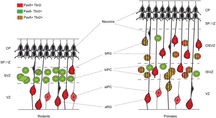
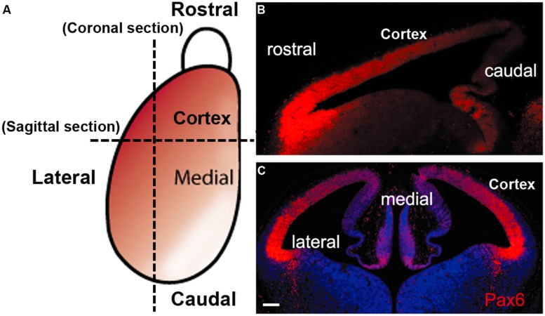
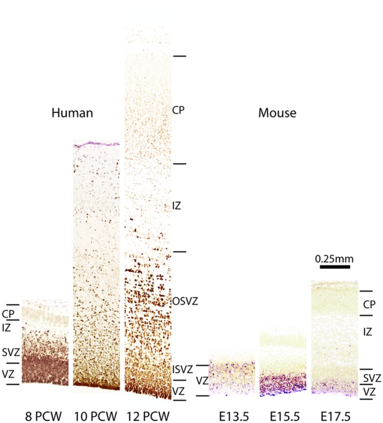
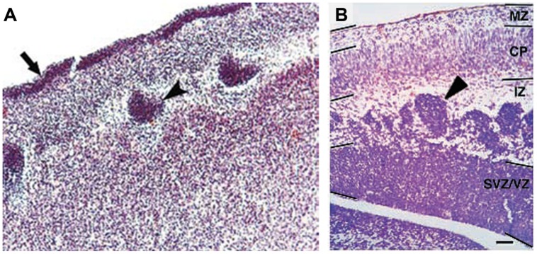
Understanding brain development remains a major challenge at the heart of understanding what makes us human. The neocortex, in evolutionary terms the newest part of the cerebral cortex, is the seat of higher cognitive functions. Its normal development requires the production, positioning, and appropriate interconnection of very large numbers of both excitatory and inhibitory neurons. Pax6 is one of a relatively small group of transcription factors that exert high-level control of cortical development, and whose mutation or deletion from developing embryos causes major brain defects and a wide range of neurodevelopmental disorders. Pax6 is very highly conserved between primate and non-primate species, is expressed in a gradient throughout the developing cortex and is essential for normal corticogenesis. Our understanding of Pax6's functions and the cellular processes that it regulates during mammalian cortical development has significantly advanced in the last decade, owing to the combined application of genetic and biochemical analyses. Here, we review the functional importance of Pax6 in regulating cortical progenitor proliferation, neurogenesis, and formation of cortical layers and highlight important differences between rodents and primates. We also review the pathological effects of PAX6 mutations in human neurodevelopmental disorders. We discuss some aspects of Pax6's molecular actions including its own complex transcriptional regulation, the distinct molecular functions of its splice variants and some of Pax6's known direct targets which mediate its actions during cortical development.
Overview
The claims and statistics of this paper haven’t been checked yet.
Run the check — free, no account needed. It reads the open-access text and checks the sources it cites.
Integrity signals
Registry
Cites
Statistics
Figures
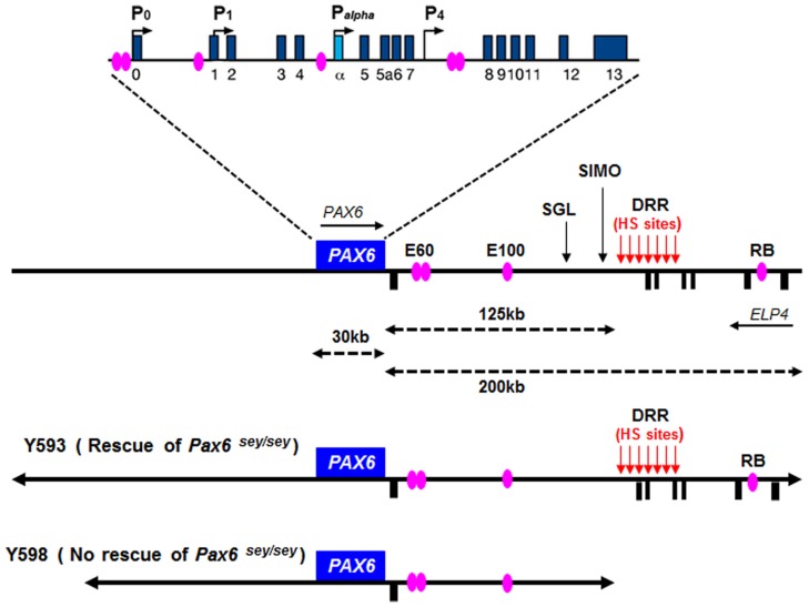
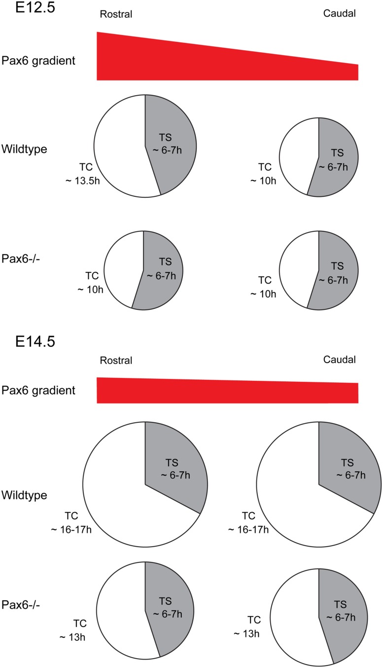
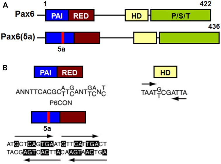
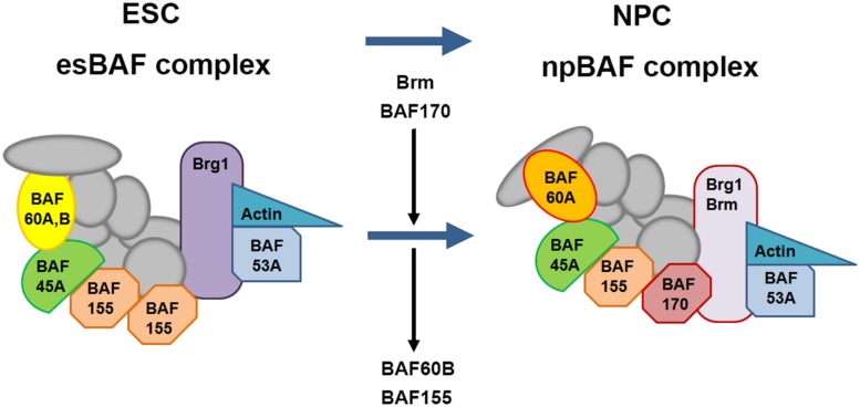
Verification
Claims the paper makes
The claims and statistics of this paper haven’t been checked yet.
Run the check — free, no account needed. It reads the open-access text and checks the sources it cites.
Claims other papers make about this one
Each is checked against this paper’s own text: does it say what the citing paper says it does?
In the citing paper
“Pax6 is one of the few “master regulator” neural TFs with a well-known role in cortical development (Manuel et al., 2015).”
From Multiscale 3D Genome Rewiring during Mouse Neural Development (2017) · cites this paper as Manuel et al., 2015
Self-check
Percentages, sums and reported differences re-computed from the paper’s own numbers.
The self-check needs the paper’s full text.
Citations
Cites
Inhibitors of mammalian G1 cyclin-dependent kinases.Preprint
Sherr, C. J., & Roberts, J. M. (1995) · Genes & Development · doi:10.1101/gad.9.10.1149 · Cited by 3,242
Specification of Cerebral Cortical Areas
Rakic, P. (1988) · Science · doi:10.1126/science.3291116 · Cited by 3,127 · cited 2×
“RGCs, whose long processes span the neuroepithelium, have been known for a long time to provide guidance for migrating neurons (Levitt and Rakic, 1980; Rakic, 1988; Hatten, 2002; Tan and Shi, 2013).”
Noctor, S. C., Martínez-Cerdeño, V., Ivic, L., & Kriegstein, A. R. (2004) · Nature Neuroscience · doi:10.1038/nn1172 · Cited by 2,229
Polyák, K., Lee, M.-H., Erdjument‐Bromage, H., Koff, A., Roberts, J. M., Tempst, P., & Massagué, J. (1994) · Cell · doi:10.1016/0092-8674(94)90572-x · Cited by 2,118
Neurons derived from radial glial cells establish radial units in neocortex
Noctor, S. C., Flint, A. C., Weissman, T. A., Dammerman, R. S., & Kriegstein, A. R. (2001) · Nature · doi:10.1038/35055553 · Cited by 2,016
The basics of brain development
Stiles, J., & Jernigan, T. L. (2010) · Neuropsychology Review · doi:10.1007/s11065-010-9148-4 · Cited by 2,009
Autoradiographic Study of Cell Migration during Histogenesis of Cerebral Cortex in the Mouse
ANGEVINE, J. B., & SIDMAN, R. L. (1961) · Nature · doi:10.1038/192766b0 · Cited by 1,530
To cycle or not to cycle: a critical decision in cancer
Malumbres, M., & Barbacid, M. (2001) · Nature reviews. Cancer · doi:10.1038/35106065 · Cited by 1,510
Neurogenic radial glia in the outer subventricular zone of human neocortex
Hansen, D. V., Lui, J. H., Parker, P. R. L., & Kriegstein, A. R. (2010) · Nature · doi:10.1038/nature08845 · Cited by 1,388
Mammalian cyclin-dependent kinases
Malumbres, M., & Barbacid, M. (2005) · Trends in Biochemical Sciences · doi:10.1016/j.tibs.2005.09.005 · Cited by 1,333
Englund, C., Fink, A., Lau, C., Pham, D., Daza, R. A. M., Bulfone, A., Kowalczyk, T., & Hevner, R. F. (2005) · The Journal of Neuroscience · doi:10.1523/jneurosci.2899-04.2005 · Cited by 1,261
Rakic, P. (1974) · Science · doi:10.1126/science.183.4123.425 · Cited by 1,161
Isolation of radial glial cells by fluorescent-activated cell sorting reveals a neuronal lineage
Malatesta, P., Hartfuss, E., & Götz, M. (2000) · Development · doi:10.1242/dev.127.24.5253 · Cited by 1,157
Haubensak, W. E., Attardo, A., Denk, W. T., & Huttner, W. Β. (2004) · Proceedings of the National Academy of Sciences · doi:10.1073/pnas.0308600100 · Cited by 1,004
The origin and specification of cortical interneurons
Wonders, C. P., & Anderson, S. A. (2006) · Nature Reviews Neuroscience · doi:10.1038/nrn1954 · Cited by 980 · cited 1×
“%) with others coming from the caudal ganglionic eminence (CGE) and areas including the preoptic area (POA; Anderson et al., 2001; Wonders and Anderson, 2006; Fogarty et al., 2007; Gelman et al., 2009; Miyoshi et al., 2010).”
Pax6 Is Required for the Multipotent State of Retinal Progenitor Cells
Marquardt, T., Ashery‐Padan, R., Andrejewski, N., Scardigli, R., Guillemot, F., & Gruß, P. (2001) · Cell · doi:10.1016/s0092-8674(01)00295-1 · Cited by 926
Asymmetric Inheritance of Radial Glial Fibers by Cortical Neurons
Miyata, T., Kawaguchi, A., Okano, H., & Ogawa, M. (2001) · Neuron · doi:10.1016/s0896-6273(01)00420-2 · Cited by 892
OSVZ progenitors of human and ferret neocortex are epithelial-like and expand by integrin signaling
Fietz, S. A., Kelava, I., Vogt, J., Wilsch-Bräuninger, M., Stenzel, D., Fish, J. L., Corbeil, D., Riehn, A., Distler, W., Nitsch, R., et al. (2010) · Nature Neuroscience · doi:10.1038/nn.2553 · Cited by 830
Asymmetric production of surface-dividing and non-surface-dividing cortical progenitor cells
Miyata, T., Kawaguchi, A., Saito, K., Kawano, M., Muto, T., & Ogawa, M. (2004) · Development · doi:10.1242/dev.01173 · Cited by 791
Glial cells generate neurons: the role of the transcription factor Pax6
Heins, N., Malatesta, P., Cecconi, F., Nakafuku, M., Tucker, K. L., Hack, M. A., Chapouton, P., Barde, Y.-A., & Götz, M. (2002) · Nature Neuroscience · doi:10.1038/nn828 · Cited by 775
Malatesta, P., Hack, M. A., Hartfuss, E., Kettenmann, H., Klinkert, W. E. F., Kirchhoff, F., & Götz, M. (2003) · Neuron · doi:10.1016/s0896-6273(03)00116-8 · Cited by 771
Lessard, J., Wu, J. I., Ranish, J. A., Wan, M., Winslow, M. M., Staahl, B. T., Wu, H., Aebersold, R., Graef, I. A., & Crabtree, G. R. (2007) · Neuron · doi:10.1016/j.neuron.2007.06.019 · Cited by 735
Origin of GABAergic neurons in the human neocortex
Letinić, K., Zoncu, R., & RAKIC, P. T. (2002) · Nature · doi:10.1038/nature00779 · Cited by 730
Glaser, T. M., Jepeal, L. I., Edwards, J. G., Young, S. R., Favor, J., & Maas, R. L. (1994) · Nature Genetics · doi:10.1038/ng0894-463 · Cited by 713
Levitt, P., & Rakic, P. (1980) · Journal of Comparative Neurology · doi:10.1002/cne.901930316 · Cited by 704
Molecular biology of the Ets family of transcription factors
Oikawa, T., & Yamada, T. (2003) · Gene · doi:10.1016/s0378-1119(02)01156-3 · Cited by 701
Smart, I. H. M. (2002) · Cerebral Cortex · doi:10.1093/cercor/12.1.37 · Cited by 687
The migration of neuroblasts in the developing cerebral cortex.
Berry, M., & Rogers, A. W. (1965) · PubMed · Cited by 674
The cell cycle of the pseudostratified ventricular epithelium of the embryonic murine cerebral wall
Takahashi, T., Nowakowski, R. S., & Caviness, V. S. (1995) · Journal of Neuroscience · doi:10.1523/jneurosci.15-09-06046.1995 · Cited by 673
Pax6 Controls Radial Glia Differentiation in the Cerebral Cortex
Götz, M., Stoykova, A., & Gruß, P. (1998) · Neuron · doi:10.1016/s0896-6273(00)80621-2 · Cited by 665
Cell-cycle control and cortical development
Dehay, C., & Kennedy, H. (2007) · Nature Reviews Neuroscience · doi:10.1038/nrn2097 · Cited by 655
Genomic structure, evolutionary conservation and aniridia mutations in the human PAX6 gene
Glaser, T. M., Walton, D. S., & Maas, R. L. (1992) · Nature Genetics · doi:10.1038/ng1192-232 · Cited by 655
Neural progenitors, neurogenesis and the evolution of the neocortex
Florio, M., & Huttner, W. Β. (2014) · Development · doi:10.1242/dev.090571 · Cited by 654
Distinct cortical migrations from the medial and lateral ganglionic eminences
Anderson, S. A., Marín, Ó., Horn, C., Jennings, K., & Rubenstein, J. L. R. (2001) · Development · doi:10.1242/dev.128.3.353 · Cited by 651
Numbers and proportions of GABA-immunoreactive neurons in different areas of monkey cerebral cortex
Hendry, S., Schwark, H. D., Jones, E. G., & Yan, J. (1987) · Journal of Neuroscience · doi:10.1523/jneurosci.07-05-01503.1987 · Cited by 601
Area Patterning of the Mammalian Cortex
O’Leary, D. D. M., Chou, S., & Sahara, S. (2007) · Neuron · doi:10.1016/j.neuron.2007.10.010 · Cited by 593
Roles of Pax-genes in developing and adult brain as suggested by expression patterns
Stoykova, A., & Gruss, P. (1994) · Journal of Neuroscience · doi:10.1523/jneurosci.14-03-01395.1994 · Cited by 577
Ho, L., Ronan, J. L., Wu, J. I., Staahl, B. T., Chen, L., Kuo, A., Lessard, J. A., Nesvizhskii, A. I., Ranish, J. A., & Crabtree, G. R. (2009) · Proceedings of the National Academy of Sciences · doi:10.1073/pnas.0812889106 · Cited by 576
Regulation of Area Identity in the Mammalian Neocortex by Emx2 and Pax6
Bishop, K. M., Goudreau, G., & O’Leary, D. D. M. (2000) · Science · doi:10.1126/science.288.5464.344 · Cited by 575
Miyoshi, G., Hjerling-Leffler, J., Karayannis, T., Sousa, V. H., Butt, S. J. B., Battiste, J., Johnson, J. E., Machold, R. P., & Fishell, G. (2010) · The Journal of Neuroscience · doi:10.1523/jneurosci.4515-09.2010 · Cited by 559
Hox cofactors in vertebrate development
Moens, C. B., & Selleri, L. (2006) · Developmental Biology · doi:10.1016/j.ydbio.2005.10.032 · Cited by 515
Betizeau, M., Cortay, V., Patti, D., Pfister, S., Gautier, E., Bellemin-Ménard, A., Afanassieff, M., Huissoud, C., Douglas, R. J., Kennedy, H., et al. (2013) · Neuron · doi:10.1016/j.neuron.2013.09.032 · Cited by 462
Murray, A. W. (1989) · American Zoologist · doi:10.1093/icb/29.2.511 · Cited by 460
Gsh2 and Pax6 play complementary roles in dorsoventral patterning of the mammalian telencephalon
Yun, K., Potter, S., & Rubenstein, J. L. R. (2001) · Development · doi:10.1242/dev.128.2.193 · Cited by 449
Pax6 induces ectopic eyes in a vertebrate
Chow, R. L., Altmann, C. R., Lang, R. A., & Hemmati‐Brivanlou, A. (1999) · Development · doi:10.1242/dev.126.19.4213 · Cited by 447
Influence of PAX6 Gene Dosage on Development: Overexpression Causes Severe Eye Abnormalities
Schedl, A., Ross, A., Lee, M., Engelkamp, D., Rashbass, P., van Heyningen, V., & Hastie, N. D. (1996) · Cell · doi:10.1016/s0092-8674(00)80078-1 · Cited by 440
Cell-cycle inhibitors: three families united by a common cause
Vidal, A., & Koff, A. (2000) · Gene · doi:10.1016/s0378-1119(00)00092-5 · Cited by 439
Ho, L., Jothi, R., Ronan, J. L., Cui, K., Zhao, K., & Crabtree, G. R. (2009) · Proceedings of the National Academy of Sciences · doi:10.1073/pnas.0812888106 · Cited by 429
Fogarty, M., Grist, M., Gelman, D., Marín, O., Pachnis, V., & Kessaris, N. (2007) · The Journal of Neuroscience · doi:10.1523/jneurosci.1629-07.2007 · Cited by 412
Compartments and their boundaries in vertebrate brain development
Kiecker, C. M., & Lumsden, A. (2005) · Nature reviews. Neuroscience · doi:10.1038/nrn1702 · Cited by 408
Glial cell lineage in the cerebral cortex: A review and synthesis
Cameron, R. S., & RAKIC, P. T. (1991) · Glia · doi:10.1002/glia.440040204 · Cited by 407
Gruß, P., & Walther, C. (1992) · Cell · doi:10.1016/0092-8674(92)90281-g · Cited by 403
Sansom, S. N., Griffiths, D. S., Faedo, A., Kleinjan, D., Ruan, Y., Smith, J. C., van Heyningen, V., Rubenstein, J. L. R., & Livesey, F. J. (2009) · PLoS Genetics · doi:10.1371/journal.pgen.1000511 · Cited by 401
Neural stem and progenitor cells shorten S-phase on commitment to neuron production
Arai, Y., Pulvers, J. N., Haffner, C., Schilling, B., Nüsslein, I., Calegari, F., & Huttner, W. Β. (2011) · Nature Communications · doi:10.1038/ncomms1155 · Cited by 399
Sequential phases of cortical specification involve Neurogenin‐dependent and ‐independent pathways
Schuurmans, C. J., Armant, O., Nieto, M., Stenman, J. M., Britz, O., Klenin, N., Brown, C. E., Langevin, L.-M., Seibt, J., Tang, H., et al. (2004) · The EMBO Journal · doi:10.1038/sj.emboj.7600278 · Cited by 399
Pax6 and SOX2 form a co-DNA-binding partner complex that regulates initiation of lens developmentPreprint
Kamachi, Y., Uchikawa, M., Tanouchi, A., Sekido, R., & Kondoh, H. (2001) · Genes & Development · doi:10.1101/gad.887101 · Cited by 394
Czerny, T., Schaffner, G., & Busslinger, M. (1993) · Genes & Development · doi:10.1101/gad.7.10.2048 · Cited by 392
Genetic control of dorsal-ventral identity in the telencephalon: opposing roles for Pax6 and Gsh2
Toresson, H., Potter, S. S., & Campbell, K. (2000) · Development · doi:10.1242/dev.127.20.4361 · Cited by 387
Czerny, T., Halder, G. A., Kloter, U., Souabni, A., Gehring, W. J., & Busslinger, M. (1999) · Molecular Cell · doi:10.1016/s1097-2765(00)80457-8 · Cited by 385
Cooperative dimerization of paired class homeo domains on DNA.Preprint
Wilson, D., Sheng, G., Lecuit, T., Dostatni, N., & Desplan, C. (1993) · Genes & Development · doi:10.1101/gad.7.11.2120 · Cited by 381
Chromatin-Remodeling Components of the BAF Complex Facilitate Reprogramming
Singhal, N., Graumann, J., Wu, G., Araúzo-Bravo, M. J., Han, D. W., Greber, B., Gentile, L., Mann, M., & Schöler, H. R. (2010) · Cell · doi:10.1016/j.cell.2010.04.037 · Cited by 378 · cited 1×
“In mammals, BAF complexes contain two ATPase subunits Brg1 or Brm (Brahma), which are mutually exclusive and essential for remodeling activity, in combination with at least 15 different BAF (Brg1/Brm-associated factor) subunits (Ho et al., 2009a; Singhal et al., 2010).”
Epstein, J. A., Cai, J., Glaser, T. M., Jepeal, L. I., & Maas, R. L. (1994) · Journal of Biological Chemistry · doi:10.1016/s0021-9258(17)37201-0 · Cited by 368
Osumi, N., Shinohara, H. M., Numayama‐Tsuruta, K., & Maekawa, M. (2008) · Stem Cells · doi:10.1634/stemcells.2007-0884 · Cited by 360
Gal, J. S., Morozov, Y. M., Ayoub, A. E., Chatterjee, M., RAKIC, P. T., & Haydar, T. F. (2006) · Journal of Neuroscience · doi:10.1523/jneurosci.4499-05.2006 · Cited by 358
What is the restriction point?
Zetterberg, A., Larsson, O., & Wiman, K. G. (1995) · Current Opinion in Cell Biology · doi:10.1016/0955-0674(95)80067-0 · Cited by 351
Primate Origins and Evolution. A phylogenetic reconstruction
Dagosto, M. (1991) · International Journal of Primatology · doi:10.1007/bf02547560 · Cited by 349
Takahashi, T., Nowakowski, R. S., & Caviness, V. S. (1993) · Journal of Neuroscience · doi:10.1523/jneurosci.13-02-00820.1993 · Cited by 339
Cortical upper layer neurons derive from the subventricular zone as indicated bySvet1gene expression
Tarabykin, V. S., Stoykova, A., Usman, N. Y., & Gruß, P. (2001) · Development · doi:10.1242/dev.128.11.1983 · Cited by 336
Meis1, a PBX1-Related Homeobox Gene Involved in Myeloid Leukemia in BXH-2 Mice
Moskow, J. J., Bullrich, F., Huebner, K., Daar, I. O., & Buchberg, A. M. (1995) · Molecular and Cellular Biology · doi:10.1128/mcb.15.10.5434 · Cited by 335
Paige, S. L., Thomas, S., Stoick-Cooper, C. L., Wang, H., Maves, L., Sandstrom, R. S., Pabon, L., Reinecke, H., Pratt, G. A., Keller, G. M., et al. (2012) · Cell · doi:10.1016/j.cell.2012.08.027 · Cited by 332
Non-epithelial stem cells and cortical interneuron production in the human ganglionic eminences
Hansen, D. V., Lui, J. H., Flandin, P., Yoshikawa, K., Rubenstein, J. L., Alvarez-Buylla, A., & Kriegstein, A. R. (2013) · Nature Neuroscience · doi:10.1038/nn.3541 · Cited by 328
Conserved regulation of proximodistal limb axis development by Meis1/Hth
Mercader, N., Leonardo, E., Azpiazu, N., Serrano, A., Morata, G., Martínez‐A, C., & Torres, M. (1999) · Nature · doi:10.1038/46580 · Cited by 322
Pax6Modulates the Dorsoventral Patterning of the Mammalian Telencephalon
Stoykova, A., Treichel, D., Hallonet, M. E. R., & Gruß, P. (2000) · Journal of Neuroscience · doi:10.1523/jneurosci.20-21-08042.2000 · Cited by 322
New Directions in Neuronal Migration
Hatten, M. E. (2002) · Science · doi:10.1126/science.1074572 · Cited by 321
G1 Phase Regulation, Area-Specific Cell Cycle Control, and Cytoarchitectonics in the Primate Cortex
Lukaszewicz, A., Savatier, P., Cortay, V., Giroud, P., Huissoud, C., Berland, M., Kennedy, H., & Dehay, C. (2005) · Neuron · doi:10.1016/j.neuron.2005.06.032 · Cited by 315
Xu, H. E., Rould, M. A., Xu, W., Epstein, J. A., Maas, R. L., & Pabo, C. O. (1999) · Genes & Development · doi:10.1101/gad.13.10.1263 · Cited by 311
Hematopoietic, angiogenic and eye defects in Meis1 mutant animals
Hisa, T., Spence, S. E., Rachel, R. A., Fujita, M., Nakamura, T., Ward, J. M., Devor-Henneman, D. E., Saiki, Y., Kutsuna, H., Tessarollo, L., et al. (2004) · The EMBO Journal · doi:10.1038/sj.emboj.7600038 · Cited by 306
Huttner, W. Β., & Kosodo, Y. (2005) · Current Opinion in Cell Biology · doi:10.1016/j.ceb.2005.10.005 · Cited by 305
Cubelos, B., Sebastián‐Serrano, Á., Beccari, L. L., Calcagnotto, M. E., Cisneros, E., Kim, S., Dopazo, A., Álvarez‐Dolado, M., Redondo, J. M., Bovolenta, P., et al. (2010) · Neuron · doi:10.1016/j.neuron.2010.04.038 · Cited by 301
Numerical data on neocortical neurons in adult rat, with special reference to the GABA population
Beaulieu, C. (1993) · Brain Research · doi:10.1016/0006-8993(93)90884-p · Cited by 301
Czerny, T., & Busslinger, M. (1995) · Molecular and Cellular Biology · doi:10.1128/mcb.15.5.2858 · Cited by 298
Regionalization and fate specification in neurospheres: the role of Olig2 and Pax6.
Hack, M., Sugimori, M., Lundberg, C., Nakafuku, M., & Götz, M. (2004) · Molecular and cellular neurosciences · doi:10.1016/j.mcn.2003.12.012 · Cited by 294
Changes in cell-cycle kinetics during the development and evolution of primate neocortex
Kornack, D. R., & RAKIC, P. T. (1998) · Proceedings of the National Academy of Sciences · doi:10.1073/pnas.95.3.1242 · Cited by 292
Cell cycle control of mammalian neural stem cells: putting a speed limit on G1
Salomoni, P., & Calegari, F. (2010) · Trends in Cell Biology · doi:10.1016/j.tcb.2010.01.006 · Cited by 286
Distinctcis-Essential Modules Direct the Time–Space Pattern of thePax6Gene Activity
Kammandel, B., Chowdhury, K., Stoykova, A., Aparicio, S. A. J. R., Brenner, S., & Gruß, P. (1999) · Developmental Biology · doi:10.1006/dbio.1998.9128 · Cited by 281
The embryonic preoptic area is a novel source of cortical GABAergic interneurons
Gelman, D. M., Martini, F. J., Nóbrega-Pereira, S., Pierani, A., Kessaris, N., & Marín, O. (2009) · The Journal of Neuroscience · doi:10.1523/jneurosci.0604-09.2009 · Cited by 280
Contributions of cortical subventricular zone to the development of the human cerebral cortex
Zecevic, N., Chen, Y., & Filipovic, R. (2005) · The Journal of Comparative Neurology · doi:10.1002/cne.20714 · Cited by 269
Farkas, L. M., & Huttner, W. Β. (2008) · Current Opinion in Cell Biology · doi:10.1016/j.ceb.2008.09.008 · Cited by 263
Sequence and expression pattern ofpax-6are highly conserved between zebrafish and mice
Püschel, A. W., Gruß, P., & Westerfield, M. (1992) · Development · doi:10.1242/dev.114.3.643 · Cited by 261
Bishop, K. M., Rubenstein, J. L. R., & O’Leary, D. D. M. (2002) · Journal of Neuroscience · doi:10.1523/jneurosci.22-17-07627.2002 · Cited by 260
Meis Proteins are Major In Vivo DNA Binding Partners for Wild-Type but Not Chimeric Pbx Proteins
Chang, C., Jacobs, Y., Nakamura, T., Jenkins, N. A., Copeland, N. G., & Cleary, M. L. (1997) · Molecular and Cellular Biology · doi:10.1128/mcb.17.10.5679 · Cited by 253
Estivill‐Torrús, G., Pearson, H., van Heyningen, V., Price, D. J., & Rashbass, P. (2002) · Development · doi:10.1242/dev.129.2.455 · Cited by 251
Olig2 Directs Astrocyte and Oligodendrocyte Formation in Postnatal Subventricular Zone Cells
Marshall, C. A. G., Novitch, B. G., & Goldman, J. (2005) · Journal of Neuroscience · doi:10.1523/jneurosci.1924-05.2005 · Cited by 249
Mitotic spindle rotation and mode of cell division in the developing telencephalon
Haydar, T. F., Ang, E. S. B. C., & RAKIC, P. T. (2003) · Proceedings of the National Academy of Sciences · doi:10.1073/pnas.0437969100 · Cited by 242
Direct and concentration-dependent regulation of the proneural gene Neurogenin2 by Pax6
Scardigli, R., Bäumer, N., Gruss, P., Guillemot, F., & Roux, I. L. (2003) · Development · doi:10.1242/dev.00539 · Cited by 239
PAX6 haploinsufficiency causes cerebral malformation and olfactory dysfunction in humans
Sisodiya, S. M., Free, S. L., Williamson, K. A., Mitchell, T. N., Willis, C., Stevens, J., Kendall, B. E., Shorvon, S. D., Hanson, I. M., Moore, A. T., et al. (2001) · Nature Genetics · doi:10.1038/90042 · Cited by 238
Quinn, J. C., Molinek, M., Martynoga, B., Zaki, P. A., Faedo, A., Bulfone, A., Hevner, R. F., West, J. D., & Price, D. J. (2006) · Developmental Biology · doi:10.1016/j.ydbio.2006.08.035 · Cited by 231
The Pax-6 homeobox gene is expressed throughout the corneal and conjunctival epithelia.
Koroma, B. M., Yang, J.-M., & Sundin, O. H. (1997) · PubMed · Cited by 222
Frequent Chromosome Aberrations Revealed by Molecular Cytogenetic Studies in Patients with Aniridia
Crolla, J. A., & van Heyningen, V. (2002) · The American Journal of Human Genetics · doi:10.1086/344396 · Cited by 221
The evolution of the neocortex in mammals: how is phenotypic diversity generated?
Krubitzer, L. A., & Kaas, J. H. (2005) · Current Opinion in Neurobiology · doi:10.1016/j.conb.2005.07.003 · Cited by 218
Ninkovic, J., Steiner-Mezzadri, A., Jawerka, M., Akinci, U., Masserdotti, G., Petricca, S., Fischer, J., von Holst, A., Beckers, J., Lie, D. C., et al. (2013) · Cell stem cell · doi:10.1016/j.stem.2013.07.002 · Cited by 216
Fantes, J. A., Redeker, B., Breen, M., Boyle, S., Brown, J., Fletcher, J. M., Jones, S., Bickmore, W. A., Fukushima, Y., Mannens, M. M. A. M., et al. (1995) · Human Molecular Genetics · doi:10.1093/hmg/4.3.415 · Cited by 214
Cooperative interactions between paired domain and homeodomain
Jun, S., & Desplan, C. (1996) · Development · doi:10.1242/dev.122.9.2639 · Cited by 213
Kleinjan, D. A. (2001) · Human Molecular Genetics · doi:10.1093/hmg/10.19.2049 · Cited by 209
Schmahl, W., Knödlseder, M., Favor, J., & Davidson, D. R. (1993) · Acta Neuropathologica · doi:10.1007/bf00334879 · Cited by 205
Sheng, G., Thouvenot, E., Schmucker, D., Wilson, D. S., & Desplan, C. (1997) · Genes & Development · doi:10.1101/gad.11.9.1122 · Cited by 201
Haubst, N., Berger, J., Radjendirane, V., Graw, J., Favor, J., Saunders, G. F., Stoykova, A., & Götz, M. (2004) · Development · doi:10.1242/dev.01524 · Cited by 192
Detailed Ophthalmologic Evaluation of 43 Individuals with PAX6 Mutations
Hingorani, M., Williamson, K. A., Moore, A. T., & van Heyningen, V. (2009) · Investigative Ophthalmology & Visual Science · doi:10.1167/iovs.08-2827 · Cited by 190
Regulation of Pax6 expression is conserved between mice and flies
Xu, P., Zhang, X., Heaney, S., Yoon, A., Michelson, A. M., & Maas, R. L. (1999) · Development · doi:10.1242/dev.126.2.383 · Cited by 190
Generation of interneuron diversity in the mouse cerebral cortex
Gelman, D. M., & Marín, O. (2010) · European Journal of Neuroscience · doi:10.1111/j.1460-9568.2010.07267.x · Cited by 188
The role of Pax6 in forebrain development
Georgala, P. A., Carr, C. B., & Price, D. J. (2011) · Developmental Neurobiology · doi:10.1002/dneu.20895 · Cited by 186
Long, J. E., Cobos, I., Potter, G. B., & Rubenstein, J. L. R. (2009) · Cerebral Cortex · doi:10.1093/cercor/bhp045 · Cited by 178
Chromatin Regulation by BAF170 Controls Cerebral Cortical Size and Thickness
Tuoc, T. C., Boretius, S., Sansom, S. N., Pitulescu, M.-E., Frahm, J., Livesey, F. J., & Stoykova, A. (2013) · Developmental Cell · doi:10.1016/j.devcel.2013.04.005 · Cited by 177
A Role for Proneural Genes in the Maturation of Cortical Progenitor Cells
Britz, O., Mattar, P. A., Nguyen, L., Langevin, L.-M., Zimmer, C., Alam, S., Guillemot, F., & Schuurmans, C. (2006) · Cerebral Cortex · doi:10.1093/cercor/bhj168 · Cited by 175
The Groucho/TLE/Grg family of transcriptional co-repressors
Jennings, B. H., & Ish‐Horowicz, D. (2008) · Genome biology · doi:10.1186/gb-2008-9-1-205 · Cited by 175
Emx2 and Pax6 Control Regionalization of the Pre-neuronogenic Cortical Primordium
Muzio, L., Benedetto, B. D., Stoykova, A., Boncinelli, E., Gruss, P., & Mallamaci, A. (2002) · Cerebral Cortex · doi:10.1093/cercor/12.2.129 · Cited by 172
Warren, N. (1999) · Cerebral Cortex · doi:10.1093/cercor/9.6.627 · Cited by 171
Yan, Z., Wang, Z., Sharova, L. V., Sharov, A. A., Ling, C., Piao, Y., Aiba, K., Matoba, R., Wang, W., & Ko, M. S. H. (2008) · Stem Cells · doi:10.1634/stemcells.2007-0846 · Cited by 168
3′ deletions cause aniridia by preventing PAX6 gene expression
Lauderdale, J. D., Wilensky, J. S., Oliver, E. R., Walton, D. S., & Glaser, T. M. (2000) · Proceedings of the National Academy of Sciences · doi:10.1073/pnas.240398797 · Cited by 165
Polymicrogyria and absence of pineal gland due to PAX6 mutation
Mitchell, T. N., Free, S. L., Williamson, K. A., Stevens, J., Churchill, A., Hanson, I. M., Shorvon, S. D., Moore, A. T., van Heyningen, V., & Sisodiya, S. M. (2003) · Annals of Neurology · doi:10.1002/ana.10576 · Cited by 163
Bentivoglio, M., & Mazzarello, P. (1999) · Brain Research Bulletin · doi:10.1016/s0361-9230(99)00065-9 · Cited by 162
Kozmík, Z., Czerny, T., & Busslinger, M. (1997) · The EMBO Journal · doi:10.1093/emboj/16.22.6793 · Cited by 157
Long, J. E., Swan, C. H., Liang, W. S., Cobos, I., Potter, G. B., & Rubenstein, J. L. R. (2008) · The Journal of Comparative Neurology · doi:10.1002/cne.21854 · Cited by 156
Carić, D., Gooday, D. J., Hill, R. E., McConnell, S. K., & Price, D. J. (1997) · Development · doi:10.1242/dev.124.24.5087 · Cited by 150
Gradients in the Brain: The Control of the Development of Form and Function in the Cerebral Cortex
Sansom, S. N., & Livesey, F. J. (2009) · Cold Spring Harbor Perspectives in Biology · doi:10.1101/cshperspect.a002519 · Cited by 148
Strategies for the generation of neuronal diversity in the developing central nervous system
McConnell, S. K. (1995) · Journal of Neuroscience · doi:10.1523/jneurosci.15-11-06987.1995 · Cited by 147
Domínguez, M., Ferrés-Marcó, D., Gutiérrez-Aviñó, F. J., Speicher, S. A., & Beneyto, M. (2003) · Nature Genetics · doi:10.1038/ng1281 · Cited by 146
A highly conserved lens transcriptional control element from the Pax-6 gene
Williams, S., Altmann, C. R., Chow, R. L., Hemmati‐Brivanlou, A., & Lang, R. A. (1998) · Mechanisms of Development · doi:10.1016/s0925-4773(98)00057-4 · Cited by 142
Dual Roles for Pax-6: a Transcriptional Repressor of Lens Fiber Cell-Specific β-Crystallin Genes
Duncan, M. K., Haynes, J. I., Cvekl, A., & Piatigorsky, J. (1998) · Molecular and Cellular Biology · doi:10.1128/mcb.18.9.5579 · Cited by 141
Hevner, R. F. (2005) · Molecular Neurobiology · doi:10.1385/mn:33:1:033 · Cited by 138
The ‘Marx’ of Groucho on development and disease
Buscarlet, M., & Stifani, S. (2007) · Trends in Cell Biology · doi:10.1016/j.tcb.2007.07.002 · Cited by 138
Functional analysis of an eye specific enhancer of the eyeless gene in Drosophila
Hauck, B., Gehring, W. J., & Walldorf, U. (1999) · Proceedings of the National Academy of Sciences · doi:10.1073/pnas.96.2.564 · Cited by 136
Long-range downstream enhancers are essential for Pax6 expression
Kleinjan, D. A., Seawright, A., Mella, S., Carr, C. B., Tyas, D. A., Simpson, T. I., Mason, J. O., Price, D. J., & van Heyningen, V. (2006) · Developmental Biology · doi:10.1016/j.ydbio.2006.08.060 · Cited by 136
Pax-6 is essential for lens-specific expression of zeta-crystallin.
Richardson, J. C., Cvekl, A., & Wistow, G. J. (1995) · Proceedings of the National Academy of Sciences · doi:10.1073/pnas.92.10.4676 · Cited by 135
Stenman, J. M., Yu, R. T., Evans, R. M., & Campbell, K. (2003) · Development · doi:10.1242/dev.00328 · Cited by 129
Pax6 3′ deletion results in aniridia, autism and mental retardation
Davis, L. K., Meyer, K. J., Rudd, D. S., Librant, A. L., Epping, E. A., Sheffield, V. C., & Wassink, T. H. (2008) · Human Genetics · doi:10.1007/s00439-008-0484-x · Cited by 128
Jakovcevski, I., Mayer, N., & Zecevic, N. (2011) · Cerebral Cortex · doi:10.1093/cercor/bhq245 · Cited by 124
The on/off of Pax6 controls the tempo of neuronal differentiation in the developing spinal cord
Bel-Vialar, S., Médevielle, F., & Pituello, F. (2007) · Developmental Biology · doi:10.1016/j.ydbio.2007.02.012 · Cited by 123
Agoston, Z., Heine, P., Brill, M. S., Grebbin, B. M., Hau, A., Kallenborn-Gerhardt, W., Schramm, J., Götz, M., & Schulte, D. (2013) · Development · doi:10.1242/dev.097295 · Cited by 123
Aota, S., Nakajima, N., Sakamoto, R., Watanabe, S., Ibaraki, N., & Okazaki, K. (2003) · Developmental Biology · doi:10.1016/s0012-1606(03)00058-7 · Cited by 118
Manuel, M. N., Georgala, P. A., Carr, C. B., Chanas, S. A., Kleinjan, D. A., Martynoga, B., Mason, J. O., Molinek, M., Pinson, J., Pratt, T., et al. (2007) · Development · doi:10.1242/dev.02764 · Cited by 117
Knoepfler, P. S., Bergstrom, D. A., Uetsuki, T., Dac-Korytko, L., Sun, Y., Wright, W. E., Tapscott, S. J., & Kamps, M. P. (1999) · Nucleic Acids Research · doi:10.1093/nar/27.18.3752 · Cited by 117
Loss- and gain-of-function analyses reveal targets of Pax6 in the developing mouse telencephalon
Holm, P. C., Mader, M. T., Haubst, N., Wizenmann, A., Sigvardsson, M., & Götz, M. (2006) · Molecular and Cellular Neuroscience · doi:10.1016/j.mcn.2006.10.008 · Cited by 117
Tuoc, T., Radyushkin, K., Tonchev, A. B., Piñon, M. C., Ashery‐Padan, R., Molnár, Z., Davidoff, M. S., & Stoykova, A. (2009) · Journal of Neuroscience · doi:10.1523/jneurosci.5669-08.2009 · Cited by 115
Tuoc, T., & Stoykova, A. (2008) · Genes & Development · doi:10.1101/gad.471708 · Cited by 115
Enhancer identification through comparative genomics
Visel, A., Bristow, J. D., & Pennacchio, L. A. (2007) · Seminars in Cell and Developmental Biology · doi:10.1016/j.semcdb.2006.12.014 · Cited by 114
Conserved elements in Pax6 intron 7 involved in (auto)regulation and alternative transcription
Kleinjan, D. A., Seawright, A., Childs, A. J., & van Heyningen, V. (2003) · Developmental Biology · doi:10.1016/j.ydbio.2003.09.011 · Cited by 111
Kroll, T. T., & O’Leary, D. D. M. (2005) · Proceedings of the National Academy of Sciences · doi:10.1073/pnas.0500819102 · Cited by 111
Characterization of Quail Pax-6 (Pax-QNR) Proteins Expressed in the Neuroretina
Carrière, C., Plaza, S., Martin, P., Quatannens, B., Bailly, M., Stéhelin, D., & Saule, S. (1993) · Molecular and Cellular Biology · doi:10.1128/mcb.13.12.7257-7266.1993 · Cited by 109
Precocious retinal neurons: Pax6 controls timing of differentiation and determination of cell type
Philips, G. T., Stair, C. N., Lee, H. Y., Wroblewski, E. E., Berberoglu, M. A., Brown, N. L., & Mastick, G. S. (2005) · Developmental Biology · doi:10.1016/j.ydbio.2004.12.018 · Cited by 109
Er81 is expressed in a subpopulation of layer 5 neurons in rodent and primate neocortices
Yoneshima, H., Yamasaki, S., Voelker, C., Molnár, Z., Christophe, E., Audinat, É., Takemoto, M., Nishiwaki, M., Tsuji, S., Fujita, I., et al. (2005) · Neuroscience · doi:10.1016/j.neuroscience.2005.08.075 · Cited by 107
Emx1, Emx2 and Pax6 in Specification, Regionalization and Arealization of the Cerebral Cortex
Muzio, L. (2003) · Cerebral Cortex · doi:10.1093/cercor/13.6.641 · Cited by 106
Cvekl, A., Yang, Y., Chauhan, B. K., & Cveklová, K. (2004) · The International Journal of Developmental Biology · doi:10.1387/ijdb.041866ac · Cited by 105
Diverse functions of ATP-dependent chromatin remodeling complexes in development and cancer
Wu, J. I. (2011) · Acta Biochimica et Biophysica Sinica · doi:10.1093/abbs/gmr099 · Cited by 101
Evidence for an evolutionary conserved role ofhomothorax/Meis1/2during vertebrate retina development
Heine, P., Dohle, E., O’Brien, K. M. B., Engelkamp, D., & Schulte, D. (2008) · Development · doi:10.1242/dev.012088 · Cited by 100
Identification of Pax6-Dependent Gene Regulatory Networks in the Mouse Lens
Wolf, L. V., Yang, Y., Wang, J., Xie, Q. M., Braunger, B. M., Tamm, E. R., Zavadil, J., & Cvekl, A. (2009) · PLoS ONE · doi:10.1371/journal.pone.0004159 · Cited by 98
choe, S., Lu, P., Nakamura, M., Lee, J., & Sagerström, C. G. (2009) · Developmental Cell · doi:10.1016/j.devcel.2009.08.007 · Cited by 98
Bessa, J., Tavares, M. J., Santos, J., Kikuta, H., Laplante, M. A., Becker, T. S., Gómez-Skármeta, J. L., & Casares, F. (2008) · Development · doi:10.1242/dev.011932 · Cited by 97
Solomon, B. D., Pineda‐Alvarez, D., Balog, J. Z., Hadley, D. W., Gropman, A. L., Nandagopal, R., Han, J. C., Hahn, J. S., Blain, D., Brooks, B. P., et al. (2009) · American Journal of Medical Genetics Part A · doi:10.1002/ajmg.a.33081 · Cited by 95
Agoston, Z., & Schulte, D. (2009) · Development · doi:10.1242/dev.037770 · Cited by 95
Truncation Mutations in the Transactivation Region of PAX6 Result in Dominant-Negative Mutants
Singh, S., Tang, H. K., Lee, J.-Y., & Saunders, G. F. (1998) · Journal of Biological Chemistry · doi:10.1074/jbc.273.34.21531 · Cited by 91
PAX6 suppresses growth of human glioblastoma cells
Zhou, Y.-H., Wu, X., Tan, F., Shi, Y.-X., Glass, T. L., Liu, T. J., Wathen, K., Hess, K. R., Gumin, J., Lang, F. F., et al. (2005) · Journal of Neuro-Oncology · doi:10.1007/s11060-004-1720-4 · Cited by 91
TlxControls Proliferation and Patterning of Lateral Telencephalic Progenitor Domains
Stenman, J. M., Wang, B., & Campbell, K. (2003) · Journal of Neuroscience · doi:10.1523/jneurosci.23-33-10568.2003 · Cited by 90
Griffin, C., Kleinjan, D. A., Doe, B., & van Heyningen, V. (2002) · Mechanisms of Development · doi:10.1016/s0925-4773(01)00646-3 · Cited by 89
Tang, H. K., Singh, S., & Saunders, G. F. (1998) · Journal of Biological Chemistry · doi:10.1074/jbc.273.13.7210 · Cited by 88
Kim, J., & Lauderdale, J. D. (2006) · Developmental Biology · doi:10.1016/j.ydbio.2005.12.041 · Cited by 86
Neocortical neurogenesis and neuronal migration
Tan, X., & Shi, S. (2012) · Wiley Interdisciplinary Reviews Developmental Biology · doi:10.1002/wdev.88 · Cited by 85
Loss of Gsx1 and Gsx2 function rescues distinct phenotypes in Dlx1/2 mutants
Wang, B., Long, J. E., Flandin, P., Pla, R., Waclaw, R. R., Campbell, K., & Rubenstein, J. L. R. (2013) · Journal of Comparative Neurology · doi:10.1002/cne.23242 · Cited by 85
Mikkola, I., Bruun, J., Holm, T., & Johansen, T. (2001) · Journal of Biological Chemistry · doi:10.1074/jbc.m008882200 · Cited by 84
Is Pax6 Critical for Neurogenesis in the Human Fetal Brain?
Mo, Z., & Zečević, N. (2007) · Cerebral Cortex · doi:10.1093/cercor/bhm181 · Cited by 82
Bamiou, D., Free, S. L., Sisodiya, S. M., Chong, W. K., Musiek, F. E., Williamson, K. A., van Heyningen, V., Moore, A. T., Gadian, D. G., & LUXON, L. M. (2007) · Archives of Pediatrics and Adolescent Medicine · doi:10.1001/archpedi.161.5.463 · Cited by 82
Xie, Q. M., Yang, Y., Huang, J., Ninkovic, J., Walcher, T., Wolf, L. V., Vitenzon, A., Zheng, D., Götz, M., Beebe, D. C., et al. (2013) · PLoS ONE · doi:10.1371/journal.pone.0054507 · Cited by 79
Mi, D., Carr, C. B., Georgala, P. A., Huang, Y.-T., Manuel, M. N., Jeanes, E. C., Niisato, E., Sansom, S. N., Livesey, F. J., Theil, T., et al. (2013) · Neuron · doi:10.1016/j.neuron.2013.02.012 · Cited by 78
Mansouri, A., Stoykova, A., & Gruß, P. (1994) · Journal of Cell Science · doi:10.1242/jcs.1994.supplement_18.5 · Cited by 72
Walcher, T., Xie, Q. M., Sun, J., Irmler, M., Beckers, J., Öztürk, T., Niessing, D., Stoykova, A., Cvekl, A., Ninkovic, J., et al. (2013) · Development · doi:10.1242/dev.082875 · Cited by 72
Georgala, P. A., Manuel, M. N., & Price, D. J. (2010) · Cerebral Cortex · doi:10.1093/cercor/bhq061 · Cited by 71
Small eye (Sey): Cloning and characterization of the murine homolog of the human aniridia gene
Ton, C. C. T., Miwa, H., & Saunders, G. F. (1992) · Genomics · doi:10.1016/0888-7543(92)90239-o · Cited by 70
Ellison‐Wright, Z., Heyman, I., Frampton, I. J., Rubia, K., Chitnis, X. A., Ellison-Wright, I., Williams, S., Suckling, J., Simmons, A., & Bullmore, E. T. (2004) · European Journal of Neuroscience · doi:10.1111/j.1460-9568.2004.03236.x · Cited by 67
Azuma, N., Tadokoro, K., Asaka, A., Yamada, M., Yamaguchi, Y., Handa, H., Matsushima, S., Watanabe, T., Kohsaka, S., Kida, Y. S., et al. (2005) · Human Molecular Genetics · doi:10.1093/hmg/ddi069 · Cited by 67
Truncated forms of Pax-6 disrupt lens morphology in transgenic mice.
Duncan, M. K., Cvekl, A., Li, X., & Piatigorsky, J. (2000) · PubMed · Cited by 66
Snf2l Regulates Foxg1-Dependent Progenitor Cell Expansion in the Developing Brain
Yip, D. J., Corcoran, C. P., Alvarez-Saavedra, M., DeMaria, A., Rennick, S., Mears, A. J., Rudnicki, M. A., Messier, C., & Picketts, D. J. (2012) · Developmental Cell · doi:10.1016/j.devcel.2012.01.020 · Cited by 66
Eberhard, D., & Busslinger, M. (1999) · PubMed · Cited by 65
Gillies, K., & Price, D. J. (1993) · European Journal of Neuroscience · doi:10.1111/j.1460-9568.1993.tb00207.x · Cited by 65
The fates of cells generated at the end of neurogenesis in developing mouse cortex
Levers, T. E., Edgar, J. M., & Price, D. J. (2001) · Journal of Neurobiology · doi:10.1002/neu.1056 · Cited by 65
Conditional activation ofPax6in the developing cortex of transgenic mice causes progenitor apoptosis
Berger, J., Berger, S., Tuoc, T., D’Amelio, M., Cecconi, F., Gorski, J. A., Jones, K. R., Gruß, P., & Stoykova, A. (2007) · Development · doi:10.1242/dev.02809 · Cited by 65
McBride, D. J., Buckle, A., van Heyningen, V., & Kleinjan, D. A. (2011) · PLoS ONE · doi:10.1371/journal.pone.0028616 · Cited by 64
The Transcription Factor Cux1 Regulates Dendritic Morphology of Cortical Pyramidal Neurons
Li, N., Zhao, C., Wang, Y., & Yuan, X. (2010) · PLoS ONE · doi:10.1371/journal.pone.0010596 · Cited by 62
The Orchestration of Mammalian Tissue Morphogenesis through a Series of Coherent Feed-forward Loops
Xie, Q. M., & Cvekl, A. (2011) · Journal of Biological Chemistry · doi:10.1074/jbc.m111.264580 · Cited by 61
PAX6 mutation in a family with aniridia, congenital ptosis, and mental retardation
Malandrini, A., Mari, F., Palmeri, S., Gambelli, S., Berti, G., Bruttini, M., Am, B., Williamson, K. A., van Heyningen, V., & Renieri, A. (2001) · Clinical Genetics · doi:10.1034/j.1399-0004.2001.600210.x · Cited by 59
Ouyang, J., Shen, Y.-C., Yeh, L., Li, W. S., Coyle, B. M., Liu, C., & Fini, M. E. (2006) · Investigative Ophthalmology & Visual Science · doi:10.1167/iovs.05-1083 · Cited by 59
Cubelos, B., Briz, C. G., Esteban-Ortega, G. M., & Nieto, M. (2014) · Developmental Neurobiology · doi:10.1002/dneu.22215 · Cited by 59
In vivo requirement for the paired domain and homeodomain of the paired segmentation gene product
Bertuccioli, C., Fasano, L., Jun, S., Wang, S., Sheng, G., & Desplan, C. (1996) · Development · doi:10.1242/dev.122.9.2673 · Cited by 59
Pax genes, mutants and molecular function
Chalepakis, G., Tremblay, P. J., & Gruß, P. (1992) · Journal of Cell Science · doi:10.1242/jcs.1992.supplement_16.8 · Cited by 55
Missense mutation at the C-terminus of PAX6 negatively modulates homeodomain function
Singh, S. (2001) · Human Molecular Genetics · doi:10.1093/hmg/10.9.911 · Cited by 55
Genetic and physical interaction of Meis2, Pax3 and Pax7 during dorsal midbrain development
Agoston, Z., Li, N., Haslinger, A., Wizenmann, A., & Schulte, D. (2012) · BMC Developmental Biology · doi:10.1186/1471-213x-12-10 · Cited by 54
Kerwin, J. M., Yang, Y., Merchán, P., Sarma, S., Thompson, J., Wang, X., Sandoval, J. E., Puelles, L., Baldock, R., & Lindsay, S. (2010) · Journal of Anatomy · doi:10.1111/j.1469-7580.2010.01290.x · Cited by 53
Skogh, C., Eriksson, C., Kokaia, M., Meijer, X., Wahlberg, L. U., Wictorin, K., & Campbell, K. (2001) · Molecular and Cellular Neuroscience · doi:10.1006/mcne.2001.0973 · Cited by 53
Alternative Splicing ofPax6in Bovine Eye and Evolutionary Conservation of Intron Sequences
Jaworski, C. J., Sperbeck, S. J., Graham, C., & Wistow, G. J. (1997) · Biochemical and Biophysical Research Communications · doi:10.1006/bbrc.1997.7623 · Cited by 49
Quantitation of PAX6 and PAX6(5a) transcript levels in adult human lens, cornea, and monkey retina.
Zhang, W., Cveklová, K., Oppermann, B., Kantorow, M., & Cvekl, A. (2001) · PubMed · Cited by 49
Quantitative MR image analysis in subjects with defects in the PAX6 gene
Free, S. L. (2003) · NeuroImage · doi:10.1016/j.neuroimage.2003.07.001 · Cited by 45
Research works printed but not linked: 1
- Price, 1997, Fates of the earliest generated cells in the developing murine neocortex., J. Comp. Neurol., 377, 414, 10.1002/(SICI)1096-9861(19970120)377:3414::AID-CNE83.0.CO;2-5 · DOI
Cited by
Sorted by citations
Multiscale 3D Genome Rewiring during Mouse Neural Development
Bonev, B., Mendelson Cohen, N., Szabo, Q., Fritsch, L., Papadopoulos, G. L., Lubling, Y., Xu, X., Lv, X., Hugnot, J.-P., Tanay, A., et al. (2017) · Cell · doi:10.1016/j.cell.2017.09.043 · Cited by 1,338
Xiang, Y., Tanaka, Y., Patterson, B., Kang, Y., Govindaiah, G., Roselaar, N., Çakır, B., Kim, K.-Y., Lombroso, A., Hwang, S., et al. (2017) · Cell stem cell · doi:10.1016/j.stem.2017.07.007 · Cited by 711
Huntington’s disease alters human neurodevelopment
Barnat, M., Capizzi, M., Aparicio, E., Boluda, S., Wennagel, D., Kacher, R., Kassem, R., Lenoir, S., Agasse, F., Braz, B. Y., et al. (2020) · Science · doi:10.1126/science.aax3338 · Cited by 326
PAX6 maintains β cell identity by repressing genes of alternative islet cell types
Swisa, A., Avrahami, D., Eden, N., Zhang, J., Feleke, E., Dahan, T., Cohen‐Tayar, Y., Stolovich-Rain, M., Kaestner, K. H., Gläser, B., et al. (2016) · Journal of Clinical Investigation · doi:10.1172/jci88015 · Cited by 155
PAX6: 25th anniversary and more to learn
Cvekl, A., & Callaerts, P. (2016) · Experimental Eye Research · doi:10.1016/j.exer.2016.04.017 · Cited by 148
Metadata
- Authors
- Manuel, M. N., Mi, D., Mason, J. O., & Price, D. J.
- Affiliations
- University of Edinburgh (GB)
- Corresponding author
- Price, David J.
- Venue
- Frontiers in Cellular Neuroscience
- Publisher
- Frontiers Media SA
- Published
- 2015-03-10
- Volume
- 9
- Pages
- 70-70
- ISSN
- 1662-5102 (ISSN-L)
- PMC
- PMC4354436
- OpenAlex
- W2054346966
- Cited by
- 153
- References
- 249
- Citations by year
- 2026: 6 · 2025: 11 · 2024: 8 · 2023: 15 · 2022: 25 · 2021: 11 · 2020: 22 · 2019: 16 · 2018: 18 · 2017: 6 · 2016: 8 · 2015: 7
- FWCI
- 5.26
- Type
- journal article
- Language
- en
- Open access
- gold
- doi.org
- Licence
- cc-by
- Locations
- Frontiers in Cellular Neuroscience (publishedVersion, cc-by)
- Europe PMC (PubMed Central) (publishedVersion, cc-by)
- PubMed Central (publishedVersion, cc-by)
- PubMed (publishedVersion)
- Edinburgh Research Explorer (University of Edinburgh) (submittedVersion, cc-by)
- DOAJ (DOAJ: Directory of Open Access Journals) (submittedVersion, cc-by-sa)
- Funders
- Wellcome Trust (099175/Z/12/Z, 099175)
- Medical Research Council (MC_PC_15004, J003662/1, MR/J003662/1, J003662, 099175/Z/12/Z)
- Topics
- Life Sciences › Neuroscience › Developmental Neuroscience › Neurogenesis and neuroplasticity mechanisms
- Life Sciences › Biochemistry, Genetics and Molecular Biology › Molecular Biology › Developmental Biology and Gene Regulation
- Life Sciences › Biochemistry, Genetics and Molecular Biology › Genetics › Genetics and Neurodevelopmental Disorders
- Keywords
- Pax6, cortical development, neocortex, cortical neurogenesis, cortical lamination, transcriptional regulation, PAX6 mutations, neurodevelopmental disorders, excitatory and inhibitory neurons
About these checks
Registry, retraction, Cites and statistics results are computed, not judged, with one exception: a printed reference that no index lists is read once by a model before it counts as a citation error. Claims are extracted by a model and kept only when their quote appears verbatim in the text; each cited source is then checked, with a model’s help, for whether it says what the claim relies on. Those verdicts can be wrong — read the claim’s quote and the cited source before relying on one.
Statistics and arithmetic are recomputed, not judged.
Confidence bands are shown only for fields calibrated on our annotated test set. None is calibrated yet, so no band is shown.
What counts as a claim ›How we measure confidence ›How we check this ›
Are you an author, or do you see a problem with this page? Request a review
