

The DOI matches its Crossref record; 620 of the 625 references it cites were found in a registry or an index.
Neuropeptide and Small Transmitter Coexistence: Fundamental Studies and Relevance to Mental Illness
Open accessAbstract
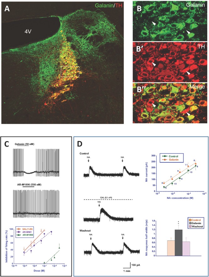
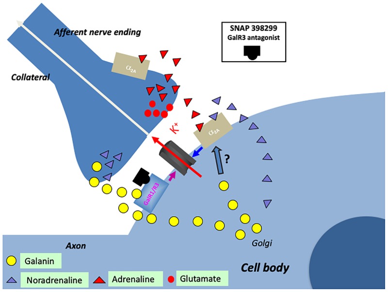
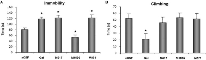
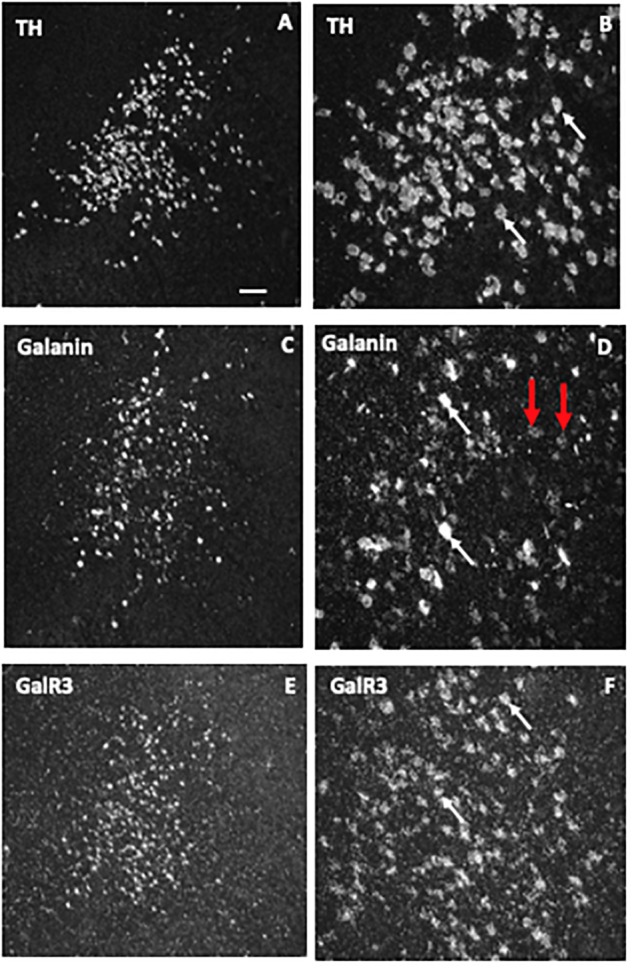
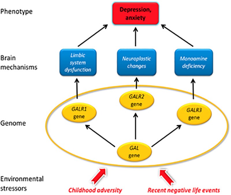
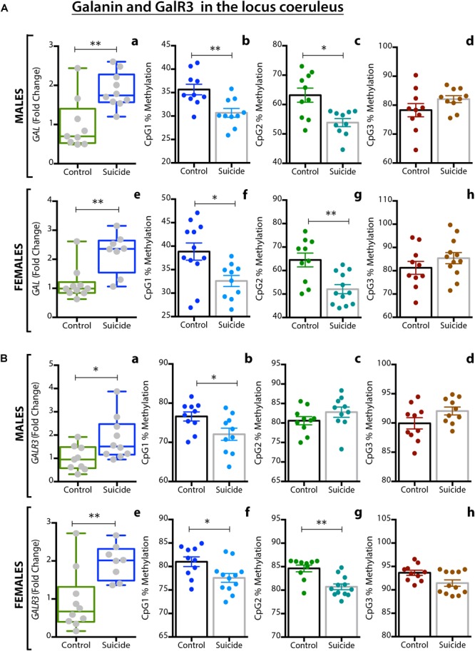
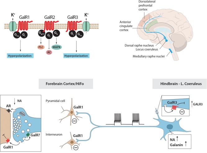
Neuropeptide are auxiliary messenger molecules that always co-exist in nerve cells with one or more small-molecule (classic) neurotransmitters. Neuropeptides act both as transmitters and trophic factors, and play a role especially when the nervous system is challenged, as by injury, pain or stress. Here neuropeptides and coexistence in mammals are reviewed, but with special focus on the 29/30 amino acid galanin and its three receptors GalR1, -R2 and -R3. In particular, galanin’s role as a co-transmitter in both rodent and human noradrenergic locus coeruleus (LC) neurons is addressed. Extensive experimental animal data strongly suggest a role for the galanin system in depression–like behavior. The translational potential of these results was tested by studying the galanin system in human postmortem brains, first normal brains, and then by comparing five regions of brains obtained from depressed people who committed suicide, and from matched controls. It is hypothesized that, when exposed to severe stress, the noradrenergic LC neurons fire in bursts and release galanin from their soma/dendrites. Galanin then acts on somato-dendritic, inhibitory GalR3 autoreceptors, opening potassium channels and inhibiting firing. The purpose of these autoreceptors is to act as a ‘brake’ to prevent overexcitation, a brake that also is part of resilience to stress that protects against depression. Depression then arises when the inhibition is too strong and long lasting - a maladaption, allostatic load, leading to depletion of NA levels in the forebrain. It is suggested that disinhibition by a galanin GalR3 antagonist may have antidepressant activity by restituting forebrain NA levels. A role of galanin in depression is also supported by a recent candidate gene study, showing that variants in genes for galanin and its three receptors confer increased risk of depression and anxiety in people who experienced childhood adversity or recent negative life events. Taken together, the neuropeptide galanin, coexisting in LC neurons, may participate in the mechanism underlying resilience against a serious and common disorder, MDD. These results may lead to an increased understanding of how this illness develops which in turn can provide a basis for its treatment.
Overview
The claims and statistics of this paper haven’t been checked yet.
Run the check — free, no account needed. It reads the open-access text and checks the sources it cites.
Integrity signals
Registry
Cites
Statistics
Figures
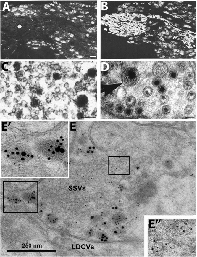
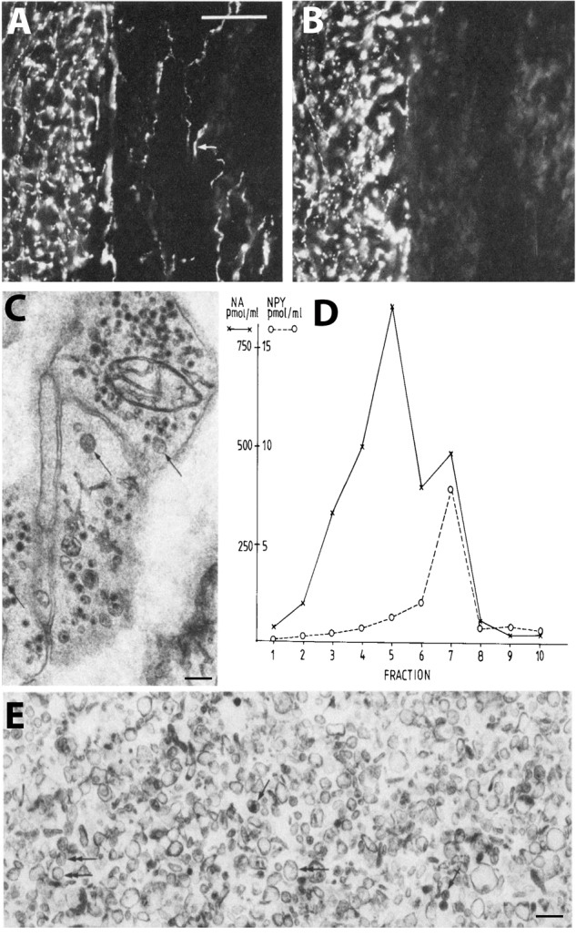
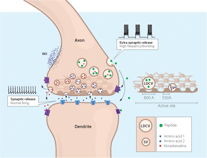
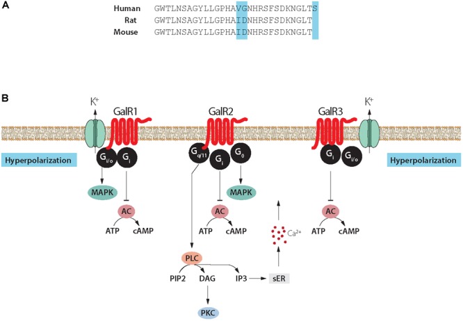
Verification
Claims the paper makes
The claims and statistics of this paper haven’t been checked yet.
Run the check — free, no account needed. It reads the open-access text and checks the sources it cites.
Claims other papers make about this one
Each is checked against this paper’s own text: does it say what the citing paper says it does?
In the citing paper
“Diverse neuropeptides are expressed and released in regions of anxiety/fear neurocircuitries, often together with classical transmitters (Hökfelt et al., 2018).”
From Novel pharmacological targets in drug development for the treatment of anxiety and anxiety-related disorders (2019) · cites this paper as Hökfelt et al., 2018
Self-check
Percentages, sums and reported differences re-computed from the paper’s own numbers.
The self-check needs the paper’s full text.
Citations
Cites
Continuous cultures of fused cells secreting antibody of predefined specificity
Köhler, G., & Milstein, C. (1975) · Nature · doi:10.1038/256495a0 · Cited by 17,200
Molecular Mechanisms of Depression: Perspectives on New Treatment Strategies
Lang, U. E., & Borgwardt, S. (2013) · Cellular Physiology and Biochemistry · doi:10.1159/000350094 · Cited by 8,526
Influence of life stress on depression: moderation by a polymorphism in the 5-HTT gene
Caspi, A., Sugden, K., Moffitt, T. E., Taylor, A., Craig, I. W., Harrington, H., McClay, J., Mill, J., Martin, J., Braithwaite, A., et al. (2003) · Science · doi:10.1126/science.1083968 · Cited by 8,120
Kessler, R. C., Berglund, P., Demler, O., Jin, R., Koretz, D., Merikangas, K. R., Rush, A. J., Walters, E. E., & Wang, P. S. (2003) · JAMA · doi:10.1001/jama.289.23.3095 · Cited by 7,988
Genome-wide atlas of gene expression in the adult mouse brain
Lein, E. S., Hawrylycz, M. J., Ao, N., Ayres, M., Bensinger, A., Bernard, A., Boe, A. F., Boguski, M. S., Brockway, K. S., Byrnes, E. J., et al. (2007) · Nature · doi:10.1038/nature05453 · Cited by 5,572
Sakurai, T., Amemiya, A., Ishii, M., Matsuzaki, I., Chemelli, R. M., Tanaka, H., Williams, S. C., Richardson, J. A., Kozlowski, G. P., Wilson, S., et al. (1998) · Cell · doi:10.1016/s0092-8674(00)80949-6 · Cited by 5,501
Physiology and neurobiology of stress and adaptation: central role of the brain
McEwen, B. S. (2007) · Physiological Reviews · doi:10.1152/physrev.00041.2006 · Cited by 5,429 · cited 16×
“A very large GWAS failed to identify risk genes (Major Depressive Disorder Working Group of the Psychiatric Gwas Consortium, 2013), but recent advances in large MDD GWAS studies resulted in several SNPs being associated with MDD (e.g., Hyde et al., 2016; Okbay et al., 2016; Xiao et al., 2017; Wray et al., 2018), and p…”
BackgroundInfluentialLesch, K.-P., Bengel, D., Heils, A., Sabol, S. Z., Greenberg, B. D., Petri, S., Benjamin, J., Müller, C. R., Hamer, D. H., & Murphy, D. L. (1996) · Science · doi:10.1126/science.274.5292.1527 · Cited by 5,162
Dahlstroem, A., & Fuxé, K. (1964) · PubMed · Cited by 5,133
DNA methylation and its basic function
Moore, L. D., Le, T., & Fan, G. (2013) · Neuropsychopharmacology · doi:10.1038/npp.2012.112 · Cited by 4,906
Stress and the brain: from adaptation to disease
de Kloet, E. R., Joëls, M., & Holsboer, F. (2005) · Nature Reviews Neuroscience · doi:10.1038/nrn1683 · Cited by 4,621
Global mortality, disability, and the contribution of risk factors: Global Burden of Disease Study
Murray, C. J., & Lopez, A. D. (1997) · The Lancet · doi:10.1016/s0140-6736(96)07495-8 · Cited by 4,492
Vale, W., Spiess, J., Rivier, C., & Rivier, J. (1981) · Science · doi:10.1126/science.6267699 · Cited by 3,965
Vale, W., Spiess, J., RIVIER, C. L., & Rivier, J. (1982) · Obstetrical & Gynecological Survey · doi:10.1097/00006254-198205000-00013 · Cited by 3,904
Trivedi, M. H., Rush, A. J., Wisniewski, S. R., Nierenberg, A. A., Warden, D., Ritz, L., Norquist, G., Howland, R. H., Lebowitz, B., McGrath, P. J., et al. (2006) · American Journal of Psychiatry · doi:10.1176/appi.ajp.163.1.28 · Cited by 3,879
The hypocretins: hypothalamus-specific peptides with neuroexcitatory activity
de Lecea, L., Kilduff, T. S., Peyron, C., Gao, X.-B., Foye, P. E., Danielson, P. E., Fukuhara, C., Battenberg, E. L. F., Gautvik, V. T., Bartlett, F. S., et al. (1998) · Proceedings of the National Academy of Sciences · doi:10.1073/pnas.95.1.322 · Cited by 3,878
Stereotaxic mapping of the monoamine pathways in the rat brain
Ungerstedt, U. (1971) · Acta Physiologica Scandinavica · doi:10.1111/j.1365-201x.1971.tb10998.x · Cited by 3,819
Identification of two related pentapeptides from the brain with potent opiate agonist activity
Hughes, J., Smith, T. W., Kosterlitz, H. W., Fothergill, L. A., Morgan, B. A., & Morris, H. R. (1975) · Nature · doi:10.1038/258577a0 · Cited by 3,806
Neural systems of reinforcement for drug addiction: from actions to habits to compulsion
Everitt, B. J., & Robbins, T. W. (2005) · Nature Neuroscience · doi:10.1038/nn1579 · Cited by 3,785
A randomized trial of an N-methyl-D-aspartate antagonist in treatment-resistant major depression.
Zarate, C. A., Singh, J. B., Carlson, P. J., Brutsche, N. E., Ameli, R., Luckenbaugh, D. A., Charney, D. S., & Manji, H. K. (2006) · Archives of General Psychiatry · doi:10.1001/archpsyc.63.8.856 · Cited by 3,766
The size and burden of mental disorders and other disorders of the brain in Europe 2010
Wittchen, H. U., Jacobi, F., Rehm, J., Gustavsson, A., Svensson, M., Jönsson, B., Olesen, J., Allgulander, C., Alonso, J., Faravelli, C., et al. (2011) · European Neuropsychopharmacology · doi:10.1016/j.euroneuro.2011.07.018 · Cited by 3,754
The Neuroendocrinology of Stress: A Never Ending Story
Lightman, S. L. (2008) · Journal of Neuroendocrinology · doi:10.1111/j.1365-2826.2008.01711.x · Cited by 3,588
THE CATECHOLAMINE HYPOTHESIS OF AFFECTIVE DISORDERS: A REVIEW OF SUPPORTING EVIDENCE
Schildkraut, J. J. (1965) · American Journal of Psychiatry · doi:10.1176/ajp.122.5.509 · Cited by 3,568
Hypothalamic Polypeptide That Inhibits the Secretion of Immunoreactive Pituitary Growth Hormone
Brazeau, P., Vale, W., Burgus, R. C., LING, N. C., Butcher, M., Rivier, J., & Guillemin, R. (1973) · Science · doi:10.1126/science.179.4068.77 · Cited by 3,536
Genetic Epidemiology of Major Depression: Review and Meta-Analysis
Sullivan, P. F., Neale, M. C., & Kendler, K. S. (2000) · American Journal of Psychiatry · doi:10.1176/appi.ajp.157.10.1552 · Cited by 3,431
Nestler, E. J., Barrot, M., DiLeone, R. J., Eisch, A. J., Gold, S. J., & Monteggia, L. M. (2002) · Neuron · doi:10.1016/s0896-6273(02)00653-0 · Cited by 3,217
Ferrari, A. J., Charlson, F. J., Norman, R. E., Patten, S. B., Freedman, G., Murray, C. J. L., Vos, T., & Whiteford, H. A. (2013) · PLoS Medicine · doi:10.1371/journal.pmed.1001547 · Cited by 3,115
eQTLGen, Wray, N. R., 23andMe, the Major Depressive Disorder Working Group of the Psychiatric Genomics Consortium, Ripke, S., Mattheisen, M., Trzaskowski, M., Byrne, E. M., Abdellaoui, A., Adams, M. J., et al. (2018) · Nature Genetics · doi:10.1038/s41588-018-0090-3 · Cited by 3,005
Trends in GPCR drug discovery: new agents, targets and indications
Hauser, A. S., Attwood, M. M., Rask‐Andersen, M., Schiöth, H. B., & Gloriam, D. E. I. (2017) · Nature Reviews Drug Discovery · doi:10.1038/nrd.2017.178 · Cited by 2,714
FLUORESCENCE OF CATECHOL AMINES AND RELATED COMPOUNDS CONDENSED WITH FORMALDEHYDE
FALCK, B., Hillarp, N. ‐., THIEME, G., & Torp, A. (1962) · Journal of Histochemistry & Cytochemistry · doi:10.1177/10.3.348 · Cited by 2,697
Berridge, C. W., & Waterhouse, B. D. (2003) · Brain Research Reviews · doi:10.1016/s0165-0173(03)00143-7 · Cited by 2,529
A comprehensive map of molecular drug targets
Santos, R., Ursu, O., Gaulton, A., Bento, A. P., Donadi, R. S., Bologa, C. G., Karlsson, A., Al-Lazikani, B., Hersey, A., Oprea, T. I., et al. (2017) · Nature Reviews Drug Discovery · doi:10.1038/nrd.2016.230 · Cited by 2,427
Krishnan, V., Han, M.-H., Graham, D. L., Berton, O., Renthal, W., Russo, S. J., LaPlant, Q., Graham, A., Lutter, M., Lagace, D. C., et al. (2007) · Cell · doi:10.1016/j.cell.2007.09.018 · Cited by 2,307
Production of a novel neuropeptide encoded by the calcitonin gene via tissue-specific RNA processing
Rosenfeld, M. G., Mermod, J.-J., Amara, S. G., Swanson, L. W., Sawchenko, P. E., Rivier, J., Vale, W. W., & Evans, R. M. (1983) · Nature · doi:10.1038/304129a0 · Cited by 2,302
Tatemoto, K., Carlquist, M., & Mutt, V. (1982) · Nature · doi:10.1038/296659a0 · Cited by 2,254
The mesolimbic dopamine reward circuit in depression
Nestler, E. J., & Carlezon, W. A. (2006) · Biological Psychiatry · doi:10.1016/j.biopsych.2005.09.018 · Cited by 2,059
Willner, P., Towell, A., Sampson, D., Sophokleous, S., & Muscat, R. (1987) · Psychopharmacology · doi:10.1007/bf00187257 · Cited by 1,993
Nucleus locus ceruleus: new evidence of anatomical and physiological specificity
Foote, S. L., Bloom, F. E., & Aston-Jones, G. (1983) · Physiological Reviews · doi:10.1152/physrev.1983.63.3.844 · Cited by 1,901
Burnstock, G. (1972) · Pharmacological Reviews · doi:10.1016/s0031-6997(25)06930-3 · Cited by 1,852
Charney, D. S. (2004) · American Journal of Psychiatry · doi:10.1176/appi.ajp.161.2.195 · Cited by 1,605
New approaches to antidepressant drug discovery: beyond monoamines
Berton, O., & Nestler, E. J. (2006) · Nature Reviews Neuroscience · doi:10.1038/nrn1846 · Cited by 1,589
An unidentified depressor substance in certain tissue extracts
von Euler, U. S., & Gaddum, J. H. (1931) · The Journal of Physiology · doi:10.1113/jphysiol.1931.sp002763 · Cited by 1,584
Hökfelt, T. G. M., Johansson, O., Ljungdahl, Å., Lundberg, J. M., & Schultzberg, M. (1980) · Nature · doi:10.1038/284515a0 · Cited by 1,552
Galanin — a novel biologically active peptide from porcine intestine
Tatemoto, K., Rökaeus, Å., Jörnvall, H., McDonald, T. J., & Mutt, V. (1983) · FEBS Letters · doi:10.1016/0014-5793(83)80033-7 · Cited by 1,490
Swanson, L. W., & Hartman, B. K. (1975) · The Journal of Comparative Neurology · doi:10.1002/cne.901630406 · Cited by 1,475
Ljungdahl, Å., Hökfelt, T. G. M., & Nilsson, G. (1978) · Neuroscience · doi:10.1016/0306-4522(78)90116-1 · Cited by 1,425
Calcium-binding proteins in the nervous system
Baimbridge, K. G., Celio, M. R., & ROGERS, J. H. (1992) · Trends in Neurosciences · doi:10.1016/0166-2236(92)90081-i · Cited by 1,329
Psychobiology and molecular genetics of resilience
Feder, A., Nestler, E. J., & Charney, D. S. (2009) · Nature Reviews Neuroscience · doi:10.1038/nrn2649 · Cited by 1,272
Russo, S. J., Murrough, J. W., Han, M.-H., Charney, D. S., & Nestler, E. J. (2012) · Nature Neuroscience · doi:10.1038/nn.3234 · Cited by 1,206
Lundberg, J. M., Terenius, L. Y., Hökfelt, T. G. M., Martling, C., Tatemoto, K., Mutt, V., Polak, J. M., Bloom, S. R., & Goldstein, M. (1982) · Acta Physiologica Scandinavica · doi:10.1111/j.1748-1716.1982.tb07171.x · Cited by 1,199
Cholinergic and inhibitory synapses in a pathway from motor‐axon collaterals to motoneurones
Eccles, J. C., Fatt, P., & KOKETSU, K. (1954) · The Journal of Physiology · doi:10.1113/jphysiol.1954.sp005226 · Cited by 1,198
Norepinephrine in Depressive Reactions
Bunney, W. E. (1965) · Archives of General Psychiatry · doi:10.1001/archpsyc.1965.01730060001001 · Cited by 1,166
The Psychobiology of Depression and Resilience to Stress: Implications for Prevention and Treatment
Southwick, S. M., Vythilingam, M., & Charney, D. S. (2005) · Annual Review of Clinical Psychology · doi:10.1146/annurev.clinpsy.1.102803.143948 · Cited by 1,160
Cellular localization of brain monoamines.
Carlsson, A. C., FALCK, B., & Hillarp, N. ‐. (1962) · PubMed · Cited by 1,144
Mood disorders and allostatic load
McEwen, B. S. (2003) · Biological Psychiatry · doi:10.1016/s0006-3223(03)00177-x · Cited by 1,135
A mega-analysis of genome-wide association studies for major depressive disorder
Major Depressive Disorder Working Group of the Psychiatric GWAS Consortium, Ripke, S., Wray, N. R., Lewis, C. M., Hamilton, S. P., Weissman, M. M., Breen, G., Byrne, E. M., Blackwood, D. H. R., Boomsma, D. I., et al. (2013) · Molecular Psychiatry · doi:10.1038/mp.2012.21 · Cited by 1,120
Characterization of a growth hormone-releasing factor from a human pancreatic islet tumour
Rivier, J., Spiess, J., Thorner, M. O., & Vale, W. (1982) · Nature · doi:10.1038/300276a0 · Cited by 1,102
Assay of Plasma Insulin in Human Subjects by Immunological Methods
Yalow, R. S., & Berson, S. A. (1959) · Nature · doi:10.1038/1841648b0 · Cited by 1,098
Immunohistochemical evidence for the existence of adrenaline neurons in the rat brain
Ho¨kfelt, T., Fuxé, K., Goldstein, M., & Johansson, O. (1974) · Brain Research · doi:10.1016/0006-8993(74)90143-7 · Cited by 1,093
The anatomy of neuropeptide-y-containing neurons in rat brain
Chronwall, B. M., DiMaggio, D. A., Massari, V. J., Pickel, V. M., Ruggiero, D. A., & O’Donohue, T. L. (1985) · Neuroscience · doi:10.1016/0306-4522(85)90260-x · Cited by 1,088
Distinct Mechanism for Antidepressant Activity by Blockade of Central Substance P Receptors
Kramer, M. S., Cutler, N. R., Feighner, J. P., Shrivastava, R. K., Carman, J. S., Sramek, J. J., Reines, S. A., Liu, G. F., Snavely, D. B., Wyatt-Knowles, E. S., et al. (1998) · Science · doi:10.1126/science.281.5383.1640 · Cited by 1,071
Landgraf, R., & Neumann, I. D. (2004) · Frontiers in Neuroendocrinology · doi:10.1016/j.yfrne.2004.05.001 · Cited by 1,059
Substance P: Localization in the Central Nervous System and in Some Primary Sensory Neurons
Hökfelt, T. G. M., Kellerth, J. ‐., Nilsson, G., & Pernow, B. (1975) · Science · doi:10.1126/science.242075 · Cited by 1,024
CGRP as the target of new migraine therapies — successful translation from bench to clinic
Edvinsson, L. I., Haanes, K. A., Warfvinge, K., & Krause, D. N. (2018) · Nature Reviews Neurology · doi:10.1038/s41582-018-0003-1 · Cited by 1,021
Svensson, T. H., Bunney, B. S., & Aghajanian, G. K. (1975) · Brain Research · doi:10.1016/0006-8993(75)90276-0 · Cited by 1,015
Study, L. C., Okbay, A., Baselmans, B. M. L., De Neve, J.-E., Turley, P., Nivard, M. G., Fontana, M. A., Meddens, S. F. W., Linnér, R. K., Rietveld, C. A., et al. (2016) · Nature Genetics · doi:10.1038/ng.3552 · Cited by 1,013
The oxytocin receptor: from intracellular signaling to behavior
Jurek, B., & Neumann, I. D. (2018) · Physiological Reviews · doi:10.1152/physrev.00031.2017 · Cited by 999
Dendritic peptide release and peptide-dependent behaviours
Ludwig, M., & Leng, G. (2006) · Nature Reviews Neuroscience · doi:10.1038/nrn1845 · Cited by 945
Defined types of cortical interneurone structure space and spike timing in the hippocampus
Somogyi, P., & Klausberger, T. (2005) · The Journal of Physiology · doi:10.1113/jphysiol.2004.078915 · Cited by 927
Hökfelt, T. G. M., Ljungdahl, Å., Terenius, L. Y., Elde, R., & Nilsson, G. (1977) · Proceedings of the National Academy of Sciences · doi:10.1073/pnas.74.7.3081 · Cited by 887
Everitt, B. J., Hökfelt, T. G. M., Terenius, L. Y., Tatemoto, K., Mutt, V., & Goldstein, M. (1984) · Neuroscience · doi:10.1016/0306-4522(84)90036-8 · Cited by 871
Locus coeruleus and dopaminergic consolidation of everyday memory
Takeuchi, T., Duszkiewicz, A. J., Sonneborn, A., Spooner, P. A., Yamasaki, M., Watanabe, M., Smith, C. C., Fernández, G., Deisseroth, K., Greene, R. W., et al. (2016) · Nature · doi:10.1038/nature19325 · Cited by 827
Hökfelt, T. G. M., Ljungdahl, Å., Steinbusch, H. W. M., Verhofstad, A. A. J., Nilsson, G., Brodin, E., Pernow, B., & Goldstein, M. (1978) · Neuroscience · doi:10.1016/0306-4522(78)90017-9 · Cited by 814
Neuropeptide Transmission in Brain Circuits
van den Pol, A. N. (2012) · Neuron · doi:10.1016/j.neuron.2012.09.014 · Cited by 810
Fremanezumab for the Preventive Treatment of Chronic Migraine
Silberstein, S. D., Dodick, D. W., BIGAL, M. E., Yeung, P. P., Goadsby, P. J., Blankenbiller, T., Grozinski‐Wolff, M., Yang, R., Ma, Y., & Aycardi, E. (2017) · New England Journal of Medicine · doi:10.1056/nejmoa1709038 · Cited by 809
Broberger, C., Johansen, J., Johansson, C., Schalling, M., & Hökfelt, T. (1998) · Proceedings of the National Academy of Sciences · doi:10.1073/pnas.95.25.15043 · Cited by 798
Self-citationAmino-acid Sequence of Substance P
CHANG, M. M., Leeman, S. E., & Niall, H. D. (1971) · Nature New Biology · doi:10.1038/newbio232086a0 · Cited by 790
Corelease of Two Fast Neurotransmitters at a Central Synapse
Jonas, P., Bischofberger, J., & Sandkühler, J. (1998) · Science · doi:10.1126/science.281.5375.419 · Cited by 784
Hyde, C. L., Nagle, M. W., Tian, C., Chen, X., Paciga, S. A., Wendland, J. R., Tung, J. Y., Hinds, D. A., Perlis, R. H., & Winslow, A. R. (2016) · Nature Genetics · doi:10.1038/ng.3623 · Cited by 784
Seidah, N. G., & Chrétien, M. (1999) · Brain Research · doi:10.1016/s0006-8993(99)01909-5 · Cited by 770
Sex-specific transcriptional signatures in human depression
Labonté, B., Engmann, O., Purushothaman, I., Menard, C., Wang, J., Tan, C., Scarpa, J. R., Moy, G., Loh, Y.-H. E., Cahill, M., et al. (2017) · Nature Medicine · doi:10.1038/nm.4386 · Cited by 748
Neuropeptide Y co-exists and co-operates with noradrenaline in perivascular nerve fibers
Ekblad, E., Edvinsson, L. I., Wahlestedt, C., Uddman, R., Håkanson, R., & Sundler, F. (1984) · Regulatory Peptides · doi:10.1016/0167-0115(84)90064-8 · Cited by 741
Distribution of galaninlike immunoreactivity in the rat central nervous system
Melander, T., Hökfelt, T. G. M., & Rökaeus, Å. (1986) · The Journal of Comparative Neurology · doi:10.1002/cne.902480404 · Cited by 724
Ho¨kfelt, T., Skirboll, L. R., Rehfeld, J. F., Goldstein, M., MARKEY, K., & Dann, O. (1980) · Neuroscience · doi:10.1016/0306-4522(80)90127-x · Cited by 713
Calcium-binding proteins: selective markers of nerve cells
Andressen, C., Bl�mcke, I., & Celio, M. R. (1993) · Cell and Tissue Research · doi:10.1007/bf00318606 · Cited by 701
Chang, M. M., & Leeman, S. E. (1970) · Journal of Biological Chemistry · doi:10.1016/s0021-9258(18)62860-1 · Cited by 699
The Demonstration of Pneumococcal Antigen in Tissues by the Use of Fluorescent Antibody
Coons, A. H., Creech, H. J., Jones, R. N., & Berliner, E. (1942) · The Journal of Immunology · doi:10.4049/jimmunol.45.3.159 · Cited by 674
Oligomerization of G-protein-coupled transmitter receptors
Bouvier, M. (2001) · Nature reviews. Neuroscience · doi:10.1038/35067575 · Cited by 668
Tatemoto, K., & Mutt, V. (1980) · Nature · doi:10.1038/285417a0 · Cited by 663
Weiss, J. M., Goodman, P. A., Losito, B. G., Corrigan, S. A., Charry, J. M., & Bailey, W. H. (1981) · Brain Research Reviews · doi:10.1016/0165-0173(81)90005-9 · Cited by 662
Effects of substance P on functionally identified units in cat spinal cord
Henry, J. L. (1976) · Brain Research · doi:10.1016/0006-8993(76)90965-3 · Cited by 660
Immunohistochemical mapping of galanin-like neurons in the rat central nervous system
Skofitsch, G., & Jacobowitz, D. M. (1985) · Peptides · doi:10.1016/0196-9781(85)90118-4 · Cited by 659
Choudary, P. V., Molnar, M., Evans, S. J., Tomita, H., Li, J., Vawter, M. P., Myers, R., Bunney, W. E., Akil, H., Watson, S. J., et al. (2005) · Proceedings of the National Academy of Sciences · doi:10.1073/pnas.0507901102 · Cited by 653
Kempadoo, K. A., Mosharov, E. V., Choi, S. J., Sulzer, D., & Kandel, E. R. (2016) · Proceedings of the National Academy of Sciences · doi:10.1073/pnas.1616515114 · Cited by 647
Do some nerve cells release more than one transmitter?
Burnstock, G. (1976) · Neuroscience · doi:10.1016/0306-4522(76)90054-3 · Cited by 640
Cedarbaum, J. M., & Aghajanian, G. K. (1976) · Brain Research · doi:10.1016/0006-8993(76)90297-3 · Cited by 636
The NPY system in stress, anxiety and depression
Heilig, M. A. (2004) · Neuropeptides · doi:10.1016/j.npep.2004.05.002 · Cited by 617
Neuropeptides in perspective: The last ten years
Hökfelt, T. (1991) · Neuron · doi:10.1016/0896-6273(91)90333-u · Cited by 612
Galanin neurons in the medial preoptic area govern parental behaviour
Wu, Z., Autry, A. E., Bergan, J. F., Watabe‐Uchida, M., & Dulac, C. G. (2014) · Nature · doi:10.1038/nature13307 · Cited by 610
The Regional Distribution of Somatostatin in the Rat Brain
Brownstein, M. J., Arimura, A., SATO, H., Schally, A. V., & Kizer, J. S. (1975) · Endocrinology · doi:10.1210/endo-96-6-1456 · Cited by 594
Circadian patterns of gene expression in the human brain and disruption in major depressive disorder
Li, J. Z., Bunney, B. G., Meng, F., Hagenauer, M. H., Walsh, D. M., Vawter, M. P., Evans, S. J., Choudary, P. V., Cartagena, P., Barchas, J. D., et al. (2013) · Proceedings of the National Academy of Sciences · doi:10.1073/pnas.1305814110 · Cited by 589
Dopaminergic neurons inhibit striatal output through non-canonical release of GABA
Tritsch, N. X., Ding, J. B., & Sabatini, B. L. (2012) · Nature · doi:10.1038/nature11466 · Cited by 576
Hypothalamic Regulatory Hormones
Schally, A. V., Arimura, A., & Kastin, A. J. (1973) · Science · doi:10.1126/science.179.4071.341 · Cited by 575
Millan, M. J. (2006) · Pharmacology & Therapeutics · doi:10.1016/j.pharmthera.2005.11.006 · Cited by 569
Epigenetics and the environmental regulation of the genome and its function
Zhang, T.-Y., & Meaney, M. J. (2010) · Annual Review of Psychology · doi:10.1146/annurev.psych.60.110707.163625 · Cited by 565
Coexistence of peptides and classical neurotransmitters
Lundberg, J. M., & Hökfelt, T. G. M. (1983) · Trends in Neurosciences · doi:10.1016/0166-2236(83)90149-2 · Cited by 560
Afferent projections to the rat locus coeruleus as determined by a retrograde tracing technique
Cedarbaum, J. M., & Aghajanian, G. K. (1978) · The Journal of Comparative Neurology · doi:10.1002/cne.901780102 · Cited by 553
Afferent regulation of locus coeruleus neurons: anatomy, physiology and pharmacology
Aston‐Jones, G., Shipley, M., Chouvet, G., Ennis, M. J., Bockstaele, E. V., Pieribone, V. A., Shiekhattar, R., Akaoka, H., Drolet, G., Astier, B., et al. (1991) · Progress in brain research · doi:10.1016/s0079-6123(08)63799-1 · Cited by 543
Bøler, J. B., Enzmann, F., Folkers, K., Bowers, C. Y., & Schally, A. V. (1969) · Biochemical and Biophysical Research Communications · doi:10.1016/0006-291x(69)90868-7 · Cited by 532
Lundberg, J. (1996) · Pharmacological Reviews · doi:10.1016/s0031-6997(25)06937-6 · Cited by 523
Neuroplasticity in the mesolimbic dopamine system and cocaine addiction
Thomas, M. J., Kalivas, P. W., & Shaham, Y. (2008) · British Journal of Pharmacology · doi:10.1038/bjp.2008.77 · Cited by 521
Melander, T., Hökfelt, T. G. M., Rökaeus, Å., Cuello, A. C., Oertel, W., Verhofstad, A. A. J., & Goldstein, M. (1986) · Journal of Neuroscience · doi:10.1523/jneurosci.06-12-03640.1986 · Cited by 518
The locus coeruleus: A cytoarchitectonic, golgi and immunohistochemical study in the albino rat
Swanson, L. W. (1976) · Brain Research · doi:10.1016/0006-8993(76)90207-9 · Cited by 518
Todd, A. J., & Sullivan, A. C. (1990) · The Journal of Comparative Neurology · doi:10.1002/cne.902960312 · Cited by 501
Lundberg, J. M., Hökfelt, T. G. M., Schultzberg, M., Uvnäs‐Wallensten, K., Köhler, C., & Said, S. I. (1979) · Neuroscience · doi:10.1016/0306-4522(79)90018-6 · Cited by 497
Vandesande, F., & Dierickx, K. (1975) · Cell and Tissue Research · doi:10.1007/bf00218970 · Cited by 496
Branchek, T. A., Smith, K. E., Gerald, C., & Walker, M. W. (2000) · Trends in Pharmacological Sciences · doi:10.1016/s0165-6147(00)01446-2 · Cited by 495
Evidence for norepinephrine-mediated collateral inhibition of locus coeruleus neurons
Aghajanian, G. K., Cedarbaum, J. M., & Wang, R. Y. (1977) · Brain Research · doi:10.1016/0006-8993(77)90083-x · Cited by 489
Serotonin and substance P coexist in neurons of the rat's central nervous system
Chan‐Palay, V., JONSSON, G., & Palay, S. L. (1978) · Proceedings of the National Academy of Sciences · doi:10.1073/pnas.75.3.1582 · Cited by 487
Lundberg, J. M., Änggård, A., Fahrenkrug, J., Hökfelt, T. G. M., & Mutt, V. (1980) · Proceedings of the National Academy of Sciences · doi:10.1073/pnas.77.3.1651 · Cited by 482
Receptor Endocytosis and Dendrite Reshaping in Spinal Neurons After Somatosensory Stimulation
Mantyh, P. W., DeMaster, E., Malhotra, A., Ghilardi, Rogers, S. D., Mantyh, C. R., Liu, H., Basbaum, A. I., Vigna, Maggio, J. E., et al. (1995) · Science · doi:10.1126/science.7539937 · Cited by 475
(2005) · PubMed · Cited by 470
Hökfelt, T. G. M., Elfvin, L.-G., Elde, R., Schultzberg, M., Goldstein, M., & Luft, R. (1977) · Proceedings of the National Academy of Sciences · doi:10.1073/pnas.74.8.3587 · Cited by 464
Treatment resistant depression: A multi-scale, systems biology approach
Akil, H., Gordon, J. A., Hen, R., Javitch, J. A., Mayberg, H. S., McEwen, B. S., Meaney, M. J., & Nestler, E. J. (2017) · Neuroscience & Biobehavioral Reviews · doi:10.1016/j.neubiorev.2017.08.019 · Cited by 463
NK1 (substance P) receptor antagonists – why are they not analgesic in humans?
Hill, R. (2000) · Trends in Pharmacological Sciences · doi:10.1016/s0165-6147(00)01502-9 · Cited by 462
Pathways to Regulated Exocytosis in Neurons
Camilli, P. V. D., & Jahn, R. (1990) · Annual Review of Physiology · doi:10.1146/annurev.ph.52.030190.003205 · Cited by 462
Hökfelt, T. G. M., Efendić, S., Hellerström, C., Johansson, O., Luft, R., & Arimura, A. (1975) · European Journal of Endocrinology · doi:10.1530/acta.0.080s005 · Cited by 457
Genetic variation in human NPY expression affects stress response and emotion
Zhou, Z., Zhu, G., Hariri, A. R., Enoch, M.-A., Scott, D., Sinha, R., Virkkunen, M., Mash, D. C., Lipsky, R. H., Hu, X.-Z., et al. (2008) · Nature · doi:10.1038/nature06858 · Cited by 455
[The neurosecretory connection between the hypothalamus and the neurohypophysis].
Bargmann, W. (1949) · PubMed · Cited by 455
Samuels, E., & Szabadi, E. (2008) · Current Neuropharmacology · doi:10.2174/157015908785777193 · Cited by 454
Detection of viral sequences of low reiteration frequency by in situ hybridization.
Brahic, M., & Haase, A. T. (1978) · Proceedings of the National Academy of Sciences · doi:10.1073/pnas.75.12.6125 · Cited by 454
Takahashi, T., & Otsuka, M. (1975) · Brain Research · doi:10.1016/0006-8993(75)90774-x · Cited by 444
Wray, N. R., Pergadia, M. L., Blackwood, D. H. R., Penninx, B. W. J. H., Gordon, S. D., Nyholt, D. R., Ripke, S., MacIntyre, D. J., McGhee, K. A., Maclean, A. W., et al. (2012) · Molecular Psychiatry · doi:10.1038/mp.2010.109 · Cited by 444
Increase of galanin-like immunoreactivity in rat dorsal root ganglion cells after peripheral axotomy
Hökfelt, T. G. M., Wiesenfeld‐Hallin, Z., Villar, M. J., & Melander, T. (1987) · Neuroscience Letters · doi:10.1016/0304-3940(87)90088-7 · Cited by 442
A galanin-like peptide in the central nervous system and intestine of the rat
Rökaeus, Å., Melander, T., Hökfelt, T. G. M., Lundberg, J. M., Tatemoto, K., Carlquist, M., & Mutt, V. (1984) · Neuroscience Letters · doi:10.1016/0304-3940(84)90423-3 · Cited by 436
Furshpan, E. J., MacLeish, P. R., O'Lague, P. H., & Potter, D. (1976) · Proceedings of the National Academy of Sciences · doi:10.1073/pnas.73.11.4225 · Cited by 432
50 years of hurdles and hope in anxiolytic drug discovery
Griebel, G., & Holmes, A. (2013) · Nature Reviews Drug Discovery · doi:10.1038/nrd4075 · Cited by 430
Orexin (Hypocretin) Neurons Contain Dynorphin
Chou, T. C., Lee, C. E., Lü, J., Elmquist, J. K., Hara, J., Willie, J. T., Beuckmann, C. T., Chemelli, R. M., Sakurai, T., Yanagisawa, M., et al. (2001) · Journal of Neuroscience · doi:10.1523/jneurosci.21-19-j0003.2001 · Cited by 430
Depressant action of TRH, LH-RH and somatostatin on activity of central neurones
Renaud, L. P., Martin, J. B., & Brazeau, P. (1975) · Nature · doi:10.1038/255233a0 · Cited by 429
Aston, C., Jiang, L., & Sokolov, B. P. (2005) · Molecular Psychiatry · doi:10.1038/sj.mp.4001565 · Cited by 427
Primary structure of corticotropin-releasing factor from ovine hypothalamus
Spiess, J., Rivier, J., Rivier, C., & Vale, W. (1981) · Proceedings of the National Academy of Sciences · doi:10.1073/pnas.78.10.6517 · Cited by 424
Dysregulation of the fibroblast growth factor system in major depression
Evans, S. J., Choudary, P. V., Neal, C. R., Li, J. Z., Vawter, M. P., Tomita, H., Lopez, J. F., Thompson, R. C., Meng, F., Stead, J. D., et al. (2004) · Proceedings of the National Academy of Sciences · doi:10.1073/pnas.0406788101 · Cited by 424
Abdallah, C. G., Sanacora, G., Duman, R. S., & Krystal, J. H. (2015) · Annual Review of Medicine · doi:10.1146/annurev-med-053013-062946 · Cited by 422
Chen, N., Reith, M. E. A., & Quick, M. W. (2004) · Pflügers Archiv - European Journal of Physiology · doi:10.1007/s00424-003-1064-5 · Cited by 411
Vesicular glutamate transport promotes dopamine storage and glutamate corelease in vivo
Hnasko, T. S., Chuhma, N., Zhang, H., Goh, G. Y., Sulzer, D., Palmiter, R. D., Rayport, S., & Edwards, R. H. (2010) · Neuron · doi:10.1016/j.neuron.2010.02.012 · Cited by 405
Neuropeptide systems as novel therapeutic targets for depression and anxiety disorders
Holmes, A., Heilig, M. A., Rupniak, N. M. J., Steckler, T., & Griebel, G. (2003) · Trends in Pharmacological Sciences · doi:10.1016/j.tips.2003.09.011 · Cited by 405
Mongeau, R., Blier, P., & de Montigny, C. (1997) · Brain Research Reviews · doi:10.1016/s0165-0173(96)00017-3 · Cited by 399
Early life stress confers lifelong stress susceptibility in mice via ventral tegmental area OTX2
Peña, C. J., Kronman, H. G., Walker, D. M., Cates, H. M., Bagot, R. C., Purushothaman, I., Issler, O., Loh, Y.-H. E., Leong, T., Kiraly, D. D., et al. (2017) · Science · doi:10.1126/science.aan4491 · Cited by 394
Peptidergic transmission in sympathetic ganglia of the frog.
Jan, L. Y., & Jan, Y. N. (1982) · The Journal of Physiology · doi:10.1113/jphysiol.1982.sp014228 · Cited by 390
Sequeira, A., Mamdani, F., Ernst, C., Vawter, M. P., Bunney, W. E., Lebel, V., Rehal, S., Klempan, T., Gratton, A., Benkelfat, C., et al. (2009) · PLoS ONE · doi:10.1371/journal.pone.0006585 · Cited by 388
LANG, R., Gundlach, A. L., & Kofler, B. (2007) · Pharmacology & Therapeutics · doi:10.1016/j.pharmthera.2007.05.009 · Cited by 382
Electron Microscopic Identification of Autonomic Nerve Endings
RICHARDSON, K. C. (1966) · Nature · doi:10.1038/210756a0 · Cited by 381
Kendler, K. S. (2012) · Molecular Psychiatry · doi:10.1038/mp.2011.182 · Cited by 381
Characterization of Ovine Hypothalamic Hypophysiotropic TSH-releasing Factor
Burgus, R. C., Dunn, T. F., Desiderio, D. M., Ward, D. N., Vale, W., & Guillemin, R. (1970) · Nature · doi:10.1038/226321a0 · Cited by 379
The site of origin of the hormones of the posterior pituitary.
Bargmann, W., & Scharrer, E. (1951) · PubMed · Cited by 379
Abercrombie, E., & Jacobs, B. (1987) · The Journal of Neuroscience · doi:10.1523/jneurosci.07-09-02837.1987 · Cited by 377 · cited 2×
“…the LC: stress increases NA turnover, as well as tyrosine hydroxylase activity and transcription in the LC (Korf et al., 1973; Zigmond et al., 1974; Abercrombie and Jacobs, 1987; Komori et al., 1990; Smith et al., 1991; Melia et al., 1992; Aston-Jones et al., 1996; Rusnak et al., 1998; Chang et…”
BackgroundCortical gamma band synchronization through somatostatin interneurons
Veit, J., Hakim, R., Jadi, M. P., B, T. J. S. A., & Adesnik, H. (2017) · Nature Neuroscience · doi:10.1038/nn.4562 · Cited by 376
Armstrong, D. M., Ross, C. A., Pickel, V. M., JOH, T. H., & Reis, D. J. (1982) · The Journal of Comparative Neurology · doi:10.1002/cne.902120207 · Cited by 375
Bernard, R., Kerman, I. A., Thompson, R. C., Jones, E. G., Bunney, W. E., Barchas, J. D., Schatzberg, A. F., Myers, R. M., Akil, H., & Watson, S. J. (2011) · Molecular Psychiatry · doi:10.1038/mp.2010.44 · Cited by 372
Characterization of rat hypothalamic growth hormone-releasing factor
Spiess, J., Rivier, J., & Vale, W. (1983) · Nature · doi:10.1038/303532a0 · Cited by 372
Holets, V. R., Hökfelt, T. G. M., Rökaeus, Å., Terenius, L. Y., & Goldstein, M. (1988) · Neuroscience · doi:10.1016/0306-4522(88)90076-0 · Cited by 368
H 2 S: A Novel Gasotransmitter that Signals by Sulfhydration
Paul, B. D., & Snyder, S. H. (2015) · Trends in Biochemical Sciences · doi:10.1016/j.tibs.2015.08.007 · Cited by 365
Tachykinin-Expressing Neurons Control Male-Specific Aggressive Arousal in Drosophila
Asahina, K., Watanabe, K., Duistermars, B. J., Hoopfer, E. D., González, C. R., Eyjólfsdóttir, E., Perona, P., & Anderson, D. J. (2014) · Cell · doi:10.1016/j.cell.2013.11.045 · Cited by 363
The neurocircuitry and receptor subtypes mediating anxiolytic-like effects of neuropeptide Y
Kask, A., Harro, J., von Hörsten, S., Redrobe, J. P., Dumont, Y., & Quirion, R. (2002) · Neuroscience & Biobehavioral Reviews · doi:10.1016/s0149-7634(01)00066-5 · Cited by 358
Isolation and structural organization of the human preproenkephalin B gene
Horikawa, S., Takai, T., Toyosato, M., Takahashi, H., Noda, M., Kakidani, H., Kubo, T., Hirose, T., Inayama, S., Hayashida, H., et al. (1983) · Nature · doi:10.1038/306611a0 · Cited by 353
Abercrombie, E., Keller, R., & Zigmond, M. (1988) · Neuroscience · doi:10.1016/0306-4522(88)90192-3 · Cited by 342
Molecular cloning of a functional human galanin receptor.
Habert, E., Amiranoff, B., Loquet, I., Laburthe, M., & Mayaux, J. F. (1994) · Proceedings of the National Academy of Sciences · doi:10.1073/pnas.91.21.9780 · Cited by 340
Serotonin Release as a Possible Mechanism of Reserpine Action
Pletscher, A., Shore, P. A., & Brodie, B. B. (1955) · Science · doi:10.1126/science.122.3165.374 · Cited by 336
Coexistence of peptides with classical neurotransmitters
Hökfelt, T. G. M., Millhorn, D. E., Seroogy, K. B., Tsuruo, Y., Ceccatelli, S., Lindh, B., Meister, B., Melander, T., Schalling, M., Bartfai, T., et al. (1987) · Cellular and Molecular Life Sciences · doi:10.1007/bf01945354 · Cited by 335
Differential release of amino acids, neuropeptides, and catecholamines from isolated nerve terminals
Verhage, M., McMahon, H. T., Ghijsen, W. E. J. M., Boomsma, F., Scholten, G., Wiegant, V. M., & Nicholls, D. G. (1991) · Neuron · doi:10.1016/0896-6273(91)90054-4 · Cited by 335
Neuropeptides: opportunities for drug discovery
Hökfelt, T. G. M., Bartfai, T., & Bloom, F. E. (2003) · The Lancet Neurology · doi:10.1016/s1474-4422(03)00482-4 · Cited by 335
Iwamoto, K., Kakiuchi, C., Bundo, M., Ikeda, K., & Kato, T. (2004) · Molecular Psychiatry · doi:10.1038/sj.mp.4001437 · Cited by 331
Anatomy and physiology of central galanin-containing pathways
Merchenthaler, I., LÓPEZ, F. J., & Negro-Vilar, A. F. (1993) · Progress in Neurobiology · doi:10.1016/0301-0082(93)90012-h · Cited by 331
Lang, R., Gundlach, A. L., Holmes, F. E., Hobson, S. A., Wynick, D., Hökfelt, T., & Kofler, B. (2015) · Pharmacological Reviews · doi:10.1124/pr.112.006536 · Cited by 327
Culverhouse, R., Saccone, N. L., Horton, A. C., Ma, Y., Anstey, K. J., Banaschewski, T., Burmeister, M., Cohen‐Woods, S., Étain, B., Fisher, H. L., et al. (2017) · Molecular Psychiatry · doi:10.1038/mp.2017.44 · Cited by 327
Shumyatsky, G. P., Tsvetkov, E., Malleret, G., Vronskaya, S., Hatton, M., Hampton, L. L., Battey, J. F., Dulac, C. G., Kandel, E. R., & Bolshakov, V. Y. (2002) · Cell · doi:10.1016/s0092-8674(02)01116-9 · Cited by 326
Landis, S. C., & Keefe, D. (1983) · Developmental Biology · doi:10.1016/0012-1606(83)90365-2 · Cited by 324
Immunofluorescence of vasopressin and oxytocin in the rat hypothalamo-neurohypophyseal system
Swaab, D. F., Pool, C. W., & Nijveldt, F. (1975) · Journal of Neural Transmission · doi:10.1007/bf01253126 · Cited by 323
Celada, P., Bortolozzi, A., & Artigas, F. (2013) · CNS Drugs · doi:10.1007/s40263-013-0071-0 · Cited by 323
Agnati, L. F., Ferré, S., Lluı́s, C., Franco, R., & Fuxé, K. (2003) · Pharmacological Reviews · doi:10.1124/pr.55.3.2 · Cited by 323
Melander, T., Staines, W. A., Hökfelt, T. G. M., Rökaeus, Å., Eckenstein, F. P., Salvaterra, P. M., & Wainer, B. H. (1985) · Brain Research · doi:10.1016/0006-8993(85)91228-4 · Cited by 318
Molecular studies of major depressive disorder: the epigenetic perspective
Mill, J. S., & Petronis, A. (2007) · Molecular Psychiatry · doi:10.1038/sj.mp.4001992 · Cited by 311
Korf, J., Aghajanian, G. K., & Roth, R. H. (1973) · Neuropharmacology · doi:10.1016/0028-3908(73)90024-5 · Cited by 311
Lopez, J. P., Lim, R., Cruceanu, C., Crapper, L., Fasano, C., Labonte, B., Maussion, G., Yang, J. P., Yerko, V., Vigneault, E., et al. (2014) · Nature Medicine · doi:10.1038/nm.3582 · Cited by 308
How reliable are G-protein-coupled receptor antibodies?
Michel, M. C., Wieland, T., & Tsujimoto, G. (2009) · Naunyn-Schmiedeberg's Archives of Pharmacology · doi:10.1007/s00210-009-0395-y · Cited by 306
LRF producing cells of the mammalian hypothalamus
Barry, J. G., Dubois, M. P., & Poulain, P. (1973) · Cell and Tissue Research · doi:10.1007/bf02346227 · Cited by 303
Autoradiographic distribution of 125I-galanin binding sites in the rat central nervous system
Skofitsch, G., Sills, M. A., & Jacobowitz, D. M. (1986) · Peptides · doi:10.1016/0196-9781(86)90133-6 · Cited by 302
Cohen, H., Liu, T., Kozlovsky, N., Kaplan, Z., Zohar, J., & Mathé, A. A. (2012) · Neuropsychopharmacology · doi:10.1038/npp.2011.230 · Cited by 299
Functional studies of cotransmission.
Kupfermann, I. (1991) · Physiological Reviews · doi:10.1152/physrev.1991.71.3.683 · Cited by 298
Neurotrophins: a family of proteins supporting the survival of neurons.
Barde, Y. (1994) · PubMed · Cited by 296
THE ROLE OF VESICULAR TRANSPORT PROTEINS IN SYNAPTIC TRANSMISSION AND NEURAL DEGENERATION
Liu, Y., & Edwards, R. H. (1997) · Annual Review of Neuroscience · doi:10.1146/annurev.neuro.20.1.125 · Cited by 295
Klempan, T. A., Sequeira, A., Canetti, L., Lalovic, A., Ernst, C., ffrench-Mullen, J., & Turecki, G. (2009) · Molecular Psychiatry · doi:10.1038/sj.mp.4002110 · Cited by 294
Grenhoff, J., Nisell, M., Ferr�, S., Aston‐Jones, G., & Svensson, T. H. (1993) · Journal of Neural Transmission · doi:10.1007/bf01244934 · Cited by 292
The Neuropeptide Tac2 Controls a Distributed Brain State Induced by Chronic Social Isolation Stress
Zelikowsky, M., Hui, M., Karigo, T., Choe, A., Yang, B., Blanco, M. R., Beadle, K., Gradinaru, V., Deverman, B. E., & Anderson, D. J. (2018) · Cell · doi:10.1016/j.cell.2018.03.037 · Cited by 290
Functional consequences of neuropeptide and small-molecule co-transmission
Nusbaum, M. P., Blitz, D. M., & Marder, E. E. (2017) · Nature reviews. Neuroscience · doi:10.1038/nrn.2017.56 · Cited by 289
Gaus, S. E., Strecker, R. E., Tate, B. A., Parker, R. A., & Saper, C. B. (2002) · Neuroscience · doi:10.1016/s0306-4522(02)00308-1 · Cited by 288
Keller, M., Montgomery, S., Ball, W., Morrison, M., Snavely, D., Liu, G., Hargreaves, R., Hietala, J., Lines, C., Beebe, K., et al. (2006) · Biological Psychiatry · doi:10.1016/j.biopsych.2005.07.013 · Cited by 286
The discovery of central monoamine neurons gave volume transmission to the wired brain
Fuxé, K., Dahlström, A. B., JONSSON, G., Marcellino, D. J., Guescini, M., Dam, M., Manger, P. R., & Agnati, L. F. (2009) · Progress in Neurobiology · doi:10.1016/j.pneurobio.2009.10.012 · Cited by 285
Neurotransmitter Corelease: Mechanism and Physiological Role
Hnasko, T. S., & Edwards, R. H. (2012) · Annual Review of Physiology · doi:10.1146/annurev-physiol-020911-153315 · Cited by 284
Heterodimerization of G-protein-coupled receptors: pharmacology, signaling and trafficking
Devi, L. A. (2001) · Trends in Pharmacological Sciences · doi:10.1016/s0165-6147(00)01799-5 · Cited by 282
Research works printed but not linked: 10
- Bremner, Noradrenergic mechanisms in stress and anxiety: II. Clinical studies., Synapse, 23, 39, 10.1002/(SICI)1098-2396(199605)23:1<39::AID-SYN5>3.0.CO;2-I · DOI
- Fuxe, 1998, Galanin modulates 5-Hydroxytryptamine functions. Focus on Galanin and Galanin Fragment/
- Hokfelt, 1986, Coexistence of neuronal messengers: a new principle in chemical transmission., Prog. Brain Res., 68, 1
- Hokfelt, 1984, “Central catcholamine neurons as revelaed by immunohistochemistry with special reference to adrenaline neurons,” in, Handbook of Chemical Neuroanatomy. Vol. 2: Classical Transmitters in the CNS, 157
- Maes, 1995, “The serotonin hypothesis of major depression,” in, Psychopharmacology: The Fourth Generation of Progress, 933
- Mutt, 1989, Hormonal Peptides, Fidia Research Foundation Neuroscience Award Lectures., 125
- O’Donnell, 2003, “Localization of galanin receptor subtypes in the rat CNS,” in, Handbook of Chemical Neuroanatomy, 195
- Schatzberg, 1995, “Recent studies on norepinephrine systems im mood disorders,” in, Psychopharmacology: The Fourth Generation of Progress, 911
- Ungerstedt, 1984, Measurement of neurotransmitter release by intracranial dialysis., Methods Neurosci., 6, 81
- 2013, Mental Health and Older Adults.
Cited by
Sorted by citations
Multisensory gamma stimulation promotes glymphatic clearance of amyloid
Murdock, M. H., Yang, C.-Y., Sun, N., Pao, P.-C., Blanco-Duque, C., Kahn, M. C., Kim, T., Lavoie, N. S., Victor, M. B., Islam, M. R., et al. (2024) · Nature · doi:10.1038/s41586-024-07132-6 · Cited by 379
Sartori, S. B., & Singewald, N. (2019) · Pharmacology & Therapeutics · doi:10.1016/j.pharmthera.2019.107402 · Cited by 236
The physiological control of eating: signals, neurons, and networks
Watts, A. G., Kanoski, S. E., Sanchez-Watts, G., & Langhans, W. (2022) · Physiological Reviews · doi:10.1152/physrev.00028.2020 · Cited by 168
Neuropeptide Y Is an Immunomodulatory Factor: Direct and Indirect
Chen, W.-C., Liu, Y., Liu, W.-F., Zhou, Y., He, H., & Lin, S. (2020) · Frontiers in Immunology · doi:10.3389/fimmu.2020.580378 · Cited by 140
VGF as a biomarker and therapeutic target in neurodegenerative and psychiatric diseases
Quinn, J. P., Kandigian, S. E., Trombetta, B. A., Arnold, S. E., & Carlyle, B. C. (2021) · Brain Communications · doi:10.1093/braincomms/fcab261 · Cited by 124
Metadata
- Authors
- Hökfelt, T., Barde, S., Xu, Z.-Q. D., Kuteeva, E., Rüegg, J., Le Maitre, E., Risling, M., Kehr, J., Ihnatko, R., Theodorsson, E., Palkovits, M., Deakin, W., Bagdy, G., Juhasz, G., Prud’homme, H. J., Mechawar, N., Diaz-Heijtz, R., & Ögren, S. O.
- Affiliations
- Karolinska Institutet (SE)
- Linköping University (SE)
- Douglas Mental Health University Institute (CA)
- Corresponding author
- Hökfelt, Tomas
- ORCID
- Venue
- Frontiers in Neural Circuits
- Publisher
- Frontiers Media SA
- Published
- 2018-12-21
- Volume
- 12
- Pages
- 106-106
- ISSN
- 1662-5110 (ISSN-L)
- PMC
- PMC6309708
- OpenAlex
- W2906160505
- Cited by
- 139
- References
- 625
- Citations by year
- 2026: 14 · 2025: 16 · 2024: 23 · 2023: 13 · 2022: 24 · 2021: 22 · 2020: 18 · 2019: 8 · 2018: 1
- FWCI
- 1.58
- Type
- journal article
- Language
- en
- Open access
- gold
- doi.org
- Licence
- Locations
- Frontiers in Neural Circuits (publishedVersion, cc-by)
- Europe PMC (PubMed Central) (publishedVersion, cc-by)
- PubMed Central (publishedVersion, cc-by)
- PubMed (publishedVersion)
- Research Explorer (The University of Manchester) (publishedVersion, other-oa)
- Repository of the Academy's Library (Library of the Hungarian Academy of Sciences) (acceptedVersion, other-oa)
- DiVA (Linkoping University) (submittedVersion, other-oa)
- DOAJ (DOAJ: Directory of Open Access Journals) (submittedVersion, cc-by-sa)
- Funders
- Torsten Söderbergs Stiftelse
- National Alliance for Research on Schizophrenia and Depression
- Karolinska Institutet
- Vetenskapsrådet (04X-2887)
- Marcus och Amalia Wallenbergs minnesfond
- Topics
- Life Sciences › Neuroscience › Cellular and Molecular Neuroscience › Neuropeptides and Animal Physiology
- Life Sciences › Neuroscience › Behavioral Neuroscience › Stress Responses and Cortisol
- Life Sciences › Neuroscience › Cellular and Molecular Neuroscience › Neurotransmitter Receptor Influence on Behavior
- Keywords
- neuropeptides, co-transmission, galanin, galanin receptors, locus coeruleus, noradrenergic neurons, depression, major depressive disorder, stress resilience, allostatic load, postmortem brain, suicide, antidepressant mechanism, childhood adversity, anxiety, coexistence of neurotransmitters
- MeSH terms
- Animals, Mental Disorders, Humans, Locus Coeruleus, Neuropeptides, Receptors, Neurotransmitter, Neurotransmitter Agents
About these checks
Registry, retraction, Cites and statistics results are computed, not judged, with one exception: a printed reference that no index lists is read once by a model before it counts as a citation error. Claims are extracted by a model and kept only when their quote appears verbatim in the text; each cited source is then checked, with a model’s help, for whether it says what the claim relies on. Those verdicts can be wrong — read the claim’s quote and the cited source before relying on one.
Statistics and arithmetic are recomputed, not judged.
Confidence bands are shown only for fields calibrated on our annotated test set. None is calibrated yet, so no band is shown.
What counts as a claim ›How we measure confidence ›How we check this ›
Are you an author, or do you see a problem with this page? Request a review
