

The DOI matches its Crossref record; 85 of the 86 references it cites were found in a registry or an index.
Investigating white matter development in infancy and early childhood using myelin water faction and relaxation time mapping
Open accessAbstract
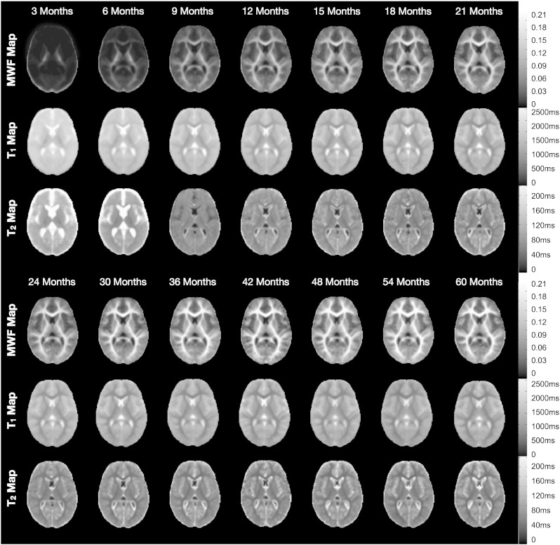
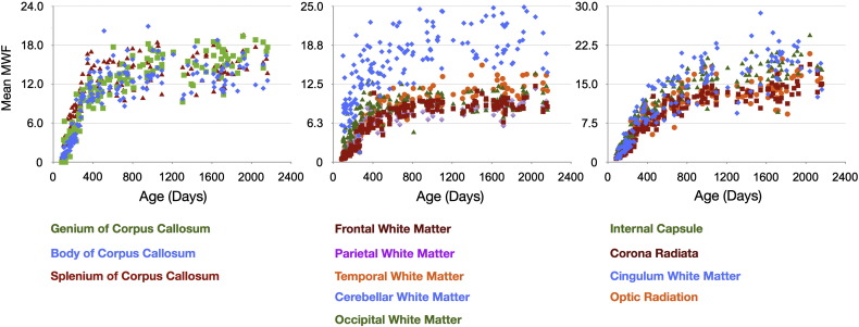
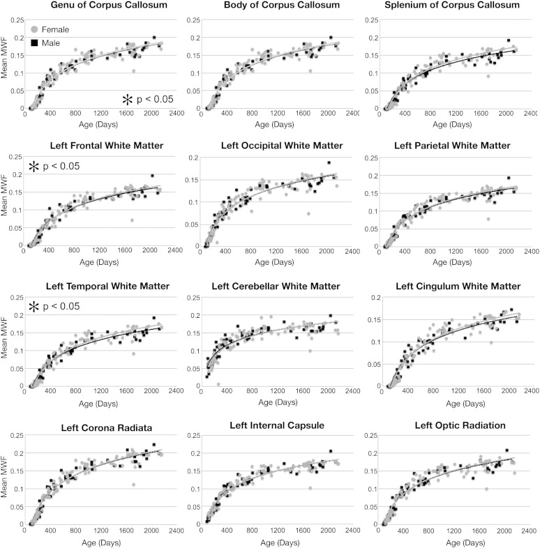
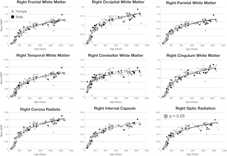
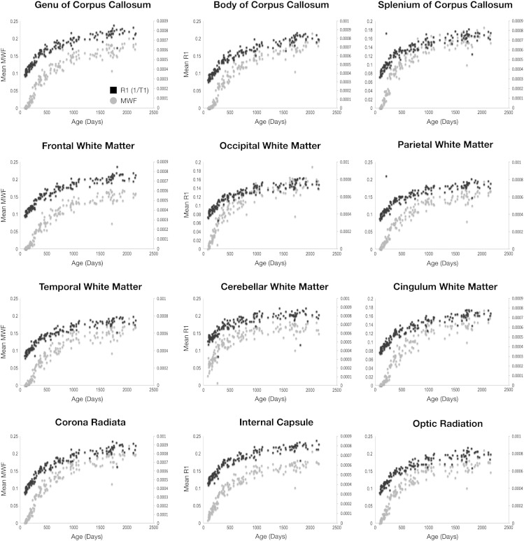
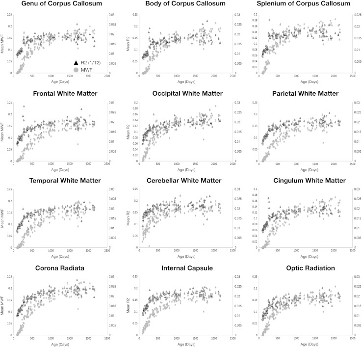
The elaboration of the myelinated white matter is essential for normal neurodevelopment, establishing and mediating rapid communication pathways throughout the brain. These pathways facilitate the synchronized communication required for higher order behavioral and cognitive functioning. Altered neural messaging (or 'disconnectivity') arising from abnormal white matter and myelin development may underlie a number of neurodevelopmental psychiatric disorders. Despite the vital role myelin plays, few imaging studies have specifically examined its maturation throughout early infancy and childhood. Thus, direct investigations of the relationship(s) between evolving behavioral and cognitive functions and the myelination of the supporting neural systems have been sparse. Further, without knowledge of the 'normative' developmental time-course, identification of early abnormalities associated with developmental disorders remains challenging. In this work, we examined the use of longitudinal (T(1)) and transverse (T(2)) relaxation time mapping, and myelin water fraction (MWF) imaging to investigate white matter and myelin development in 153 healthy male and female children, 3 months through 60 months in age. Optimized age-specific acquisition protocols were developed using the DESPOT and mcDESPOT imaging techniques; and mean T(1), T(2) and MWF trajectories were determined for frontal, temporal, occipital, parietal and cerebellar white matter, and genu, body and splenium of the corpus callosum. MWF results provided a spatio-temporal pattern in-line with prior histological studies of myelination. Comparison of T(1), T(2) and MWF measurements demonstrates dissimilar sensitivity to tissue changes associated with neurodevelopment, with each providing differential but complementary information.
Overview
The claims and statistics of this paper haven’t been checked yet.
Run the check — free, no account needed. It reads the open-access text and checks the sources it cites.
Integrity signals
Registry
Cites
Statistics
Figures
Verification
Claims the paper makes
The claims and statistics of this paper haven’t been checked yet.
Run the check — free, no account needed. It reads the open-access text and checks the sources it cites.
Claims other papers make about this one
Each is checked against this paper’s own text: does it say what the citing paper says it does?
In the citing paper
“Males also show increased global gyrification at 2 years of age compared with females (adjusting for total brain volume)”
From Imaging structural and functional brain development in early childhood (2018) · cites this paper as [127]
In the citing paper
“local or regional gyrification index is similar between males and females at each age127.”
From Imaging structural and functional brain development in early childhood (2018) · cites this paper as [127]
In the citing paper
“but not at 0 or 1 year of age”
From Imaging structural and functional brain development in early childhood (2018) · cites this paper as [127]
In the citing paper
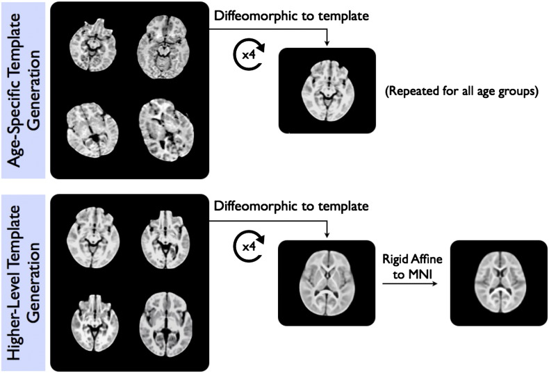
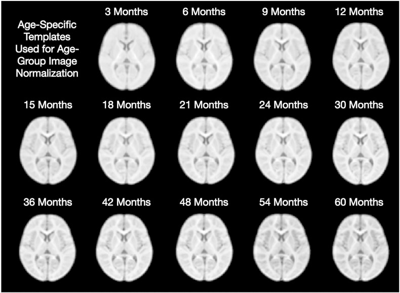
“this strategy of template creation mirrors our prior work that has been shown to successfully create structural and quantitative MRI templates of the pediatric brain (Deoni et al., 2012, Dean et al., 2014).”
From Quantifying cortical development in typically developing toddlers and young children, 1–6 years of age (2017) · cites this paper as Deoni et al., 2012
Self-check
Percentages, sums and reported differences re-computed from the paper’s own numbers.
The self-check needs the paper’s full text.
Citations
Cites
Avants, B., Epstein, C. L., Grossman, M., & Gee, J. C. (2007) · Medical Image Analysis · doi:10.1016/j.media.2007.06.004 · Cited by 5,876
The basis of anisotropic water diffusion in the nervous system—A technical review.
Beaulieu, C. (2002) · NMR in Biomedicine · doi:10.1002/nbm.782 · Cited by 4,628
The Human Connectome Project: A data acquisition perspective
Essen, D. C. V., Uǧurbil, K., Auerbach, E. J., Barch, D. M., Behrens, T. E. J., Bucholz, R. D., Chang, A., Chen, L., Corbetta, M., Curtiss, S. W., et al. (2012) · NeuroImage · doi:10.1016/j.neuroimage.2012.02.018 · Cited by 2,638
Mullen Scales of Early Learning
Bradley‐Johnson, S. (1997) · Psychology in the Schools · doi:10.1002/(sici)1520-6807(199710)34:4 · Cited by 2,329
Unbiased average age-appropriate atlases for pediatric studies
Fonov, V., Evans, A. C., Botteron, K., Almli, C. R., McKinstry, R. C., & Collins, D. L. (2011) · NeuroImage · doi:10.1016/j.neuroimage.2010.07.033 · Cited by 2,290
Regional Development of the Brain in Early Life
(1968) · Archives of Disease in Childhood · doi:10.1136/adc.43.229.388 · Cited by 2,107
Brain development in children and adolescents: Insights from anatomical magnetic resonance imaging
Lenroot, R. K., & Giedd, J. N. (2006) · Neuroscience & Biobehavioral Reviews · doi:10.1016/j.neubiorev.2006.06.001 · Cited by 1,863
(1967) · Medical Entomology and Zoology · Cited by 1,713
Behavioral manifestations of autism in the first year of life
Zwaigenbaum, L., Bryson, S., Rogers, T., Roberts, W., Brian, J., & Szatmari, P. (2005) · International Journal of Developmental Neuroscience · doi:10.1016/j.ijdevneu.2004.05.001 · Cited by 1,602
Intellectual ability and cortical development in children and adolescents
Shaw, P., Greenstein, D., Lerch, J., Clasen, L., Lenroot, R., Gogtay, N., Evans, A., Rapoport, J., & Giedd, J. (2006) · Nature · doi:10.1038/nature04513 · Cited by 1,543
Mapping Human Cortical Areas In Vivo Based on Myelin Content as Revealed by T1- and T2-Weighted MRI
Glasser, M. F., & Van Essen, D. C. (2011) · The Journal of Neuroscience · doi:10.1523/jneurosci.2180-11.2011 · Cited by 1,447
Structural maturation of neural pathways in children and adolescents: in vivo study
Paus, T., Zijdenbos, A., Worsley, K., Collins, D. L., Blumenthal, J., Giedd, J. N., Rapoport, J. L., & Evans, A. C. (1999) · Science · doi:10.1126/science.283.5409.1908 · Cited by 1,322
Sexual dimorphism of brain developmental trajectories during childhood and adolescence
Lenroot, R. K., Gogtay, N., Greenstein, D. K., Wells, E. M., Wallace, G. L., Clasen, L. S., Blumenthal, J. D., Lerch, J., Zijdenbos, A. P., Evans, A. C., et al. (2007) · NeuroImage · doi:10.1016/j.neuroimage.2007.03.053 · Cited by 1,309
Schizophrenia as a Disorder of Neurodevelopment
Lewis, D. A., & Levitt, P. (2002) · Annual Review of Neuroscience · doi:10.1146/annurev.neuro.25.112701.142754 · Cited by 1,163
Courchesne, E., Chisum, H. J., Townsend, J., Cowles, A., Covington, J., Egaas, B., Harwood, M., Hinds, S., & Press, G. A. (2000) · Radiology · doi:10.1148/radiology.216.3.r00au37672 · Cited by 1,049
Quantitative magnetic resonance imaging of human brain development: ages 4–18
Giedd, J. N., Snell, J. W., Lange, N., Rajapakse, J. C., Casey, B. J., Kozuch, P. L., Vaituzis, A. C., Vauss, Y. C., Hamburger, S. D., Kaysen, D., et al. (1996) · Cerebral Cortex · doi:10.1093/cercor/6.4.551 · Cited by 1,024
Maturation of white matter in the human brain: A review of magnetic resonance studies.
Paus, T., Collins, D. L., Evans, A. C., Leonard, G., Pike, B., & Zijdenbos, A. (2001) · Brain Research Bulletin · doi:10.1016/s0361-9230(00)00434-2 · Cited by 863
Structural MRI of pediatric brain development: what have we learned and where are we going?
Giedd, J. N., & Rapoport, J. L. (2010) · Neuron · doi:10.1016/j.neuron.2010.08.040 · Cited by 836
In vivo measurement of T 2 distributions and water contents in normal human brain
Whittall, K. P., MacKay, A. L., Graeb, D. A., Nugent, R. A., Li, D. K. B., & Paty, D. W. (1997) · Magnetic Resonance in Medicine · doi:10.1002/mrm.1910370107 · Cited by 796
Nagy, Z., Westerberg, H., & Klingberg, T. (2004) · Journal of Cognitive Neuroscience · doi:10.1162/0898929041920441 · Cited by 794
Maturation of widely distributed brain function subserves cognitive development.
Luna, B., Thulborn, K. R., Munoz, D. P., Merriam, E. P., Garver, K. E., Minshew, N. J., Keshavan, M. S., Genovese, C. R., Eddy, W. F., & Sweeney, J. A. (2001) · NeuroImage · doi:10.1006/nimg.2000.0743 · Cited by 757
Normal maturation of the neonatal and infant brain: MR imaging at 1.5 T.
Barkovich, A. J., Kjos, B. O., Jackson, D. E., & Norman, D. M. (1988) · Radiology · doi:10.1148/radiology.166.1.3336675 · Cited by 741
Raznahan, A., Shaw, P., Lalonde, F., Stockman, M., Wallace, G. L., Greenstein, D., Clasen, L., Gogtay, N., & Giedd, J. N. (2011) · The Journal of Neuroscience · doi:10.1523/jneurosci.0054-11.2011 · Cited by 712
Brain growth across the life span in autism: Age-specific changes in anatomical pathology
Courchesne, E., Campbell, K., & Solso, S. (2011) · Brain Research · doi:10.1016/j.brainres.2010.09.101 · Cited by 711
Sowell, E. R., Trauner, D. A., Gamst, A., & Jernigan, T. L. (2002) · Developmental Medicine and Child Neurology · doi:10.1017/s0012162201001591 · Cited by 701
White matter maturation reshapes structural connectivity in the late developing human brain
Hagmann, P., Sporns, O., Madan, N., Cammoun, L., Pienaar, R., Wedeen, V. J., Meuli, R., Thiran, J.-P., & Grant, P. E. (2010) · Proceedings of the National Academy of Sciences · doi:10.1073/pnas.1009073107 · Cited by 700
Wolff, J. J., Gu, H., Gerig, G., Elison, J. T., Styner, M., Gouttard, S., Botteron, K. N., Dager, S. R., Dawson, G., Estes, A. M., et al. (2012) · American Journal of Psychiatry · doi:10.1176/appi.ajp.2011.11091447 · Cited by 671
Mullen Scales of Early Learning: Ags Edition
Dumont, R., & Willis, J. O. (2008) · Encyclopedia of Special Education · doi:10.1002/9780470373699.speced1402 · Cited by 667
Diffusion tensor imaging and aging
Sullivan, E. V., & Pfefferbaum, A. (2006) · Neuroscience & Biobehavioral Reviews · doi:10.1016/j.neubiorev.2006.06.002 · Cited by 649
The optimal template effect in hippocampus studies of diseased populations
Avants, B. B., Yushkevich, P., Pluta, J., Minkoff, D., Korczykowski, M., Detre, J., & Gee, J. C. (2010) · NeuroImage · doi:10.1016/j.neuroimage.2009.09.062 · Cited by 640
Sex differences in the adolescent brain
Lenroot, R., & Giedd, J. N. (2009) · Brain and Cognition · doi:10.1016/j.bandc.2009.10.008 · Cited by 608
Autism as a neural systems disorder: A theory of frontal-posterior underconnectivity
Just, M. A., Keller, T. A., Malave, V. L., Kana, R. K., & Varma, S. (2012) · Neuroscience & Biobehavioral Reviews · doi:10.1016/j.neubiorev.2012.02.007 · Cited by 578
Oishi, K., Zilles, K., Amunts, K., Faria, A. V., Jiang, H., Li, X., Akhter, K., Hua, K., Woods, R. P., Toga, A. W., et al. (2008) · NeuroImage · doi:10.1016/j.neuroimage.2008.07.009 · Cited by 577
Mapping Infant Brain Myelination with Magnetic Resonance Imaging
Deoni, S. C. L., Mercure, E., Blasi, A., Gasston, D., Thomson, A., Johnson, M., Williams, S. C. R., & Murphy, D. G. M. (2011) · The Journal of Neuroscience · doi:10.1523/jneurosci.2106-10.2011 · Cited by 497
Self-citationMyelin water imaging in multiple sclerosis: quantitative correlations with histopathology
Laule, C., Leung, E., Li, D. K., Traboulsee, A. L., Paty, D. W., MacKay, A. L., & Moore, G. R. (2006) · Multiple Sclerosis Journal · doi:10.1177/1352458506070928 · Cited by 489
Gleaning multicomponent T 1 and T 2 information from steady‐state imaging data
Deoni, S., Rutt, B. K., Arun, T., Pierpaoli, C. M., & Jones, D. K. (2008) · Magnetic Resonance in Medicine · doi:10.1002/mrm.21704 · Cited by 473
Self-citationHermoye, L., Saint‐Martin, C., Cosnard, G., Lee, S., Kim, J., Nassogne, M., Menten, R., Clapuyt, P. R. M., Donohue, P., Hua, K., et al. (2005) · NeuroImage · doi:10.1016/j.neuroimage.2005.08.017 · Cited by 422
Anatomical MRI of the developing human brain: what have we learned?
DURSTON, S., HULSHOFF POL, H. E., CASEY, B. J., GIEDD, J. N., BUITELAAR, J. K., & VAN ENGELAND, H. (2001) · Journal of the American Academy of Child & Adolescent Psychiatry · doi:10.1097/00004583-200109000-00009 · Cited by 410 · cited 2×
“These results are in general agreement with prior literature (see Durston et al., 2001 for a thorough review) showing little male–female difference in white matter volume between brain regions.”
ResultWhite and gray matter development in human fetal, newborn and pediatric brains
Huang, H., Zhang, J.-Y., Wakana, S., Zhang, W., Ren, T., Richards, L. J., Yarowsky, P., Donohue, P., Graham, E. M., van Zijl, P., et al. (2006) · NeuroImage · doi:10.1016/j.neuroimage.2006.06.009 · Cited by 404
A Four-Dimensional Probabilistic Atlas of the Human Brain
Mazziotta, J. C., Toga, A. W., Evans, A. C., Fox, P. T., Lancaster, J. L., Zilles, K., Woods, R. P., Paus, T., Simpson, G., Pike, G. B., et al. (2001) · Journal of the American Medical Informatics Association · doi:10.1136/jamia.2001.0080401 · Cited by 384
Diffusion tensor imaging of neurodevelopment in children and young adults.
Snook, L., Paulson, L.-A., Roy, D., Phillips, L., & Beaulieu, C. (2005) · NeuroImage · doi:10.1016/j.neuroimage.2005.03.016 · Cited by 350
Insights into brain microstructure from the T2 distribution
MacKay, A. L., Laule, C., Vavasour, I. M., Bjarnason, T. A., Kolind, S., & Mädler, B. (2006) · Magnetic Resonance Imaging · doi:10.1016/j.mri.2005.12.037 · Cited by 339
Processes of change in brain and cognitive development
Johnson, M. H., & Munakata, Y. (2005) · Trends in Cognitive Sciences · doi:10.1016/j.tics.2005.01.009 · Cited by 337
Dubois, J., Hertz‐Pannier, L., Dehaene‐Lambertz, G., Cointepas, Y., & Bihan, D. L. (2006) · NeuroImage · doi:10.1016/j.neuroimage.2005.11.022 · Cited by 333
Diffusion tensor imaging and aging—A review.
Moseley, M. (2002) · NMR in Biomedicine · doi:10.1002/nbm.785 · Cited by 302
Accelerated maturation of white matter in young children with autism: A high b value DWI study
Ben Bashat, D., Kronfeld-Duenias, V., Zachor, D. A., Ekstein, P. M., Hendler, T., Tarrasch, R., Even, A., Levy, Y., & Ben Sira, L. (2007) · NeuroImage · doi:10.1016/j.neuroimage.2007.04.060 · Cited by 300
Lifespan trajectory of myelin integrity and maximum motor speed
Bartzokis, G., Lu, P. H., Tingus, K., Mendez, M. F., Richard, A. G., Peters, D. G., Oluwadara, B., Barrall, K. A., Finn, J. P., Villablanca, P. J., et al. (2008) · Neurobiology of Aging · doi:10.1016/j.neurobiolaging.2008.08.015 · Cited by 284
Changes in white matter microstructure during adolescence
Giorgio, A., Watkins, K. E., Douaud, G., James, A. C., James, S., De Stefano, N., Matthews, P. M., Smith, S. M., & Johansen-Berg, H. (2008) · NeuroImage · doi:10.1016/j.neuroimage.2007.07.043 · Cited by 278
Meda, S. A., Gill, A., Stevens, M. C. G., Lorenzoni, R. P., Glahn, D. C., Calhoun, V. D., Sweeney, J. A., Tamminga, C. A., Keshavan, M. S., Thaker, G. K., et al. (2012) · Biological Psychiatry · doi:10.1016/j.biopsych.2012.01.025 · Cited by 273
Structural Asymmetries in the Infant Language and Sensori-Motor Networks
Dubois, J., Hertz‐Pannier, L., Cachia, A., Mangin, J.-F., Bihan, D. L., & Dehaene‐Lambertz, G. (2008) · Cerebral Cortex · doi:10.1093/cercor/bhn097 · Cited by 264
Disrupted cortical connectivity theory as an explanatory model for autism spectrum disorders
Kana, R. K., Libero, L. E., & Moore, M. S. (2011) · Physics of Life Reviews · doi:10.1016/j.plrev.2011.10.001 · Cited by 263
Cheung, M. M., Hui, E. S. K., Chan, K. C., Helpern, J. A., Qi, L., & Wu, E. X. (2008) · NeuroImage · doi:10.1016/j.neuroimage.2008.12.018 · Cited by 260
Smyser, C. D., Snyder, A. Z., & Neil, J. J. (2011) · NeuroImage · doi:10.1016/j.neuroimage.2011.02.073 · Cited by 259
Deoni, S. (2007) · Journal of Magnetic Resonance Imaging · doi:10.1002/jmri.21130 · Cited by 237
Self-citationIs diffusion anisotropy an accurate monitor of myelination?
Mädler, B., Drabycz, S. A., Kolind, S. H., Whittall, K. P., & MacKay, A. L. (2008) · Magnetic Resonance Imaging · doi:10.1016/j.mri.2008.01.047 · Cited by 227
Analysis of biological NMR relaxation data with continuous distributions of relaxation times
Kroeker, R. M., & Henkelman, R. M. (1986) · Journal of Magnetic Resonance (1969) · doi:10.1016/0022-2364(86)90074-0 · Cited by 214
Is the magnetization transfer ratio a marker for myelin in multiple sclerosis?
Vavasour, I. M., Laule, C., Li, D. K. B., Traboulsee, A. L., & MacKay, A. L. (2011) · Journal of Magnetic Resonance Imaging · doi:10.1002/jmri.22441 · Cited by 201
Almli, C. R., Rivkin, M. J., & McKinstry, R. C. (2007) · NeuroImage · doi:10.1016/j.neuroimage.2006.08.058 · Cited by 193
Experience effects on brain development: possible contributions to psychopathology
Grossman, A. W., Churchill, J. D., McKinney, B. C., Kodish, I. M., Otte, S. L., & Greenough, W. T. (2002) · Journal of Child Psychology and Psychiatry · doi:10.1111/1469-7610.t01-1-00102 · Cited by 187
Motor Development during Infancy and Early Childhood: Overview and Suggested Directions for Research
Malina, R. M. (2004) · International Journal of Sport and Health Science · doi:10.5432/ijshs.2.50 · Cited by 185
Is multicomponent T2 a good measure of myelin content in peripheral nerve?
Webb, S., Munro, C. A., Midha, R., & Stanisz, G. J. (2003) · Magnetic Resonance in Medicine · doi:10.1002/mrm.10411 · Cited by 172
Early postnatal development of rat brain: In vivo diffusion tensor imaging
Bockhorst, K. H. J., Narayana, P. A., Liu, R., Ahobila‐Vijjula, P., Ramu, J., Kamel, M. R., Wosik, J., Bockhorst, T., Hahn, K. R., Hasan, K. M., et al. (2008) · Journal of Neuroscience Research · doi:10.1002/jnr.21607 · Cited by 169
Autism: The first firm finding = underconnectivity?
Hughes, J. R. (2007) · Epilepsy & Behavior · doi:10.1016/j.yebeh.2007.03.010 · Cited by 156
Sex differences in adolescent white matter architecture
Bava, S., Boucquey, V., Goldenberg, D., Thayer, R. E., Ward, M. E., Jacobus, J., & Tapert, S. F. (2010) · Brain Research · doi:10.1016/j.brainres.2010.12.051 · Cited by 148
Early Maturation of the Linguistic Dorsal Pathway in Human Infants
Leroy, F., Glasel, H., Dubois, J., Hertz‐Pannier, L., Thirion, B., Mangin, J., & Dehaene‐Lambertz, G. (2011) · Journal of Neuroscience · doi:10.1523/jneurosci.4141-10.2011 · Cited by 145
BOLD-f MRI response to single-pulse transcranial magnetic stimulation (TMS)
Bohning, D. E., Shastri, A., Wassermann, E. M., Ziemann, U., Lorberbaum, J. P., Nahas, Z. H., Lomarev, M. P., & George, M. S. (2000) · Journal of Magnetic Resonance Imaging · doi:10.1002/1522-2586(200006)11:6 · Cited by 138
Establishing norms for age-related changes in proton T1 of human brain tissue in vivo
Cho, S., Jones, D., Reddick, W. E., Ogg, R. J., & Steen, R. G. (1997) · Magnetic Resonance Imaging · doi:10.1016/s0730-725x(97)00202-6 · Cited by 132
Deoni, S. (2010) · Magnetic Resonance in Medicine · doi:10.1002/mrm.22685 · Cited by 111
Self-citationMills, K. L., Bathula, D., Dias, T. G. C., Iyer, S. P., Fenesy, M. C., Musser, E. D., Stevens, C. A., Thurlow, B. L., Carpenter, S. D., Nagel, B. J., et al. (2012) · Frontiers in Psychiatry · doi:10.3389/fpsyt.2012.00002 · Cited by 108
MR assessment of myelination in infants and children: usefulness of marker sites.
Bird, C. R., Hedberg, M., Drayer, B. P., Keller, P. J., Flom, R. A., & Hodak, J. A. (1989) · PubMed Central · Cited by 105
Diffusion Tensor Imaging Assessment of Brain White Matter Maturation During the First Postnatal Year
Provenzale, J. M., Liang, L., DeLong, D. M., & WHITE, L. E. (2007) · American Journal of Roentgenology · doi:10.2214/ajr.07.2132 · Cited by 103
Sex differences in frontal lobe white matter microstructure: a DTI study
Szeszko, P. R., Vogel, J., Ashtari, M., Malhotra, A. K., Bates, J., Kane, J. M., Bilder, R. M., Frevert, T. A., & Lim, K. O. (2003) · Neuroreport · doi:10.1097/00001756-200312190-00035 · Cited by 96
MR Relaxation in Multiple Sclerosis
MacKay, A. L., Vavasour, I. M., Rauscher, A., Kolind, S., Mädler, B., Moore, G. R. W., Traboulsee, A. L., Li, D. K. B., & Laule, C. (2008) · Neuroimaging Clinics of North America · doi:10.1016/j.nic.2008.09.007 · Cited by 95
Somatosensory lateralization in the newborn brain
Erberich, S. G., Panigrahy, A., Friedlich, P. S., Seri, I., Nelson, M. D., & Gilles, F. H. (2005) · NeuroImage · doi:10.1016/j.neuroimage.2005.07.024 · Cited by 94
T2 relaxometry of normal pediatric brain development
Leppert, I. R., Almli, C. R., McKinstry, R. C., Mulkern, R. V., Pierpaoli, C. M., Rivkin, M. J., & Pike, G. B. (2009) · Journal of Magnetic Resonance Imaging · doi:10.1002/jmri.21646 · Cited by 86
On the inherent precision of mcDESPOT
Lankford, C. L., & Does, M. D. (2013) · Magnetic Resonance in Medicine · doi:10.1002/mrm.24241 · Cited by 83
Saito, N., Sakai, O., Ozonoff, A., & Jara, H. J. (2009) · Magnetic Resonance Imaging · doi:10.1016/j.mri.2009.05.006 · Cited by 70
Delayed myelination in children with developmental delay detected by volumetric MRI
Pujol, J., Sala, A. L., Sebastián‐Gallés, N., Deus, J., Cardoner, N., Soriano‐Mas, C., Moreno, A., & Sans, A. (2004) · NeuroImage · doi:10.1016/j.neuroimage.2004.01.029 · Cited by 69
Raznahan, A., GREENSTEIN, D. K., Lee, Y., Long, R., CLASEN, L. S., Gochman, P., Addington, A. M., Giedd, J. N., Rapoport, J. L., & Gogtay, N. (2011) · NeuroImage · doi:10.1016/j.neuroimage.2011.05.032 · Cited by 50
Multiexponential proton relaxation in model cellular systems
Menon, R. S., Rusinko, M. S., & Allen, P. S. (1991) · Magnetic Resonance in Medicine · doi:10.1002/mrm.1910200204 · Cited by 49
Regional variation, hemispheric asymmetries and gender differences in pericortical white matter
Kang, X., Herron, T. J., & Woods, D. L. (2011) · NeuroImage · doi:10.1016/j.neuroimage.2011.03.016 · Cited by 41
MRI assessment of myelination: an age standardization
Staudt, M., Schropp, C., Staudt, F., Obletter, N., Bise, K., Breit, A., & Weinmann, H.-M. (1994) · Pediatric Radiology · doi:10.1007/bf02020169 · Cited by 40
The Differentiated Maturation of the Cerebral Cortex
Rabinowicz, T. (1986) · Springer eBooks · doi:10.1007/978-1-4899-0522-2_14 · Cited by 33
Supportive neurodevelopmental evidence for ADHD as a developmental disorder
Panzer, A., & Viljoen, M. (2004) · Medical Hypotheses · doi:10.1016/j.mehy.2003.12.060 · Cited by 20
Imaging white matter diffusion changes with development and recovery from brain injury
Bosnell, R., Giorgio, A., & Johansen‐Berg, H. (2008) · Developmental Neurorehabilitation · doi:10.1080/17518420802289065 · Cited by 18
Cited by
Sorted by citations
Imaging structural and functional brain development in early childhood
Gilmore, J. H., Knickmeyer, R. C., & Gao, W. (2018) · Nature Reviews Neuroscience · doi:10.1038/nrn.2018.1 · Cited by 1,013
Dubois, J., Dehaene-Lambertz, G., Kulikova, S., Poupon, C., Hüppi, P. S., & Hertz-Pannier, L. (2014) · Neuroscience · doi:10.1016/j.neuroscience.2013.12.044 · Cited by 851
Biological embedding of childhood adversity: from physiological mechanisms to clinical implications
Berens, A. E., Jensen, S. K. G., & Nelson, C. A. (2017) · BMC Medicine · doi:10.1186/s12916-017-0895-4 · Cited by 657
“Dramatic growth in gray and white matter occurs in the first 2 years of life, when the brain attains 80–90% of its adult volume before continuing to grow at an attenuated rate [22, 23].”
BackgroundAn open resource for transdiagnostic research in pediatric mental health and learning disorders
Alexander, L. M., Escalera, J., Ai, L., Andreotti, C., Febre, K., Mangone, A., Potler, N. V., Langer, N., Alexander, A., Kovacs, M., et al. (2017) · Scientific Data · doi:10.1038/sdata.2017.181 · Cited by 681
The development of brain white matter microstructure
Lebel, C., & Deoni, S. (2018) · NeuroImage · doi:10.1016/j.neuroimage.2017.12.097 · Cited by 602
Self-citation
Metadata
- Authors
- Deoni, S. C. L., Dean, D. C., O'Muircheartaigh, J., Dirks, H., & Jerskey, B. A.
- Affiliations
- Brown University (US)
- Corresponding author
- Deoni, Sean C.L.
- Venue
- NeuroImage
- Publisher
- Elsevier BV
- Published
- 2012-11
- Volume
- 63
- Issue
- 3
- Pages
- 1038-1053
- ISSN
- 1053-8119 (ISSN-L)
- PMC
- PMC3711836
- OpenAlex
- W2131940336
- Cited by
- 390
- References
- 86
- Citations by year
- 2026: 18 · 2025: 28 · 2024: 25 · 2023: 32 · 2022: 27 · 2021: 36 · 2020: 64 · 2019: 32 · 2018: 27 · 2017: 27 · 2016: 25 · 2015: 21 · 2014: 14 · 2013: 12 · 2012: 2
- FWCI
- 11.84
- Type
- journal article
- Language
- en
- Open access
- hybrid
- doi.org
- Licence
- Locations
- NeuroImage (publishedVersion, cc-by)
- Europe PMC (PubMed Central) (publishedVersion, cc-by)
- PubMed Central (publishedVersion, cc-by)
- PubMed (publishedVersion)
- Research Portal (King's College London) (submittedVersion)
- Funders
- National Institutes of Mental Health (R01 MH087510)
- Sir Henry Wellcome Postdoctoral Fellowship (No 096195)
- Topics
- Health Sciences › Medicine › Radiology, Nuclear Medicine and Imaging › Advanced Neuroimaging Techniques and Applications
- Health Sciences › Medicine › Radiology, Nuclear Medicine and Imaging › Advanced MRI Techniques and Applications
- Physical Sciences › Chemistry › Spectroscopy › NMR spectroscopy and applications
- Keywords
- white matter development, myelination, myelin water fraction, T1 relaxation time, T2 relaxation time, corpus callosum, brain development, infancy, early childhood, despots, cerebellum
- MeSH terms
- Brain, Brain Mapping, Child, Preschool, Female, Humans, Infant, Male, Myelin Sheath, Neurogenesis
About these checks
Registry, retraction, Cites and statistics results are computed, not judged, with one exception: a printed reference that no index lists is read once by a model before it counts as a citation error. Claims are extracted by a model and kept only when their quote appears verbatim in the text; each cited source is then checked, with a model’s help, for whether it says what the claim relies on. Those verdicts can be wrong — read the claim’s quote and the cited source before relying on one.
Statistics and arithmetic are recomputed, not judged.
Confidence bands are shown only for fields calibrated on our annotated test set. None is calibrated yet, so no band is shown.
What counts as a claim ›How we measure confidence ›How we check this ›
Are you an author, or do you see a problem with this page? Request a review
