

The DOI matches its Crossref record; 65 of the 65 references it cites were found in a registry or an index; 19 of the 33 sources its claims cite were checked for support.
TFEB regulates lysosomal positioning by modulating TMEM55B expression and JIP4 recruitment to lysosomes
Open accessAbstract
Lysosomal distribution is linked to the role of lysosomes in many cellular functions, including autophagosome degradation, cholesterol homeostasis, antigen presentation, and cell invasion. Alterations in lysosomal positioning contribute to different human pathologies, such as cancer, neurodegeneration, and lysosomal storage diseases. Here we report the identification of a novel mechanism of lysosomal trafficking regulation. We found that the lysosomal transmembrane protein TMEM55B recruits JIP4 to the lysosomal surface, inducing dynein-dependent transport of lysosomes toward the microtubules minus-end. TMEM55B overexpression causes lysosomes to collapse into the cell center, whereas depletion of either TMEM55B or JIP4 results in dispersion toward the cell periphery. TMEM55B levels are transcriptionally upregulated following TFEB and TFE3 activation by starvation or cholesterol-induced lysosomal stress. TMEM55B or JIP4 depletion abolishes starvation-induced retrograde lysosomal transport and prevents autophagosome–lysosome fusion. Overall our data suggest that the TFEB/TMEM55B/JIP4 pathway coordinates lysosome movement in response to a variety of stress conditions.
Overview
AI snapshot
Written from the paper’s full text.
TL;DR
The paper identifies TMEM55B as a lysosomal transmembrane protein that recruits JIP4 to lysosomes, promoting dynein-dependent retrograde transport and perinuclear lysosome clustering. TFEB/TFE3 upregulate TMEM55B under starvation or cholesterol stress, and TMEM55B/JIP4 depletion blocks starvation-induced lysosomal clustering and autophagosome–lysosome fusion.
Key findings
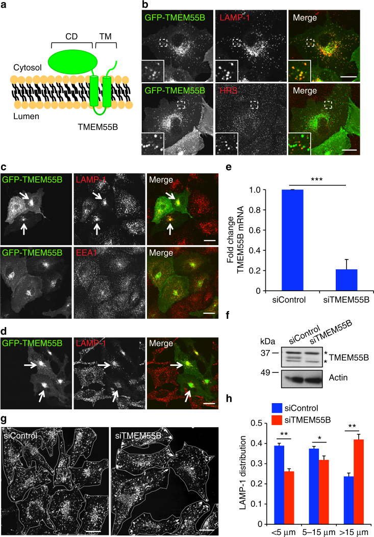
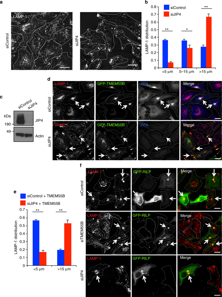
- TMEM55B overexpression clusters lysosomes in the cell center, while TMEM55B depletion causes over 80% reduction in TMEM55B mRNA/protein and shifts lysosomes toward the cell periphery; JIP4 depletion causes more than 60% of LAMP-1 signal to fall >15 μm from the nuclear rim.
- TMEM55B’s cytosolic domain recruits JIP4 to lysosomal membranes; in JIP4-depleted cells, GFP-TMEM55B fails to cluster lysosomes, with distal LAMP-1 signal >15 μm changing from 20% in control cells to more than 50%.
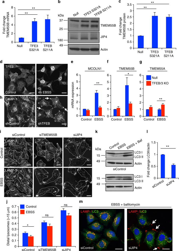
- Starvation increases TMEM55B mRNA/protein in a TFEB/TFE3-dependent manner; starvation-induced lysosomal clustering is reduced in TFEB-depleted cells and blocked by TMEM55B or JIP4 depletion, and JIP4 depletion reduces LC3II accumulation after EBSS plus bafilomycin.
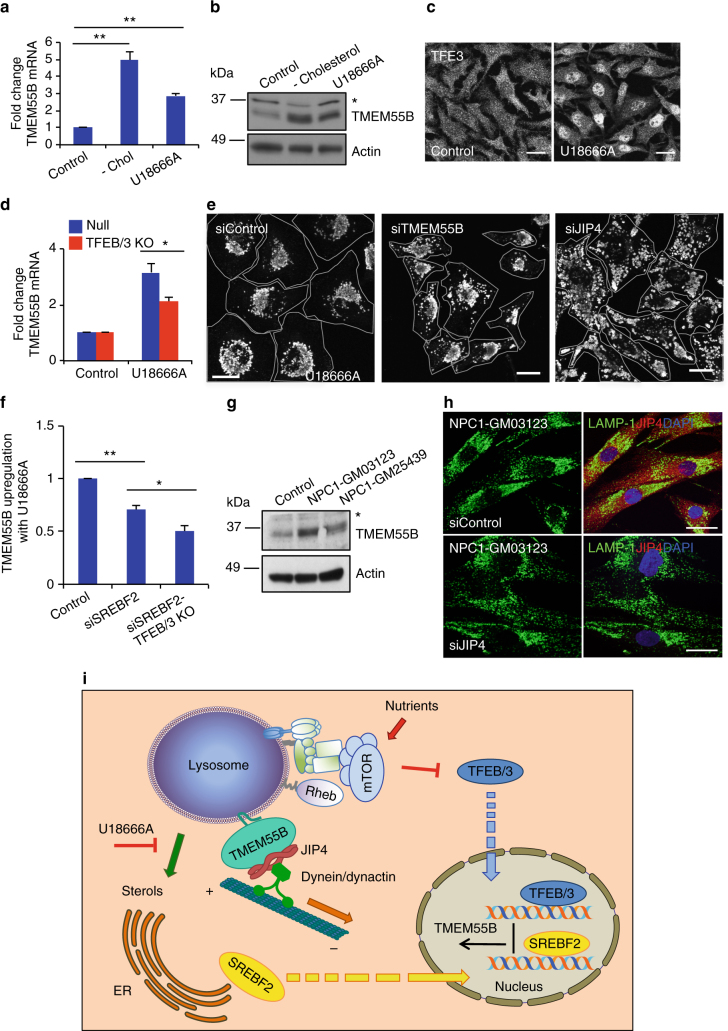
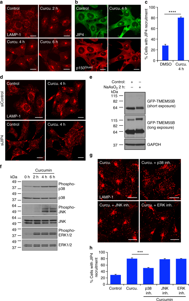
- U18666A-induced cholesterol stress and NPC1 patient fibroblasts upregulate TMEM55B; JIP4 depletion reduces perinuclear clustering in NPC fibroblasts, and acute curcumin/NaAsO2 stress induces JIP4/p150Glued recruitment and lysosomal clustering via p38, not JNK or ERK.
Methods
- Cell culture and perturbations: ARPE-19, HeLa, U2OS, MEFs, and NPC1/control fibroblasts were subjected to siRNA/shRNA knockdown, adenoviral expression, rapamycin-induced dimerization, nocodazole, starvation, U18666A, curcumin/NaAsO2, and MAPK inhibitors.
- Imaging and quantification: immunofluorescence for LAMP-1, JIP4, p150Glued, and other markers; lysosome distribution measured by linear profile, radial profile, and cumulative intensity distribution; time-lapse confocal microscopy; statistics by two-tailed t-test or one-way ANOVA.
- Biochemical assays: qRT-PCR, immunoblotting, GFP/RFP pull-downs, mass spectrometry, subcellular fractionation, and phosphatidylinositol phosphatase assays including malachite green and PH-PLCD1 imaging.
Limitations
- The paper states that it remains to be determined whether TMEM55B functions as a mere JIP4 recruiter or also stimulates the interaction between JIP4 and the dynein–dynactin complex, and how the TMEM55B/JIP4 pathway works in conjunction with other retrograde pathways such as Rab7/RILP/ORP1L and MCOLN1/ALG-2.
- Evident from the text: mechanistic conclusions rely largely on cultured cell lines with overexpression, RNAi, and pharmacological stress, with no in vivo animal model or direct structural mapping of the TMEM55B–JIP4 interface presented.
Model-written summary. Check it against the paper before relying on it.
Integrity signals
Registry
Cites
Statistics
Figures
Verification
Claims the paper makes
74 Own result · 36 Own interpretation · 10 Partly supported · 9 Own method · 3 No basis given · 2 No full text of the sources · 2 Not checked yet · 1 Supported so far · 1 Couldn’t confirm · accuracy not yet measured
The cited sources support only part of this claim — read each source’s verdict below.
In the paper
“we observed that the upregulation of TMEM55B induced by U18666A was significantly decreased in cells depleted of SREBF2 (Fig. 7f)”
§ TFEB regulates lysosomal positioning by modulating TMEM55B expression and JIP4 recruitment to lysosomes > Results > TMEM55B responds to changes in cholesterol levels
What the sources say
- Partly supportedcites Medina et al. [48] Medina, M. W. et al. Transmembrane protein 55B is a novel regulator of cellular cholesterol metabolism. Arterioscler. Thromb. Vasc. Biol. 34, 1917–1923 (2014). sourceExperimental — accuracy not yet measured
Results section II reports that SREBF2 knockdown suppressed sterol-dependent upregulation of TMEM55B in HeLa cells. It does not report treatment with U18666A, so it does not establish the claim for that specific condition. (LLM, conf=0.86)
plaindirect- Asserted: passes — The authors put it forward as true.
- Checkable: passes — It can be checked against evidence.
- Load-bearing: unclear — It is unclear whether the paper’s conclusions depend on it.
- Based: passes — The paper gives a reason why it holds.
- Partly supported
The cited sources that could be checked couldn’t confirm or dispute this claim.
In the paper
“TMEM55B is a target of TFEB and TFE3”
- Also stated in: § TMEM55B promotes lysosomal retrograde transport · hedged
“ChIP-seq analysis identified TMEM55B as a putative TFE3 target both in RAW 264.7 macrophages and MEFs5,6.”
§ TFEB regulates lysosomal positioning by modulating TMEM55B expression and JIP4 recruitment to lysosomes > Introduction
What the sources say
- Could not verifycites [5] Pastore, N. et al. TFEB and TFE3 cooperate in the regulation of the innate immune response in activated macrophages. Autophagy 12, 1240–1258 (2016). sourceExperimental — accuracy not yet measured
The Introduction says TFE3 regulates a set of genes similar to TFEB’s, and the ‘Identification of TFE3 direct-targets using ChIP-seq’ section discusses target genes generally. Neither passage identifies TMEM55B as a target. Searches for ‘TMEM55B’ and ‘transmembrane protein 55B’ found no passage addressing that specific gene; page numbers were unavailable in our copy. (LLM, conf=0.73)
- Could not verifycites [6] Martina, J. A., Diab, H. I., Brady, O. A. & Puertollano, R. TFEB and TFE3 are novel components of the integrated stress response. EMBO J. 35, 479–495 (2016). sourceExperimental — accuracy not yet measured
The Results section on lysosomal and autophagic gene expression identifies several TFEB- and TFE3-dependent genes, but not TMEM55B. Searches for “TMEM55B,” “TMEM55,” and “transmembrane protein 55B” found no passage in our copy establishing that TMEM55B is a target of either factor. (LLM, conf=0.73)
plaindirect- Asserted: passes — The authors put it forward as true.
- Checkable: passes — It can be checked against evidence.
- Load-bearing: passes — The paper’s conclusions depend on it.
- Based: passes — The paper gives a reason why it holds.
- Also stated in: § TMEM55B promotes lysosomal retrograde transport · hedged
A result the paper reports from its own data — it cites no other source for it, so there is none to check. The quote shows it is in the paper.
In the paper
“depletion of either TMEM55B or JIP4 results in dispersion toward the cell periphery”
- Also stated in: § TMEM55B promotes lysosomal retrograde transport · plain
“In cells depleted of TMEM55B the distribution significantly shifted, showing a decrease in the concentration of LAMP-1-positive structures in the perinuclear region, concomitant with a distinct increase in the number of lysosomes localized towards the cell periphery”
§ TFEB regulates lysosomal positioning by modulating TMEM55B expression and JIP4 recruitment to lysosomes > Abstract
plaindirect- Asserted: passes — The authors put it forward as true.
- Checkable: passes — It can be checked against evidence.
- Load-bearing: passes — The paper’s conclusions depend on it.
- Based: passes — The paper gives a reason why it holds.
- Also stated in: § TMEM55B promotes lysosomal retrograde transport · plain
A result the paper reports from its own data — it cites no other source for it, so there is none to check. The quote shows it is in the paper.
In the paper
“TMEM55B levels are transcriptionally upregulated following TFEB and TFE3 activation by starvation or cholesterol-induced lysosomal stress.”
- Also stated in: § Introduction · plain
“Interestingly, TMEM55B is a target of TFEB and TFE3 and its mRNA and protein levels increase following TFEB/3 activation by starvation or cholesterol accumulation in lysosomes.”
§ TFEB regulates lysosomal positioning by modulating TMEM55B expression and JIP4 recruitment to lysosomes > Abstract
plaindirect- Asserted: passes — The authors put it forward as true.
- Checkable: passes — It can be checked against evidence.
- Load-bearing: passes — The paper’s conclusions depend on it.
- Based: passes — The paper gives a reason why it holds.
- Rests on an assumption: Starvation activates TFEB and TFE3.
- Rests on an assumption: Cholesterol accumulation in lysosomes activates TFEB and TFE3.
- Also stated in: § Introduction · plain
A result the paper reports from its own data — it cites no other source for it, so there is none to check. The quote shows it is in the paper.
In the paper
“TMEM55B overexpression causes lysosomes to collapse into the cell center”
- Also stated in: § TMEM55B promotes lysosomal retrograde transport · plain
“upon overexpression of GFP-TMEM55B, we observed a dramatic repositioning of lysosomes, changing from dispersed throughout the cell to tightly clustered in the cell center”
§ TFEB regulates lysosomal positioning by modulating TMEM55B expression and JIP4 recruitment to lysosomes > Abstract
plaindirect- Asserted: passes — The authors put it forward as true.
- Checkable: passes — It can be checked against evidence.
- Load-bearing: passes — The paper’s conclusions depend on it.
- Based: passes — The paper gives a reason why it holds.
- Also stated in: § TMEM55B promotes lysosomal retrograde transport · plain
A result the paper reports from its own data — it cites no other source for it, so there is none to check. The quote shows it is in the paper.
In the paper
“Additionally, this lysosome clustering was not cell-type specific as evidenced by TMEM55B-induced lysosomal repositioning in HeLa cells (Fig. 1d).”
§ TFEB regulates lysosomal positioning by modulating TMEM55B expression and JIP4 recruitment to lysosomes > Results > TMEM55B promotes lysosomal retrograde transport
boosteddirectSingle case- Asserted: passes — The authors put it forward as true.
- Checkable: passes — It can be checked against evidence.
- Load-bearing: unclear — It is unclear whether the paper’s conclusions depend on it.
- Based: passes — The paper gives a reason why it holds.
A result the paper reports from its own data — it cites no other source for it, so there is none to check. The quote shows it is in the paper.
In the paper
“expression of GFP-TMEM55B in cells depleted of JIP4 failed to induce clustering of lysosomes into the perinuclear region (Fig. 5d, bottom).”
§ TFEB regulates lysosomal positioning by modulating TMEM55B expression and JIP4 recruitment to lysosomes > Results > JIP4 depletion causes peripheral accumulation of lysosomes
plain- Asserted: passes — The authors put it forward as true.
- Checkable: passes — It can be checked against evidence.
- Load-bearing: passes — The paper’s conclusions depend on it.
- Based: passes — The paper gives a reason why it holds.
A result the paper reports from its own data — it cites no other source for it, so there is none to check. The quote shows it is in the paper.
In the paper
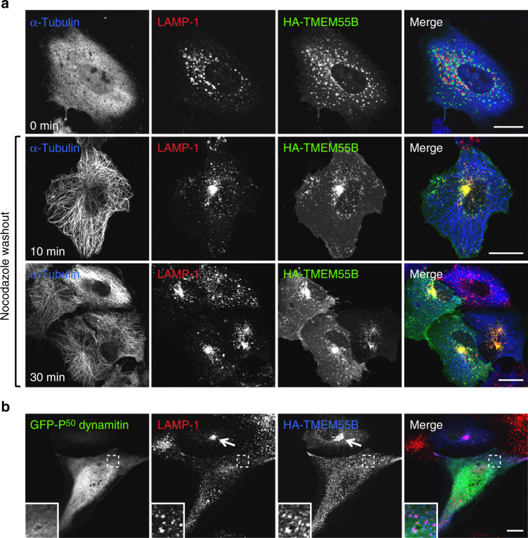
“disruption of the dynein–dynactin complex by overexpression of the dynactin complex subunit P50-dynamitin was sufficient to completely block TMEM55B-induced lysosome clustering (Fig. 2b).”
§ TFEB regulates lysosomal positioning by modulating TMEM55B expression and JIP4 recruitment to lysosomes > Results > TMEM55B promotes lysosomal retrograde transport
plaindirect- Asserted: passes — The authors put it forward as true.
- Checkable: passes — It can be checked against evidence.
- Load-bearing: passes — The paper’s conclusions depend on it.
- Based: passes — The paper gives a reason why it holds.
A result the paper reports from its own data — it cites no other source for it, so there is none to check. The quote shows it is in the paper.
In the paper
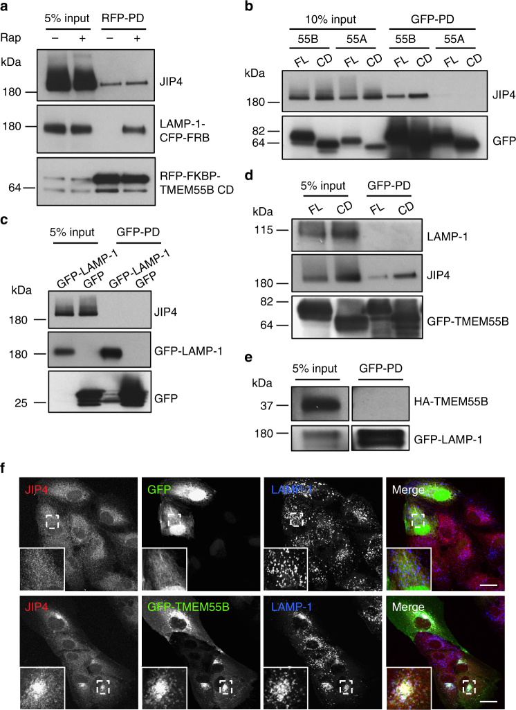
“overexpression of GFP-TMEM55B resulted in a very noticeable accumulation of JIP4 on clustered lysosomes (Fig. 4f)”
§ TFEB regulates lysosomal positioning by modulating TMEM55B expression and JIP4 recruitment to lysosomes > Results > TMEM55B recruits JIP4 to lysosomal membranes
plaindirect- Asserted: passes — The authors put it forward as true.
- Checkable: passes — It can be checked against evidence.
- Load-bearing: passes — The paper’s conclusions depend on it.
- Based: passes — The paper gives a reason why it holds.
A result the paper reports from its own data — it cites no other source for it, so there is none to check. The quote shows it is in the paper.
In the paper
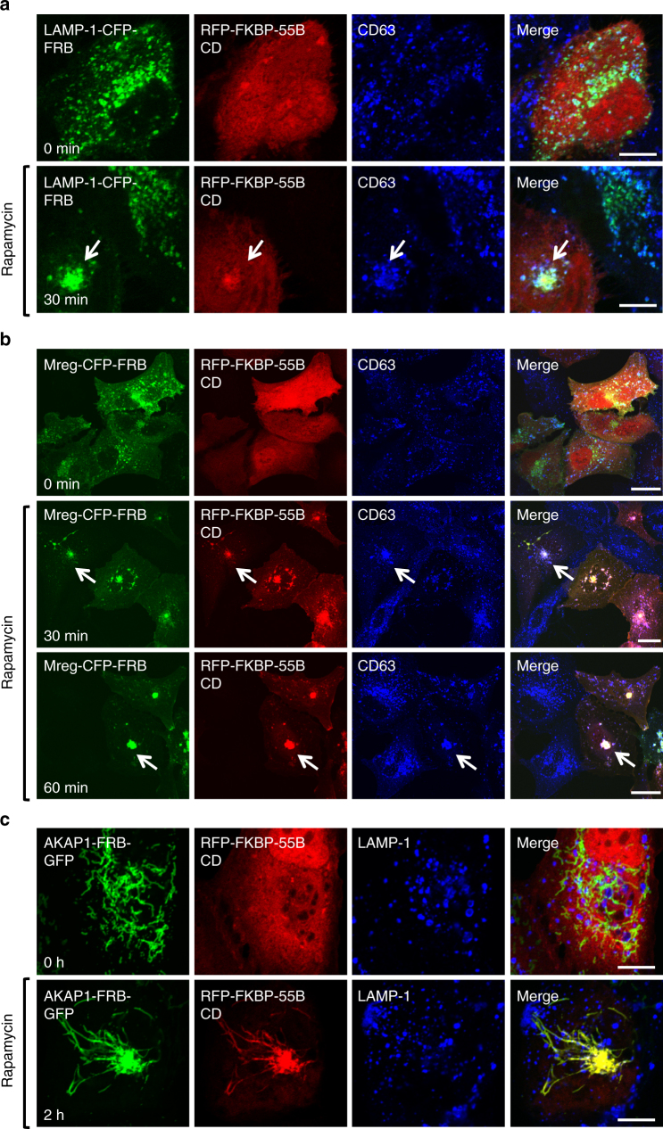
“Mitochondria expressing the targeting sequence AKAP1-FRB-GFP experienced dramatic changes in mitochondria morphology after recruitment of RFP-FKBP-TMEM55B CD to outer membranes, changing from dispersed, inter-connected membranes (Fig. 3c, top panel) to a tight cluster in the cell center (Fig. 3c, bottom panel).”
§ TFEB regulates lysosomal positioning by modulating TMEM55B expression and JIP4 recruitment to lysosomes > Results > TMEM55B CD induces organelle retrograde transport
plaindirect- Asserted: passes — The authors put it forward as true.
- Checkable: passes — It can be checked against evidence.
- Load-bearing: passes — The paper’s conclusions depend on it.
- Based: passes — The paper gives a reason why it holds.
- Probably not a substantial claim. 24
- It may state two separate claims as one. 32
- The quote only partly states this claim. 1
The cited sources support only part of this claim — read each source’s verdict below.
In the paper
“under starvation conditions, autophagosomes and lysosomes move toward the cell center, facilitating the fusion between both organelles and degradation of the autophagosomal content9,10.”
§ TFEB regulates lysosomal positioning by modulating TMEM55B expression and JIP4 recruitment to lysosomes > Introduction
What the sources say
- Partly supportedcites [9] Kimura, S., Noda, T. & Yoshimori, T. Dynein-dependent movement of autophagosomes mediates efficient encounters with lysosomes. Cell Struct. Funct. 33, 109–122 (2008). sourceExperimental — accuracy not yet measured
The sections “Autophagosomes move toward the cell center” and “Significance of the rapid vectorial movement for autophagy” support movement of autophagosomes toward centrally clustered lysosomes and its role in efficient targeting. But “Anti-LC3 antibody microinjection inhibits autophagosome movement” says lysosome localization did not significantly change under starvation; the paper does not support the claim that lysosomes also move toward the cell center under those conditions. Page numbers are unavailable in our copy. (LLM, conf=0.90)
- Supportedcites [10] Korolchuk, V. I. et al. Lysosomal positioning coordinates cellular nutrient responses. Nat. Cell Biol. 13, 453–460 (2011). source
The paper states that starvation causes lysosomes to cluster near the microtubule-organizing center, facilitating fusion with autophagosomes (Abstract, page unavailable). It also describes autophagosomes moving toward that center and reports increased autolysosome formation during starvation. The paper does not say starvation itself causes autophagosomes to move inward, but the claim does not require that causal interpretation. (LLM, conf=0.90)
plaindirect- Asserted: passes — The authors put it forward as true.
- Checkable: passes — It can be checked against evidence.
- Load-bearing: passes — The paper’s conclusions depend on it.
- Based: passes — The paper gives a reason why it holds.
Probably not a substantial claim.
- Partly supported
The cited sources support only part of this claim — read each source’s verdict below.
In the paper
“Redistribution of lysosomes toward the cell periphery is critical for cancer growth, invasion and metastasis.”
§ TFEB regulates lysosomal positioning by modulating TMEM55B expression and JIP4 recruitment to lysosomes > Introduction
What the sources say
- Not checked — no full text of the sourcecites [11] Nishimura, Y., Itoh, K., Yoshioka, K., Tokuda, K. & Himeno, M. Overexpression of ROCK in human breast cancer cells: evidence that ROCK activity mediates intracellular membrane traffic of lysosomes. Pathol. Oncol. Res. 9, 83–95 (2003). source
- Not checked — no full text of the sourcecites [12] Macpherson, I. R. et al. CLIC3 controls recycling of late endosomal MT1-MMP and dictates invasion and metastasis in breast cancer. J. Cell Sci. 127, 3893–3901 (2014). source
- Not checked yetcites [13] Marchesin, V. et al. ARF6-JIP3/4 regulate endosomal tubules for MT1-MMP exocytosis in cancer invasion. J. Cell Biol. 211, 339–358 (2015). source
Checked before — its earlier verdict is held back until a new check runs.
- Partly supportedcites [14] Dykes, S. S. et al. The Arf-like GTPase Arl8b is essential for three-dimensional invasive growth of prostate cancer in vitro and xenograft formation and growth in vivo. Oncotarget 7, 31037–31052 (2016). sourceExperimental — accuracy not yet measured
The Introduction says peripheral lysosome trafficking facilitates protease secretion and invasion, and the Discussion links it to invasive growth and, ultimately, metastasis (pages not provided). But the Discussion distinguishes this trafficking role from Arl8b’s metabolic role in tumor growth; the paper does not establish that peripheral redistribution itself is critical for cancer growth or directly demonstrate metastasis. (LLM, conf=0.86)
plaindirect- Asserted: passes — The authors put it forward as true.
- Checkable: passes — It can be checked against evidence.
- Load-bearing: passes — The paper’s conclusions depend on it.
- Based: passes — The paper gives a reason why it holds.
Probably not a substantial claim.
The cited sources support only part of this claim — read each source’s verdict below.
In the paper
“kinesin-dependent tubulation of lysosomes is required for efficient MHCII-mediated antigen presentation in dendritic cells16–18.”
§ TFEB regulates lysosomal positioning by modulating TMEM55B expression and JIP4 recruitment to lysosomes > Introduction
What the sources say
- Not checked — no full text of the sourcecites [16] Chow, A., Toomre, D., Garrett, W. & Mellman, I. Dendritic cell maturation triggers retrograde MHC class II transport from lysosomes to the plasma membrane. Nature 418, 988–994 (2002). source
- Partly supportedcites [17] Vyas, J. M. et al. Tubulation of class II MHC compartments is microtubule dependent and involves multiple endolysosomal membrane proteins in primary dendritic cells. J. Immunol. 178, 7199–7210 (2007). sourceExperimental — accuracy not yet measured
The Discussion (page unavailable) reports that endolysosomal tubules carry class II MHC molecules toward the cell surface and depend on microtubules. It says kinesin is a possible motor but that direct evidence is lacking. The paper also does not establish that tubulation is required for efficient MHCII-mediated antigen presentation. (LLM, conf=0.88)
- Partly supportedcites [18] Mrakovic, A., Kay, J. G., Furuya, W., Brumell, J. H. & Botelho, R. J. Rab7 and Arl8 GTPases are necessary for lysosome tubulation in macrophages. Traffic 13, 1667–1679 (2012). sourceExperimental — accuracy not yet measured
The paper reports in “Dynein and kinesin are required for lysosome tubulation” (page unavailable) that disrupting kinesin reduced tubular lysosomes in macrophages. Its Discussion (page unavailable) says tubular MHCII compartments in dendritic cells have been proposed to deliver antigen-loaded MHCII to immune synapses. It does not establish that kinesin-dependent tubulation is required for efficient antigen presentation in dendritic cells. (LLM, conf=0.89)
plaindirect- Asserted: passes — The authors put it forward as true.
- Checkable: passes — It can be checked against evidence.
- Load-bearing: fails — The paper’s conclusions may not depend on it.
- Based: passes — The paper gives a reason why it holds.
Probably not a substantial claim. The paper’s conclusions may not depend on it.
The cited sources support only part of this claim — read each source’s verdict below.
In the paper
“Anterograde movement of lysosomes is regulated by the multi-subunit complex BORC, the small GTPase Arl8, and its effector SKIP, which directly interacts with the kinesin light chain, thus linking lysosomes to the plus-end-directed microtubule motor kinesin19,20.”
§ TFEB regulates lysosomal positioning by modulating TMEM55B expression and JIP4 recruitment to lysosomes > Introduction
What the sources say
- Partly supportedcites [19] Rosa-Ferreira, C. & Munro, S. Ar18 and SKIP act together to link lysosomes to kinesin-1. Dev. Cell 21, 1171–1178 (2011). sourceExperimental — accuracy not yet measured
The Discussion states that Arl8 and SKIP recruit kinesin-1 to lysosomes and direct movement toward microtubule plus ends; it also says SKIP binds kinesin light chain. The cited paper does not identify BORC as a regulator of this movement (searched: BORC), so that part of the attributed claim is unsupported. (LLM, conf=0.88)
- Supportedcites [20] Pu, J. et al. BORC, a multisubunit complex that regulates lysosome positioning. Dev. Cell 33, 176–188 (2015). source
The Results section on Kinesin-1-dependent movement states that BORC engages the Arl8b–SKIP–Kinesin-1 pathway to regulate outward lysosome movement. The same section identifies a kinesin light-chain-binding sequence in SKIP that links lysosomes to Kinesin-1; the Discussion describes BORC recruiting Arl8b to enable that coupling. (LLM, conf=0.92)
plaindirect- Asserted: passes — The authors put it forward as true.
- Checkable: passes — It can be checked against evidence.
- Load-bearing: fails — The paper’s conclusions may not depend on it.
- Based: passes — The paper gives a reason why it holds.
Probably not a substantial claim. The paper’s conclusions may not depend on it.
- Partly supported
The sources with a verdict support this claim; the rest weren’t checked yet, have no full text we could read, or their verdicts aren’t rated yet.
In the paper
“The transcription factors TFEB and TFE3 exhibit the unique ability to promote expression of multiple lysosomal genes and are considered master regulators of lysosomal biogenesis2,3.”
§ TFEB regulates lysosomal positioning by modulating TMEM55B expression and JIP4 recruitment to lysosomes > Introduction
What the sources say
- Not checked — no full text of the sourcecites [2] Sardiello, M. et al. A gene network regulating lysosomal biogenesis and function. Science 325, 473–477 (2009). source
- Supportedcites [3] Martina, J. A. et al. The nutrient-responsive transcription factor TFE3 promotes autophagy, lysosomal biogenesis, and clearance of cellular debris. Sci. Signal. 7, ra9 (2014). source
The Results report that TFE3 increased expression of 16 of 17 lysosomal genes tested. The Discussion says TFEB induces expression of multiple lysosomal genes and identifies TFE3 as another regulator of lysosomal biogenesis; the Results also describe either factor as a potential “master” regulator, depending on its relative abundance. (LLM, conf=0.92)
plaindirect- Asserted: passes — The authors put it forward as true.
- Checkable: passes — It can be checked against evidence.
- Load-bearing: fails — The paper’s conclusions may not depend on it.
- Based: passes — The paper gives a reason why it holds.
Probably not a substantial claim. The paper’s conclusions may not depend on it. It may state two separate claims as one.
The cited sources support only part of this claim — read each source’s verdict below.
In the paper
“LAMP-1 and LAMP-2 promote coupling of lysosomes to dynein–dynactin26,27”
§ TFEB regulates lysosomal positioning by modulating TMEM55B expression and JIP4 recruitment to lysosomes > Introduction
What the sources say
- Partly supportedcites [26] Huynh, K. K. et al. LAMP proteins are required for fusion of lysosomes with phagosomes. EMBO J. 26, 313–324 (2007). sourceExperimental — accuracy not yet measured
The Distribution and motility section reports reduced dynein-mediated lysosome transport in cells lacking LAMP-1 and LAMP-2. The Discussion proposes that LAMPs help lysosomes associate with dynein–dynactin, but says the physical link is unclear; it does not establish coupling as a demonstrated mechanism. No page numbers are available in our copy. (LLM, conf=0.86)
- Not checked — no full text of the sourcecites [27] Krzewski, K., Gil-Krzewska, A., Nguyen, V., Peruzzi, G. & Coligan, J. E. LAMP1/CD107a is required for efficient perforin delivery to lytic granules and NK-cell cytotoxicity. Blood 121, 4672–4683 (2013). source
plaindirect- Asserted: passes — The authors put it forward as true.
- Checkable: passes — It can be checked against evidence.
- Load-bearing: fails — The paper’s conclusions may not depend on it.
- Based: passes — The paper gives a reason why it holds.
Probably not a substantial claim. The paper’s conclusions may not depend on it.
- Partly supported
The cited sources support only part of this claim — read each source’s verdict below.
In the paper
“We could not detect robust hydrolysis of PtdIns-4,5-P2 by either TMEM55, though we could detect hydrolysis by SHIP2, despite the fact that this enzyme has by far the weakest activity against this substrate of the 5-phosphatases44 (Supplementary Fig. 8e).”
§ TFEB regulates lysosomal positioning by modulating TMEM55B expression and JIP4 recruitment to lysosomes > Results > Assessing TMEM55B phosphatidylinositol phosphatase activity
What the sources say
- Partly supportedcites [44] Tresaugues, L. et al. Structural basis for phosphoinositide substrate recognition, catalysis, and membrane interactions in human inositol polyphosphate 5-phosphatases. Structure 22, 744–755 (2014). sourceExperimental — accuracy not yet measured
The Results (page unavailable) report that SHIP2 had low activity relative to the other 5-phosphatases assayed, supporting the comparison attached to citation 44. The study tested soluble, short-chain phosphoinositide analogs and the isolated SHIP2 catalytic domain; it does not establish that intact SHIP2 has the weakest activity against native PtdIns(4,5)P2 among all 5-phosphatases. The TMEM55 observations are the citing authors’ own results, not a finding attributed to this source. (LLM, conf=0.84)
plain- Asserted: passes — The authors put it forward as true.
- Checkable: passes — It can be checked against evidence.
- Load-bearing: passes — The paper’s conclusions depend on it.
- Based: passes — The paper gives a reason why it holds.
It may state two separate claims as one.
- Partly supported
The cited sources support only part of this claim — read each source’s verdict below.
In the paper
“ALG-2 interacts with the lysosomal transient receptor potential channel MCOLN1 in a calcium-dependent manner to recruit dynein–dynactin to lysosomes28,29.”
§ TFEB regulates lysosomal positioning by modulating TMEM55B expression and JIP4 recruitment to lysosomes > Introduction
What the sources say
- Partly supportedcites [28] Li, X. et al. A molecular mechanism to regulate lysosome motility for lysosome positioning and tubulation. Nat. Cell Biol. 18, 404–417 (2016). sourceExperimental — accuracy not yet measured
The Results section states that ALG-2 interacts directly with TRPML1 (MCOLN1) in a Ca²⁺-dependent manner and pulls down dynamitin, a dynactin component. It proposes that ALG-2 may link lysosomal Ca²⁺ release to dynein motors, but does not establish the stronger claim that this interaction recruits dynein–dynactin to lysosomes. (LLM, conf=0.84)
- Partly supportedcites [29] Vergarajauregui, S., Martina, J. A. & Puertollano, R. Identification of the penta-EF-hand protein ALG-2 as a Ca2+-dependent interactor of mucolipin-1. J. Biol. Chem. 284, 36357–36366 (2009). sourceExperimental — accuracy not yet measured
The Abstract states that ALG-2 binds MCOLN1 in a Ca2+-dependent manner. The Discussion proposes that ALG-2 may recruit other proteins involved in endosomal fusion or fission, but does not identify dynein–dynactin or establish its recruitment to lysosomes. Page numbers are not available in our copy. (LLM, conf=0.90)
plaindirect- Asserted: passes — The authors put it forward as true.
- Checkable: passes — It can be checked against evidence.
- Load-bearing: fails — The paper’s conclusions may not depend on it.
- Based: passes — The paper gives a reason why it holds.
Probably not a substantial claim. The paper’s conclusions may not depend on it.
- Partly supported
The cited sources support only part of this claim — read each source’s verdict below.
In the paper
“Retrograde movement is regulated by Rab7 and its effector RILP. RILP interacts with the p150-glued subunit of dynactin, thus recruiting the minus-end-directed microtubule motor dynein to lysosomes23–25.”
§ TFEB regulates lysosomal positioning by modulating TMEM55B expression and JIP4 recruitment to lysosomes > Introduction
What the sources say
- Partly supportedcites [23] Cantalupo, G., Alifano, P., Roberti, V., Bruni, C. B. & Bucci, C. Rab-interacting lysosomal protein (RILP): the Rab7 effector required for transport to lysosomes. EMBO J. 20, 683–693 (2001). sourceExperimental — accuracy not yet measured
The Abstract and Discussion identify RILP as a Rab7 effector involved in late endocytic transport and lysosomal organization. The Discussion only proposes that RILP might link Rab7-containing organelles to a motor complex; it does not establish that RILP binds p150-Glued or recruits dynein to lysosomes. Page numbers are unavailable in our copy. (LLM, conf=0.88)
- Partly supportedcites [24] Johansson, M. et al. Activation of endosomal dynein motors by stepwise assembly of Rab7-RILP-p150Glued, ORP1L, and the receptor betalll spectrin. J. Cell Biol. 176, 459–471 (2007). sourceExperimental — accuracy not yet measured
The Discussion states that Rab7 acts through RILP to recruit dynein–dynactin for minus-end transport, and that RILP directly binds dynactin’s p150Glued subunit. The paper demonstrates this recruitment on late endosomes (LEs), rather than specifically on lysosomes; it also says ORP1L and βIII spectrin are needed for transport after recruitment. (LLM, conf=0.82)
- Partly supportedcites [25] Jordens, I. et al. The Rab7 effector protein RILP controls lysosomal transport by inducing the recruitment of dynein-dynactin motors. Curr. Biol. 11, 1680–1685 (2001). sourceExperimental — accuracy not yet measured
The supplied passages report that Rab7-associated RILP recruits dynein–dynactin complexes to late endosomes and lysosomes, producing minus-end-directed transport. They also report recruitment of p150glued, but the cited sentence’s claim that RILP interacts with that subunit is not established: the paper says its assays revealed no direct interaction with motor-complex subunits. (LLM, conf=0.88)
plaindirect- Asserted: passes — The authors put it forward as true.
- Checkable: passes — It can be checked against evidence.
- Load-bearing: fails — The paper’s conclusions may not depend on it.
- Based: passes — The paper gives a reason why it holds.
Probably not a substantial claim. The paper’s conclusions may not depend on it. It may state two separate claims as one.
- Partly supported
The cited sources support only part of this claim — read each source’s verdict below.
In the paper
“overexpression of Rab34, Rab36, Rabring7, or Rapsyn causes clustering of lysosomes in the perinuclear area.”
§ TFEB regulates lysosomal positioning by modulating TMEM55B expression and JIP4 recruitment to lysosomes > Introduction
What the sources say
- Partly supportedcites [30] Chen, L., Hu, J., Yun, Y. & Wang, T. Rab36 regulates the spatial distribution of late endosomes and lysosomes through a similar mechanism to Rab34. Mol. Membr. Biol. 27, 23–30 (2010). sourceExperimental — accuracy not yet measured
The Abstract and Figure 3 caption (page not provided) report that Rab36 overexpression clusters lysosomes around the Golgi in the perinuclear region. The Introduction describes Rab34 as regulating lysosome distribution near the Golgi, and Figure 3 compares Rab34 overexpression with Rab36. Our copy does not address Rabring7 or Rapsyn, so it does not support the full list. (LLM, conf=0.88)
- Not checked — no full text of the sourcecites [31] Wang, T. & Hong, W. Interorganellar regulation of lysosome positioning by the Golgi apparatus through Rab34 interaction with Rab-interacting lysosomal protein. Mol. Biol. Cell 13, 4317–4332 (2002). source
- Not checked — no full text of the sourcecites [32] Mizuno, K., Kitamura, A. & Sasaki, T. Rabring7, a novel Rab7 target protein with a RING finger motif. Mol. Biol. Cell 14, 3741–3752 (2003). source
- Not checked — no full text of the sourcecites [33] Aittaleb, M., Chen, P. J. & Akaaboune, M. Failure of lysosome clustering and positioning in the juxtanuclear region in cells deficient in rapsyn. J. Cell Sci. 128, 3744–3756 (2015). source
plaindirect- Asserted: passes — The authors put it forward as true.
- Checkable: passes — It can be checked against evidence.
- Load-bearing: fails — The paper’s conclusions may not depend on it.
- Based: passes — The paper gives a reason why it holds.
Probably not a substantial claim. The paper’s conclusions may not depend on it. It may state two separate claims as one.
- Partly supported
A result the paper reports from its own data — it cites no other source for it, so there is none to check. The quote shows it is in the paper.
In the paper
“TMEM55B or JIP4 depletion abolishes starvation-induced retrograde lysosomal transport and prevents autophagosome–lysosome fusion.”
§ TFEB regulates lysosomal positioning by modulating TMEM55B expression and JIP4 recruitment to lysosomes > Abstract
plaindirect- Asserted: passes — The authors put it forward as true.
- Checkable: passes — It can be checked against evidence.
- Load-bearing: passes — The paper’s conclusions depend on it.
- Based: passes — The paper gives a reason why it holds.
It may state two separate claims as one.
A result the paper reports from its own data — it cites no other source for it, so there is none to check. The quote shows it is in the paper.
In the paper
“A modest, although statistically significant, increase in JIP4 mRNA levels was also observed in nutrient-deprived cells (Supplementary Fig. 9a).”
§ TFEB regulates lysosomal positioning by modulating TMEM55B expression and JIP4 recruitment to lysosomes > Results > TFEB and TFE3 upregulate TMEM55B in response to starvation
plaindirect- Asserted: passes — The authors put it forward as true.
- Checkable: passes — It can be checked against evidence.
- Load-bearing: fails — The paper’s conclusions may not depend on it.
- Based: passes — The paper gives a reason why it holds.
Probably not a substantial claim. The paper’s conclusions may not depend on it.
None of the sources cited for this claim have been checked yet.
In the paper
“recruitment of INPP5E to the cell surface, which was used as a positive control43, induced a nearly complete loss of plasma membrane PtdIns-4,5-P2 (Supplementary Fig. 8c, d).”
§ TFEB regulates lysosomal positioning by modulating TMEM55B expression and JIP4 recruitment to lysosomes > Results > Assessing TMEM55B phosphatidylinositol phosphatase activity
What the sources say
- Not checked yetcites [43] Varnai, P., Thyagarajan, B., Rohacs, T. & Balla, T. Rapidly inducible changes in phosphatidylinositol 4,5-bisphosphate levels influence multiple regulatory functions of the lipid in intact living cells. J. Cell Biol. 175, 377–382 (2006). source
Checked before — its earlier verdict is held back until a new check runs.
plaindirect- Asserted: passes — The authors put it forward as true.
- Checkable: passes — It can be checked against evidence.
- Load-bearing: fails — The paper’s conclusions may not depend on it.
- Based: passes — The paper gives a reason why it holds.
Probably not a substantial claim. The paper’s conclusions may not depend on it.
- Not checked yet
A result the paper reports from its own data — it cites no other source for it, so there is none to check. The quote shows it is in the paper.
In the paper
“In agreement with this idea, we found that induction of acute stress by treatment with NaAsO2 or curcumin resulted in post-transcriptional regulation of TMEM55B, increased JIP4 and p150Glued recruitment to lysosomes, and rapid lysosomal retrograde transport via a mechanism that required JIP4 and p38 activation.”
§ TFEB regulates lysosomal positioning by modulating TMEM55B expression and JIP4 recruitment to lysosomes > Discussion
plaindirect- Asserted: passes — The authors put it forward as true.
- Checkable: passes — It can be checked against evidence.
- Load-bearing: passes — The paper’s conclusions depend on it.
- Based: passes — The paper gives a reason why it holds.
It may state two separate claims as one.
A result the paper reports from its own data — it cites no other source for it, so there is none to check. The quote shows it is in the paper.
In the paper
“Accordingly, incubation of MEFs in EBSS (starvation medium) caused significant MCOLN1 upregulation, a lysosomal protein and TFEB/TFE3 target used in this experiment as a positive control (Fig. 6e).”
§ TFEB regulates lysosomal positioning by modulating TMEM55B expression and JIP4 recruitment to lysosomes > Results > TFEB and TFE3 upregulate TMEM55B in response to starvation
plaindirect- Asserted: passes — The authors put it forward as true.
- Checkable: passes — It can be checked against evidence.
- Load-bearing: fails — The paper’s conclusions may not depend on it.
- Based: passes — The paper gives a reason why it holds.
Probably not a substantial claim. The paper’s conclusions may not depend on it.
A result the paper reports from its own data — it cites no other source for it, so there is none to check. The quote shows it is in the paper.
In the paper
“Golgi morphology and early endosome distribution was unaffected in cells depleted of either TMEM55B or JIP4 (Supplementary Fig. 5c, d), demonstrating the specificity of TMEM55B/JIP4 in regulating late-endosome/lysosome trafficking.”
§ TFEB regulates lysosomal positioning by modulating TMEM55B expression and JIP4 recruitment to lysosomes > Results > JIP4 depletion causes peripheral accumulation of lysosomes
boosted- Asserted: passes — The authors put it forward as true.
- Checkable: passes — It can be checked against evidence.
- Load-bearing: passes — The paper’s conclusions depend on it.
- Based: passes — The paper gives a reason why it holds.
It may state two separate claims as one.
A result the paper reports from its own data — it cites no other source for it, so there is none to check. The quote shows it is in the paper.
In the paper
“In cells depleted of TMEM55B the distribution significantly shifted, showing a decrease in the concentration of LAMP-1-positive structures in the perinuclear region, concomitant with a distinct increase in the number of lysosomes localized towards the cell periphery (Fig. 1g, h).”
§ TFEB regulates lysosomal positioning by modulating TMEM55B expression and JIP4 recruitment to lysosomes > Results > TMEM55B promotes lysosomal retrograde transport
plaindirect- Asserted: passes — The authors put it forward as true.
- Checkable: passes — It can be checked against evidence.
- Load-bearing: passes — The paper’s conclusions depend on it.
- Based: passes — The paper gives a reason why it holds.
It may state two separate claims as one.
A result the paper reports from its own data — it cites no other source for it, so there is none to check. The quote shows it is in the paper.
In the paper
“In control RNAi-treated cells, lysosomes were broadly distributed throughout the cell, with a slight concentration in the perinuclear region (Fig. 1g, h).”
§ TFEB regulates lysosomal positioning by modulating TMEM55B expression and JIP4 recruitment to lysosomes > Results > TMEM55B promotes lysosomal retrograde transport
plain- Asserted: passes — The authors put it forward as true.
- Checkable: passes — It can be checked against evidence.
- Load-bearing: passes — The paper’s conclusions depend on it.
- Based: passes — The paper gives a reason why it holds.
It may state two separate claims as one.
A result the paper reports from its own data — it cites no other source for it, so there is none to check. The quote shows it is in the paper.
In the paper
“Starvation also significantly increased TMEM55B mRNA levels, and this increase was TFEB/TFE3-dependent since it was not observed in TFEB/TFE3 double knockout MEFs (Fig. 6f).”
§ TFEB regulates lysosomal positioning by modulating TMEM55B expression and JIP4 recruitment to lysosomes > Results > TFEB and TFE3 upregulate TMEM55B in response to starvation
plaindirect- Asserted: passes — The authors put it forward as true.
- Checkable: passes — It can be checked against evidence.
- Load-bearing: passes — The paper’s conclusions depend on it.
- Based: passes — The paper gives a reason why it holds.
It may state two separate claims as one.
A result the paper reports from its own data — it cites no other source for it, so there is none to check. The quote shows it is in the paper.
In the paper
“Transport was exceedingly rapid, with retrograde movement detectable as early as 5 min following rapamycin induction (Supplementary Movie 1), and near complete clustering of endosomes after 30 min (Fig. 3b).”
§ TFEB regulates lysosomal positioning by modulating TMEM55B expression and JIP4 recruitment to lysosomes > Results > TMEM55B CD induces organelle retrograde transport
plaindirect- Asserted: passes — The authors put it forward as true.
- Checkable: passes — It can be checked against evidence.
- Load-bearing: unclear — It is unclear whether the paper’s conclusions depend on it.
- Based: passes — The paper gives a reason why it holds.
It may state two separate claims as one.
A result the paper reports from its own data — it cites no other source for it, so there is none to check. The quote shows it is in the paper.
In the paper
“we found that TMEM55B co-localized with the lysosomal marker LAMP-1 (Fig. 1b, top) but not with early endosome marker hepatocyte growth factor-regulated tyrosine kinase substrate (HRS) (Fig. 1b, bottom).”
§ TFEB regulates lysosomal positioning by modulating TMEM55B expression and JIP4 recruitment to lysosomes > Results > TMEM55B promotes lysosomal retrograde transport
plaincorrelational- Asserted: passes — The authors put it forward as true.
- Checkable: passes — It can be checked against evidence.
- Load-bearing: passes — The paper’s conclusions depend on it.
- Based: passes — The paper gives a reason why it holds.
It may state two separate claims as one.
A result the paper reports from its own data — it cites no other source for it, so there is none to check. The quote shows it is in the paper.
In the paper
“Interestingly, lysosomal clustering and JIP4 recruitment to lysosomes was prevented by incubation with p38 inhibitors, but not JNK or Erk1/2 inhibitors (Fig. 8g, h), thus revealing a novel and unexpected role of p38 in retrograde organelle transport.”
§ TFEB regulates lysosomal positioning by modulating TMEM55B expression and JIP4 recruitment to lysosomes > Results > MAPK-dependent regulation of the TMEM55B/JIP4 pathway
plaindirect- Asserted: passes — The authors put it forward as true.
- Checkable: passes — It can be checked against evidence.
- Load-bearing: passes — The paper’s conclusions depend on it.
- Based: passes — The paper gives a reason why it holds.
It may state two separate claims as one.
A result the paper reports from its own data — it cites no other source for it, so there is none to check. The quote shows it is in the paper.
In the paper
“full length TMEM55A, as well as its N-terminal domain (TMEM55A CD), lacked the ability to bind JIP4 (Fig. 4b).”
§ TFEB regulates lysosomal positioning by modulating TMEM55B expression and JIP4 recruitment to lysosomes > Results > TMEM55B recruits JIP4 to lysosomal membranes
plain- Asserted: passes — The authors put it forward as true.
- Checkable: passes — It can be checked against evidence.
- Load-bearing: passes — The paper’s conclusions depend on it.
- Based: passes — The paper gives a reason why it holds.
It may state two separate claims as one.
A result the paper reports from its own data — it cites no other source for it, so there is none to check. The quote shows it is in the paper.
In the paper
“JIP4-depleted cells exhibited almost complete peripheral accumulation of both lysosomes (Fig. 5a) and late endosomes (Supplementary Fig. 5c), with more than 60% of the LAMP-1 signal falling in the distal region of the cell (>15 μm from the nuclear rim) (Fig. 5b).”
§ TFEB regulates lysosomal positioning by modulating TMEM55B expression and JIP4 recruitment to lysosomes > Results > JIP4 depletion causes peripheral accumulation of lysosomes
plaindirect- Asserted: passes — The authors put it forward as true.
- Checkable: passes — It can be checked against evidence.
- Load-bearing: passes — The paper’s conclusions depend on it.
- Based: passes — The paper gives a reason why it holds.
It may state two separate claims as one.
A result the paper reports from its own data — it cites no other source for it, so there is none to check. The quote shows it is in the paper.
In the paper
“Interestingly, lysosomal clustering and JIP4 recruitment to lysosomes was prevented by incubation with p38 inhibitors, but not JNK or Erk1/2 inhibitors (Fig. 8g, h), thus revealing a novel and unexpected role of p38 in retrograde organelle transport.”
§ TFEB regulates lysosomal positioning by modulating TMEM55B expression and JIP4 recruitment to lysosomes > Results > MAPK-dependent regulation of the TMEM55B/JIP4 pathway
plain- Asserted: passes — The authors put it forward as true.
- Checkable: passes — It can be checked against evidence.
- Load-bearing: passes — The paper’s conclusions depend on it.
- Based: passes — The paper gives a reason why it holds.
It may state two separate claims as one.
A result the paper reports from its own data — it cites no other source for it, so there is none to check. The quote shows it is in the paper.
In the paper
“Treatment of U2OS cells with NaAsO2 resulted in a clear shift in TMEM55B electrophoretic mobility, as well as an increase in TMEM55B dimerization (Fig. 8e), suggesting that TMEM55B might undergo post-translational modifications in response to acute stress.”
§ TFEB regulates lysosomal positioning by modulating TMEM55B expression and JIP4 recruitment to lysosomes > Results > MAPK-dependent regulation of the TMEM55B/JIP4 pathway
boosteddirect- Asserted: passes — The authors put it forward as true.
- Checkable: passes — It can be checked against evidence.
- Load-bearing: unclear — It is unclear whether the paper’s conclusions depend on it.
- Based: passes — The paper gives a reason why it holds.
It may state two separate claims as one.
A result the paper reports from its own data — it cites no other source for it, so there is none to check. The quote shows it is in the paper.
In the paper
“Mutation of the Cx5R motif in TMEM55B full-length did not alter the ability of this protein to induce lysosomal clustering or interact with JIP4 (Supplementary Fig. 8a, b).”
§ TFEB regulates lysosomal positioning by modulating TMEM55B expression and JIP4 recruitment to lysosomes > Results > Assessing TMEM55B phosphatidylinositol phosphatase activity
plain- Asserted: passes — The authors put it forward as true.
- Checkable: passes — It can be checked against evidence.
- Load-bearing: passes — The paper’s conclusions depend on it.
- Based: passes — The paper gives a reason why it holds.
It may state two separate claims as one.
A result the paper reports from its own data — it cites no other source for it, so there is none to check. The quote shows it is in the paper.
In the paper
“alanine substitutions of Leucine10 and Leucine11, residues of a putative acidic-dileucine motif at the amino termini of TMEM55B, caused accumulation of the full-length protein at the cell surface, with a minor fraction co-localizing with LAMP-1-positive structures (Supplementary Fig. 2b).”
§ TFEB regulates lysosomal positioning by modulating TMEM55B expression and JIP4 recruitment to lysosomes > Results > TMEM55B CD induces organelle retrograde transport
plaindirect- Asserted: passes — The authors put it forward as true.
- Checkable: passes — It can be checked against evidence.
- Load-bearing: passes — The paper’s conclusions depend on it.
- Based: passes — The paper gives a reason why it holds.
It may state two separate claims as one.
A result the paper reports from its own data — it cites no other source for it, so there is none to check. The quote shows it is in the paper.
In the paper
“GFP-TMEM55B CD exhibited a cytosolic distribution, whereas TMEM55B TM localized predominantly to the plasma membrane.”
§ TFEB regulates lysosomal positioning by modulating TMEM55B expression and JIP4 recruitment to lysosomes > Results > TMEM55B CD induces organelle retrograde transport
plain- Asserted: passes — The authors put it forward as true.
- Checkable: passes — It can be checked against evidence.
- Load-bearing: passes — The paper’s conclusions depend on it.
- Based: passes — The paper gives a reason why it holds.
It may state two separate claims as one.
A result the paper reports from its own data — it cites no other source for it, so there is none to check. The quote shows it is in the paper.
In the paper
“As expected, TFEB and TFE3 caused a significant increase in TMEM55B mRNA and protein levels (Fig. 6a–c), thus confirming that these transcription factors modulate TMEM55B expression.”
§ TFEB regulates lysosomal positioning by modulating TMEM55B expression and JIP4 recruitment to lysosomes > Results > TFEB and TFE3 upregulate TMEM55B in response to starvation
boosteddirect- Asserted: passes — The authors put it forward as true.
- Checkable: passes — It can be checked against evidence.
- Load-bearing: passes — The paper’s conclusions depend on it.
- Based: passes — The paper gives a reason why it holds.
The quote only partly states this claim.
A result the paper reports from its own data — it cites no other source for it, so there is none to check. The quote shows it is in the paper.
In the paper
“JIP4 displayed no interaction with either RILP or ORP1L (Supplementary Fig. 6b)”
§ TFEB regulates lysosomal positioning by modulating TMEM55B expression and JIP4 recruitment to lysosomes > Results > JIP4 depletion causes peripheral accumulation of lysosomes
plain- Asserted: passes — The authors put it forward as true.
- Checkable: passes — It can be checked against evidence.
- Load-bearing: passes — The paper’s conclusions depend on it.
- Based: passes — The paper gives a reason why it holds.
It may state two separate claims as one.
A result the paper reports from its own data — it cites no other source for it, so there is none to check. The quote shows it is in the paper.
In the paper
“we detected binding of both proteins to endogenous JIP4 (Fig. 4b).”
§ TFEB regulates lysosomal positioning by modulating TMEM55B expression and JIP4 recruitment to lysosomes > Results > TMEM55B recruits JIP4 to lysosomal membranes
plaincorrelational- Asserted: passes — The authors put it forward as true.
- Checkable: passes — It can be checked against evidence.
- Load-bearing: passes — The paper’s conclusions depend on it.
- Based: passes — The paper gives a reason why it holds.
It may state two separate claims as one.
In the paper
“Lysosome retrograde transport toward the immunological synapse is essential for the ability of natural killer and cytotoxic T lymphocytes to kill pathogen infected cells15”
§ TFEB regulates lysosomal positioning by modulating TMEM55B expression and JIP4 recruitment to lysosomes > Introduction
What the sources say
- Not checked — no full text of the sourcecites [15] Stinchcombe, J. C., Majorovits, E., Bossi, G., Fuller, S. & Griffiths, G. M. Centrosome polarization delivers secretory granules to the immunological synapse. Nature 443, 462–465 (2006). source
plaindirect- Asserted: passes — The authors put it forward as true.
- Checkable: passes — It can be checked against evidence.
- Load-bearing: fails — The paper’s conclusions may not depend on it.
- Based: passes — The paper gives a reason why it holds.
Probably not a substantial claim. The paper’s conclusions may not depend on it.
What the paper did — it cites no source for this, so there is none to check. The quote shows it is in the paper.
In the paper
“Thirty or more cells were quantified per cell treatment and averaged to quantify the population lysosome distribution.”
§ TFEB regulates lysosomal positioning by modulating TMEM55B expression and JIP4 recruitment to lysosomes > Methods > Lysosome distribution quantification
plain- Asserted: passes — The authors put it forward as true.
- Checkable: passes — It can be checked against evidence.
- Load-bearing: passes — The paper’s conclusions depend on it.
- Based: passes — The paper gives a reason why it holds.
Probably not a substantial claim.
What the paper did — it cites no source for this, so there is none to check. The quote shows it is in the paper.
In the paper
“No portion of the cell was measured more than once.”
§ TFEB regulates lysosomal positioning by modulating TMEM55B expression and JIP4 recruitment to lysosomes > Methods > Lysosome distribution quantification
plain- Asserted: passes — The authors put it forward as true.
- Checkable: passes — It can be checked against evidence.
- Load-bearing: fails — The paper’s conclusions may not depend on it.
- Based: passes — The paper gives a reason why it holds.
Probably not a substantial claim. The paper’s conclusions may not depend on it.
A conclusion the paper draws from its own results — it cites no other source for it, so there is none to check. The quote shows it is in the paper.
In the paper
“In this regard, the TMEM55B/JIP4/dynein–dynactin pathway is particularly interesting, since it exhibits a dual function. It regulates the constitutive bi-directional transport of lysosomes but it also acts “on-demand” to promote retrograde lysosomal transport under certain stress conditions.”
§ TFEB regulates lysosomal positioning by modulating TMEM55B expression and JIP4 recruitment to lysosomes > Discussion
plaindirect- Asserted: passes — The authors put it forward as true.
- Checkable: passes — It can be checked against evidence.
- Load-bearing: passes — The paper’s conclusions depend on it.
- Based: passes — The paper gives a reason why it holds.
It may state two separate claims as one.
- Rests on an assumption: Lysosomes undergo bidirectional transport along microtubules through the opposite activities of dynein and kinesins.
A conclusion the paper draws from its own results — it cites no other source for it, so there is none to check. The quote shows it is in the paper.
In the paper
“Increased TMEM55B levels will favor movement of lysosomes toward the center of the cell, promoting fusion between lysosomes and autophagosomes to ensure efficient autophagic flux.”
§ TFEB regulates lysosomal positioning by modulating TMEM55B expression and JIP4 recruitment to lysosomes > Discussion
hedgeddirect- Asserted: passes — The authors put it forward as true.
- Checkable: passes — It can be checked against evidence.
- Load-bearing: passes — The paper’s conclusions depend on it.
- Based: passes — The paper gives a reason why it holds.
It may state two separate claims as one.
- Rests on an assumption: Lysosome transport to the perinuclear region following starvation is critical to facilitate autophagosome–lysosome interactions and controls the rate of autophagosome degradation.
A conclusion the paper draws from its own results — it cites no other source for it, so there is none to check. The quote shows it is in the paper.
In the paper
“Based on these results, we concluded that TMEM55B CD is both necessary and sufficient to drive retrograde trafficking of TMEM55B-targeted membranes, and does so by linking organelles to dynein motors.”
§ TFEB regulates lysosomal positioning by modulating TMEM55B expression and JIP4 recruitment to lysosomes > Results > TMEM55B CD induces organelle retrograde transport
plaindirect- Asserted: passes — The authors put it forward as true.
- Checkable: passes — It can be checked against evidence.
- Load-bearing: passes — The paper’s conclusions depend on it.
- Based: passes — The paper gives a reason why it holds.
It may state two separate claims as one.
A conclusion the paper draws from its own results — it cites no other source for it, so there is none to check. The quote shows it is in the paper.
In the paper
“The resulting increase in TMEM55B levels promoted transport of lysosomes to the perinuclear region and likely facilitated lysosome/ER interactions.”
§ TFEB regulates lysosomal positioning by modulating TMEM55B expression and JIP4 recruitment to lysosomes > Discussion
hedgeddirect- Asserted: passes — The authors put it forward as true.
- Checkable: passes — It can be checked against evidence.
- Load-bearing: passes — The paper’s conclusions depend on it.
- Based: passes — The paper gives a reason why it holds.
It may state two separate claims as one.
- Rests on an assumption: Free cholesterol is transferred from lysosomes to other cellular compartments in a process that requires transport and tethering of late endosomes/lysosomes with ER membranes.
What the paper did — it cites no source for this, so there is none to check. The quote shows it is in the paper.
In the paper
“Amplification of the sequence of interest was normalized with a reference endogenous gene Glyceraldehyde 3-phosphate dehydrogenase (GAPDH).”
§ TFEB regulates lysosomal positioning by modulating TMEM55B expression and JIP4 recruitment to lysosomes > Methods > RNA isolation and relative quantitative real-time PCR
plain- Asserted: passes — The authors put it forward as true.
- Checkable: passes — It can be checked against evidence.
- Load-bearing: fails — The paper’s conclusions may not depend on it.
- Based: passes — The paper gives a reason why it holds.
Probably not a substantial claim. The paper’s conclusions may not depend on it.
What the paper did — it cites no source for this, so there is none to check. The quote shows it is in the paper.
In the paper
“Twenty or more cells were quantified per cell treatment and averaged to quantify the population lysosome distribution.”
§ TFEB regulates lysosomal positioning by modulating TMEM55B expression and JIP4 recruitment to lysosomes > Methods > Lysosome distribution quantification
plain- Asserted: passes — The authors put it forward as true.
- Checkable: passes — It can be checked against evidence.
- Load-bearing: passes — The paper’s conclusions depend on it.
- Based: passes — The paper gives a reason why it holds.
Probably not a substantial claim.
What the paper did — it cites no source for this, so there is none to check. The quote shows it is in the paper.
In the paper
“Line scans were drawn in various regions of the cell to accurately sample total LAMP-1 distribution.”
§ TFEB regulates lysosomal positioning by modulating TMEM55B expression and JIP4 recruitment to lysosomes > Methods > Lysosome distribution quantification
plain- Asserted: passes — The authors put it forward as true.
- Checkable: passes — It can be checked against evidence.
- Load-bearing: passes — The paper’s conclusions depend on it.
- Based: passes — The paper gives a reason why it holds.
Probably not a substantial claim.
A conclusion the paper draws from its own results — it cites no other source for it, so there is none to check. The quote shows it is in the paper.
In the paper
“Inhibition of cholesterol efflux from lysosomes in U18666A-treated cells was sensed by different cellular compartments, promoting activation of SREBF2 at the ER and TFEB/3 at the lysosomes.”
§ TFEB regulates lysosomal positioning by modulating TMEM55B expression and JIP4 recruitment to lysosomes > Discussion
plaindirect- Asserted: passes — The authors put it forward as true.
- Checkable: passes — It can be checked against evidence.
- Load-bearing: passes — The paper’s conclusions depend on it.
- Based: passes — The paper gives a reason why it holds.
It may state two separate claims as one.
- Rests on an assumption: LDL-bound cholesterol enters cells via endocytosis and is delivered to lysosomes to be converted into unesterified cholesterol.
- Rests on an assumption: Free cholesterol is transferred from lysosomes to other cellular compartments in a process that requires transport and tethering of late endosomes/lysosomes with ER membranes.
A conclusion the paper draws from its own results — it cites no other source for it, so there is none to check. The quote shows it is in the paper.
In the paper
“Thus, it was concluded that TMEM55B clusters lysosomes by recruiting the microtubule minus-end motor dynein to lysosomes, disrupting the balance of positive- and minus-end microtubule motors directing lysosomal trafficking.”
§ TFEB regulates lysosomal positioning by modulating TMEM55B expression and JIP4 recruitment to lysosomes > Results > TMEM55B promotes lysosomal retrograde transport
boosteddirect- Asserted: passes — The authors put it forward as true.
- Checkable: passes — It can be checked against evidence.
- Load-bearing: passes — The paper’s conclusions depend on it.
- Based: passes — The paper gives a reason why it holds.
It may state two separate claims as one.
- Rests on an assumption: In non-polarized cells, microtubule minus-ends localize to the perinuclear region, close to the MTOC, whereas plus-ends are directed toward the cell periphery.
None of the sources cited for this claim have been checked yet.
In the paper
“a tripartite complex between Rab7, FYCO1, and kinesin has also been shown to promote outward lysosomal movement21,22.”
§ TFEB regulates lysosomal positioning by modulating TMEM55B expression and JIP4 recruitment to lysosomes > Introduction
What the sources say
- Not checked yetcites [21] Matsuzaki, F., Shirane, M., Matsumoto, M. & Nakayama, K. I. Protrudin serves as an adaptor molecule that connects KIF5 and its cargoes in vesicular transport during process formation. Mol. Biol. Cell 22, 4602–4620 (2011). source
Checked before — its earlier verdict is held back until a new check runs.
- Not checked — no full text of the sourcecites [22] Raiborg, C., Wenzel, E. M., Pedersen, N. M. & Stenmark, H. ER-endosome contact sites in endosome positioning and protrusion outgrowth. Biochem. Soc. Trans. 44, 441–446 (2016). source
plaindirect- Asserted: passes — The authors put it forward as true.
- Checkable: passes — It can be checked against evidence.
- Load-bearing: fails — The paper’s conclusions may not depend on it.
- Based: passes — The paper gives a reason why it holds.
Probably not a substantial claim. The paper’s conclusions may not depend on it.
- Not checked yet
What the paper did — it cites no source for this, so there is none to check. The quote shows it is in the paper.
In the paper
“Western blots are representative of three or more independent experiments.”
§ TFEB regulates lysosomal positioning by modulating TMEM55B expression and JIP4 recruitment to lysosomes > Methods > Electrophoresis and immunoblotting
plain- Asserted: passes — The authors put it forward as true.
- Checkable: passes — It can be checked against evidence.
- Load-bearing: fails — The paper’s conclusions may not depend on it.
- Based: passes — The paper gives a reason why it holds.
Probably not a substantial claim. The paper’s conclusions may not depend on it.
A conclusion the paper draws from its own results — it cites no other source for it, so there is none to check. The quote shows it is in the paper.
In the paper
“Therefore, our results suggest that TMEM55B does not function as a bona fide PtdIns-4,5-P2 4-phosphatase and its role in retrograde trafficking is independent of its Cx5R motif.”
§ TFEB regulates lysosomal positioning by modulating TMEM55B expression and JIP4 recruitment to lysosomes > Results > Assessing TMEM55B phosphatidylinositol phosphatase activity
hedged- Asserted: passes — The authors put it forward as true.
- Checkable: passes — It can be checked against evidence.
- Load-bearing: passes — The paper’s conclusions depend on it.
- Based: passes — The paper gives a reason why it holds.
It may state two separate claims as one.
What the paper did — it cites no source for this, so there is none to check. The quote shows it is in the paper.
In the paper
“For each measurement, blank measurements (20 µM substrate with no enzyme, as well as 250 ng protein with no substrate) were subtracted.”
§ TFEB regulates lysosomal positioning by modulating TMEM55B expression and JIP4 recruitment to lysosomes > Methods > Malachite green phosphatase assay
plain- Asserted: passes — The authors put it forward as true.
- Checkable: passes — It can be checked against evidence.
- Load-bearing: fails — The paper’s conclusions may not depend on it.
- Based: passes — The paper gives a reason why it holds.
Probably not a substantial claim. The paper’s conclusions may not depend on it.
What the paper did — it cites no source for this, so there is none to check. The quote shows it is in the paper.
In the paper
“Cell images are representative from three or more independent experiments.”
§ TFEB regulates lysosomal positioning by modulating TMEM55B expression and JIP4 recruitment to lysosomes > Methods > Immunofluorescence staining
plain- Asserted: passes — The authors put it forward as true.
- Checkable: passes — It can be checked against evidence.
- Load-bearing: fails — The paper’s conclusions may not depend on it.
- Based: passes — The paper gives a reason why it holds.
Probably not a substantial claim. The paper’s conclusions may not depend on it.
In the paper
“JIP4 is a scaffold protein that interacts with c-Jun NH2-terminal kinase (JNK) and functions as an activator of the p38 mitogen-activated protein (MAP) kinase pathway56, suggesting that JIP4 might link transport of lysosomes to signaling.”
§ TFEB regulates lysosomal positioning by modulating TMEM55B expression and JIP4 recruitment to lysosomes > Discussion
What the sources say
- Not checked — no full text of the sourcecites [56] Kelkar, N., Standen, C. L. & Davis, R. J. Role of the JIP4 scaffold protein in the regulation of mitogen-activated protein kinase signaling pathways. Mol. Cell Biol. 25, 2733–2743 (2005). source
plaindirect- Asserted: passes — The authors put it forward as true.
- Checkable: passes — It can be checked against evidence.
- Load-bearing: passes — The paper’s conclusions depend on it.
- Based: passes — The paper gives a reason why it holds.
It may state two separate claims as one.
What the paper did — it cites no source for this, so there is none to check. The quote shows it is in the paper.
In the paper
“Experiments involving siRNA-induced knockdown of indicated proteins are representative of three or more independent experiments.”
§ TFEB regulates lysosomal positioning by modulating TMEM55B expression and JIP4 recruitment to lysosomes > Methods > RNA interference
plain- Asserted: passes — The authors put it forward as true.
- Checkable: passes — It can be checked against evidence.
- Load-bearing: passes — The paper’s conclusions depend on it.
- Based: passes — The paper gives a reason why it holds.
Probably not a substantial claim.
A conclusion the paper draws from its own results — it cites no other source for it, so there is none to check. The quote shows it is in the paper.
In the paper
“Here we postulate that the interaction of TMEM55B with the JIP4–dynactin complex helps bring dynein to the lysosomal surface, thus promoting retrograde transport.”
§ TFEB regulates lysosomal positioning by modulating TMEM55B expression and JIP4 recruitment to lysosomes > Discussion
hedgeddirect- Asserted: fails — The authors may not put it forward as true.
- Checkable: passes — It can be checked against evidence.
- Load-bearing: passes — The paper’s conclusions depend on it.
- Based: fails — The paper may give no reason why it holds.
Probably not a substantial claim. The authors may not put it forward as true. The paper may give no reason why it holds.
A conclusion the paper draws from its own results — it cites no other source for it, so there is none to check. The quote shows it is in the paper.
In the paper
“Altogether, these results indicate that TMEM55B CD is required for targeting to lysosomes, and that a significant fraction of TMEM55B traffics through the plasma membrane before reaching the endo-lysosomal compartment.”
§ TFEB regulates lysosomal positioning by modulating TMEM55B expression and JIP4 recruitment to lysosomes > Results > TMEM55B CD induces organelle retrograde transport
hedgeddirect- Asserted: passes — The authors put it forward as true.
- Checkable: passes — It can be checked against evidence.
- Load-bearing: passes — The paper’s conclusions depend on it.
- Based: passes — The paper gives a reason why it holds.
It may state two separate claims as one.
A conclusion the paper draws from its own results — it cites no other source for it, so there is none to check. The quote shows it is in the paper.
In the paper
“These results suggest that the full-length protein is required for proper localization to the endo-lysosomal compartment and that one or more endo/lysosomal targeting motifs likely reside within TMEM55B CD.”
§ TFEB regulates lysosomal positioning by modulating TMEM55B expression and JIP4 recruitment to lysosomes > Results > TMEM55B CD induces organelle retrograde transport
hedgeddirect- Asserted: passes — The authors put it forward as true.
- Checkable: passes — It can be checked against evidence.
- Load-bearing: passes — The paper’s conclusions depend on it.
- Based: passes — The paper gives a reason why it holds.
It may state two separate claims as one.
The paper relies on this without support.
Starvation activates TFEB and TFE3.
“TMEM55B levels are transcriptionally upregulated following TFEB and TFE3 activation by starvation or cholesterol-induced lysosomal stress.”
The paper relies on this without support.
Cholesterol accumulation in lysosomes activates TFEB and TFE3.
“TMEM55B levels are transcriptionally upregulated following TFEB and TFE3 activation by starvation or cholesterol-induced lysosomal stress.”
The paper relies on this without support.
In non-polarized cells, microtubule minus-ends localize to the perinuclear region, close to the MTOC, whereas plus-ends are directed toward the cell periphery.
“In non-polarized cells, microtubule minus-ends generally localize to the perinuclear region, close to the microtubule-organizing center (MTOC), whereas the microtubule plus-ends are directed toward the cell periphery.”
The paper relies on this without support.
JIP4 is a dynein adaptor/scaffold that links cargo to dynein–dynactin.
“we identified the dynein scaffold JIP4 as a major interacting protein of TMEM55B CD”
The paper relies on this without support.
Lysosome transport to the perinuclear region following starvation is critical to facilitate autophagosome–lysosome interactions and controls the rate of autophagosome degradation.
“Lysosome transport to the perinuclear region following starvation is critical to facilitate autophagosome–lysosome interactions and controls the rate of autophagosome degradation.”
The paper relies on this without support.
U18666A mimics the lack of functional Niemann-Pick type C protein responsible for NPC disease.
“It is well established that U18666A mimics the lack of functional Niemann-Pick type C protein responsible for the NPC disease.”
The paper relies on this without support.
In NPC1 and other lysosomal diseases, persistent accumulation of cholesterol in late endosomes/lysosomes leads to aberrant lysosomal clustering in the cell center.
“In NPC1 and other lysosomal diseases, the persistent accumulation of cholesterol in late endosomes/lysosomes leads to aberrant lysosomal clustering in the cell center.”
The paper relies on this without support.
Lysosomes undergo bidirectional transport along microtubules through the opposite activities of dynein and kinesins.
“Lysosomes undergo bidirectional transport along microtubules through the opposite activities of dynein and kinesins.”
The paper relies on this without support.
LDL-bound cholesterol enters cells via endocytosis and is delivered to lysosomes to be converted into unesterified cholesterol.
“LDL-bound cholesterol enters cells via endocytosis and it is delivered to lysosomes to be converted into unesterified cholesterol.”
The paper relies on this without support.
Free cholesterol is transferred from lysosomes to other cellular compartments in a process that requires transport and tethering of late endosomes/lysosomes with ER membranes.
“Free cholesterol is then transferred from lysosomes to other cellular compartments in a process that requires transport and tethering of late endosomes/lysosomes with the endoplasmic reticulum (ER) membranes.”
Claims other papers make about this one
Each is checked against this paper’s own text: does it say what the citing paper says it does?
The source backs part of the claim, not all of it — or backs it for a different population, condition or comparison.
In the citing paper
“JIP4-mediated retrograde transport of lysosomes facilitates interaction and fusion between autophagosomes and lysosomes, resulting in degradation of autophagosomal content and restoration of energy homeostasis7–9.”
From TMEM55B links autophagy flux, lysosomal repair, and TFE3 activation in response to oxidative stress (2024) · cites this paper as [7]
Experimental — accuracy not yet measuredThe source backs part of the claim, not all of it — or backs it for a different population, condition or comparison.
In the citing paper
“Interestingly, while TMEM55B expression is dependent on TFEB (Willett et al., 2017), TMEM55B can actually inhibit TFEB nuclear translocation (Hashimoto et al., 2018) thus suppressing expression of lysosomal genes.”
From Hippocampal TMEM55B overexpression in the 5XFAD mouse model of Alzheimer's disease (2024) · cites this paper as Willett et al., 2017
Experimental — accuracy not yet measuredThe cited source says what the claim attributes to it.
In the citing paper
“TMEM55B is localized to the late endosome and lysosome7, and has been shown to impact lysosome function23, 28.”
From Phosphatidylinositol-(4,5)-Bisphosphate Regulates Plasma Cholesterol Through LDL (Low-Density Lipoprotein) Receptor Lysosomal Degradation (2020) · cites this paper as [28]
The cited source says what the claim attributes to it.
In the citing paper
“TMEM55B is localized to the late endosome and lysosome7, and has been shown to impact lysosome function23, 28.”
From Phosphatidylinositol-(4,5)-Bisphosphate Regulates Plasma Cholesterol Through LDL (Low-Density Lipoprotein) Receptor Lysosomal Degradation (2020) · cites this paper as [28]
The cited source says what the claim attributes to it.
In the citing paper
“very recently TMEM55B was reported to impact lysosome positioning in AREP-19 and HeLa cells28.”
From Phosphatidylinositol-(4,5)-Bisphosphate Regulates Plasma Cholesterol Through LDL (Low-Density Lipoprotein) Receptor Lysosomal Degradation (2020) · cites this paper as [28]
The cited source says what the claim attributes to it.
In the citing paper
“Although TMEM55B was first identified as a PI(4,5)P2 phosphatase by Ungewickell et al7, a recent study by Willett et al failed to detect PI(4,5)P2 hydrolysis activity of TMEM55B28.”
From Phosphatidylinositol-(4,5)-Bisphosphate Regulates Plasma Cholesterol Through LDL (Low-Density Lipoprotein) Receptor Lysosomal Degradation (2020) · cites this paper as [28]
The cited source says what the claim attributes to it.
In the citing paper
“Overexpression of TMEM55B in ARPE19 or HeLa cells leads to perinuclear clustering of lysosomes, and KD drives microtubule-dependent transport of lysosomes to the cell's periphery.”
From TMEM55B promotes anterograde transport and distal axonal distribution of LAMP1-positive organelles (2026) · cites this paper as Willett et al., 2017
The cited source says what the claim attributes to it.
In the citing paper
“JIP4 binds dynein/dynactin and mediates retrograde lysosomal transport (Willett et al., 2017).”
From TMEM55B promotes anterograde transport and distal axonal distribution of LAMP1-positive organelles (2026) · cites this paper as Willett et al., 2017
The cited source says what the claim attributes to it.
In the citing paper
“TMEM55B plays a critical role in the microtubule-dependent retrograde positioning of lysosomes upon overexpression and knockdown (KD) in non-neuronal cell-based models (Willett et al., 2017).”
From TMEM55B promotes anterograde transport and distal axonal distribution of LAMP1-positive organelles (2026) · cites this paper as Willett et al., 2017
The cited source says what the claim attributes to it.
In the citing paper
“TMEM55B, an integral lysosomal membrane protein with two transmembrane segments, a large N-terminal cytoplasmic domain, and a short cytoplasmic C-terminus, interacts with JIP4.”
From TMEM55B promotes anterograde transport and distal axonal distribution of LAMP1-positive organelles (2026) · cites this paper as Willett et al., 2017
The cited source says what the claim attributes to it.
In the citing paper
“This function was later challenged (Pal et al., 2023;Willett et al., 2017), and phosphatase activity could not be reproduced.”
From TMEM55B promotes anterograde transport and distal axonal distribution of LAMP1-positive organelles (2026) · cites this paper as Willett et al., 2017
The cited source says what the claim attributes to it.
In the citing paper
“TMEM55B recruits the cytosolic scaffold protein JIP4 (Willett et al., 2017), which serves as a platform for both microtubule motor proteins dynein and kinesin-1.”
From TMEM55B promotes anterograde transport and distal axonal distribution of LAMP1-positive organelles (2026) · cites this paper as Willett et al., 2017
The cited source says what the claim attributes to it.
In the citing paper
“Likewise, the KD of JIP4 drives lysosomes into the cell's periphery (Willett et al., 2017).”
From TMEM55B promotes anterograde transport and distal axonal distribution of LAMP1-positive organelles (2026) · cites this paper as Willett et al., 2017
The cited source says what the claim attributes to it.
In the citing paper
“Overexpression of TMEM55B in ARPE19 or HeLa cells leads to perinuclear clustering of lysosomes, and KD drives microtubule-dependent transport of lysosomes to the cell's periphery.”
From TMEM55B promotes anterograde transport and distal axonal distribution of LAMP1-positive organelles (2026) · cites this paper as Willett et al., 2017
The cited source says what the claim attributes to it.
In the citing paper
“TMEM55A overexpression does not affect lysosomal positioning (Willett et al., 2017), suggesting it may have functions independent of lysosomal positioning.”
From TMEM55B promotes anterograde transport and distal axonal distribution of LAMP1-positive organelles (2026) · cites this paper as Willett et al., 2017
The cited source says what the claim attributes to it.
In the citing paper
“As we have previously described, expression of recombinant TMEM55B induced lysosomal clustering in the perinuclear region (Fig. 1g)7.”
From TMEM55B links autophagy flux, lysosomal repair, and TFE3 activation in response to oxidative stress (2024) · cites this paper as [7]
The cited source says what the claim attributes to it.
In the citing paper
“Activation of TFEB/TFE3 following starvation increases TMEM55B protein levels, leading to increased JIP4 recruitment to the lysosomal surface7.”
From TMEM55B links autophagy flux, lysosomal repair, and TFE3 activation in response to oxidative stress (2024) · cites this paper as [7]
The cited source says what the claim attributes to it.
In the citing paper
“TMEM55B is a lysosomal transmembrane protein that binds to the dynein adapter JIP4, thus promoting dynein-dynactin-dependent lysosomal trafficking to the perinuclear region7.”
From TMEM55B links autophagy flux, lysosomal repair, and TFE3 activation in response to oxidative stress (2024) · cites this paper as [7]
The cited source says what the claim attributes to it.
In the citing paper
“Our laboratory previously identified TMEM55B as an important regulator of lysosomal positioning.”
From TMEM55B links autophagy flux, lysosomal repair, and TFE3 activation in response to oxidative stress (2024) · cites this paper as [7]
The cited source says what the claim attributes to it.
In the citing paper
“Paradoxically, treatment with NaAsO2 causes robust JIP4-dependent clustering of lysosomes at the cell center7.”
From TMEM55B links autophagy flux, lysosomal repair, and TFE3 activation in response to oxidative stress (2024) · cites this paper as [7]
The cited source says what the claim attributes to it.
In the citing paper
“TMEM55B over-expression causes lysosomes to collapse into the cell center, whereas depletion of either TMEM55B or JIP4 results in dispersion toward the cell periphery.”
From TMEM55B downregulation promotes neurodegeneration in Alzheimer’s disease models (2026) · cites this paper as Willett et al. 2017
The cited source says what the claim attributes to it.
In the citing paper
“They also found that reduction of either TMEM55B or JIP4 caused a dramatic accumulation of lysosomes at the cell periphery without affecting the distribution of early endosomes or the Golgi apparatus.”
From TMEM55B downregulation promotes neurodegeneration in Alzheimer’s disease models (2026) · cites this paper as Willett et al. 2017
The cited source says what the claim attributes to it.
In the citing paper
“TMEM55B transcription is regulated by the master autophagic and lysosomal regulator TFEB (Willett et al., 2017) which is dysregulated in AD (Zhang & Zhao, 2015).”
From Hippocampal TMEM55B overexpression in the 5XFAD mouse model of Alzheimer's disease (2024) · cites this paper as Willett et al., 2017
In the citing paper
“It recruits the dynein adaptor JIP4 to lysosomes, promoting retrograde transport, while depletion of TMEM55B or JIP4 causes peripheral lysosome accumulation (Willett et al. 2017).”
From TMEM55B downregulation promotes neurodegeneration in Alzheimer’s disease models (2026) · cites this paper as Willett et al. 2017
In the citing paper
“Activation of TFEB and TFE3 resulted in a significant increase in TMEM55B mRNA and protein levels, supporting a role for these transcription factors in modulating TMEM55B expression (Willett et al. 2017) .”
From TMEM55B downregulation promotes neurodegeneration in Alzheimer’s disease models (2026) · cites this paper as Willett et al. 2017
In the citing paper
“They showed that TMEM55B promotes lysosomal retrograde transport.”
From TMEM55B downregulation promotes neurodegeneration in Alzheimer’s disease models (2026) · cites this paper as Willett et al. 2017
In the citing paper
“Since TMEM55B is a target of TFEB and TFE3, they proposed that, by regulating TMEM55B, expression levels [either by TFEB/3 activation by starvation or cholesterol accumulation in lysosomes]; TFEB and TFE3 modulate lysosomal positioning in response to nutrient deprivation and lysosomal stress.”
From TMEM55B downregulation promotes neurodegeneration in Alzheimer’s disease models (2026) · cites this paper as Willett et al. 2017
In the citing paper
“TMEM55B is a TFEB/TFE3 target induced by starvation and cholesterol stress (Willett et al. 2017).”
From TMEM55B downregulation promotes neurodegeneration in Alzheimer’s disease models (2026) · cites this paper as Willett et al. 2017
In the citing paper
“They found that the lysosomal protein TMEM55B interacts and recruits the dynein adaptor JIP4 to lysosomal membranes, thus inducing retrograde transport of lysosomes along microtubules.”
From TMEM55B downregulation promotes neurodegeneration in Alzheimer’s disease models (2026) · cites this paper as Willett et al. 2017
In the citing paper
“Puertollano and coworkers (Willett et al. 2017) showed TMEM55B recruits JIP4 to lysosomes, promoting dynein-dependent retrograde transport, and depletion causes peripheral lysosome accumulation without affecting endosomes or Golgi.”
From TMEM55B downregulation promotes neurodegeneration in Alzheimer’s disease models (2026) · cites this paper as Willett et al. 2017
In the citing paper
“Transmembrane protein 55B (TMEM55B), also known as phosphatidylinositol-4,5bisphosphate 4-phosphatase 1 (PIP4P1), is a key player in the retrograde trafficking of lysosomes in multiple cell types (Araki & Kontani, 2021;Rudnik et al., 2022;Takemasu et al., 2019;Willett et al., 2017).”
From Hippocampal TMEM55B overexpression in the 5XFAD mouse model of Alzheimer's disease (2024) · cites this paper as Willett et al., 2017
In the citing paper
“TMEM55B regulates lysosome positioning and trafficking via interaction with the JIP4 motor protein adapter (Willett et al., 2017) independently from its phosphatase function (Takemasu et al., 2019) but dependent on S-palmitoylation (Rudnik et al., 2022).”
From Hippocampal TMEM55B overexpression in the 5XFAD mouse model of Alzheimer's disease (2024) · cites this paper as Willett et al., 2017
In the citing paper
“This suggest that TMEM55B activity may mitigate Aβ plaque-associated lysosomal dysregulation perhaps by promoting retrograde trafficking (Willett et al., 2017).”
From Hippocampal TMEM55B overexpression in the 5XFAD mouse model of Alzheimer's disease (2024) · cites this paper as Willett et al., 2017
Self-check
Percentages, sums and reported differences re-computed from the paper’s own numbers.
Each contradiction between two passages listed here was confirmed by an independent check.
Rated major: it would touch a result — a value a result rests on, reported and recomputed p-values on opposite sides of the significance level, two passages that conflict with no sign which is right, or a cited work retracted before this paper was published.
In the paper
§ TFEB regulates lysosomal positioning by modulating TMEM55B expression and JIP4 recruitment to lysosomes > Results > MAPK-dependent regulation of the TMEM55B/JIP4 pathway
“e Immunoblot of U2OS expressing GFP-TMEM55B cells untreated or treated with 150 µM NaAsO2.”
§ TFEB regulates lysosomal positioning by modulating TMEM55B expression and JIP4 recruitment to lysosomes > Methods > Cell lines and treatments
“For acute oxidative stress in U2OS, cells were treated with 40 µM curcumin for 6 h, or 300 µM NaAsO2 for 2 h.”
Rated minor: it would not touch a result — a value no result rests on, a p-value that stays on the same side of the significance level, or a conflict that is clerical, resolved elsewhere in the paper, or outside its own findings.
In the paper
§ TFEB regulates lysosomal positioning by modulating TMEM55B expression and JIP4 recruitment to lysosomes > Results > TFEB and TFE3 upregulate TMEM55B in response to starvation
“To confirm this, HeLa cells were infected with adenovirus expressing constitutive active versions of TFEB (Ad-TFEB-S211A) and TFE3 (Ad-TFE3-S321A)3,45.”
§ TFEB regulates lysosomal positioning by modulating TMEM55B expression and JIP4 recruitment to lysosomes > Results > TFEB and TFE3 upregulate TMEM55B in response to starvation
“a–c ARPE-19 cells were infected with adenovirus expressing TFE3-S321A-Myc, TFEB-S211A-FLAG, or null virus for 36 h.”
Rated minor: it would not touch a result — a value no result rests on, a p-value that stays on the same side of the significance level, or a conflict that is clerical, resolved elsewhere in the paper, or outside its own findings.
In the paper
§ TFEB regulates lysosomal positioning by modulating TMEM55B expression and JIP4 recruitment to lysosomes > Results > TMEM55B responds to changes in cholesterol levels
“Interestingly, U18666A induced TFE3 activation and TFEB/TFE3-dependent TMEM55B upregulation in MEFs, suggesting that the TFEB/TMEM55B/JIP4 pathway may also participate in the adaptation of lysosomal function and distribution in response to cholesterol (Fig. 7c, d).”
§ TFEB regulates lysosomal positioning by modulating TMEM55B expression and JIP4 recruitment to lysosomes > Results > TMEM55B responds to changes in cholesterol levels
“d Relative quantitative real-time PCR analysis of TMEM55B mRNA transcript levels from null- or TFE3/TFEB knockout MEFs, untreated or starved in EBSS for 4 h”
Rated minor: it would not touch a result — a value no result rests on, a p-value that stays on the same side of the significance level, or a conflict that is clerical, resolved elsewhere in the paper, or outside its own findings.
In the paper
§ TFEB regulates lysosomal positioning by modulating TMEM55B expression and JIP4 recruitment to lysosomes > Results > TMEM55B responds to changes in cholesterol levels
“Accordingly, depletion of TMEM55B, or more dramatically JIP4, prevented U18666A-mediated lysosomal clustering without affecting U18666A-induced lysosomal enlargement (Fig. 7e and Supplementary Fig. 9e).”
§ TFEB regulates lysosomal positioning by modulating TMEM55B expression and JIP4 recruitment to lysosomes > Results > TMEM55B responds to changes in cholesterol levels
“e HeLa cells depleted of TMEM55B, JIP4, or control RNAi were starved in EBSS for 4 h and then immunostained with antibody against LAMP-1.”
Citations
Cites
TFEB Links Autophagy to Lysosomal Biogenesis
Settembre, C., Di Malta, C., Polito, V. A., Arencibia, M. G., Vetrini, F., Erdin, S., Erdin, S. U., Huynh, T., Medina, D., Colella, P., et al. (2011) · Science · doi:10.1126/science.1204592 · Cited by 3,365
A Gene Network Regulating Lysosomal Biogenesis and Function
Sardiello, M., Palmieri, M., di Ronza, A., Medina, D. L., Valenza, M., Gennarino, V. A., Di Malta, C., Donaudy, F., Embrione, V., Polishchuk, R. S., et al. (2009) · Science · doi:10.1126/science.1174447 · Cited by 2,543
A lysosome‐to‐nucleus signalling mechanism senses and regulates the lysosome via mTOR and TFEB
Settembre, C., Zoncu, R., Medina, D. L., Vetrini, F., Erdin, S., Erdin, S., Huynh, T., Ferron, M., Karsenty, G., Vellard, M. C., et al. (2012) · The EMBO Journal · doi:10.1038/emboj.2012.32 · Cited by 1,920
Roczniak-Ferguson, A., Petit, C. S. V., Froehlich, F., Qian, S., Ky, J., Angarola, B. L., Walther, T. C., & Ferguson, S. M. (2012) · Science Signaling · doi:10.1126/scisignal.2002790 · Cited by 1,393
MTORC1 functions as a transcriptional regulator of autophagy by preventing nuclear transport of TFEB
Martina, J. A., Chen, Y., Gucek, M., & Puertollano, R. (2012) · Autophagy · doi:10.4161/auto.19653 · Cited by 1,333
Self-citationLysosomal positioning coordinates cellular nutrient responses
Korolchuk, V. I., Saiki, S., Lichtenberg, M., Siddiqi, F. H., Roberts, E. A., Imarisio, S., Jahreiss, L., Sarkar, S., Futter, M., Menzies, F. M., et al. (2011) · Nature Cell Biology · doi:10.1038/ncb2204 · Cited by 891
Jordens, I., Fernandez-Borja, M., Marsman, M., Dusseljee, S., Janssen, L., Calafat, J., Janssen, H., Wubbolts, R., & Neefjes, J. (2001) · Current Biology · doi:10.1016/s0960-9822(01)00531-0 · Cited by 811
Transcriptional Activation of Lysosomal Exocytosis Promotes Cellular Clearance
Medina, D. L., Fraldi, A., Bouchè, V., Annunziata, F., Mansueto, G., Spampanato, C., Puri, C., Pignata, A., Martina, J. A., Sardiello, M., et al. (2011) · Developmental Cell · doi:10.1016/j.devcel.2011.07.016 · Cited by 729
Self-citationMartina, J. A., Diab, H. I., Lishu, L., Jeong-A, L., Patange, S., Raben, N., & Puertollano, R. (2014) · Science Signaling · doi:10.1126/scisignal.2004754 · Cited by 677
Self-citationLAMP proteins are required for fusion of lysosomes with phagosomes
Huynh, K. K., Eskelinen, E., Scott, C. C., Malevanets, A., Saftig, P., & Grinstein, S. (2007) · The EMBO Journal · doi:10.1038/sj.emboj.7601511 · Cited by 619
Cited by
Sorted by citations
Lysosomes as dynamic regulators of cell and organismal homeostasis
Ballabio, A., & Bonifacino, J. S. (2020) · Nature Reviews Molecular Cell Biology · doi:10.1038/s41580-019-0185-4 · Cited by 1,469
Lim, C.-Y., Davis, O. B., Shin, H. R., Zhang, J., Berdan, C. A., Jiang, X., Counihan, J. L., Ory, D. S., Nomura, D. K., & Zoncu, R. (2019) · Nature Cell Biology · doi:10.1038/s41556-019-0391-5 · Cited by 312
Lysosomal Biology and Function: Modern View of Cellular Debris Bin
Trivedi, P. C., Bartlett, J. J., & Pulinilkunnil, T. (2020) · Cells · doi:10.3390/cells9051131 · Cited by 309
A genome-wide CRISPR screen identifies host factors that regulate SARS-CoV-2 entry
Zhu, Y., Feng, F., Hu, G., Wang, Y., Yin, Y., Zhu, Y., Xü, W., Cai, X., Sun, Z., Han, W., et al. (2021) · Nature Communications · doi:10.1038/s41467-021-21213-4 · Cited by 307
Lysosomes as coordinators of cellular catabolism, metabolic signalling and organ physiology
Settembre, C., & Perera, R. M. (2023) · Nature Reviews Molecular Cell Biology · doi:10.1038/s41580-023-00676-x · Cited by 303
Metadata
- Authors
- Willett, R., Martina, J. A., Zewe, J. P., Wills, R., Hammond, G. R. V., & Puertollano, R.
- Affiliations
- National Institutes of Health (US)
- University of Pittsburgh (US)
- Corresponding author
- Puertollano, Rosa
- Venue
- Nature Communications
- Published
- 2017-11-17
- Volume
- 8
- Issue
- 1
- Pages
- 1580-1580
- ISSN
- 2041-1723 (ISSN-L)
- PMC
- PMC5691037
- OpenAlex
- W2770609313
- Cited by
- 201
- References
- 65
- Citations by year
- 2026: 15 · 2025: 31 · 2024: 27 · 2023: 25 · 2022: 28 · 2021: 29 · 2020: 27 · 2019: 10 · 2018: 8
- FWCI
- 4.73
- Type
- journal article
- Language
- en
- Open access
- gold
- doi.org
- Locations
- Nature Communications (publishedVersion, cc-by)
- PubMed Central (publishedVersion, cc-by)
- PubMed (publishedVersion)
- RePEc: Research Papers in Economics (submittedVersion)
- DOAJ (DOAJ: Directory of Open Access Journals) (submittedVersion, cc-by-sa)
- DOAJ (DOAJ: Directory of Open Access Journals) (submittedVersion)
- Funders
- National Institutes of Health
- National Heart, Lung, and Blood Institute
- National Institute of General Medical Sciences (R35GM119412)
- Topics
- Life Sciences › Biochemistry, Genetics and Molecular Biology › Cell Biology › Autophagy in Disease and Therapy
- Life Sciences › Biochemistry, Genetics and Molecular Biology › Physiology › Calcium signaling and nucleotide metabolism
- Life Sciences › Biochemistry, Genetics and Molecular Biology › Cell Biology › Cellular transport and secretion
- Keywords
- TFEB, JIP4, autophagosome-lysosome fusion, lysosomal trafficking, starvation, lysosomal storage disease, microtubules
- MeSH terms
- Phosphoinositide Phosphatases, Animals, Chlorocebus aethiops, Gene Expression Regulation, HeLa Cells, Humans, Lysosomes, Microtubules, COS Cells, Protein Transport, Vesicular Transport Proteins, RNA Interference, RNA, Small Interfering, Cell Line, Tumor, Adaptor Proteins, Signal Transducing, Basic Helix-Loop-Helix Leucine Zipper Transcription Factors, Lysosomal Membrane Proteins
About these checks
Registry, retraction, Cites and statistics results are computed, not judged, with one exception: a printed reference that no index lists is read once by a model before it counts as a citation error. Claims are extracted by a model and kept only when their quote appears verbatim in the text; each cited source is then checked, with a model’s help, for whether it says what the claim relies on. Those verdicts can be wrong — read the claim’s quote and the cited source before relying on one.
A language model drew the claims from the paper on 2026-11-09, under version 3 of the claim definition (model deepseek-v4.1-flash, prompt extract-v3).
A second model labelled each claim on 2026-11-09 (model jev-1.13-20260917, definition revision gold-guideline-v0.2).
Cited sources are checked with a language model’s help (gpt-6-sol).
Statistics and arithmetic are recomputed, not judged.
Confidence bands are shown only for fields calibrated on our annotated test set. None is calibrated yet, so no band is shown.
What counts as a claim ›How we measure confidence ›How we check this ›
Are you an author, or do you see a problem with this page? Request a review
