

The DOI matches its Crossref record; 224 of the 224 references it cites were found in a registry or an index.
The cell biology of systemic insulin function
Open accessAbstract
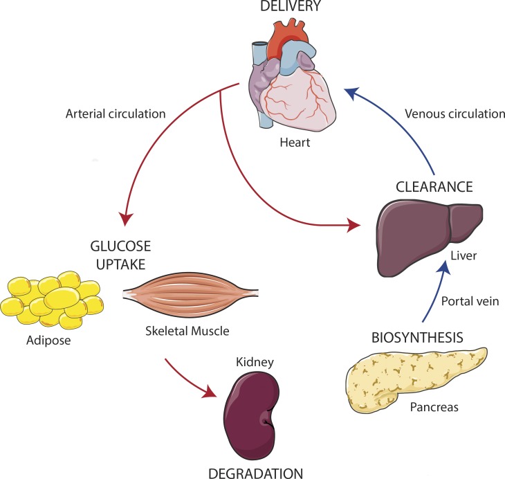
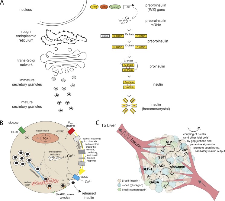
Insulin is the paramount anabolic hormone, promoting carbon energy deposition in the body. Its synthesis, quality control, delivery, and action are exquisitely regulated by highly orchestrated intracellular mechanisms in different organs or “stations” of its bodily journey. In this Beyond the Cell review, we focus on these five stages of the journey of insulin through the body and the captivating cell biology that underlies the interaction of insulin with each organ. We first analyze insulin’s biosynthesis in and export from the β-cells of the pancreas. Next, we focus on its first pass and partial clearance in the liver with its temporality and periodicity linked to secretion. Continuing the journey, we briefly describe insulin’s action on the blood vasculature and its still-debated mechanisms of exit from the capillary beds. Once in the parenchymal interstitium of muscle and adipose tissue, insulin promotes glucose uptake into myofibers and adipocytes, and we elaborate on the intricate signaling and vesicle traffic mechanisms that underlie this fundamental function. Finally, we touch upon the renal degradation of insulin to end its action. Cellular discernment of insulin’s availability and action should prove critical to understanding its pivotal physiological functions and how their failure leads to diabetes.
Overview
The claims and statistics of this paper haven’t been checked yet.
Run the check — free, no account needed. It reads the open-access text and checks the sources it cites.
Integrity signals
Registry
Cites
Statistics
Figures
Verification
Claims the paper makes
The claims and statistics of this paper haven’t been checked yet.
Run the check — free, no account needed. It reads the open-access text and checks the sources it cites.
Claims other papers make about this one
Each is checked against this paper’s own text: does it say what the citing paper says it does?
In the citing paper
“An increase in circulating glucose levels is sensed by pancreatic β‐cells through the glucose transporter GLUT2.”
From Inter‐organ communication: a gatekeeper for metabolic health (2019) · cites this paper as [24]
Self-check
Percentages, sums and reported differences re-computed from the paper’s own numbers.
The self-check needs the paper’s full text.
Citations
Cites
Central nervous system control of food intake
Schwartz, M. W., Woods, S. C., Porte, D., Seeley, R. J., & Baskin, D. G. (2000) · Nature · doi:10.1038/35007534 · Cited by 6,451 · cited 1×
“Insulin regulates appetite by reducing expression of neuropeptide Y and Agouti-related peptide (orexigenic) and, conversely, elevating expression of pro-opiomelanocortin (anorexigenic; Schwartz et al., 2000).”
BackgroundVascular endothelial cells synthesize nitric oxide from L-arginine
Palmer, R. M. J., Ashton, D., & Moncada, S. (1988) · Nature · doi:10.1038/333664a0 · Cited by 4,786 · cited 1×
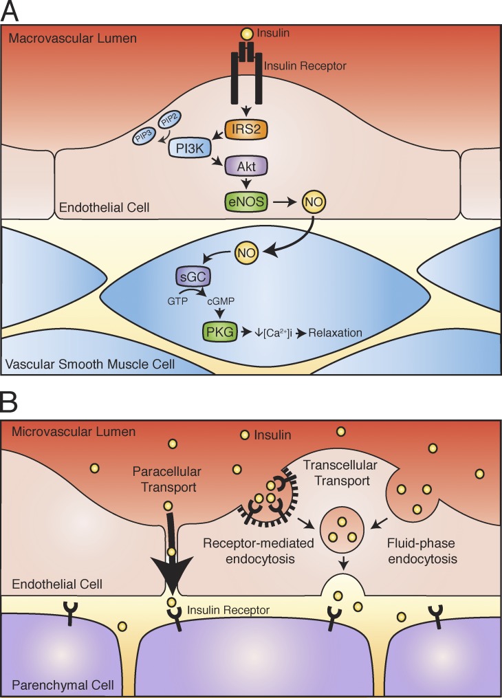
“In turn, Akt activates endothelial NO synthase to catalyze the conversion of l-arginine to NO (Palmer et al., 1988; Zeng et al., 2000).”
BackgroundCritical nodes in signalling pathways: insights into insulin action
Taniguchi, C. M., Emanuelli, B., & Kahn, C. R. (2006) · Nature Reviews Molecular Cell Biology · doi:10.1038/nrm1837 · Cited by 2,760 · cited 1×
“…of the hormone’s journey in the body have been correlated and often causally related to insulin resistance, hypertension, and type 2 diabetes (Taniguchi et al., 2006; Hoehn et al., 2008; Odegaard and Chawla, 2013; Boucher et al., 2014; DeFronzo et al., 2015; Samuel and Shulman, 2016;…”
BackgroundDeFronzo, R. A., Ferrannini, E., Groop, L., Henry, R. R., Herman, W. H., Holst, J. J., Hu, F. B., Kahn, C. R., Raz, I., Shulman, G. I., et al. (2015) · Nature Reviews Disease Primers · doi:10.1038/nrdp.2015.19 · Cited by 2,611 · cited 1×
“…2018) and CEA CAM1 expression (Lee, 2011) in obesity • Impaired vasoactive effects of insulin during insulin resistance, including capillary recruitment (de Jongh et al., 2004; Clerk et al., 2006; Keske et al., 2009); reduced insulin delivery to muscle in obesity and diabetes (Broussard et al.,…”
BackgroundMechanisms for Insulin Resistance: Common Threads and Missing Links
Samuel, V. T., & Shulman, G. I. (2012) · Cell · doi:10.1016/j.cell.2012.02.017 · Cited by 2,294 · cited 1×
“, 2013) as well as a number of proteins of the GLUT4 vesicle fusion machinery (Aslamy and Thurmond, 2017); the underlying defects include alterations in the maintenance of the storage compartment (Foley et al., 2011; Samuel and Shulman, 2012) and in the insulin-derived signals that trigger GLUT4 vesicle release from s…”
BackgroundRole of Brain Insulin Receptor in Control of Body Weight and Reproduction
Brüning, J. C., Gautam, D., Burks, D. J., Gillette, J., Schubert, M., Orban, P. C., Klein, R., Krone, W., Müller-Wieland, D., & Kahn, C. R. (2000) · Science · doi:10.1126/science.289.5487.2122 · Cited by 2,235
Insulin Receptor Signaling in Normal and Insulin-Resistant StatesPreprint
Boucher, J., Kleinridders, A., & Kahn, C. R. (2014) · Cold Spring Harbor Perspectives in Biology · doi:10.1101/cshperspect.a009191 · Cited by 1,550 · cited 1×
“…often causally related to insulin resistance, hypertension, and type 2 diabetes (Taniguchi et al., 2006; Hoehn et al., 2008; Odegaard and Chawla, 2013; Boucher et al., 2014; DeFronzo et al., 2015; Samuel and Shulman, 2016; Haeusler et al., 2018; also see other important highlights in the text box).”
BackgroundFarris, W., Mansourian, S., Chang, Y., Lindsley, L., Eckman, E. A., Frosch, M. P., Eckman, C. B., Tanzi, R. E., Selkoe, D. J., & Guénette, S. Y. (2003) · Proceedings of the National Academy of Sciences · doi:10.1073/pnas.0230450100 · Cited by 1,526 · cited 1×
“…the role of IDE in hepatic insulin clearance in vivo is controversial, as some studies report that loss of IDE results in hyperinsulinemia (Farris et al., 2003; Abdul-Hay et al., 2011), whereas others observed no changes in systemic circulating insulin in the absence of the enzyme…”
BackgroundThe pathogenesis of insulin resistance: integrating signaling pathways and substrate flux
Samuel, V. T., & Shulman, G. I. (2016) · Journal of Clinical Investigation · doi:10.1172/jci77812 · Cited by 1,484 · cited 1×
“…often causally related to insulin resistance, hypertension, and type 2 diabetes (Taniguchi et al., 2006; Hoehn et al., 2008; Odegaard and Chawla, 2013; Boucher et al., 2014; DeFronzo et al., 2015; Samuel and Shulman, 2016; Haeusler et al., 2018; also see other important highlights in the text box).”
BackgroundArnold, W. P., Mittal, C. K., Katsuki, S., & Murad, F. (1977) · Proceedings of the National Academy of Sciences · doi:10.1073/pnas.74.8.3203 · Cited by 1,459 · cited 1×
“NO is a potent vasodilator that rapidly diffuses to the vessels’ outer layer of smooth muscle cells, where it activates intracellular guanylate cyclase to increase cyclic guanosine monophosphate production (Arnold et al., 1977).”
BackgroundMichael, M. D., Kulkarni, R. N., Postic, C., Previs, S. F., Shulman, G. I., Magnuson, M. A., & Kahn, C. R. (2000) · Molecular Cell · doi:10.1016/s1097-2765(05)00015-8 · Cited by 1,296 · cited 4×
“Consistent with earlier studies, liver-specific IR knockout mice provided direct evidence that receptor-mediated degradation regulates systemic insulin levels and that impairments in this process lead to severe hyperinsulinemia that, in turn, contributes to whole-body insulin resistance (Michael et al., 2000).”
BackgroundResultInfluentialInsulin action and resistance in obesity and type 2 diabetes
Czech, M. P. (2017) · Nature Medicine · doi:10.1038/nm.4350 · Cited by 1,258 · cited 2×
“Notably, alterations in insulin renal clearance prolong the permanence of insulin in the blood (Dahl et al., 1989), evincing the importance of this process to insulin’s half-life in the circulation.”
BackgroundSteinberg, H. O., Brechtel, G., Johnson, A., Fineberg, N. S., & Baron, A. (1994) · Journal of Clinical Investigation · doi:10.1172/jci117433 · Cited by 1,216 · cited 1×
“As a consequence of endothelial NO production, insulin stimulates dilation of arteries and arterioles (Steinberg et al., 1994; Vincent et al., 2002).”
BackgroundTriggering and amplifying pathways of regulation of insulin secretion by glucose.
Henquin, J. C. (2000) · Diabetes · doi:10.2337/diabetes.49.11.1751 · Cited by 1,201 · cited 1×
“Hence, glucose not only controls the Ca2+ signals that trigger insulin secretion but also generates signals that improve the efficacy of Ca2+ on the secretory process, likely by acting on the Ca2+ sensitivity of diverse components in the pathway (Henquin, 2000).”
BackgroundCopps, K. D., & White, M. F. (2012) · Diabetologia · doi:10.1007/s00125-012-2644-8 · Cited by 1,017 · cited 1×
“…autophosphorylation by inducing structural rearrangement of the transmembrane domains to bring them into close proximity with each other (Gutmann et al., 2018), and the consequent activation of the IR tyrosine kinase toward phosphorylation of its major substrates IRS1,2 (Copps and White, 2012).”
BackgroundInsulin-stimulated Phosphorylation of a Rab GTPase-activating Protein Regulates GLUT4 Translocation
Sano, H., Kane, S., Sano, E., Mı̂inea, C. P., Asara, J. M., Lane, W. S., Garner, C. W., & Lienhard, G. E. (2003) · Journal of Biological Chemistry · doi:10.1074/jbc.c300063200 · Cited by 964 · cited 7×
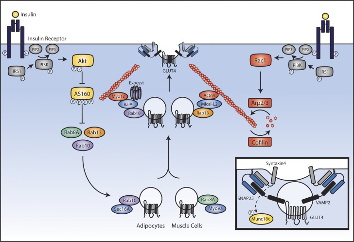
“The TBC domain of AS160/TBC1D4 defines its GAP activity toward Rab family small GTPases.”
BackgroundInfluentialRegulation of glucose transport by insulin: traffic control of GLUT4
Leto, D., & Saltiel, A. R. (2012) · Nature Reviews Molecular Cell Biology · doi:10.1038/nrm3351 · Cited by 848
Insulin granule dynamics in pancreatic beta cells
Rorsman, P., & Renstr�m, E. (2003) · Diabetologia · doi:10.1007/s00125-003-1153-1 · Cited by 844 · cited 1×
“Insulin granule pools and their intracellular traffic Most insulin granules (perhaps 75–95% of an estimated 10,000) are stored within the β-cell cytoplasm at some distance away from the cell membrane (Rorsman and Renström, 2003).”
BackgroundZeng, G., Nystrom, F. H., Ravichandran, L. V., Cong, L.-N., Kirby, M., Mostowski, H., & Quon, M. J. (2000) · Circulation · doi:10.1161/01.cir.101.13.1539 · Cited by 745 · cited 1×
“In turn, Akt activates endothelial NO synthase to catalyze the conversion of l-arginine to NO (Palmer et al., 1988; Zeng et al., 2000).”
BackgroundNonalcoholic fatty liver disease as a nexus of metabolic and hepatic diseases
Samuel, V. T., & Shulman, G. I. (2018) · Cell Metabolism · doi:10.1016/j.cmet.2017.08.002 · Cited by 745 · cited 1×
“This ensures that a portion of dietary glucose is effectively stored in the liver and is only released to the rest of the body upon cessation of insulin action (between meals) or upon metabolic demand enacted by other “counterregulatory” hormones (Samuel and Shulman, 2018).”
BackgroundBiochemical and cellular properties of insulin receptor signalling
Haeusler, R. A., McGraw, T. E., & Accili, D. (2018) · Nature Reviews Molecular Cell Biology · doi:10.1038/nrm.2017.89 · Cited by 739 · cited 1×
“…often causally related to insulin resistance, hypertension, and type 2 diabetes (Taniguchi et al., 2006; Hoehn et al., 2008; Odegaard and Chawla, 2013; Boucher et al., 2014; DeFronzo et al., 2015; Samuel and Shulman, 2016; Haeusler et al., 2018; also see other important highlights in the text box).”
BackgroundInsulin Action in AgRP-Expressing Neurons Is Required for Suppression of Hepatic Glucose Production
Könner, A. C., Janoschek, R., Plum, L., Jordan, S. D., Rother, E., Ma, X., Xu, C., Enriori, P., Hampel, B., Barsh, G. S., et al. (2007) · Cell Metabolism · doi:10.1016/j.cmet.2007.05.004 · Cited by 695
An electron microscopic study of the fenestrated endothelial lining of rat liver sinusoids
Wisse, E. (1970) · Journal of Ultrastructure Research · doi:10.1016/s0022-5320(70)90150-4 · Cited by 655 · cited 2×
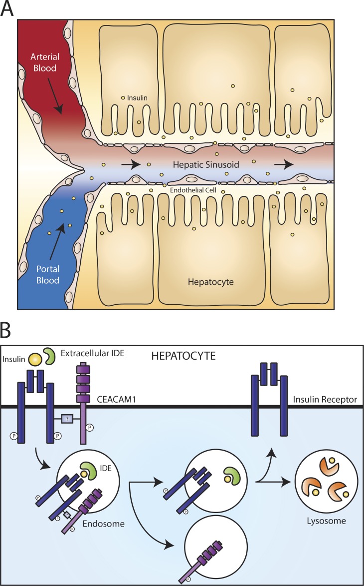
“The portal circulation delivers insulin into the capillaries of the sinusoids, which are not supported by a basement membrane and their endothelial cells contain fenestrations (Wisse, 1970; Braet et al., 1995), together permitting the exchange of contents between the blood and the surrounding liver cells.”
BackgroundPleiotropic actions of insulin resistance and inflammation in metabolic homeostasis
Odegaard, J. I., & Chawla, A. (2013) · Science · doi:10.1126/science.1230721 · Cited by 643
Insulin Action in Brain Regulates Systemic Metabolism and Brain Function
Kleinridders, A., Ferris, H. A., Cai, W., & Kahn, C. R. (2014) · Diabetes · doi:10.2337/db14-0568 · Cited by 625 · cited 3×
“Conversely, central defects in insulin action are emerging as a potential contributor to the development of Alzheimer’s disease (Griffith et al., 2018), possibly as a result of abnormal phosphorylation of tau protein (Kleinridders et al., 2014).”
BackgroundProtein Kinase B/Akt Participates in GLUT4 Translocation by Insulin in L6 Myoblasts
Wang, Q., Somwar, R., Bilan, P. J., Liu, Z., Jin, J., Woodgett, J. R., & Klip, A. (1999) · Molecular and Cellular Biology · doi:10.1128/mcb.19.6.4008 · Cited by 595 · cited 2×
“Two major consequences of the PIP3 burst relevant for GLUT4 translocation are activation of the kinases Akt1, 2 (Brozinick and Birnbaum, 1998; Wang et al., 1999) and of the Rho-family GTPase Rac1 (Chiu et al., 2011).”
BackgroundSelf-citationAstrocytic Insulin Signaling Couples Brain Glucose Uptake with Nutrient Availability
García‐Cáceres, C., Quarta, C., Varela, L., Gao, Y., Gruber, T., Legutko, B., Jastroch, M., Johansson, P., Ninkovic, J., Yi, C., et al. (2016) · Cell · doi:10.1016/j.cell.2016.07.028 · Cited by 533 · cited 1×
“The journey of insulin in the body Journal of Cell Biology https://doi.org/10.1083/jcb.201802095 11 PTP-1B (Yamada et al., 2005; Fukuda et al., 2009; Bakke et al., 2013; Garrido-Sanchez et al., 2013; Fig.”
BackgroundInfluentialActivation of phosphatidylinositol 3-kinase by insulin.
Ruderman, N. B., Kapeller, R., White, M. F., & Cantley, L. C. (1990) · Proceedings of the National Academy of Sciences · doi:10.1073/pnas.87.4.1411 · Cited by 511 · cited 1×
“Phosphorylation sites on IRS1,2 constitute entropic information to attract class I PI3K, which rapidly generates membrane domains enriched in PI(3,4,5)P3 (PIP3) within minutes (Ruderman et al., 1990).”
BackgroundProinsulin, insulin, and C-peptide concentrations in human portal and peripheral blood.
Horwitz, D. L., Starr, J. I., Mako, M. E., Blackard, W. G., & Rubenstein, A. H. (1975) · Journal of Clinical Investigation · doi:10.1172/jci108047 · Cited by 473
Dong, X. C., Copps, K. D., Guo, S., Li, Y., Kollipara, R., DePinho, R. A., & White, M. F. (2008) · Cell Metabolism · doi:10.1016/j.cmet.2008.06.006 · Cited by 463
Insulin resistance in chronic kidney disease: a systematic review
Spoto, B., Pisano, A., & Zoccali, C. (2016) · American Journal of Physiology-Renal Physiology · doi:10.1152/ajprenal.00340.2016 · Cited by 463
Garvey, W. T., Maianu, L., Zhu, J. H., Brechtel-Hook, G., Wallace, P., & Baron, A. D. (1998) · Journal of Clinical Investigation · doi:10.1172/jci1557 · Cited by 459
Beta cell hubs dictate pancreatic islet responses to glucose
Johnston, N. R., Mitchell, R. K., Haythorne, E., Pessoa, M. P., Semplici, F., Ferrer, J., Piemonti, L., Marchetti, P., Bugliani, M., Bosco, D., et al. (2016) · Cell Metabolism · doi:10.1016/j.cmet.2016.06.020 · Cited by 454
Kubota, T., Kubota, N., Kumagai, H., Yamaguchi, S., Kozono, H., Takahashi, T., Inoue, M., Itoh, S., Takamoto, I., Sasako, T., et al. (2011) · Cell Metabolism · doi:10.1016/j.cmet.2011.01.018 · Cited by 435 · cited 1×
“…MacDonald, 2017; Gandasi et al., 2017) and impaired pulsatile secretion of insulin in diabetic individuals (Lang et al., 1981; Hollingdal et al., 2000; Laedtke et al., 2000) • Reduced hepatic insulin clearance (Jung et al., 2018) and CEA CAM1 expression (Lee, 2011) in obesity • Impaired vasoactive…”
BackgroundPdx1 Maintains β Cell Identity and Function by Repressing an α Cell Program
Gao, T., McKenna, B. D., Li, C., Reichert, M., Nguyen, J., Singh, T., Yang, C., Pannikar, A., Doliba, N. M., Zhang, T., et al. (2014) · Cell Metabolism · doi:10.1016/j.cmet.2013.12.002 · Cited by 431
Hormonal regulation of hepatic glucose production in health and disease
Lin, H. V., & Accili, D. (2011) · Cell Metabolism · doi:10.1016/j.cmet.2011.06.003 · Cited by 429 · cited 2×
“GLUT4 vesicle retention near the membrane also involves the exocyst subunit Exo70 (Lizunov et al., 2012).”
BackgroundDiscovery, characterization, and clinical development of the glucagon-like peptides
Drucker, D. J., Habener, J. F., & Holst, J. J. (2017) · Journal of Clinical Investigation · doi:10.1172/jci97233 · Cited by 418 · cited 2×
“HID-1 is required for homotypic fusion of immature secretory granules during maturation. eLife.”
Backgroundde Jongh, R. T., Serné, E. H., IJzerman, R. G., de Vries, G., & Stehouwer, C. D. A. (2004) · Circulation · doi:10.1161/01.cir.0000129772.26647.6f · Cited by 407 · cited 1×
“Insulin acting centrally also evokes efferent inputs into peripheral tissue metabolism (Ferris and Kahn, 2016), contributing to the suppression of gluconeogenesis in the liver and the counterregulatory response to hypoglycemia (Diggs-Andrews et al., 2010).”
BackgroundZierath, J. R., He, L., Gumà, A., Wahlström, E. O., Klip, A., & Wallberg-Henriksson, H. (1996) · Diabetologia · doi:10.1007/bf02658504 · Cited by 402 · cited 1×
“…in obesity and diabetes (Broussard et al., 2016) • Diminished GLUT4 translocation to the muscle membrane in diabetic rodents and humans (Klip et al., 1990; Zierath et al., 1996; Garvey et al., 1998; Hoehn et al., 2008; Czech, 2017) and lowered expression of Rac1 (Sylow et al., 2013) as well as a…”
BackgroundSelf-citationRetinal and choroidal neovascularization
Campochiaro, P. A. (2000) · Journal of Cellular Physiology · doi:10.1002/1097-4652(200009)184:3 · Cited by 393
Wang, Z., & Thurmond, D. C. (2009) · Journal of Cell Science · doi:10.1242/jcs.034355 · Cited by 386 · cited 1×
“…phase resulting from fusion and secretion by already “docked and primed” secretory granules that in human lasts up to 10 min, and a subsequent second-phase secretion that is associated with actin reorganization thought to allow granule recruitment to the plasma membrane (Wang and Thurmond, 2009).”
BackgroundGlucose‐induced electrical activity in pancreatic islet cells
Dean, P. M., & Matthews, E. K. (1970) · The Journal of Physiology · doi:10.1113/jphysiol.1970.sp009207 · Cited by 365 · cited 1×
“…often causally related to insulin resistance, hypertension, and type 2 diabetes (Taniguchi et al., 2006; Hoehn et al., 2008; Odegaard and Chawla, 2013; Boucher et al., 2014; DeFronzo et al., 2015; Samuel and Shulman, 2016; Haeusler et al., 2018; also see other important highlights in the text box).”
BackgroundRavier, M. A., Güldenagel, M., Charollais, A., Gjinovci, A., Caille, D., Söhl, G., Wollheim, C. B., Willecke, K., Henquin, J.-C., & Meda, P. (2005) · Diabetes · doi:10.2337/diabetes.54.6.1798 · Cited by 360 · cited 1×
“Gap junction coupling between β-cells via connexin36 plays a critical role, the loss of which results in dysregulation of insulin secretion (Ravier et al., 2005).”
BackgroundThe role of endothelial insulin signaling in the regulation of vascular tone and insulin resistance
Vicent, D., Ilany, J., Kondo, T., Naruse, K., Fisher, S. J., Kisanuki, Y. Y., Bursell, S. E., Yanagisawa, M., King, G. L., & Kahn, C. R. (2003) · Journal of Clinical Investigation · doi:10.1172/jci15211 · Cited by 354
Sano, H., Eguez, L., Teruel, M. N., Fukuda, M., Chuang, T. D., Chavez, J. A., Lienhard, G. E., & McGraw, T. E. (2007) · Cell Metabolism · doi:10.1016/j.cmet.2007.03.001 · Cited by 340 · cited 1×
“In adipocytes, Rab10 promotes GLUT4 mobilization from the perinuclear region toward the plasma membrane (Sano et al., 2007; Bruno et al., 2016), specifically by interacting with Sec16A (Sano et al., 2007; Bruno et al., 2016).”
BackgroundInsulin Signaling in the Central Nervous System
Porte, D., Baskin, D. G., & Schwartz, M. W. (2005) · Diabetes · doi:10.2337/diabetes.54.5.1264 · Cited by 339 · cited 1×
“Thought to be insulin unresponsive in the past, the central nervous system is well recognized to be exquisitely responsive to the incoming hormone (Porte et al., 2005).”
BackgroundLemaire, K., Ravier, M. A., Schraenen, A., Creemers, J. W. M., Van de Plas, R., Granvik, M., Van Lommel, L., Waelkens, E., Chimienti, F., Rutter, G. A., et al. (2009) · Proceedings of the National Academy of Sciences · doi:10.1073/pnas.0906587106 · Cited by 330 · cited 2×
“…the initial gain in surface GLUT4 emanates from the storage compartment, whereas maintenance of the steady state involves the endosomal route (Bryant and Gould, 2011; Kandror and Pilch, 2011; Stöckli et al., 2011; Bogan, 2012; Leto and Saltiel, 2012; Klip et al., 2014; Jaldin-Fincati et al., 2017).”
BackgroundBarrett, E. J., Eggleston, E. M., Inyard, A. C., Wang, H., Li, G., Chai, W., & Liu, Z. (2009) · Diabetologia · doi:10.1007/s00125-009-1313-z · Cited by 323 · cited 4×
“Insulin action in target tissues is temporally linked to these vascular effects (Barrett et al., 2009); in particular, the full stimulation of skeletal muscle glucose uptake in vivo is contingent on prior NO-mediated vasodilation (Vincent et al., 2003; Bradley et al., 2013).”
BackgroundInfluentialInsulin and the Blood-Brain Barrier
Woods, S. C., Seeley, R. J., Baskin, D. G., & Schwartz, M. W. (2003) · Current Pharmaceutical Design · doi:10.2174/1381612033455323 · Cited by 321 · cited 1×
“As outlined above, insulin crosses the blood–brain barrier through a receptor-mediated process (Woods et al., 2003).”
BackgroundRegulation of ATP production by mitochondrial Ca2+
Tarasov, A. I., Griffiths, E. J., & Rutter, G. A. (2012) · Cell Calcium · doi:10.1016/j.ceca.2012.03.003 · Cited by 319 · cited 1×
“For example, feedback from oscillatory Ca2+ signals controls mitochondrial ATP generation (Tarasov et al., 2012), and recent work shows that Ca2+ oscillations and the ER protein TMEM24 interact at ER–plasma membrane contact sites to maintain phosphatidylinositol levels required for β-cell signaling…”
BackgroundIRS1-Independent Defects Define Major Nodes of Insulin Resistance
Hoehn, K. L., Hohnen-Behrens, C., Cederberg, A., Wu, L. E., Turner, N. A., Yuasa, T., Ebina, Y., & James, D. E. (2008) · Cell Metabolism · doi:10.1016/j.cmet.2008.04.005 · Cited by 318 · cited 2×
“…journey in the body have been correlated and often causally related to insulin resistance, hypertension, and type 2 diabetes (Taniguchi et al., 2006; Hoehn et al., 2008; Odegaard and Chawla, 2013; Boucher et al., 2014; DeFronzo et al., 2015; Samuel and Shulman, 2016; Haeusler et al., 2018; also…”
BackgroundInsulin Degradation: Mechanisms, Products, and Significance*
Duckworth, W. C. (1988) · Endocrine Reviews · doi:10.1210/edrv-9-3-319 · Cited by 315 · cited 3×
“The majority of insulin that binds to hepatic IRs is degraded (Duckworth, 1988).”
BackgroundObesity Blunts Insulin-Mediated Microvascular Recruitment in Human Forearm Muscle
Clerk, L. H., Keske, M. A., Jahn, L. A., Liu, Z., Lindner, J. R., & Barrett, E. J. (2006) · Diabetes · doi:10.2337/db05-1373 · Cited by 305 · cited 2×
“, 2018) and CEA CAM1 expression (Lee, 2011) in obesity • Impaired vasoactive effects of insulin during insulin resistance, including capillary recruitment (de Jongh et al., 2004; Clerk et al., 2006; Keske et al., 2009); reduced insulin delivery to muscle in obesity and diabetes (Broussard et al.”
BackgroundVincent, M. A., Barrett, E. J., Lindner, J. R., Clark, M. G., & Rattigan, S. (2003) · American Journal of Physiology-Endocrinology and Metabolism · doi:10.1152/ajpendo.00021.2003 · Cited by 305
The renal metabolism of insulin
Rabkin, R., Ryan, M. P., & Duckworth, W. C. (1984) · Diabetologia · doi:10.1007/bf00304849 · Cited by 294 · cited 3×
“In addition, these IR are important sites sensing the hormone to stimulate important functions such as reabsorption of sodium, phosphate, and glucose (Rabkin et al., 1984; Hale and Coward, 2013).”
BackgroundMurphy, R. F., Powers, S., & Cantor, C. R. (1984) · The Journal of Cell Biology · doi:10.1083/jcb.98.5.1757 · Cited by 287 · cited 1×
“As endosomes acidify, any remaining insulin or partially degraded insulin that escaped complete degradation by IDE dissociates from the IR (Murphy et al., 1984; Fig.”
BackgroundUpdate on GLUT4 Vesicle Traffic: A Cornerstone of Insulin Action
Jaldín‐Fincati, J. R., Pavarotti, M. A., Frendo‐Cumbo, S., Bilan, P. J., & Klip, A. (2017) · Trends in Endocrinology and Metabolism · doi:10.1016/j.tem.2017.05.002 · Cited by 287 · cited 3×
“…the initial gain in surface GLUT4 emanates from the storage compartment, whereas maintenance of the steady state involves the endosomal route (Bryant and Gould, 2011; Kandror and Pilch, 2011; Stöckli et al., 2011; Bogan, 2012; Leto and Saltiel, 2012; Klip et al., 2014; Jaldin-Fincati et al., 2017).”
BackgroundSelf-citationRegulation of Glucose Transporter Translocation in Health and Diabetes
Bogan, J. S. (2012) · Annual Review of Biochemistry · doi:10.1146/annurev-biochem-060109-094246 · Cited by 279 · cited 3×
“…the initial gain in surface GLUT4 emanates from the storage compartment, whereas maintenance of the steady state involves the endosomal route (Bryant and Gould, 2011; Kandror and Pilch, 2011; Stöckli et al., 2011; Bogan, 2012; Leto and Saltiel, 2012; Klip et al., 2014; Jaldin-Fincati et al., 2017).”
BackgroundBrief, Irregular Oscillations of Basal Plasma Insulin and Glucose Concentrations in Diabetic Man
Lang, D. A., Matthews, D. R., Burnett, M., & Turner, R. C. (1981) · Diabetes · doi:10.2337/diab.30.5.435 · Cited by 279 · cited 1×
“Activated Akt1,2 migrates to the cytosol and intracellular membranes (Zheng and Cartee, 2016), where it phosphorylates AS160, a substrate of 160 kD more appropriately named TBC1D4 (Sano et al., 2003; Lansey et al., 2012).”
BackgroundVan Der Heide, L. P., Kamal, A., Artola, A., Gispen, W. H., & Ramakers, G. M. J. (2005) · Journal of Neurochemistry · doi:10.1111/j.1471-4159.2005.03269.x · Cited by 278 · cited 1×
“Although rich information is being gathered through electrophysiological approaches (van der Heide et al., 2005; Könner et al., 2007; Korol et al., 2018), there is a rich opportunity to explore additional mechanisms through the advent of real-time intravital imaging of the central nervous system…”
BackgroundStöckli, J., Fazakerley, D. J., & James, D. E. (2011) · Journal of Cell Science · doi:10.1242/jcs.097063 · Cited by 276 · cited 3×
“…the initial gain in surface GLUT4 emanates from the storage compartment, whereas maintenance of the steady state involves the endosomal route (Bryant and Gould, 2011; Kandror and Pilch, 2011; Stöckli et al., 2011; Bogan, 2012; Leto and Saltiel, 2012; Klip et al., 2014; Jaldin-Fincati et al., 2017).”
BackgroundCEACAM1 regulates insulin clearance in liver
Poy, M. N., Yang, Y., Rezaei, K., Fernström, M. A., Lee, A. D., Kido, Y., Erickson, S. K., & Najjar, S. M. (2002) · Nature Genetics · doi:10.1038/ng840 · Cited by 269 · cited 12×
“Hence, the level of CEA CAM1 at the plasma membrane and its phosphorylation may impart physiological fine-tuning regulation to the process of insulin clearance.”
BackgroundInfluentialLee, M. R., Lin, L., & Kitazawa, T. (1997) · Journal of Biological Chemistry · doi:10.1074/jbc.272.8.5063 · Cited by 265 · cited 2×
“, 2000) prevent phosphorylation of myosin light chain required for cytoskeletal cross-bridge formation and contraction (Lee et al., 1997; Mizuno et al., 2008), thereby resulting in vessel relaxation (Fig.”
BackgroundGlucose transporter recycling in response to insulin is facilitated by myosin Myo1c
Bose, A., Guilherme, A., Robida, S., Nicoloro, S. M. C., Zhou, Q. L., Jiang, Z. Y., Pomerleau, D. P., & Czech, M. (2002) · Nature · doi:10.1038/nature01246 · Cited by 265 · cited 1×
“In addition to the Rab13–MIC AL-L2–α-actinin4 connection, GLUT4 vesicles tether to actin filaments via Myosin 1c (Bose et al., 2002; Boguslavsky et al., 2012).”
BackgroundMeier, J. J., Veldhuis, J. D., & Butler, P. C. (2005) · Diabetes · doi:10.2337/diabetes.54.6.1649 · Cited by 261 · cited 4×
“This degradation is coupled to pulsatile delivery, such that the liver preferentially clears insulin that arrives in pulses (Meier et al., 2005).”
BackgroundInfluentialBergsten, P., Grapengiesser, E., Gylfe, E., Tengholm, A., & Hellman, B. (1994) · Journal of Biological Chemistry · doi:10.1016/s0021-9258(17)37032-1 · Cited by 260 · cited 1×
“Importantly, this translates into oscillations of insulin secretion from isolated islets, again with a periodicity of 1–5 min (Bergsten et al., 1994).”
BackgroundPulsatile insulin secretion, impaired glucose tolerance and type 2 diabetes
Satin, L. S., Butler, P. C., Ha, J., & Sherman, A. (2015) · Molecular Aspects of Medicine · doi:10.1016/j.mam.2015.01.003 · Cited by 243 · cited 1×
“Thus, communication between cells within an islet likely contributes to the well-described phenomenon of insulin secretory oscillations, which occur in healthy humans with a periodicity of 5–10 min (Satin et al., 2015).”
BackgroundRegulation of calcium in pancreatic α- and β-cells in health and disease
Rorsman, P., Braun, M., & Zhang, Q. (2012) · Cell Calcium · doi:10.1016/j.ceca.2011.11.006 · Cited by 220 · cited 1×
“When the membrane potential depolarizes sufficiently (approximately −50 mV), the activation of voltage-dependent Na+ and Ca2+ channels cause repetitive action potential spiking and a rise in intracellular Ca2+ (Rorsman et al., 2012).”
BackgroundVincent, M. A., Dawson, D. K., Clark, A. D. H., Lindner, J. R., Rattigan, S., Clark, M. G., & Barrett, E. J. (2002) · Diabetes · doi:10.2337/diabetes.51.1.42 · Cited by 216 · cited 2×
“As a consequence of endothelial NO production, insulin stimulates dilation of arteries and arterioles (Steinberg et al., 1994; Vincent et al., 2002).”
BackgroundDissecting Multiple Steps of GLUT4 Trafficking and Identifying the Sites of Insulin Action
Bai, L., Wang, Y., Fan, J., Chen, Y., Ji, W., Qu, A., Xu, P., James, D. E., & Xu, T. (2007) · Cell Metabolism · doi:10.1016/j.cmet.2006.11.013 · Cited by 216 · cited 1×
“This restricts GLUT4 mobility beneath the membrane, a phenomenon nicely documented through total internal reflection fluorescence microscopy of muscle and adipose cells (Bai et al., 2007; Xiong et al., 2010; Boguslavsky et al., 2012; Lizunov et al., 2012).”
BackgroundGlucose regulation of insulin gene expression in pancreatic β-cells
Andrali, S. S., Sampley, M. L., Vanderford, N. L., & Özcan, S. (2008) · Biochemical Journal · doi:10.1042/bj20081029 · Cited by 213 · cited 1×
“The journey of insulin in the body Journal of Cell Biology https://doi.org/10.1083/jcb.201802095 2 required for insulin gene expression and contribute to the regulation of INS transcription in response to glucose and autocrine insulin signaling (Andrali et al., 2008).”
BackgroundInfluentialSylow, L., Jensen, T. E., Kleinert, M., Højlund, K., Kiens, B., Wojtaszewski, J. F. P., Prats, C., Schjerling, P., & Richter, E. A. (2013) · Diabetes · doi:10.2337/db12-1148 · Cited by 210 · cited 10×
“This includes the glucose- and Cdc42-dependent activation of Rac1, which, when released from an inhibitory RhoGDI and in its GTP-bound form, promotes cortical actin remodeling, perhaps via an interaction with gelsolin (Kalwat and Thurmond, 2013).”
BackgroundInfluentialLipid transport by TMEM24 at ER–plasma membrane contacts regulates pulsatile insulin secretion
Lees, J. A., Messa, M., Sun, E. W., Wheeler, H., Torta, F. T., Wenk, M. R., Camilli, P. V. D., & Reinisch, K. M. (2017) · Science · doi:10.1126/science.aah6171 · Cited by 208
McCulloch, L. J., van de Bunt, M., Braun, M., Frayn, K. N., Clark, A., & Gloyn, A. L. (2011) · Molecular Genetics and Metabolism · doi:10.1016/j.ymgme.2011.08.026 · Cited by 207
Rab8A and Rab13 are activated by insulin and regulate GLUT4 translocation in muscle cells
Sun, Y., Bilan, P. J., Liu, Z., & Klip, A. (2010) · Proceedings of the National Academy of Sciences · doi:10.1073/pnas.1009523107 · Cited by 197
Self-citationEffect of Endothelium-Specific Insulin Resistance on Endothelial Function In Vivo
Duncan, E. R., Crossey, P. A., Walker, S., Anilkumar, N. P., Poston, L., Douglas, G., Ezzat, V., Wheatcroft, S. B., Shah, A. M., & Kearney, M. T. (2008) · Diabetes · doi:10.2337/db07-1111 · Cited by 188 · cited 1×
“…the role of IDE in hepatic insulin clearance in vivo is controversial, as some studies report that loss of IDE results in hyperinsulinemia (Farris et al., 2003; Abdul-Hay et al., 2011), whereas others observed no changes in systemic circulating insulin in the absence of the enzyme…”
BackgroundGLUT4 Is Retained by an Intracellular Cycle of Vesicle Formation and Fusion with Endosomes
Karylowski, O., Zeigerer, A., Cohen, A., & McGraw, T. E. (2003) · Molecular Biology of the Cell · doi:10.1091/mbc.e03-07-0517 · Cited by 182
Insulin in the nervous system and the mind: Functions in metabolism, memory, and mood
Lee, S.-H., Zabolotny, J. M., Huang, H., Lee, H., & Kim, Y.-B. (2016) · Molecular Metabolism · doi:10.1016/j.molmet.2016.06.011 · Cited by 176
Insulin-stimulated translocation of GLUT4 glucose transporters requires SNARE-complex proteins
Cheatham, B., Volchuk, A., Kahn, C. R., Wang, L., Rhodes, C. J., & Klip, A. (1996) · Proceedings of the National Academy of Sciences · doi:10.1073/pnas.93.26.15169 · Cited by 174 · cited 1×
“This is brought about through formation of a SNA RE complex between VAMP2 on the vesicles and synatxin4 and SNAP23 on the plasma membrane (Cheatham et al., 1996; Foster and Klip, 2000; Thurmond and Pessin, 2001).”
BackgroundSelf-citationDirect Measurement of Pulsatile Insulin Secretion from the Portal Vein in Human Subjects1
Song, S. H., McIntyre, S. S., Shah, H., Veldhuis, J. D., Hayes, P. C., & Butler, P. C. (2000) · The Journal of Clinical Endocrinology & Metabolism · doi:10.1210/jcem.85.12.7043 · Cited by 173 · cited 3×
“5–1 nmol/liter in the fasted state and rises to ∼5 nmol/liter after a meal (Pørksen et al., 1995; Song et al., 2000).”
BackgroundInfluentialSignal transduction meets vesicle traffic: the software and hardware of GLUT4 translocation
Klip, A., Sun, Y., Chiu, T. T., & Foley, K. P. (2014) · American Journal of Physiology-Cell Physiology · doi:10.1152/ajpcell.00069.2014 · Cited by 170 · cited 1×
“…classical G-protein–coupled receptors via Gαs-activation of adenylate cyclase to increase cAMP, which causes PKA-dependent phosphorylation of the exocytic machinery and PKA-independent effects mediated by Epac2A to promote the release of Ca2+ from intracellular stores (Kolic and MacDonald, 2015).”
BackgroundSelf-citationGloyn, A. L., Noordam, C., Willemsen, M. A. A. P., Ellard, S., Lam, W. W. K., Campbell, I. W., Midgley, P., Shiota, C., Buettger, C. W., Magnuson, M. A., et al. (2003) · Diabetes · doi:10.2337/diabetes.52.9.2433 · Cited by 168 · cited 1×
“Mutations that alter the glucose-dependent activity of GK effectively adjust the set point for whole-body glucose homeostasis (Gloyn et al., 2003).”
BackgroundForli, A., Vecchia, D., Binini, N., Succol, F., Bovetti, S., Moretti, C., Nespoli, F., Mahn, M., Baker, C. A., Bolton, M. M., et al. (2018) · Cell Reports · doi:10.1016/j.celrep.2018.02.063 · Cited by 168 · cited 1×
“…is being gathered through electrophysiological approaches (van der Heide et al., 2005; Könner et al., 2007; Korol et al., 2018), there is a rich opportunity to explore additional mechanisms through the advent of real-time intravital imaging of the central nervous system (Forli et al., 2018).”
BackgroundMembrane Receptors for Endocytosis in the Renal Proximal Tubule
Christensen, E. L., Birn, H., Verroust, P. J., & Moestrup, S. K. (1998) · International review of cytology · doi:10.1016/s0074-7696(08)61772-6 · Cited by 165 · cited 2×
“…be not the IR (Meezan et al., 1988; Sato et al., 1991; Nielsen, 1993, 1994) but possibly scavenger receptors such as megalin (member of the low-density lipoprotein receptor family; Christensen et al., 1998; Kolman et al., 2009) and cubilin, proteins that recover a number of proteins by endocytosis.”
BackgroundEndocytosis, Recycling, and Regulated Exocytosis of Glucose Transporter 4
Foley, K. P., Boguslavsky, S., & Klip, A. (2011) · Biochemistry · doi:10.1021/bi2000356 · Cited by 163 · cited 1×
“…of the GLUT4 vesicle fusion machinery (Aslamy and Thurmond, 2017); the underlying defects include alterations in the maintenance of the storage compartment (Foley et al., 2011; Samuel and Shulman, 2012) and in the insulin-derived signals that trigger GLUT4 vesicle release from storage and…”
BackgroundSelf-citationPulsatile Portal Vein Insulin Delivery Enhances Hepatic Insulin Action and Signaling
Matveyenko, A. V., Liuwantara, D., Gurlo, T., Kirakossian, D., Dalla Man, C., Cobelli, C., White, M. F., Copps, K. D., Volpi, E., Fujita, S., et al. (2012) · Diabetes · doi:10.2337/db11-1462 · Cited by 163 · cited 1×
“Pulsatile insulin delivery to the liver is an important physiological signal that regulates both hepatic insulin action (Matveyenko et al., 2012) and insulin clearance (Meier et al.”
BackgroundIshikura, S., Bilan, P. J., & Klip, A. (2006) · Biochemical and Biophysical Research Communications · doi:10.1016/j.bbrc.2006.12.140 · Cited by 158
Self-citationSignaling mechanisms of glucose-induced F-actin remodeling in pancreatic islet β cells
Kalwat, M. A., & Thurmond, D. C. (2013) · Experimental & Molecular Medicine · doi:10.1038/emm.2013.73 · Cited by 158 · cited 1×
“…insulin-derived signals that trigger GLUT4 vesicle release from storage and interaction with the plasma membrane • Compromised glomerular function in obesity (Kanasaki et al., 2013) that may alter insulin bioavailability; sodium retention and down-regulation of the natriuretic peptide system in…”
BackgroundRac1 signalling towards GLUT4/glucose uptake in skeletal muscle
Chiu, T. T., Jensen, T. E., Sylow, L., Richter, E. A., & Klip, A. (2011) · Cellular Signalling · doi:10.1016/j.cellsig.2011.05.022 · Cited by 149 · cited 2×
“Two major consequences of the PIP3 burst relevant for GLUT4 translocation are activation of the kinases Akt1, 2 (Brozinick and Birnbaum, 1998; Wang et al., 1999) and of the Rho-family GTPase Rac1 (Chiu et al., 2011).”
BackgroundSelf-citationInsulin, but Not Contraction, Activates Akt/PKB in Isolated Rat Skeletal Muscle
Brozinick, J. T., & Birnbaum, M. J. (1998) · Journal of Biological Chemistry · doi:10.1074/jbc.273.24.14679 · Cited by 149
Li, G., Rungger‐Brändle, E., Just, I., Jonas, J., Aktories, K., & Wollheim, C. B. (1994) · Molecular Biology of the Cell · doi:10.1091/mbc.5.11.1199 · Cited by 149 · cited 1×
“Loss of key SNA RE proteins results in impaired insulin secretion (Liang et al., 2017).”
BackgroundRecruitment of GLUT-4 glucose transporters by insulin in diabetic rat skeletal muscle
Klip, A., Ramlal, T., Bilan, P. J., Cartee, G. D., Gulve, E. A., & Holloszy, J. O. (1990) · Biochemical and Biophysical Research Communications · doi:10.1016/0006-291x(90)90735-6 · Cited by 142 · cited 2×
“, 2016) • Diminished GLUT4 translocation to the muscle membrane in diabetic rodents and humans (Klip et al., 1990; Zierath et al., 1996; Garvey et al., 1998; Hoehn et al., 2008; Czech, 2017) and lowered expression of Rac1 (Sylow et al.”
BackgroundSelf-citationSteil, G. M., Ader, M., Moore, D., Rebrin, K., & Bergman, R. N. (1996) · Journal of Clinical Investigation · doi:10.1172/jci118572 · Cited by 139 · cited 1×
“…insulin egress from the circulation toward tissues (Kubota et al., 2011; Majumdar et al., 2012; Meijer et al., 2016; Konishi et al., 2017), opposing studies challenge a receptor-mediated (Vicent et al., 2003; Duncan et al., 2008; Williams et al., 2018) or saturable process (Steil et al., 1996).”
BackgroundKonishi, M., Sakaguchi, M., Lockhart, S. M., Cai, W., Li, M. E., Homan, E. P., Rask‐Madsen, C., & Kahn, C. R. (2017) · Proceedings of the National Academy of Sciences · doi:10.1073/pnas.1710625114 · Cited by 139
Obesity Blunts Microvascular Recruitment in Human Forearm Muscle After a Mixed Meal
Keske, M. A., Clerk, L. H., Price, W. J., Jahn, L. A., & Barrett, E. J. (2009) · Diabetes Care · doi:10.2337/dc09-0206 · Cited by 133 · cited 2×
“Conversely, central defects in insulin action are emerging as a potential contributor to the development of Alzheimer’s disease (Griffith et al., 2018), possibly as a result of abnormal phosphorylation of tau protein (Kleinridders et al., 2014).”
BackgroundLaedtke, T., Kjems, L., Pørksen, N. K., Schmitz, O., Veldhuis, J. D., Kao, P. C., & Butler, P. C. (2000) · American Journal of Physiology-Endocrinology and Metabolism · doi:10.1152/ajpendo.2000.279.3.e520 · Cited by 132 · cited 3×
“Intriguingly, there are studies of additional participation of Ca2+-regulated proteins such as Doc2b, Tctex1d2, and E-Syt1 (Lalioti et al., 2009; Friedrich et al., 2010; Shimoda et al., 2015), and insulin-dependent Ca2+-spikes have been recorded in muscle cells (Contreras-Ferrat et al., 2014),…”
BackgroundContact-induced clustering of syntaxin and munc18 docks secretory granules at the exocytosis site
Gandasi, N. R., & Barg, S. (2014) · Nature Communications · doi:10.1038/ncomms4914 · Cited by 130 · cited 1×
“The formation and fidelity of the SNA RE complex mediating granule docking is regulated by a number of proteins such as Munc18 and syntaxin isoforms (Gandasi and Barg, 2014; Zhu et al., 2015).”
BackgroundJansson, P., Fowelin, J., von Schenck, H., Smith, U., & Lönnroth, P. (1993) · Diabetes · doi:10.2337/diab.42.10.1469 · Cited by 129 · cited 1×
“…insulin in the circulation (Kolka and Bergman, 2012), and several studies have documented that the capillary endothelium is a barrier to insulin delivery to muscle in vivo, maintaining a disequilibrium between circulating and interstitial insulin levels (Jansson et al., 1993; Herkner et al., 2003).”
BackgroundDual-mode of insulin action controls GLUT4 vesicle exocytosis
Xu, Y., Rubin, B. R., Orme, C. M., Karpikov, A., Yu, C., Bogan, J. S., & Toomre, D. K. (2011) · The Journal of Cell Biology · doi:10.1083/jcb.201008135 · Cited by 129 · cited 1×
“These emanate directly from the storage compartment; however, insulin also appears to redirect vesicles from this compartment toward the general recycling endosomes, from whence they reach the cell periphery in the form of somewhat larger vesicles (Xu et al., 2011).”
BackgroundVisualization of ligand-induced transmembrane signaling in the full-length human insulin receptor
Gutmann, T., Kim, K. H., Grzybek, M., Walz, T., & Coskun, Ü. (2018) · The Journal of Cell Biology · doi:10.1083/jcb.201711047 · Cited by 129 · cited 13×
“Subsequent increases in the cytosolic ATP/ADP ratio control cell membrane potential by inhibiting ATP-sensitive K+ (KATP) channels, eliciting a membrane depolarization that is modulated by a number of additional ion channels (Fig.”
BackgroundInfluentialBraet, F., Zanger, R. D., Baekeland, M., Crabbé, E., Smissen, P. V. D., & Wisse, E. (1995) · Hepatology · doi:10.1002/hep.1840210130 · Cited by 128 · cited 2×
“The portal circulation delivers insulin into the capillaries of the sinusoids, which are not supported by a basement membrane and their endothelial cells contain fenestrations (Wisse, 1970; Braet et al., 1995), together permitting the exchange of contents between the blood and the surrounding liver cells.”
BackgroundRegulation of insulin action by CEACAM1
Najjar, S. M. (2002) · Trends in Endocrinology and Metabolism · doi:10.1016/s1043-2760(02)00608-2 · Cited by 123 · cited 2×
“…clearance Although hepatocytes are not exclusive in their ability to internalize insulin, they highly express the transmembrane glycoprotein CEA CAM1 (carcinoembryonic antigen–related cell adhesion molecule 1), which mediates rapid and effective IR-mediated insulin endocytosis (Najjar, 2002).”
BackgroundMechanism and regulation of GLUT-4 vesicle fusion in muscle and fat cells
Foster, L. J., & Klip, A. (2000) · American Journal of Physiology-Cell Physiology · doi:10.1152/ajpcell.2000.279.4.c877 · Cited by 121 · cited 1×
“Intriguingly, there are studies of additional participation of Ca2+-regulated proteins such as Doc2b, Tctex1d2, and E-Syt1 (Lalioti et al., 2009; Friedrich et al., 2010; Shimoda et al., 2015), and insulin-dependent Ca2+-spikes have been recorded in muscle cells (Contreras-Ferrat et al., 2014),…”
BackgroundSelf-citationRab10 in insulin-stimulated GLUT4 translocation
Sano, H., Roach, W. G., Peck, G. R., Fukuda, M., & Lienhard, G. E. (2008) · Biochemical Journal · doi:10.1042/bj20071318 · Cited by 117 · cited 2×
“In adipocytes, Rab10 is the preferred GTPase in GLUT4 translocation (Sano et al., 2008), whereas in muscle cells, it is Rab8A and Rab13 (Ishikura et al., 2007; Sun et al., 2010, 2014, 2016).”
BackgroundRandhawa, V. K., Bilan, P. J., Khayat, Z. A., Daneman, N., Liu, Z., Ramlal, T., Volchuk, A., Peng, X., Coppola, T., Regazzi, R., et al. (2000) · Molecular Biology of the Cell · doi:10.1091/mbc.11.7.2403 · Cited by 116 · cited 1×
“However, another prevailing model proposes that the majority of vesicles furnishing the cell membrane with GLUT4 contain the fusogenic protein VAMP2, which segregates away with the storage compartment and is largely absent from recycling endosomes (Randhawa et al., 2000, 2004; Török et al., 2004).”
BackgroundSelf-citationA Role for the CHC22 Clathrin Heavy-Chain Isoform in Human Glucose Metabolism
Vassilopoulos, S., Esk, C., Hoshino, S., Funke, B., Chen, C., Plocik, A. M., Wright, W. E., Kucherlapati, R., & Brodsky, F. M. (2009) · Science · doi:10.1126/science.1171529 · Cited by 114
Pulsatile insulin secretion accounts for 70% of total insulin secretion during fasting
Pørksen, N. K., Munn, S. R., Steers, J. L., Vore, S. J., Veldhuis, J. D., & Butler, P. C. (1995) · American Journal of Physiology-Endocrinology and Metabolism · doi:10.1152/ajpendo.1995.269.3.e478 · Cited by 114 · cited 1×
“The portal vein delivers insulin from the pancreas to the liver in discrete pulses that occur every ∼5 min (Song et al., 2000), where the amplitude of these insulin pulses is 0.5–1 nmol/liter in the fasted state and rises to ∼5 nmol/liter after a meal (Pørksen et al., 1995; Song et al., 2000).”
BackgroundInfluentialAbdul‐Hay, S. O., Kang, D., McBride, M., Li, L., Zhao, J., & Leissring, M. A. (2011) · PLoS ONE · doi:10.1371/journal.pone.0020818 · Cited by 113 · cited 2×
“…of IDE in hepatic insulin clearance in vivo is controversial, as some studies report that loss of IDE results in hyperinsulinemia (Farris et al., 2003; Abdul-Hay et al., 2011), whereas others observed no changes in systemic circulating insulin in the absence of the enzyme (Steneberg et al., 2013).”
ResultMyosin light chain kinase activation and calcium sensitization in smooth muscle in vivo
Mizuno, Y., Isotani, E., Huang, J., Ding, H., Stull, J. T., & Kamm, K. E. (2008) · American Journal of Physiology-Cell Physiology · doi:10.1152/ajpcell.90645.2007 · Cited by 113 · cited 1×
“…monophosphate–dependent reductions in intracellular Ca2+ concentration (Carvajal et al., 2000) prevent phosphorylation of myosin light chain required for cytoskeletal cross-bridge formation and contraction (Lee et al., 1997; Mizuno et al., 2008), thereby resulting in vessel relaxation (Fig.”
BackgroundRole of the Kidney in Insulin Metabolism and Excretion
Rubenstein, A. H., & Spitz, I. (1968) · Diabetes · doi:10.2337/diab.17.3.161 · Cited by 111 · cited 1×
“Nonetheless, renal insulin clearance may explain the curious fact that type 1 diabetic patients with onset renal failure can end up reducing their requirement for injected insulin (Rubenstein and Spitz, 1968; Rabkin et al., 1984).”
BackgroundBergeron, J. J. M., Guglielmo, G. M. D., Dahan, S., Dominguez, M., & Posner, B. I. (2016) · Annual Review of Biochemistry · doi:10.1146/annurev-biochem-060815-014659 · Cited by 110 · cited 1×
“Endosomal signaling may have a differential impact from that emanating exclusively from the cell surface, akin to the differential location-based signaling of the EGF receptor (Bergeron et al., 2016).”
BackgroundLansey, M. N., Walker, N. N., Hargett, S. R., Stevens, J. R., & Keller, S. R. (2012) · American Journal of Physiology-Endocrinology and Metabolism · doi:10.1152/ajpendo.00316.2012 · Cited by 109 · cited 7×
“The TBC domain of AS160/TBC1D4 defines its GAP activity toward Rab family small GTPases.”
BackgroundInfluentialUnravelling the regulation of insulin transport across the brain endothelial cell
Gray, S. M., Aylor, K. W., & Barrett, E. J. (2017) · Diabetologia · doi:10.1007/s00125-017-4285-4 · Cited by 109
Sorting Tubules Regulate Blood-Brain Barrier Transcytosis
Villaseñor, R., Schilling, M. R., Sundaresan, J., Lutz, Y., & Collin, L. (2017) · Cell Reports · doi:10.1016/j.celrep.2017.11.055 · Cited by 109
Steneberg, P., Bernardo, L. M. D., Edfalk, S., Lundberg, L. A. C., Backlund, F., Östenson, C., & Edlund, H. (2013) · Diabetes · doi:10.2337/db12-1045 · Cited by 107 · cited 2×
“, 2011), whereas others observed no changes in systemic circulating insulin in the absence of the enzyme (Steneberg et al., 2013).”
BackgroundInsulin secretory granule biogenesis and the proinsulin-processing endopeptidases
HUTTON, J. C. (1994) · Diabetologia · doi:10.1007/bf00400826 · Cited by 105 · cited 1×
“Subsequently, carboxypeptidase E removes C-terminal basic amino acids from the resulting peptide chains, yielding mature insulin consisting of A- and B-peptide chains linked by disulfide bonds (Hutton, 1994).”
BackgroundChiu, T. T., Patel, N., Shaw, A. E., Bamburg, J. R., & Klip, A. (2010) · Molecular Biology of the Cell · doi:10.1091/mbc.e10-04-0316 · Cited by 105 · cited 2×
“The resulting Rac1 activation leads to a dynamic remodeling of cortical actin filaments via cycles of actin filament branching enacted by Arp2/3 and actin severing enacted by cofilin, which is best mapped in muscle cells (Chiu et al., 2010) and tissue (Sylow et al., 2013; Fig.”
BackgroundSelf-citationInsulin Signaling Stimulates Insulin Transport by Bovine Aortic Endothelial Cells
Wang, H., Wang, A. X., Liu, Z., & Barrett, E. J. (2007) · Diabetes · doi:10.2337/db07-0967 · Cited by 103 · cited 2×
“…scrutiny in vitro, as cell culture studies have rendered inconsistent results regarding the precise role of the endothelial IR in the uptake of fluorescently conjugated insulin, potentially dependent on their niche origin: microvascular (Azizi et al., 2015) or macrovascular (Wang et al., 2008).”
BackgroundResultFehlmann, M., Carpentier, J. L., Cam, A. L., Thamm, P., Saunders, D., Brandenburg, D., Orci, L., & Freychet, P. (1982) · The Journal of Cell Biology · doi:10.1083/jcb.93.1.82 · Cited by 102 · cited 1×
“After binding, 125I-insulin–IR complexes move to the base of the microvilli, where they associate with clathrin-coated pits (Pilch et al., 1983) and internalize by clathrin-mediated endocytosis (Fehlmann et al., 1982).”
BackgroundDiggs‐Andrews, K. A., Zhang, X., Song, Z., Daphna-Iken, D., Routh, V. H., & Fisher, S. J. (2010) · Diabetes · doi:10.2337/db10-0401 · Cited by 101 · cited 1×
“…1 and 2 (IRS1,2; adaptor proteins that can bind to the IR to initiate signal transduction) have less severe hyperinsulinemia (Dong et al., 2008) than mice lacking the IR (Michael et al., 2000; Cohen et al., 2007), suggesting that canonical insulin signaling via IRS1,2 may…”
BackgroundCoated vesicles participate in the receptor-mediated endocytosis of insulin.
Pilch, P. F., Shia, M. A., Benson, R. J. J., & Fine, R. E. (1983) · The Journal of Cell Biology · doi:10.1083/jcb.96.1.133 · Cited by 100 · cited 2×
“After binding, 125I-insulin–IR complexes move to the base of the microvilli, where they associate with clathrin-coated pits (Pilch et al., 1983) and internalize by clathrin-mediated endocytosis (Fehlmann et al.”
BackgroundPatterson, J. N., Cousteils, K., Lou, J. W., Manning Fox, J. E., MacDonald, P. E., & Joseph, J. W. (2014) · Journal of Biological Chemistry · doi:10.1074/jbc.m113.521666 · Cited by 98 · cited 2×
“Pyruvate generated from glycolysis enters the mitochondria via mitochondrial pyruvate carriers (Patterson et al., 2014), where the TCA cycle–dependent generation of NADH promotes the export of H+ from the mitochondrial matrix by the electron transport chain, and then generation of ATP from ADP by ATP synthase, which i…”
BackgroundSelf-citationCyclic variations of glucose-induced electrical activity in pancreatic B cells
Henquin, J. C., Meissner, H. P., & Schmeer, W. (1982) · Pflügers Archiv - European Journal of Physiology · doi:10.1007/bf00581418 · Cited by 96
Effect of perfusion rate on the time course of insulin-mediated skeletal muscle glucose uptake
Baron, A., Brechtel, G., Johnson, A., Cronin, J., Leaming, R., & Steinberg, H. O. (1996) · American Journal of Physiology-Endocrinology and Metabolism · doi:10.1152/ajpendo.1996.271.6.e1067 · Cited by 96 · cited 1×
“With continued insulin circulation (∼30 min), the hormone induces relaxation of larger, upstream resistance vessels to further promote limb blood flow (Baron et al., 1996).”
BackgroundDirect Measurement of Pulsatile Insulin Secretion from the Portal Vein in Human Subjects
Song, S. H. (2000) · The Journal of Clinical Endocrinology & Metabolism · doi:10.1210/jc.85.12.4491 · Cited by 96
Ca2+ channel clustering with insulin-containing granules is disturbed in type 2 diabetes
Gandasi, N. R., Yin, P., Riz, M., Chibalina, M. V., Cortese, G., Lund, P.-E., Matveev, V., Rorsman, P., Sherman, A., Pedersen, M. G., et al. (2017) · Journal of Clinical Investigation · doi:10.1172/jci88491 · Cited by 94 · cited 4×
“• Defective insulin exocytosis from diabetic β-cells (Ferdaoussi and MacDonald, 2017; Gandasi et al., 2017) and impaired pulsatile secretion of insulin in diabetic individuals (Lang et al.”
BackgroundInfluentialProcessing of Insulin by Bovine Endothelial Cells in Culture: Internalization Without Degradation
Jialal, I., King, G. L., Buchwald, S., Kahn, C. R., & Crettaz, M. (1984) · Diabetes · doi:10.2337/diab.33.8.794 · Cited by 93 · cited 2×
“This includes the transfer of insulin across cells of the blood– brain barrier (Jialal et al., 1984; Gray et al., 2017), an important conduit for the now-recognized neuronal actions of the hormone evinced by the neuron-specific IR gene depletion (Brüning et al., 2000).”
BackgroundAkt2 phosphorylates Synip to regulate docking and fusion of GLUT4-containing vesicles
Yamada, E., Okada, S., Saito, T., Ohshima, K., Sato, M., Tsuchiya, T., Uehara, Y., Shimizu, H., & Mori, M. (2005) · The Journal of Cell Biology · doi:10.1083/jcb.200408182 · Cited by 93 · cited 1×
“The journey of insulin in the body Journal of Cell Biology https://doi.org/10.1083/jcb.201802095 11 PTP-1B (Yamada et al., 2005; Fukuda et al., 2009; Bakke et al., 2013; Garrido-Sanchez et al., 2013; Fig.”
BackgroundInfluentialMartin, O. J., Lee, A. V., & McGraw, T. E. (2005) · Journal of Biological Chemistry · doi:10.1074/jbc.m505944200 · Cited by 92
Najjar, S. M., Philippe, N., Suzuki, Y., Ignacio, G. A., Formisano, P., Accili, D., & Taylor, S. I. (1995) · Biochemistry · doi:10.1021/bi00029a009 · Cited by 90 · cited 1×
“Mechanistically, CEA CAM1 is phosphorylated by the IR, enhancing the formation of an insulin– IR–CEA CAM1 complex (Najjar et al., 1995; Fig.”
BackgroundAzizi, P., Zyla, R. E., Guan, S., Wang, C., Liu, J., Bolz, S., Heit, B., Klip, A., & Lee, W. L. (2014) · Molecular Biology of the Cell · doi:10.1091/mbc.e14-08-1307 · Cited by 89 · cited 3×
“Controversy about the mechanism of insulin transit across the microvasculature also arises upon scrutiny in vitro, as cell culture studies have rendered inconsistent results regarding the precise role of the endothelial IR in the uptake of fluorescently conjugated insulin, potentially dependent on their niche origin:…”
BackgroundResultSelf-citationIdentification of a Distal GLUT4 Trafficking Event Controlled by Actin Polymerization
Lopez, J. A., Burchfield, J. G., Blair, D. H., Mele, K., Ng, Y., Vallotton, P., James, D. E., & Hughes, W. E. (2009) · Molecular Biology of the Cell · doi:10.1091/mbc.e09-03-0187 · Cited by 87 · cited 1×
“…expression of the IR or vascular insulin signaling is required for overall insulin egress from the circulation toward tissues (Kubota et al., 2011; Majumdar et al., 2012; Meijer et al., 2016; Konishi et al., 2017), opposing studies challenge a receptor-mediated (Vicent et al., 2003; Duncan et…”
BackgroundSelective Activation of the Rat Hepatic Endosomal Insulin Receptor Kinase
Bevan, A. P., Burgess, J. W., Drake, P. G., Shaver, A. G., Bergeron, J. J. M., & Posner, B. I. (1995) · Journal of Biological Chemistry · doi:10.1074/jbc.270.18.10784 · Cited by 86 · cited 1×
“Moreover, although insulin internalization is not required for many of the metabolic actions of insulin in the liver, the internalized insulin receptor (IR) continues to signal at least within early endosomes (Bevan et al., 1995).”
BackgroundThe Sugar Is sIRVed: Sorting Glut4 and Its Fellow Travelers
Kandror, K. V., & Pilch, P. F. (2011) · Traffic · doi:10.1111/j.1600-0854.2011.01175.x · Cited by 85 · cited 1×
“The story of its discovery is legendary and was awarded the Nobel Prize in Physiology or Medicine in 1923 (Karamitsos, 2011), but the journey of this hormone in the body has not been “romanced” as much.”
BackgroundHuman pancreatic neuro-insular network in health and fatty infiltration
Tang, S.-C., Baeyens, L., Shen, C.-N., Peng, S.-J., Chien, H.-J., Scheel, D. W., Chamberlain, C. E., & German, M. (2017) · Diabetologia · doi:10.1007/s00125-017-4409-x · Cited by 85 · cited 1×
“This “neuroinsular network” was most recently demonstrated by elegant 3D imaging techniques in rodent (Tang et al., 2018a) and human (Tang et al., 2018b) pancreata.”
BackgroundDegradation of Intraendosomal Insulin by Insulin-Degrading Enzyme Without Acidification
Hamel, F. G., Mahoney, M. J., & Duckworth, W. C. (1991) · Diabetes · doi:10.2337/diab.40.4.436 · Cited by 84 · cited 2×
“, 1988) before acidification occurs (Hamel et al., 1991).”
BackgroundClosing in on the mechanisms of pulsatile insulin secretion
Bertram, R., Satin, L. S., & Sherman, A. S. (2018) · Diabetes · doi:10.2337/dbi17-0004 · Cited by 83 · cited 2×
“2 B and in recent modeling that integrates metabolic, electrical, and Ca2+ feedback to produce these oscillations (Bertram et al., 2018).”
BackgroundSano, H., Peck, G. R., Kettenbach, A. N., Gerber, S. A., & Lienhard, G. E. (2011) · Journal of Biological Chemistry · doi:10.1074/jbc.c111.228908 · Cited by 81 · cited 16×
“In adipocytes, Rab10 is the preferred GTPase in GLUT4 translocation (Sano et al., 2008), whereas in muscle cells, it is Rab8A and Rab13 (Ishikura et al., 2007; Sun et al., 2010, 2014, 2016).”
BackgroundInfluentialMyosin Va mediates Rab8A-regulated GLUT4 vesicle exocytosis in insulin-stimulated muscle cells
Sun, Y., Chiu, T. T., Foley, K. P., Bilan, P. J., & Klip, A. (2014) · Molecular Biology of the Cell · doi:10.1091/mbc.e13-08-0493 · Cited by 81 · cited 2×
“In adipocytes, Rab10 is the preferred GTPase in GLUT4 translocation (Sano et al., 2008), whereas in muscle cells, it is Rab8A and Rab13 (Ishikura et al., 2007; Sun et al., 2010, 2014, 2016).”
BackgroundSelf-citationFrequency-dependent mitochondrial Ca2+ accumulation regulates ATP synthesis in pancreatic β cells
Tarasov, A. I., Semplici, F., Li, D., Rizzuto, R., Ravier, M. A., Gilon, P., & Rutter, G. A. (2013) · Pflügers Archiv - European Journal of Physiology · doi:10.1007/s00424-012-1177-9 · Cited by 81 · cited 1×
“…et al., 2014), where the TCA cycle–dependent generation of NADH promotes the export of H+ from the mitochondrial matrix by the electron transport chain, and then generation of ATP from ADP by ATP synthase, which itself appears dependent on mitochondrial Ca2+ uptake (Tarasov et al., 2013).”
BackgroundInsulin Degradation by Hepatocytes in Primary Culture*
Duckworth, W. C., Runyan, K. R., Wright, R. K., Halban, P. A., & Solomon, S. S. (1981) · Endocrinology · doi:10.1210/endo-108-4-1142 · Cited by 80 · cited 1×
“…insulin egress from the circulation toward tissues (Kubota et al., 2011; Majumdar et al., 2012; Meijer et al., 2016; Konishi et al., 2017), opposing studies challenge a receptor-mediated (Vicent et al., 2003; Duncan et al., 2008; Williams et al., 2018) or saturable process (Steil et al., 1996).”
BackgroundInsulin Stimulates the Entry of GLUT4 into the Endosomal Recycling Pathway by a Quantal Mechanism
Coster, A. C. F., Govers, R., & James, D. E. (2004) · Traffic · doi:10.1111/j.1600-0854.2004.00218.x · Cited by 77
Recent new insights into the role of SNARE and associated proteins in insulin granule exocytosis
Gaisano, H. Y. (2017) · Diabetes Obesity and Metabolism · doi:10.1111/dom.13001 · Cited by 75 · cited 1×
“Insulin granules in apposition to the plasma membrane dock with the membrane through the coordinated interaction and recruitment of exocytic SNA RE proteins that include SNAP-25, VAMP-8, and syntaxins 1A and 3 (Gaisano, 2017).”
BackgroundHepatic insulin clearance is the primary determinant of insulin sensitivity in the normal dog
Ader, M., Stefanovski, D., Kim, S. P., Richey, J. M., Ionut, V., Catalano, K. J., Hücking, K., Ellmerer, M., Citters, G. V., Hsu, I. R., et al. (2013) · Obesity · doi:10.1002/oby.20625 · Cited by 74
Goodner, C. J., Sweet, I. R., & Harrison, H. C. (1988) · Diabetes · doi:10.2337/diab.37.10.1316 · Cited by 74
Chromatographic resolution and kinetic characterization of glucokinase from islets of Langerhans.
Meglasson, M. D., Burch, P. T., Berner, D. K., Najafi, H., Vogin, A. P., & Matschinsky, F. M. (1983) · Proceedings of the National Academy of Sciences · doi:10.1073/pnas.80.1.85 · Cited by 74 · cited 2×
“, ATP and pyruvate) within a range of glucose concentrations that matches the normal physiological range for plasma glucose homeostasis (Meglasson et al., 1983).”
BackgroundMyo1c binding to submembrane actin mediates insulin-induced tethering of GLUT4 vesicles
Boguslavsky, S., Chiu, T. T., Foley, K. P., Osorio‐Fuentealba, C., Antonescu, C. N., Bayer, K. U., Bilan, P. J., & Klip, A. (2012) · Molecular Biology of the Cell · doi:10.1091/mbc.e12-04-0263 · Cited by 74 · cited 2×
“In addition to the Rab13–MIC AL-L2–α-actinin4 connection, GLUT4 vesicles tether to actin filaments via Myosin 1c (Bose et al., 2002; Boguslavsky et al., 2012).”
BackgroundSelf-citationAdapting to insulin resistance in obesity: role of insulin secretion and clearance
Jung, S.-H., Jung, C., Reaven, G. M., & Kim, S. H. (2017) · Diabetologia · doi:10.1007/s00125-017-4511-0 · Cited by 73 · cited 5×
“, 2000) • Reduced hepatic insulin clearance (Jung et al., 2018) and CEA CAM1 expression (Lee, 2011) in obesity • Impaired vasoactive effects of insulin during insulin resistance, including capillary recruitment (de Jongh et al.”
BackgroundInfluentialCarpentier, J. L., Paccaud, J. P., Görden, P., Rutter, W. J., & Orci, L. (1992) · Proceedings of the National Academy of Sciences · doi:10.1073/pnas.89.1.162 · Cited by 72 · cited 1×
“Although still unknown for hepatocytes, IR autophosphorylation is required for insulin uptake by CHO cells (Carpentier et al., 1992).”
BackgroundHollingdal, M., Juhl, C. B., Pincus, S. M., Sturis, J., Veldhuis, J. D., Polonsky, K. S., Pørksen, N., & Schmitz, O. (2000) · Diabetes · doi:10.2337/diabetes.49.8.1334 · Cited by 71 · cited 1×
“…β-cells (Ferdaoussi and MacDonald, 2017; Gandasi et al., 2017) and impaired pulsatile secretion of insulin in diabetic individuals (Lang et al., 1981; Hollingdal et al., 2000; Laedtke et al., 2000) • Reduced hepatic insulin clearance (Jung et al., 2018) and CEA CAM1 expression (Lee, 2011) in…”
BackgroundThe story of insulin discovery
Karamitsos, D. T. (2011) · Diabetes Research and Clinical Practice · doi:10.1016/s0168-8227(11)70007-9 · Cited by 70 · cited 1×
“In addition, active Rab10 binds to Exoc6/6b (Sano et al., 2015), and the Rab10 effector RalA and its GEF, Rlf, interact with exocyst components (Karunanithi et al., 2014).”
BackgroundPancreatic neuro-insular network in young mice revealed by 3D panoramic histology
Tang, S., Shen, C., Lin, P.-Y., Peng, S.-J., Chien, H., Chou, Y.-H., Chamberlain, C. E., & Pasricha, P. J. (2017) · Diabetologia · doi:10.1007/s00125-017-4408-y · Cited by 69
Cohen, S. E., Kokkotou, E. G., Biddinger, S. B., Kondo, T., Gebhardt, R., Kratzsch, J., Mantzoros, C. S., & Kahn, C. R. (2007) · Journal of Biological Chemistry · doi:10.1074/jbc.m704053200 · Cited by 67 · cited 1×
“…adaptor proteins that can bind to the IR to initiate signal transduction) have less severe hyperinsulinemia (Dong et al., 2008) than mice lacking the IR (Michael et al., 2000; Cohen et al., 2007), suggesting that canonical insulin signaling via IRS1,2 may not participate in insulin clearance.”
BackgroundRABIF/MSS4 is a Rab-stabilizing holdase chaperone required for GLUT4 exocytosis
Gulbranson, D. R., Davis, E. M., Demmitt, B. A., Ouyang, Y., Ye, Y., Yu, H., & Shen, J. (2017) · Proceedings of the National Academy of Sciences · doi:10.1073/pnas.1712176114 · Cited by 67 · cited 2×
“RAB IF/MSS4 is a Rab-stabilizing holdase chaperone required for GLUT4 exocytosis.”
BackgroundThe Barrier Within: Endothelial Transport of Hormones
Kolka, C. M., & Bergman, R. N. (2012) · Physiology · doi:10.1152/physiol.00012.2012 · Cited by 66 · cited 1×
“…be not the IR (Meezan et al., 1988; Sato et al., 1991; Nielsen, 1993, 1994) but possibly scavenger receptors such as megalin (member of the low-density lipoprotein receptor family; Christensen et al., 1998; Kolman et al., 2009) and cubilin, proteins that recover a number of proteins by endocytosis.”
BackgroundThurmond, D. C., & Pessin, J. E. (2001) · Molecular Membrane Biology · doi:10.1080/09687680110082400 · Cited by 66 · cited 1×
“This is brought about through formation of a SNA RE complex between VAMP2 on the vesicles and synatxin4 and SNAP23 on the plasma membrane (Cheatham et al., 1996; Foster and Klip, 2000; Thurmond and Pessin, 2001).”
BackgroundYokono, K., Roth, R. A., & Baba, S. (1982) · Endocrinology · doi:10.1210/endo-111-4-1102 · Cited by 65 · cited 1×
“The degradation process begins on the membrane immediately after insulin binding, where some insulin is reported to be partially degraded by extracellular IDE before internalization (Yokono et al., 1982).”
BackgroundInsulin responsiveness of glucose transporter 4 in 3T3-L1 cells depends on the presence of sortilin
Huang, G., Buckler-Pena, D., Nauta, T. D., Singh, M., Asmar, A., Shi, J., Kim, J. Y., & Kandror, K. V. (2013) · Molecular Biology of the Cell · doi:10.1091/mbc.e12-10-0765 · Cited by 65 · cited 1×
“Several elements contribute to the genesis and maintenance of this storage compartment, including sortilin (Huang et al., 2013), the Rab GTPase-activating protein (GAP) AS160/TBC1D4, syntaxin 6/16 (Bryant and Gould, 2011; Klip et al., 2014), the cleavable tether protein TUG (Belman et al., 2015),…”
BackgroundSEC16A is a RAB10 effector required for insulin-stimulated GLUT4 trafficking in adipocytes
Bruno, J., Brumfield, A., Chaudhary, N., Iaea, D. B., & McGraw, T. E. (2016) · The Journal of Cell Biology · doi:10.1083/jcb.201509052 · Cited by 65 · cited 4×
“, 2016), specifically by interacting with Sec16A (Sano et al., 2007; Bruno et al., 2016).”
BackgroundInfluentialXiong, W., Jordens, I., Gonzalez, E., & McGraw, T. E. (2010) · Molecular Biology of the Cell · doi:10.1091/mbc.e09-08-0751 · Cited by 64 · cited 1×
“This restricts GLUT4 mobility beneath the membrane, a phenomenon nicely documented through total internal reflection fluorescence microscopy of muscle and adipose cells (Bai et al., 2007; Xiong et al., 2010; Boguslavsky et al., 2012; Lizunov et al., 2012).”
BackgroundEvidence for altered insulin receptor signaling in Alzheimer's disease
Griffith, C. M., Eid, T., Rose, G. M., & Patrylo, P. R. (2018) · Neuropharmacology · doi:10.1016/j.neuropharm.2018.01.008 · Cited by 64
Randhawa, V. K., Thong, F. S. L., Lim, D. Y., Li, D., Garg, R. R., Rudge, R. E., Galli, T., Rudich, A., & Klip, A. (2004) · Molecular Biology of the Cell · doi:10.1091/mbc.e04-03-0266 · Cited by 63 · cited 1×
“However, another prevailing model proposes that the majority of vesicles furnishing the cell membrane with GLUT4 contain the fusogenic protein VAMP2, which segregates away with the storage compartment and is largely absent from recycling endosomes (Randhawa et al., 2000, 2004; Török et al., 2004).”
BackgroundSelf-citationZhao, X., & Kandror, K. V. (2002) · Journal of Biological Chemistry · doi:10.1074/jbc.c200486200 · Cited by 62 · cited 1×
“It is understood that insulin promotes GLUT4 exit from retention in the storage compartment (Xu and Kandror, 2002; Coster et al., 2004; Martin et al., 2006; Bogan et al., 2012) and also regulates GLUT4 vesicle tethering, docking, and fusion with the plasma membrane through mechanisms that borrow…”
BackgroundLalioti, V. S., Muruáis, G., Dinarina, A., Damme, J. V., Vandekerckhove, J. S., & Sandoval, I. V. (2009) · Proceedings of the National Academy of Sciences · doi:10.1073/pnas.0900218106 · Cited by 61 · cited 4×
“Intriguingly, there are studies of additional participation of Ca2+-regulated proteins such as Doc2b, Tctex1d2, and E-Syt1 (Lalioti et al., 2009; Friedrich et al., 2010; Shimoda et al., 2015), and insulin-dependent Ca2+-spikes have been recorded in muscle cells (Contreras-Ferrat et al., 2014),…”
BackgroundInfluentialA complex of Rab13 with MICAL-L2 and α-actinin-4 is essential for insulin-dependent GLUT4 exocytosis
Sun, Y., Jaldín‐Fincati, J. R., Liu, Z., Bilan, P. J., & Klip, A. (2015) · Molecular Biology of the Cell · doi:10.1091/mbc.e15-05-0319 · Cited by 61 · cited 2×
“In adipocytes, Rab10 is the preferred GTPase in GLUT4 translocation (Sano et al., 2008), whereas in muscle cells, it is Rab8A and Rab13 (Ishikura et al., 2007; Sun et al., 2010, 2014, 2016).”
BackgroundSelf-citationInsulin but not PDGF relies on actin remodeling and on VAMP2 for GLUT4 translocation in myoblasts
Török, D., Patel, N., JeBailey, L., Thong, F. S. L., Randhawa, V. K., Klip, A., & Rudich, A. (2004) · Journal of Cell Science · doi:10.1242/jcs.01421 · Cited by 60
Self-citationThe CEACAM1 expression is decreased in the liver of severely obese patients with or without diabetes
Lee, W. (2011) · Diagnostic Pathology · doi:10.1186/1746-1596-6-40 · Cited by 59 · cited 1×
“…(Lang et al., 1981; Hollingdal et al., 2000; Laedtke et al., 2000) • Reduced hepatic insulin clearance (Jung et al., 2018) and CEA CAM1 expression (Lee, 2011) in obesity • Impaired vasoactive effects of insulin during insulin resistance, including capillary recruitment (de Jongh et al.,…”
BackgroundEndoproteolytic Cleavage of TUG Protein Regulates GLUT4 Glucose Transporter Translocation
Bogan, J. S., Rubin, B. R., Yu, C., Löffler, M. G., Orme, C. M., Belman, J., McNally, L. J., Hao, M., & Cresswell, J. A. (2012) · Journal of Biological Chemistry · doi:10.1074/jbc.m112.339457 · Cited by 59 · cited 1×
“…that insulin promotes GLUT4 exit from retention in the storage compartment (Xu and Kandror, 2002; Coster et al., 2004; Martin et al., 2006; Bogan et al., 2012) and also regulates GLUT4 vesicle tethering, docking, and fusion with the plasma membrane through mechanisms that borrow…”
BackgroundSNARE Proteins Underpin Insulin‐Regulated GLUT4 Traffic
Bryant, N. J., & Gould, G. W. (2011) · Traffic · doi:10.1111/j.1600-0854.2011.01163.x · Cited by 57 · cited 3×
“…of this storage compartment, including sortilin (Huang et al., 2013), the Rab GTPase-activating protein (GAP) AS160/TBC1D4, syntaxin 6/16 (Bryant and Gould, 2011; Klip et al., 2014), the cleavable tether protein TUG (Belman et al., 2015), and, in human muscle, clathrin heavy chain 22…”
BackgroundInfluentialBelman, J., Bian, R. R., Habtemichael, E. N., Li, D. T., Jurczak, M. J., Alcázar-Román, A. R., McNally, L. J., Shulman, G. I., & Bogan, J. S. (2015) · Journal of Biological Chemistry · doi:10.1074/jbc.m114.603977 · Cited by 56 · cited 3×
“…compartment, including sortilin (Huang et al., 2013), the Rab GTPase-activating protein (GAP) AS160/TBC1D4, syntaxin 6/16 (Bryant and Gould, 2011; Klip et al., 2014), the cleavable tether protein TUG (Belman et al., 2015), and, in human muscle, clathrin heavy chain 22 (Vassilopoulos et al., 2009).”
BackgroundKebede, M. A., Oler, A. T., Gregg, T., Balloon, A. J., Johnson, A., Mitok, K. A., Rabaglia, M. E., Schueler, K. L., Stapleton, D. S., Thorstenson, C. A., et al. (2014) · Journal of Clinical Investigation · doi:10.1172/jci74072 · Cited by 55 · cited 2×
“SOR CS1 is necessary for normal insulin secretory granule biogenesis in metabolically stressed β cells.”
BackgroundTranscapillary insulin transfer in human skeletal muscle
Herkner, H., Klein, N., Joukhadar, C., Lackner, E., Langenberger, H., Frossard, M., Bieglmayer, C., Wagner, O. F., Roden, M., & Müller, M. (2003) · European Journal of Clinical Investigation · doi:10.1046/j.1365-2362.2003.01106.x · Cited by 54 · cited 2×
“first reported a delay in muscle insulin action relative to the rise in insulin in the circulation (Kolka and Bergman, 2012), and several studies have documented that the capillary endothelium is a barrier to insulin delivery to muscle in vivo, maintaining a disequilibrium between circulating and interstitial insulin…”
BackgroundFukuda, N., Emoto, M., Nakamori, Y., Taguchi, A., Miyamoto, S., Uraki, S., Oka, Y., & Tanizawa, Y. (2008) · Diabetes · doi:10.2337/db08-0303 · Cited by 52 · cited 1×
“The journey of insulin in the body Journal of Cell Biology https://doi.org/10.1083/jcb.201802095 11 PTP-1B (Yamada et al., 2005; Fukuda et al., 2009; Bakke et al., 2013; Garrido-Sanchez et al., 2013; Fig.”
BackgroundInfluentialRetroendocytosis of insulin in a cultured kidney epithelial cell line
Dahl, D. C., Tsao, T., Duckworth, W. C., Mahoney, M. J., & Rabkin, R. (1989) · American Journal of Physiology-Cell Physiology · doi:10.1152/ajpcell.1989.257.2.c190 · Cited by 52 · cited 2×
“Notably, alterations in insulin renal clearance prolong the permanence of insulin in the blood (Dahl et al., 1989), evincing the importance of this process to insulin’s half-life in the circulation.”
BackgroundCalcium signaling in insulin action on striated muscle
Contreras‐Ferrat, A. E., Lavandero, S., Jaimovich, E., & Klip, A. (2014) · Cell Calcium · doi:10.1016/j.ceca.2014.08.012 · Cited by 49
Self-citationLizunov, V. A., Stenkula, K. G., Lisinski, I., Gavrilova, O., Yver, D. R., Chadt, A., Al‐Hasani, H., Zimmerberg, J. J., & Cushman, S. W. (2012) · American Journal of Physiology-Endocrinology and Metabolism · doi:10.1152/ajpendo.00466.2011 · Cited by 49 · cited 1×
“Lastly, and importantly, the fusion event requires insulin-induced actin polymerization, evincing the contribution of the actin cytoskeleton at different steps in the process of GLUT4 translocation (Lopez et al., 2009).”
BackgroundHID-1 is required for homotypic fusion of immature secretory granules during maturation
Du, W., Zhou, M., Zhao, W. M., Cheng, D., Wang, L., Lu, J., Song, E., Feng, W., Xue, Y., Xu, P., et al. (2016) · eLife · doi:10.7554/elife.18134 · Cited by 49 · cited 2×
“The majority of insulin that binds to hepatic IRs is degraded (Duckworth, 1988).”
BackgroundPathways for insulin access to the brain: the role of the microvascular endothelial cell
Meijer, R. I., Gray, S. M., Aylor, K. W., & Barrett, E. J. (2016) · American Journal of Physiology-Heart and Circulatory Physiology · doi:10.1152/ajpheart.00081.2016 · Cited by 48
Insulin exits skeletal muscle capillaries by fluid-phase transport
Williams, I. M., Valenzuela, F. A., Kahl, S. D., Ramkrishna, D., Mezo, A. R., Young, J. D., Wells, K. S., & Wasserman, D. H. (2018) · Journal of Clinical Investigation · doi:10.1172/jci94053 · Cited by 48 · cited 4×
“…imaging of the muscle distribution of somewhat high doses of fluorescent insulin injected into the circulation was best fitted to a model of distribution that does not obey saturation kinetics, suggesting that the IR may not be a major conduit under these conditions (Williams et al., 2018).”
BackgroundInfluentialExocytosis proteins as novel targets for diabetes prevention and/or remediation?
Aslamy, A., & Thurmond, D. C. (2017) · American Journal of Physiology-Regulatory, Integrative and Comparative Physiology · doi:10.1152/ajpregu.00002.2017 · Cited by 46 · cited 1×
“, 2013) as well as a number of proteins of the GLUT4 vesicle fusion machinery (Aslamy and Thurmond, 2017); the underlying defects include alterations in the maintenance of the storage compartment (Foley et al.”
BackgroundInsulin signalling to the kidney in health and disease
Hale, L. J., & Coward, R. J. (2012) · Clinical Science · doi:10.1042/cs20120378 · Cited by 44 · cited 1×
“In addition, these IR are important sites sensing the hormone to stimulate important functions such as reabsorption of sodium, phosphate, and glucose (Rabkin et al., 1984; Hale and Coward, 2013).”
BackgroundInsulin and glucagon signaling in the central nervous system
Filippi, B., Abraham, M. A., Yue, J. T. Y., & Lam, T. K. T. (2013) · Reviews in Endocrine and Metabolic Disorders · doi:10.1007/s11154-013-9258-4 · Cited by 43 · cited 1×
“…of the GLUT4 vesicle fusion machinery (Aslamy and Thurmond, 2017); the underlying defects include alterations in the maintenance of the storage compartment (Foley et al., 2011; Samuel and Shulman, 2012) and in the insulin-derived signals that trigger GLUT4 vesicle release from storage and…”
BackgroundA Rab10:RalA G protein cascade regulates insulin-stimulated glucose uptake in adipocytes
Karunanithi, S., Xiong, T., Uhm, M., Leto, D. E., Sun, J., Chen, X., & Saltiel, A. R. (2014) · Molecular Biology of the Cell · doi:10.1091/mbc.e14-06-1060 · Cited by 42 · cited 1×
“In addition, active Rab10 binds to Exoc6/6b (Sano et al., 2015), and the Rab10 effector RalA and its GEF, Rlf, interact with exocyst components (Karunanithi et al., 2014).”
BackgroundRedistribution of 125I-Insulin on the Surface of Rat Hepatocytes as a Function of Dissociation Time
Carpenter, J.-L., Fehlmann, M., OBBERGHEN, E. V., Görden, P., & Orci, L. (1985) · Diabetes · doi:10.2337/diab.34.10.1002 · Cited by 41 · cited 1×
“Early electron microscopy studies revealed that IRs bind 125I-insulin on microvilli (interdigitations) of the hepatocyte membrane (Carpentier et al., 1985).”
BackgroundThe biological consequence of obesity on the kidney
Kanasaki, K., Kitada, M., Kanasaki, M., & Koya, D. (2013) · Nephrology Dialysis Transplantation · doi:10.1093/ndt/gft098 · Cited by 41 · cited 3×
“The storage compartment is in dynamic communication with recycling endosomes (Coster et al., 2004; Karylowski et al., 2004; Kandror and Pilch, 2011).”
BackgroundLocal NOS inhibition impairs vascular and metabolic actions of insulin in rat hindleg muscle in vivo
Bradley, E. A., Richards, S. M., Keske, M. A., & Rattigan, S. (2013) · American Journal of Physiology-Endocrinology and Metabolism · doi:10.1152/ajpendo.00289.2013 · Cited by 41 · cited 2×
“, 2009); in particular, the full stimulation of skeletal muscle glucose uptake in vivo is contingent on prior NO-mediated vasodilation (Vincent et al., 2003; Bradley et al., 2013).”
BackgroundNew Roles of Syntaxin-1A in Insulin Granule Exocytosis and Replenishment
Liang, T., Qin, T., Xie, L., Dolai, S., Zhu, D. Y., Prentice, K. J., Wheeler, M. B., Kang, Y., Osborne, L. R., & Gaisano, H. Y. (2016) · Journal of Biological Chemistry · doi:10.1074/jbc.m116.769885 · Cited by 39 · cited 1×
“Beyond first- and second-pass insulin clearance, the hepatocytes are essential metabolic responders to insulin, where one of the major actions of the hormone is to suppress gluconeogenesis and glycogenolysis (Lin and Accili, 2011).”
BackgroundDOC2B, C2 Domains, and Calcium: A Tale of Intricate Interactions
Friedrich, R., Yeheskel, A., & Ashery, U. (2010) · Molecular Neurobiology · doi:10.1007/s12035-009-8094-8 · Cited by 38 · cited 1×
“The journey of insulin in the body Journal of Cell Biology https://doi.org/10.1083/jcb.201802095 11 PTP-1B (Yamada et al., 2005; Fukuda et al., 2009; Bakke et al., 2013; Garrido-Sanchez et al., 2013; Fig.”
BackgroundInfluentialEndothelial Transcytosis of Insulin: Does It Contribute to Insulin Resistance?
Lee, W. L., & Klip, A. (2016) · Physiology · doi:10.1152/physiol.00010.2016 · Cited by 38 · cited 1×
“paracellular) and supporting mechanisms remain a matter of debate (Lee and Klip, 2016).”
BackgroundSelf-citationInsulin uptake and action in microvascular endothelial cells of lymphatic and blood origin
Jaldín‐Fincati, J. R., Pereira, R. V. S., Bilan, P. J., & Klip, A. (2018) · American Journal of Physiology-Endocrinology and Metabolism · doi:10.1152/ajpendo.00008.2018 · Cited by 37
Self-citationPhosphorylation of the exocyst protein Exo84 by TBK1 promotes insulin-stimulated GLUT4 trafficking
Uhm, M., Bazuine, M., Zhao, P., Chiang, S.-H., Xiong, T., Karunanithi, S., Chang, L. C., & Saltiel, A. R. (2017) · Science Signaling · doi:10.1126/scisignal.aah5085 · Cited by 36 · cited 2×
“Tethering may be regulated by insulin, as stimulation leads to phosphorylation of Exo84 (Uhm et al., 2017).”
BackgroundRecruitment of Epac2A to Insulin Granule Docking Sites Regulates Priming for Exocytosis
Alenkvist, I., Gandasi, N. R., Barg, S., & Tengholm, A. (2017) · Diabetes · doi:10.2337/db17-0050 · Cited by 36 · cited 1×
“Recently, Epac2A was also shown to regulate insulin granule priming (Alenkvist et al., 2017).”
BackgroundSano, H., Peck, G. R., Blachon, S., & Lienhard, G. E. (2015) · Biochemical and Biophysical Research Communications · doi:10.1016/j.bbrc.2015.08.069 · Cited by 35 · cited 3×
“In addition, active Rab10 binds to Exoc6/6b (Sano et al., 2015), and the Rab10 effector RalA and its GEF, Rlf, interact with exocyst components (Karunanithi et al.”
BackgroundRusso, L., Muturi, H. T., Ghadieh, H. E., Ghanem, S. S., Bowman, T. A., Noh, H. L., Dağdeviren, S., Dogbey, G. Y., Kim, J. K., Heinrich, G., et al. (2017) · Diabetologia · doi:10.1007/s00125-017-4432-y · Cited by 34 · cited 2×
“…the crucial role of CEA CAM1 in hepatic insulin clearance, impairments in insulin-stimulated hepatic CEA CAM1 phosphorylation or whole-body depletion of hepatic CEA CAM1 cause severe hyperinsulinemia and, consequently, insulin resistance and hyperglycemia (Poy et al., 2002; Russo et al., 2017).”
BackgroundInsulin access to skeletal muscle is impaired during the early stages of diet‐induced obesity
Broussard, J. L., Castro, A. V., Iyer, M. S., Paszkiewicz, R. L., Bediako, I. A., Szczepaniak, L. S., Szczepaniak, E. W., Bergman, R. N., & Kolka, C. M. (2016) · Obesity · doi:10.1002/oby.21562 · Cited by 33 · cited 2×
“, 2009); reduced insulin delivery to muscle in obesity and diabetes (Broussard et al., 2016) • Diminished GLUT4 translocation to the muscle membrane in diabetic rodents and humans (Klip et al.”
BackgroundZhu, D., Xie, L., Karimian, N., Liang, T., Kang, Y., Huang, Y.-C., & Gaisano, H. (2015) · Molecular Metabolism · doi:10.1016/j.molmet.2015.02.004 · Cited by 32 · cited 1×
“The formation and fidelity of the SNA RE complex mediating granule docking is regulated by a number of proteins such as Munc18 and syntaxin isoforms (Gandasi and Barg, 2014; Zhu et al., 2015).”
BackgroundToward Connecting Metabolism to the Exocytotic Site
Ferdaoussi, M., & MacDonald, P. E. (2016) · Trends in Cell Biology · doi:10.1016/j.tcb.2016.10.003 · Cited by 32 · cited 1×
“Insulin acting centrally also evokes efferent inputs into peripheral tissue metabolism (Ferris and Kahn, 2016), contributing to the suppression of gluconeogenesis in the liver and the counterregulatory response to hypoglycemia (Diggs-Andrews et al., 2010).”
BackgroundSelf-citationInsulin entry into muscle involves a saturable process in the vascular endothelium
Majumdar, S., Genders, A. J., Inyard, A. C., Frison, V., & Barrett, E. J. (2011) · Diabetologia · doi:10.1007/s00125-011-2343-x · Cited by 31 · cited 1×
“The end: Insulin degradation in the kidney Insulin is no longer detectable in the circulation 30 min after its release from the pancreas, and its half-life once in the circulation is ∼6 min (Robbins et al., 1985; Marino, 2009).”
BackgroundLuminal and basolateral uptake of insulin in isolated, perfused, proximal tubules
Nielsen, S., Nielsen, J. T., & Christensen, E. (1987) · American Journal of Physiology-Renal Physiology · doi:10.1152/ajprenal.1987.253.5.f857 · Cited by 30 · cited 1×
“Here, IRs on the epithelial cells bind insulin and transport it intracellularly for degradation (Nielsen et al., 1987).”
BackgroundInsulin-degrading enzyme is capable of degrading receptor-bound insulin
Yonezawa, K., Yokono, K., Shii, K., Hari, J., Yaso, S., Amano, K., Sakamoto, T., Kawase, Y., Akiyama, H., Nagata, M., et al. (1988) · Biochemical and Biophysical Research Communications · doi:10.1016/0006-291x(88)90436-6 · Cited by 30 · cited 2×
“, 2017), where it begins to degrade receptor-bound insulin in endosomes (Yonezawa et al., 1988) before acidification occurs (Hamel et al.”
Background
Cited by
Sorted by citations
Molecular Mechanisms Linking Oxidative Stress and Diabetes Mellitus
Yaribeygi, H., Sathyapalan, T., Atkin, S. L., & Sahebkar, A. (2020) · Oxidative Medicine and Cellular Longevity · doi:10.1155/2020/8609213 · Cited by 749
“The normal process of glucose uptake via GLUT-4 is closely dependent on the physiological functioning of healthy mitochondria [134] that produce the energy needed for glucose uptake in peripheral tissues [134].”
BackgroundSchalkwijk, C., & Stehouwer, C. D. A. (2019) · Physiological Reviews · doi:10.1152/physrev.00001.2019 · Cited by 649
The Short-Chain Fatty Acid Acetate in Body Weight Control and Insulin Sensitivity
Hernández, M. A. G., Canfora, E. E., Jocken, J. W. E., & Blaak, E. E. (2019) · Nutrients · doi:10.3390/nu11081943 · Cited by 650
“Other acetate mediated signals may indirectly contribute to insulin secretion, such as via gut-derived GLP-1 and vagal activation of the parasympathetic nervous system [176].”
BackgroundSarcopenia – Molecular mechanisms and open questions
Wiedmer, P., Jung, T., Castro, J. P., Pomatto, L. C. D., Sun, P. Y., Davies, K. J. A., & Grune, T. (2020) · Ageing Research Reviews · doi:10.1016/j.arr.2020.101200 · Cited by 524
“Insulin is another anabolic hormone acting on myocytes (Tokarz et al., 2018).”
The many actions of insulin in skeletal muscle, the paramount tissue determining glycemia
Sylow, L., Tokarz, V. L., Richter, E. A., & Klip, A. (2021) · Cell Metabolism · doi:10.1016/j.cmet.2021.03.020 · Cited by 366
Self-citation
Metadata
- Authors
- Tokarz, V. L., MacDonald, P. E., & Klip, A.
- Affiliations
- University of Toronto (CA)
- University of Alberta (CA)
- Corresponding author
- Klip, Amira
- Venue
- Journal of Cell Biology
- Publisher
- Rockefeller University Press
- Published
- 2018-04-05
- Volume
- 217
- Issue
- 7
- Pages
- 2273-2289
- ISSN
- 0021-9525 (ISSN-L)
- 1540-8140
- PMC
- PMC6028526
- OpenAlex
- W2796003794
- Cited by
- 471
- References
- 223
- Citations by year
- 2026: 37 · 2025: 63 · 2024: 46 · 2023: 64 · 2022: 80 · 2021: 88 · 2020: 41 · 2019: 41 · 2018: 10
- FWCI
- 4.41
- Type
- journal article
- Language
- en
- Open access
- hybrid
- doi.org
- Licence
- Locations
- The Journal of Cell Biology (publishedVersion, cc-by-nc-sa)
- Europe PMC (PubMed Central) (publishedVersion, cc-by-nc-sa)
- PubMed Central (publishedVersion, cc-by-nc-sa)
- PubMed (publishedVersion)
- Funders
- Canadian Institutes of Health Research (FRN 148451, FRN: FND-143203)
- University of Toronto
- The Hospital for Sick Children
- Topics
- Life Sciences › Biochemistry, Genetics and Molecular Biology › Molecular Biology › Metabolism, Diabetes, and Cancer
- Health Sciences › Medicine › Endocrinology, Diabetes and Metabolism › Pancreatic function and diabetes
- Life Sciences › Biochemistry, Genetics and Molecular Biology › Cell Biology › Cellular transport and secretion
- Keywords
- insulin, insulin biosynthesis, pancreatic beta cells, insulin secretion, hepatic insulin clearance, muscle glucose uptake, adipocyte glucose uptake, insulin signaling, vesicle trafficking, diabetes mellitus
- MeSH terms
- Adipose Tissue, Diabetes Mellitus, Glucose, Humans, Insulin, Insulin Resistance, Adipocytes, Muscle, Skeletal, Testosterone Congeners, Insulin-Secreting Cells
About these checks
Registry, retraction, Cites and statistics results are computed, not judged, with one exception: a printed reference that no index lists is read once by a model before it counts as a citation error. Claims are extracted by a model and kept only when their quote appears verbatim in the text; each cited source is then checked, with a model’s help, for whether it says what the claim relies on. Those verdicts can be wrong — read the claim’s quote and the cited source before relying on one.
Statistics and arithmetic are recomputed, not judged.
Confidence bands are shown only for fields calibrated on our annotated test set. None is calibrated yet, so no band is shown.
What counts as a claim ›How we measure confidence ›How we check this ›
Are you an author, or do you see a problem with this page? Request a review
