

The DOI matches its Crossref record; 330 of the 330 references it cites were found in a registry or an index; 4 of the 192 sources its claims cite were checked for support.
Intrinsically disordered proteins play diverse roles in cell signaling
Open accessAbstract
Signaling pathways allow cells to detect and respond to a wide variety of chemical (e.g. Ca 2+ or chemokine proteins) and physical stimuli (e.g., sheer stress, light). Together, these pathways form an extensive communication network that regulates basic cell activities and coordinates the function of multiple cells or tissues. The process of cell signaling imposes many demands on the proteins that comprise these pathways, including the abilities to form active and inactive states, and to engage in multiple protein interactions. Furthermore, successful signaling often requires amplifying the signal, regulating or tuning the response to the signal, combining information sourced from multiple pathways, all while ensuring fidelity of the process. This sensitivity, adaptability, and tunability are possible, in part, due to the inclusion of intrinsically disordered regions in many proteins involved in cell signaling. The goal of this collection is to highlight the many roles of intrinsic disorder in cell signaling. Following an overview of resources that can be used to study intrinsically disordered proteins, this review highlights the critical role of intrinsically disordered proteins for signaling in widely diverse organisms (animals, plants, bacteria, fungi), in every category of cell signaling pathway (autocrine, juxtacrine, intracrine, paracrine, and endocrine) and at each stage (ligand, receptor, transducer, effector, terminator) in the cell signaling process. Thus, a cell signaling pathway cannot be fully described without understanding how intrinsically disordered protein regions contribute to its function. The ubiquitous presence of intrinsic disorder in different stages of diverse cell signaling pathways suggest that more mechanisms by which disorder modulates intra- and inter-cell signals remain to be discovered. Graphical abstract:
Overview
AI snapshot
Written from the paper’s full text.
TL;DR
This review argues that intrinsically disordered proteins/regions are pervasive and functionally critical across cell signaling, from ligands and receptors to transducers, effectors, and terminators, in all kingdoms and signaling categories. It synthesizes examples and mechanisms—binding-induced folding, fuzzy complexes, PTM/alternative-splicing integration, scaffolding, and allostery—showing that signaling pathways cannot be fully described without considering intrinsic disorder.
Key findings
- IDPs/IDRs are present in signaling proteins across animals, plants, bacteria, and fungi, and in every signaling category (autocrine, juxtacrine, intracrine, paracrine, endocrine) and at each stage (ligand, receptor, transducer, effector, terminator).
- Disorder enables key signaling properties: binding-induced folding decouples affinity from specificity; fuzzy complexes preserve disorder in bound states; low energetic barriers support reversible sensing; IDRs facilitate allosteric regulation, PTM/alternative-splicing integration (the IDP-AS-PTM toolkit), and scaffold-mediated amplification.
- In the Wnt pathway analysis of 117 proteins, no proteins were very structured and only two were mostly disordered; the remaining 115 were moderately or highly disordered. Other examples include a 111-amino-acid intrinsically disordered region in the Notch intracellular domain, a 148-amino-acid disordered connexin C-terminal domain, an 852-residue mostly disordered axin scaffold, a 2,843-residue massively disordered APC protein, and Gab2 (74 kDa) with a ~550-amino-acid disordered region in which only two ~20-amino-acid regions bind Grb2.
- Proteome predictions indicate ~15–30% of encoded residues are disordered in archaeal/eubacterial proteomes and ~30–50% in eukaryotic proteomes; top three disorder predictors achieved balanced accuracies of 76–80% on 646 proteins; >90% of transcription factors contain IDRs or are entirely disordered; and 15% of all disordered proteins bind lipid.
Methods
- Narrative review, not primary research. The stated aims are to summarize best practices for identifying intrinsically disordered regions, highlight diverse cell signaling pathways that employ disordered proteins, and identify known molecular mechanisms implemented by these proteins and regions.
- Evidence base includes literature examples across kingdoms and pathway categories, computational disorder predictions and tools (e.g., PONDR, IUPred, MoRF/ANCHOR predictors), and cited experimental studies; it includes a summary table of ID-enabled mechanisms.
- No systematic search strategy, inclusion criteria, or meta-analysis is described in the text.
Limitations
- The paper does not state a formal limitations section.
- Evident from the text: it is a narrative review with illustrative examples rather than a systematic or exhaustive survey; some analyses are described as “work in progress” (e.g., protein interaction domains binding DBRs in IDRs), and some epigenetic implications are stated as unknown to the authors.
- Evident: many mechanistic claims rely on computational predictions and cited secondary literature, and the review notes that more mechanisms by which disorder modulates signaling remain to be discovered.
Model-written summary. Check it against the paper before relying on it.
Integrity signals
Registry
Cites
Statistics
Figures
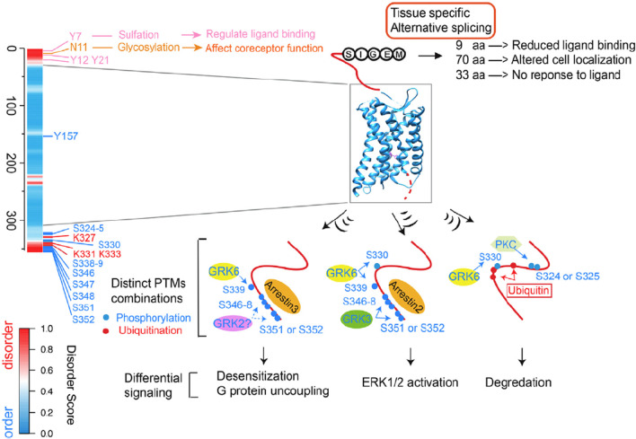
Verification
Claims the paper makes
118 Not checked yet · 15 Own interpretation · 4 No basis given · 1 Supported · 1 Supported so far · 1 Partly supported · 1 Own result · accuracy not yet measured
The cited sources support only part of this claim — read each source’s verdict below.
In the paper
“Mechanical stretching similarly unfolds talin and other mechanosensitive proteins, thus exposing cryptic binding sites [225, 226].”
§ Intrinsically disordered proteins play diverse roles in cell signaling > IDRS/IDPS pervade pathways that respond to a wide variety of signals
What the sources say
- Partly supportedcites [225] Goult BT, Yan J, Scwartz MA. Talin as a mechanosensitive signaling hub. J Cell Biol. 2018;217:3776–84. sourceExperimental — accuracy not yet measured
The section on talin rod domains as mechanochemical switches describes force-induced unfolding that changes which ligands bind talin. The Temporal interactions section also discusses an exposed vinculin-binding helix. These passages support the claim for talin, but do not establish that stretching unfolds other mechanosensitive proteins to expose their binding sites. (LLM, conf=0.84)
- Supportedcites [226] Margadant F, Chew LL, Hu X, Bate N, Zhang X, Sheetz M. Mechanotransduction in vivo by repeated Talin stretch-relaxation events depends upon Vinculin. PLoS Biol. 2011;9:e1001223. source
The Discussion (Figure 6) describes mechanically pulled talin unfolding in steps that enable vinculin binding. The Introduction also says stretching proteins can uncover binding sites in talin and phosphorylation sites in p130Cas, supporting the broader mechanosensitive-protein point. (LLM, conf=0.89)
plaindirect- Asserted: passes — The authors put it forward as true.
- Checkable: passes — It can be checked against evidence.
- Load-bearing: unclear — It is unclear whether the paper’s conclusions depend on it.
- Based: passes — The paper gives a reason why it holds.
- Partly supported
Every source the paper cites for this claim was checked and supports it.
In the paper
“Intriguingly, the disordered linker is required to transduce the pH-dependent status of the periplasmic sensing domain to the DNA binding domain via dimerization.”
§ Intrinsically disordered proteins play diverse roles in cell signaling > IDRS/IDPS pervade pathways that respond to a wide variety of signals
What the sources say
- Supportedcites [161] Buchner S, Schlundt A, Lassak J, Sattler M, Jung K. Structural and functional analysis of the signal-transducing linker in the pH-responsive one-component system CadC of Escherichia coli. J Mol Biol. 2015;427:2548–61. source
The Abstract (p. 3) proposes that the disordered CadC linker transduces the periplasmic pH response through a rearrangement that facilitates dimerization of the cytoplasmic DNA-binding domain. The manuscript (pp. 15–16) likewise describes the linker as coupling the periplasmic sensor to DNA-binding-domain dimer formation; the proposed mechanism matches the citing sentence. (LLM, conf=0.91)
plaindirect- Asserted: passes — The authors put it forward as true.
- Checkable: passes — It can be checked against evidence.
- Load-bearing: passes — The paper’s conclusions depend on it.
- Based: passes — The paper gives a reason why it holds.
- Supported
The sources with a verdict support this claim; the rest weren’t checked yet, have no full text we could read, or their verdicts aren’t rated yet.
In the paper
“The two kinases phosphorylate residues within a long IDR at β-catenin’s amino terminus [259, 260].”
§ Intrinsically disordered proteins play diverse roles in cell signaling > IDRS/IDPS are found in every category of cell signaling pathways
What the sources say
- Supportedcites [259] Xue B, Romero PR, Noutsou M, Maurice MM, Rüdiger SG, William AM Jr, et al. Stochastic machines as a colocalization mechanism for scaffold protein function. FEBS Lett. 2013;587:1587–91. source
The Introduction says CKI-α phosphorylates β-catenin first, followed by GSK-3β at three other sites. Results and discussion (‘Intrinsic disorder in the β-catenin destruction complex’; page unavailable) describes β-catenin’s termini as disordered, and ‘The Stochastic Machine Mechanism’ identifies the phosphorylation sites as Ser45, Thr41, Ser37, and Ser33—residues near its amino terminus. (LLM, conf=0.85)
- Not checked yetcites [260] Kimelman D, Xu W. beta-catenin destruction complex: insights and questions from a structural perspective. Oncogene. 2006;25:7482–91. source
plaindirect- Asserted: passes — The authors put it forward as true.
- Checkable: passes — It can be checked against evidence.
- Load-bearing: unclear — It is unclear whether the paper’s conclusions depend on it.
- Based: passes — The paper gives a reason why it holds.
- Supported
The paper states this without a source or data behind it here, so there is nothing to check it against. The quote shows it is in the paper.
In the paper
“The remaining 115 proteins are either moderately disordered or highly disordered”
§ Intrinsically disordered proteins play diverse roles in cell signaling > IDRS/IDPS are found in every step of cell signaling pathways
plain- Asserted: passes — The authors put it forward as true.
- Checkable: passes — It can be checked against evidence.
- Load-bearing: unclear — It is unclear whether the paper’s conclusions depend on it.
- Based: passes — The paper gives a reason why it holds.
A conclusion the paper draws from its own results — it cites no other source for it, so there is none to check. The quote shows it is in the paper.
In the paper
“This sensitivity, adaptability, and tunability are possible, in part, due to the inclusion of intrinsically disordered regions in many proteins involved in cell signaling.”
§ Intrinsically disordered proteins play diverse roles in cell signaling > Abstract
hedgedconditional- Asserted: passes — The authors put it forward as true.
- Checkable: passes — It can be checked against evidence.
- Load-bearing: passes — The paper’s conclusions depend on it.
- Based: unclear — It is unclear why the paper says it holds.
None of the sources cited for this claim have been checked yet.
In the paper
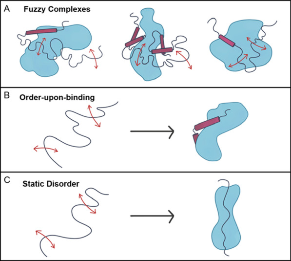
“Some IDPs/IDRs may remain unfolded and dynamic even in the bound state [19–23], creating fuzzy complexes (Fig. 1) [24, 25].”
§ Intrinsically disordered proteins play diverse roles in cell signaling > Background
What the sources say
- Not checked yetcites [19] Borgia A, Borgia MB, Bugge K, Kissling VM, Heidarsson PO, Fernandes CB, et al. Extreme disorder in an ultrahigh-affinity protein complex. Nature. 2018;55:61–6. source
- Not checked yetcites [20] Borg M, Mittag T, Pawson T, Tyers M, Forman-Kay JD, Chan HS. Polyelectrostatic interactions of disordered ligands suggest a physical basis for ultrasensitivity. Proc Natl Acad Sci USA. 2007;104:9650–5. source
- Not checked yetcites [21] Permyakov SE, Millett IS, Doniach S, Perkyakov EA, Uversky VN. Natively unfolded C-terminal domain of caldesmon remains substantially unstructured after the effective binding to calmodulin. Proteins. 2003;53:855–62. source
- Not checked yetcites [22] Sigalov AB, Zhuravleva A, Orekhov VY. Binding of intrinsically disordered proteins is not necessarily accompanied by a structural transition to a folded form. Biochimie. 2007;89:419–21. source
- Not checked yetcites [23] Sigalov AB. Protein intrinsic disorder and oligomericity in cell signaling. Mol Biosyst. 2010;6:451–61. source
- Not checked yetcites [24] Fuxreiter M, Simon I, Bondos S. Dynamic protein-DNA recognition: beyond what can be seen. Trends Biochem Sci. 2011;36:415–23. source
- Not checked yetcites [25] Fuxreiter M, Tompa P. Fuzzy complexes: a more stochastic view of protein function. Adv Exp Med Biol. 2012;725:1–14. source
hedgedconditional- Asserted: passes — The authors put it forward as true.
- Checkable: passes — It can be checked against evidence.
- Load-bearing: unclear — It is unclear whether the paper’s conclusions depend on it.
- Based: passes — The paper gives a reason why it holds.
None of the sources cited for this claim have been checked yet.
In the paper
“Many IDPs/IDRs are capable of at least partial ordering upon interaction with specific partners [7–18].”
§ Intrinsically disordered proteins play diverse roles in cell signaling > Background
What the sources say
- Not checked yetcites [7] Wright PE, Dyson HJ. Intrinsically unstructured proteins: re-assessing the protein structure-function paradigm. J Mol Biol. 1999;293:321–31. source
- Not checked yetcites [8] Dunker AK, Obradović Z. The protein trinity - linking function and disorder. Nat Biotechnol. 2001;19:805–6. source
- Not checked yetcites [9] Dunker AK, Brown CJ, Lawson JD, Iakoucheva LM, Obradović Z. Intrinsic disorder and protein function. Biochemistry. 2002;41:6573–82. source
- Not checked yetcites [10] Dunker AK, Cortese MS, Romero P, Iakoucheva LM, Uversky VN. Flexible nets: the roles of intrinsic disorder in protein interaction networks. FEBS J. 2005;272:5129–48. source
- Not checked yetcites [11] Daughdrill GW, Pielak GJ, Uversky VN, Cortese MS, Dunker AK. Natively disordered proteins. In: Buchner J, Kiefhaber T, editors. Handbook of protein folding. Weinheim: Wiley, Verlag GmbH & Co KGaA; 2005. p. 271–353.
- Not checked yetcites [12] Dyson HJ, Wright PE. Coupling of folding and binding for unstructured proteins. Curr Opin Struct Biol. 2002;12:54–60. source
- Not checked yetcites [13] Wright DHJPE. Intrinsically unstructured proteins and their functions. Nat Rev Mol Cell Biol. 2005;6:197–208. source
- Not checked yetcites [14] Iakoucheva LM, Brown CJ, Lawson JD, Obradović Z, Dunker AK. Intrinsic disorder in cell-signaling and cancer-associated proteins. J Mol Biol. 2002;323:573–84. source
- Not checked yetcites [15] Plaxco KW, Gross M. Cell biology. The importance of being unfolded. Nature. 1997;386:657–9. source
- Not checked yetcites [16] Pontius BW. Close encounters: why unstructured, polymeric domains can increase rates of specific macromolecular association. Trends Biochem Sci. 1993;18:181–6. source
plainconditional- Asserted: passes — The authors put it forward as true.
- Checkable: passes — It can be checked against evidence.
- Load-bearing: passes — The paper’s conclusions depend on it.
- Based: passes — The paper gives a reason why it holds.
A conclusion the paper draws from its own results — it cites no other source for it, so there is none to check. The quote shows it is in the paper.
In the paper
“These conflicting needs have been resolved to a large extent by including intrinsically disordered proteins in cell signaling pathways through biological evolution.”
§ Intrinsically disordered proteins play diverse roles in cell signaling > Background
plaindirect- Asserted: passes — The authors put it forward as true.
- Checkable: passes — It can be checked against evidence.
- Load-bearing: passes — The paper’s conclusions depend on it.
- Based: passes — The paper gives a reason why it holds.
A conclusion the paper draws from its own results — it cites no other source for it, so there is none to check. The quote shows it is in the paper.
In the paper
“Thus, a cell signaling pathway cannot be fully described without understanding how intrinsically disordered protein regions contribute to its function.”
§ Intrinsically disordered proteins play diverse roles in cell signaling > Abstract
plaindirect- Asserted: passes — The authors put it forward as true.
- Checkable: unclear — It is unclear whether it could be checked against evidence.
- Load-bearing: passes — The paper’s conclusions depend on it.
- Based: passes — The paper gives a reason why it holds.
None of the sources cited for this claim have been checked yet.
In the paper
“Upon binding of an IDP/IDR to a signaling partner, the free energy required to bring about the disorder-to-order transition subtracts from the interfacial, contact-free energy, resulting in a highly specific interaction that can be combined with a low net free energy of association [3, 18].”
§ Intrinsically disordered proteins play diverse roles in cell signaling > Background
What the sources say
- Not checked yetcites [3] Dunker AK, Lawson JD, Brown CJ, Williams RM, Romero P, Oh JS, et al. Intrinsically disordered protein. J Mol Graph Model. 2001;19:26–59. source
- Not checked yetcites [18] Schulz GE. Nucleotide binding proteins. In: Balaban M, editor. Molecular mechanism of biological recognition. New York: Elsevier/North-Holland Biomedical Press; 1979. p. 79–94.
plaindirect- Asserted: passes — The authors put it forward as true.
- Checkable: passes — It can be checked against evidence.
- Load-bearing: passes — The paper’s conclusions depend on it.
- Based: passes — The paper gives a reason why it holds.
- It may state two separate claims as one. 63
- Probably not a substantial claim. 11
None of the sources cited for this claim have been checked yet.
In the paper
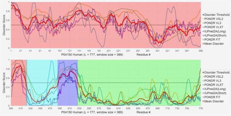
“Clearly, the N-terminal domain and a hinge region are mostly disordered”
§ Intrinsically disordered proteins play diverse roles in cell signaling > IDRS/IDPS pervade pathways that respond to a wide variety of signals
What the sources say
- Not checked yetcites [179] Obradovic Z, Peng K, Vucetic S, Radivojac P, Dunker AK. Exploiting heterogeneous sequence properties improves prediction of protein disorder. Protein. 2005;61:176–82. source
- Not checked yetcites [180] Obradovic Z, Peng K, Vucetic S, Radivojac P, Brown CJ, Dunker AK. Predicting intrinsic disorder from amino acid sequence. Protein. 2003;53:566–72. source
- Not checked yetcites [181] Xue B, Dunbrack RL, Williams RW, Dunker AK, Uversky VN. PONDR-FIT: a meta-predictor of intrinsically disordered amino acids. Biochim Biophys Acta. 2010;1804:996–1010. source
boosted- Asserted: passes — The authors put it forward as true.
- Checkable: passes — It can be checked against evidence.
- Load-bearing: unclear — It is unclear whether the paper’s conclusions depend on it.
- Based: passes — The paper gives a reason why it holds.
It may state two separate claims as one.
A result the paper reports from its own data — it cites no other source for it, so there is none to check. The quote shows it is in the paper.
In the paper
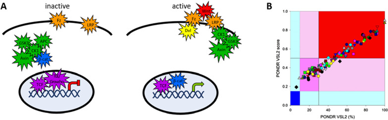
“It is noteworthy that no Wnt pathway proteins are very structured (dark blue) and only two proteins can be considered”
§ Intrinsically disordered proteins play diverse roles in cell signaling > IDRS/IDPS are found in every step of cell signaling pathways
plain- Asserted: passes — The authors put it forward as true.
- Checkable: passes — It can be checked against evidence.
- Load-bearing: unclear — It is unclear whether the paper’s conclusions depend on it.
- Based: passes — The paper gives a reason why it holds.
It may state two separate claims as one.
The paper states this without a source or data behind it here, so there is nothing to check it against. The quote shows it is in the paper.
In the paper
“So far more than 30 of these protein interaction domains have been shown by published experiments and/or by prediction to bind to DBRs in IDRs, with none so far binding to structured domains (work in progress).”
§ Intrinsically disordered proteins play diverse roles in cell signaling > IDRS/IDPS are found in every step of cell signaling pathways
plain- Asserted: passes — The authors put it forward as true.
- Checkable: passes — It can be checked against evidence.
- Load-bearing: passes — The paper’s conclusions depend on it.
- Based: passes — The paper gives a reason why it holds.
It may state two separate claims as one.
A conclusion the paper draws from its own results — it cites no other source for it, so there is none to check. The quote shows it is in the paper.
In the paper
“Thus binding-induced folding decouples binding affinity from specificity, enabling cell signaling to be reversible.”
§ Intrinsically disordered proteins play diverse roles in cell signaling > Background
plaindirect- Asserted: passes — The authors put it forward as true.
- Checkable: passes — It can be checked against evidence.
- Load-bearing: passes — The paper’s conclusions depend on it.
- Based: passes — The paper gives a reason why it holds.
It may state two separate claims as one.
In the paper
“An extreme example of this behavior is provided by two highly disordered human proteins, histone H1 and its nuclear chaperone prothymosin-α, which form a picomolar affinity complex, but in which they preserve completely their structural disorder, their long-range flexibility, and their highly dynamic character [19].”
§ Intrinsically disordered proteins play diverse roles in cell signaling > Background
What the sources say
- Not checked yetcites [19] Borgia A, Borgia MB, Bugge K, Kissling VM, Heidarsson PO, Fernandes CB, et al. Extreme disorder in an ultrahigh-affinity protein complex. Nature. 2018;55:61–6. source
plain- Asserted: passes — The authors put it forward as true.
- Checkable: passes — It can be checked against evidence.
- Load-bearing: unclear — It is unclear whether the paper’s conclusions depend on it.
- Based: passes — The paper gives a reason why it holds.
It may state two separate claims as one.
None of the sources cited for this claim have been checked yet.
In the paper
“The PTEN protein contains an N-terminal PI(4,5)P2 binding site, and a structured catalytic domain followed by an intrinsically disordered auto-inhibitory C-terminal tail which culminates in a PDZ binding domain [188, 189].”
§ Intrinsically disordered proteins play diverse roles in cell signaling > IDRS/IDPS pervade pathways that respond to a wide variety of signals
What the sources say
- Not checked yetcites [188] Lesli NR, Batty IH, Maccario H, Davidson L, Downes CP. Understanding PTEN regulation: PIP2, polarity and protein stability. Oncogene. 2008;27:5464–76. source
- Not checked yetcites [189] Odriozola L, Singh G, Hoang T, Chang AM. Regulation of PTEN activity by its carboxyl-terminal autoinhibitory domain. J Biol Chem. 2007;282:23306–15. source
plain- Asserted: passes — The authors put it forward as true.
- Checkable: passes — It can be checked against evidence.
- Load-bearing: unclear — It is unclear whether the paper’s conclusions depend on it.
- Based: passes — The paper gives a reason why it holds.
It may state two separate claims as one.
A conclusion the paper draws from its own results — it cites no other source for it, so there is none to check. The quote shows it is in the paper.
In the paper
“The concentration of both PTMs and segments encoded by alternative splicing within IDRs suggest these two sequence modifications may be co-localized, providing an opportunity to integrate two different regulatory inputs.”
§ Intrinsically disordered proteins play diverse roles in cell signaling > Background
hedgedconditional- Asserted: passes — The authors put it forward as true.
- Checkable: passes — It can be checked against evidence.
- Load-bearing: unclear — It is unclear whether the paper’s conclusions depend on it.
- Based: passes — The paper gives a reason why it holds.
It may state two separate claims as one.
None of the sources cited for this claim have been checked yet.
In the paper
“Likewise, phosphorylation [41] and many but not all other PTMs [42] show a strong preference to be localized in IDRs, likely because flexibility enhances the ability of a motif, destined for PTM addition, to enter and bind to the active site of the corresponding enzyme.”
§ Intrinsically disordered proteins play diverse roles in cell signaling > Background
What the sources say
- Not checked yetcites [41] Iakoucheva LM, Radivojac P, Brown CJ, O’Connor TR, Sikes JG, Obradovic Z, Dunker AK. The importance of intrinsic disorder for protein phosphorylation. Nucleic Acids Res. 2004;32:1037–49. source
- Not checked yetcites [42] Gao J, Xu D. Correlation between posttranslational modification and intrinsic disorder in protein. In: Pacific symposium on Biocomputing;2012. p. 94–103. source
boostedcorrelational- Asserted: passes — The authors put it forward as true.
- Checkable: passes — It can be checked against evidence.
- Load-bearing: unclear — It is unclear whether the paper’s conclusions depend on it.
- Based: passes — The paper gives a reason why it holds.
It may state two separate claims as one.
In the paper
“Consequently, PTEN is not only a powerful tumor suppressor, but also a prognostic marker that predicts response in many human cancers [187].”
§ Intrinsically disordered proteins play diverse roles in cell signaling > IDRS/IDPS pervade pathways that respond to a wide variety of signals
What the sources say
- Not checked yetcites [187] Mingo J, Luna S, Gaafar A, Nunes-Xavier E, Torices L, Mosteiro L. Precise definition of PTEN C-terminal epitopes and its implications in clinical oncology. Precision Oncol. 2019;3:11. source
plaincorrelational- Asserted: passes — The authors put it forward as true.
- Checkable: passes — It can be checked against evidence.
- Load-bearing: unclear — It is unclear whether the paper’s conclusions depend on it.
- Based: passes — The paper gives a reason why it holds.
It may state two separate claims as one.
A conclusion the paper draws from its own results — it cites no other source for it, so there is none to check. The quote shows it is in the paper.
In the paper
“Because some signaling IDPs/IDRs lack stable (secondary) structure, the energetic barriers between the bound and free states are low, allowing disordered regions to act as reversible, extremely sensitive sensors.”
§ Intrinsically disordered proteins play diverse roles in cell signaling > Background
plaindirect- Asserted: passes — The authors put it forward as true.
- Checkable: passes — It can be checked against evidence.
- Load-bearing: passes — The paper’s conclusions depend on it.
- Based: passes — The paper gives a reason why it holds.
It may state two separate claims as one.
None of the sources cited for this claim have been checked yet.
In the paper
“Finally, disordered proteins provide many avenues for integrating multiple signaling pathways [27], including providing a scaffold that binds proteins from multiple pathways [34], regulating multiple disordered substrates through PTMs, and varying pathway components through alternative mRNA splicing [35–37].”
§ Intrinsically disordered proteins play diverse roles in cell signaling > Background
What the sources say
- Not checked yetcites [27] Kreiger JM, Fusco G, Lewitzky M, Simister PC, Marchant J, Camilloni C, et al. Conformational recognition of an intrinsically disordered protein. Biophys J. 2014;106:1771–9. source
- Not checked yetcites [34] Martin-Yken H, Francois JM, Zerbib D. Knr4: a disordered hub protein at the heart of fungal cell wall signalling. Cell Microbiol. 2016;18:1217–27. source
- Not checked yetcites [35] Gough J, Dunker AK. Sequences and topology: disorder, modularity, and post/pre-translation modifications. Curr Opin Struct Biol. 2013;23:417–9. source
- Not checked yetcites [36] Kawamura H, Li X, Harper SJ, Bates DO, Claesson-Welsh L. Vascular endothelial growth factor (VEGF)-A165b is a weak in vitro agonist for VEGF receptor-2 due to lack of coreceptor binding and deficient regulation of kinase activity. Cancer Res. 2008;68:4683–92. source
- Not checked yetcites [37] Li J, White JT, Saavedra H, Wrabl JO, Motlagh HN, Liu K, Sower J, et al. Genetically tunable frustration controls allostery in an intrinsically disordered transcription factor. eLife. 2017;6:e30688. source
plaindirect- Asserted: passes — The authors put it forward as true.
- Checkable: passes — It can be checked against evidence.
- Load-bearing: unclear — It is unclear whether the paper’s conclusions depend on it.
- Based: passes — The paper gives a reason why it holds.
It may state two separate claims as one.
In the paper
“These differences can carry over to following generations, indicating that these multiple PTMs provide the basis for an epigenetic mechanism.”
§ Intrinsically disordered proteins play diverse roles in cell signaling > Background
What the sources say
- Not checked yetcites [44] Strahl BD, Allis CD. The language of covalent histone modifications. Nature. 2000;403:41–5. source
hedgeddirect- Asserted: passes — The authors put it forward as true.
- Checkable: passes — It can be checked against evidence.
- Load-bearing: unclear — It is unclear whether the paper’s conclusions depend on it.
- Based: passes — The paper gives a reason why it holds.
It may state two separate claims as one.
In the paper
“Finally, histone tails may be accessible or may adhere to the nucleosome and be inaccessible for protein interactions, demonstrating that the context provided by the nucleosome is also a key contributor [53].”
§ Intrinsically disordered proteins play diverse roles in cell signaling > Background
What the sources say
- Not checked yetcites [53] Morrison EA, Bowerman S, Sylvers KL, Wereszczynski J, Musselman CA. The conformation of the histone H3 tail inhibits association of the BPTG PHD finger with the nucleosome. eLife. 2018;7:e31481. source
boostedconditional- Asserted: passes — The authors put it forward as true.
- Checkable: passes — It can be checked against evidence.
- Load-bearing: unclear — It is unclear whether the paper’s conclusions depend on it.
- Based: passes — The paper gives a reason why it holds.
It may state two separate claims as one.
A conclusion the paper draws from its own results — it cites no other source for it, so there is none to check. The quote shows it is in the paper.
In the paper
“Whether or not stable structure results from the interaction, ligand or protein interactions shift the conformational ensemble of the IDP/IDR, linking protein function to binding.”
§ Intrinsically disordered proteins play diverse roles in cell signaling > Background
plaindirect- Asserted: passes — The authors put it forward as true.
- Checkable: passes — It can be checked against evidence.
- Load-bearing: passes — The paper’s conclusions depend on it.
- Based: passes — The paper gives a reason why it holds.
It may state two separate claims as one.
None of the sources cited for this claim have been checked yet.
In the paper
“Phosphorylation of most of these amino acids forces PTEN into a closed, more stable, inactive conformation, in which membrane association, PDZ binding, ubiquitination, and degradation are all suppressed [188, 190].”
§ Intrinsically disordered proteins play diverse roles in cell signaling > IDRS/IDPS pervade pathways that respond to a wide variety of signals
What the sources say
- Not checked yetcites [188] Lesli NR, Batty IH, Maccario H, Davidson L, Downes CP. Understanding PTEN regulation: PIP2, polarity and protein stability. Oncogene. 2008;27:5464–76. source
- Not checked yetcites [190] Rahdar M, Inoue T, Zhang J, Vazquez F, Devreotes PN. A phosphorylation-dependent intramolecular interactions regulates the membrane association and activity of the tumor suppressor PTEN. Proc Natl Acad Sci USA. 2009;106:480–5. source
plaindirect- Asserted: passes — The authors put it forward as true.
- Checkable: passes — It can be checked against evidence.
- Load-bearing: unclear — It is unclear whether the paper’s conclusions depend on it.
- Based: passes — The paper gives a reason why it holds.
It may state two separate claims as one.
In the paper
“Furthermore, the supplemental data in Zhou et al., 2018 show that all of these proteins undergo alternative splicing, suggesting the possibility that alternative splicing could be a previously unsuspected regulator of the epigenetic effects resulting from the histone code [39].”
§ Intrinsically disordered proteins play diverse roles in cell signaling > Background
What the sources say
- Not checked yetcites [39] Zhou J, Zhao S, Dunker AK. Intrinsically disordered proteins link alternative splicing and post-translational modifications to complex cell signaling and regulation. J Mol Biol. 2018;430:2342–59. source
hedgedconditional- Asserted: passes — The authors put it forward as true.
- Checkable: passes — It can be checked against evidence.
- Load-bearing: fails — The paper’s conclusions may not depend on it.
- Based: passes — The paper gives a reason why it holds.
Probably not a substantial claim. The paper’s conclusions may not depend on it. It may state two separate claims as one.
In the paper
“Inactive PTEN exists in a cytoplasmic pool and lacks substrate access [188].”
§ Intrinsically disordered proteins play diverse roles in cell signaling > IDRS/IDPS pervade pathways that respond to a wide variety of signals
What the sources say
- Not checked yetcites [188] Lesli NR, Batty IH, Maccario H, Davidson L, Downes CP. Understanding PTEN regulation: PIP2, polarity and protein stability. Oncogene. 2008;27:5464–76. source
plain- Asserted: passes — The authors put it forward as true.
- Checkable: passes — It can be checked against evidence.
- Load-bearing: unclear — It is unclear whether the paper’s conclusions depend on it.
- Based: passes — The paper gives a reason why it holds.
It may state two separate claims as one.
A conclusion the paper draws from its own results — it cites no other source for it, so there is none to check. The quote shows it is in the paper.
In the paper
“Thus, IDRs are an important feature of both the PTM code and the epigenetic effects observed for the histone code.”
§ Intrinsically disordered proteins play diverse roles in cell signaling > Background
plaincorrelational- Asserted: passes — The authors put it forward as true.
- Checkable: passes — It can be checked against evidence.
- Load-bearing: passes — The paper’s conclusions depend on it.
- Based: passes — The paper gives a reason why it holds.
It may state two separate claims as one.
None of the sources cited for this claim have been checked yet.
In the paper
“Osteopontin is a multifunctional cytokine with key roles in inflammation, cell viability, and tissue repair, which also functions as a bone matrix protein that mediates osteoclast adhesion [196, 197].”
§ Intrinsically disordered proteins play diverse roles in cell signaling > IDRS/IDPS pervade pathways that respond to a wide variety of signals
What the sources say
- Not checked yetcites [196] Icer MA, Gezmen-Karadag M. The multiple functions and mechanisms of osteopontin. Clin Biochem. 2018;59:17–24. source
- Not checked yetcites [197] O’Regan A, Berman JS. Osteopontin: a key cytokine in cell-mediated and granulomatous inflammation. Int J Exp Pathol. 2000;81:373–90. source
plaindirect- Asserted: passes — The authors put it forward as true.
- Checkable: passes — It can be checked against evidence.
- Load-bearing: fails — The paper’s conclusions may not depend on it.
- Based: passes — The paper gives a reason why it holds.
Probably not a substantial claim. The paper’s conclusions may not depend on it. It may state two separate claims as one.
None of the sources cited for this claim have been checked yet.
In the paper
“Interactions with other proteins via the PDZ binding domain or other regions of the protein both enhances PTEN stability and diversifies its function [188, 192].”
§ Intrinsically disordered proteins play diverse roles in cell signaling > IDRS/IDPS pervade pathways that respond to a wide variety of signals
What the sources say
- Not checked yetcites [188] Lesli NR, Batty IH, Maccario H, Davidson L, Downes CP. Understanding PTEN regulation: PIP2, polarity and protein stability. Oncogene. 2008;27:5464–76. source
- Not checked yetcites [192] Malaney P, Uversky VN, Davé V. Identification of intrinsically disordered region in PTEN and delineation of its function via a network approach. Methods. 2015;77–78:69–74. source
plaindirect- Asserted: passes — The authors put it forward as true.
- Checkable: passes — It can be checked against evidence.
- Load-bearing: unclear — It is unclear whether the paper’s conclusions depend on it.
- Based: passes — The paper gives a reason why it holds.
It may state two separate claims as one.
In the paper
“This trait allows IDPs to reliably sense extreme conditions and instigate responsive signaling pathways [26].”
§ Intrinsically disordered proteins play diverse roles in cell signaling > IDRS/IDPS pervade pathways that respond to a wide variety of signals
What the sources say
- Not checked yetcites [26] Launay H, Receveur-Brechot V, Carriere F, Gontero B. Ochestration of algal metabolism by protein disorder. Arch Biochem Biophys. 2019;672:108070. source
plaindirect- Asserted: passes — The authors put it forward as true.
- Checkable: passes — It can be checked against evidence.
- Load-bearing: passes — The paper’s conclusions depend on it.
- Based: passes — The paper gives a reason why it holds.
It may state two separate claims as one.
A conclusion the paper draws from its own results — it cites no other source for it, so there is none to check. The quote shows it is in the paper.
In the paper
“Signaling pathways activated by proteins often are regulated by IDPs/IDRs in multiple steps of the pathway.”
§ Intrinsically disordered proteins play diverse roles in cell signaling > IDRS/IDPS pervade pathways that respond to a wide variety of signals
plaindirect- Asserted: passes — The authors put it forward as true.
- Checkable: passes — It can be checked against evidence.
- Load-bearing: passes — The paper’s conclusions depend on it.
- Based: fails — The paper may give no reason why it holds.
Probably not a substantial claim. The paper may give no reason why it holds.
In the paper
“Thus, it is not surprising that PTEN and PTEN-L exhibit different substrate specificities and mechanisms of membrane binding [195].”
§ Intrinsically disordered proteins play diverse roles in cell signaling > IDRS/IDPS pervade pathways that respond to a wide variety of signals
What the sources say
- Not checked yetcites [195] Masson GR, Perisic O, Burke JE, Williams RL. The intrinsically disordered tails of PTEN and PTEN-L have distinct roles in regulating substrate specificity and membrane activity. Biochem J. 2016;473:135–44. source
plain- Asserted: passes — The authors put it forward as true.
- Checkable: passes — It can be checked against evidence.
- Load-bearing: unclear — It is unclear whether the paper’s conclusions depend on it.
- Based: passes — The paper gives a reason why it holds.
It may state two separate claims as one.
None of the sources cited for this claim have been checked yet.
In the paper
“The complexity of this machinery is determined by the multitude of the members of the GPCR family (in humans, there are more than 850 different GPCRs [202–205]) that are capable of being recognized and activated by more than one thousand natural and artificial extracellular ligands, ranging from photons to amines, lipids, nucleotides, organic odorants, peptides, and proteins [30, 204].”
§ Intrinsically disordered proteins play diverse roles in cell signaling > IDRS/IDPS pervade pathways that respond to a wide variety of signals
What the sources say
- Not checked yetcites [202] Bjarnadottir TK, Gloriam DE, Hellstrand SH, Kristiansson H, Fredriksson R, Schioth HB. Comprehensive repertoire and phylogenetic analysis of the G protein-coupled receptors in human and mouse. Genomics. 2006;88:263–73. source
- Not checked yetcites [203] Anantharaman V, Abhiman S, de Souza RF, Aravind L. Comparative genomics uncovers novel structural and functional features of the heterotrimeric GTPase signaling system. Gene. 2011;475:63–78. source
- Not checked yetcites [204] Fredriksson R, Lagerstrom MC, Lundin LG, Schioth HB. The G-protein-coupled receptors in the human genome form five main families. Phylogenetic analysis, paralogon groups, and fingerprints. Mol Pharmacol. 2003;63:1256–72. source
- Not checked yetcites [205] Southan C, Sharman JL, Benson HE, Faccenda E, Pawson AJ, Alexander SP, et al. The IUPHAR/BPS Guide to PHARMACOLOGY in 2016: towards curated quantitative interactions between 1300 protein targets and 6000 ligands. Nucl Acids Res. 2016;44:D1054–68. source
- Not checked yetcites [30] Flock T, Hauser AS, Lund N, Gloriam DE, Balaji S, Babu MM. Selectivity determinants of GPCR-G-protein binding. Nature. 2017;545:317–22. source
plain- Asserted: passes — The authors put it forward as true.
- Checkable: passes — It can be checked against evidence.
- Load-bearing: fails — The paper’s conclusions may not depend on it.
- Based: passes — The paper gives a reason why it holds.
Probably not a substantial claim. The paper’s conclusions may not depend on it.
In the paper
“The extent of disorder, and thus the activity, of CP12 is determined by redox conditions, although CP12 remains highly mobile in both the oxidized and reduced states.”
§ Intrinsically disordered proteins play diverse roles in cell signaling > IDRS/IDPS pervade pathways that respond to a wide variety of signals
What the sources say
- Not checked yetcites [26] Launay H, Receveur-Brechot V, Carriere F, Gontero B. Ochestration of algal metabolism by protein disorder. Arch Biochem Biophys. 2019;672:108070. source
plaindirect- Asserted: passes — The authors put it forward as true.
- Checkable: passes — It can be checked against evidence.
- Load-bearing: unclear — It is unclear whether the paper’s conclusions depend on it.
- Based: passes — The paper gives a reason why it holds.
It may state two separate claims as one.
In the paper
“Furthermore, cells contain ~ 40 of the regulator of G signaling (RGS) proteins (which are G protein effectors, modulators, and scaffold proteins) that are capable of interaction with various Gαβγ heterotrimers or their dissociated subunits [212].”
§ Intrinsically disordered proteins play diverse roles in cell signaling > IDRS/IDPS pervade pathways that respond to a wide variety of signals
What the sources say
- Not checked yetcites [212] Temple BR, Jones CD. Jones AM (2010) Evolution of a signaling nexus constrained by protein interfaces and conformational States. PLoS Comput Biol. 2010;6:e1000962. source
hedged- Asserted: passes — The authors put it forward as true.
- Checkable: passes — It can be checked against evidence.
- Load-bearing: fails — The paper’s conclusions may not depend on it.
- Based: passes — The paper gives a reason why it holds.
Probably not a substantial claim. The paper’s conclusions may not depend on it.
In the paper
“In humans, there are 23 Gα, 6 Gβ, and 12 Gγ subunits that can be assembled into numerous different heterotrimers [211].”
§ Intrinsically disordered proteins play diverse roles in cell signaling > IDRS/IDPS pervade pathways that respond to a wide variety of signals
What the sources say
- Not checked yetcites [211] Downes GB, Gautam N. The G protein subunit gene families. Genomics. 1999;62:544–52. source
plain- Asserted: passes — The authors put it forward as true.
- Checkable: passes — It can be checked against evidence.
- Load-bearing: fails — The paper’s conclusions may not depend on it.
- Based: passes — The paper gives a reason why it holds.
Probably not a substantial claim. The paper’s conclusions may not depend on it. It may state two separate claims as one.
None of the sources cited for this claim have been checked yet.
In the paper
“Multiple alternate translation and splicing start sites in the PTEN gene creates many versions of the protein, most notably PTEN-L, which contains an extra 173 amino acids on the N-terminus [187, 192, 193].”
§ Intrinsically disordered proteins play diverse roles in cell signaling > IDRS/IDPS pervade pathways that respond to a wide variety of signals
What the sources say
- Not checked yetcites [187] Mingo J, Luna S, Gaafar A, Nunes-Xavier E, Torices L, Mosteiro L. Precise definition of PTEN C-terminal epitopes and its implications in clinical oncology. Precision Oncol. 2019;3:11. source
- Not checked yetcites [192] Malaney P, Uversky VN, Davé V. Identification of intrinsically disordered region in PTEN and delineation of its function via a network approach. Methods. 2015;77–78:69–74. source
- Not checked yetcites [193] Masson GR, Williams RL. Structural mechanisms of PTEN regulation. Cold Spring Harbor Perspect Med. 2020;10:a036152. source
plaindirect- Asserted: passes — The authors put it forward as true.
- Checkable: passes — It can be checked against evidence.
- Load-bearing: unclear — It is unclear whether the paper’s conclusions depend on it.
- Based: passes — The paper gives a reason why it holds.
It may state two separate claims as one.
In the paper
“This region is also primarily disordered and post-translationally modified [192].”
§ Intrinsically disordered proteins play diverse roles in cell signaling > IDRS/IDPS pervade pathways that respond to a wide variety of signals
What the sources say
- Not checked yetcites [192] Malaney P, Uversky VN, Davé V. Identification of intrinsically disordered region in PTEN and delineation of its function via a network approach. Methods. 2015;77–78:69–74. source
plain- Asserted: passes — The authors put it forward as true.
- Checkable: passes — It can be checked against evidence.
- Load-bearing: unclear — It is unclear whether the paper’s conclusions depend on it.
- Based: passes — The paper gives a reason why it holds.
It may state two separate claims as one.
None of the sources cited for this claim have been checked yet.
In the paper
“This addition reverses the function of VEGF: while VEGF165 is a potent stimulator of angiogenesis, VEGF165b binds VEGF receptors but fails to activate them in a robust, sustained manner.”
§ Intrinsically disordered proteins play diverse roles in cell signaling > IDRS/IDPS pervade pathways that respond to a wide variety of signals
What the sources say
- Not checked yetcites [36] Kawamura H, Li X, Harper SJ, Bates DO, Claesson-Welsh L. Vascular endothelial growth factor (VEGF)-A165b is a weak in vitro agonist for VEGF receptor-2 due to lack of coreceptor binding and deficient regulation of kinase activity. Cancer Res. 2008;68:4683–92. source
- Not checked yetcites [200] Woolard J, Wang WY, Bevan HS, Qiu Y, Morbidelli L, Pritchard-Jones RO, et al. VEGF165b, an inhibitory vascular endothelial growth factor splice variant: mechanism of action in vivo effect on aniogenesis and endogenous protein expression. Cancer Res. 2004;64:7822–35. source
plaindirect- Asserted: passes — The authors put it forward as true.
- Checkable: passes — It can be checked against evidence.
- Load-bearing: unclear — It is unclear whether the paper’s conclusions depend on it.
- Based: passes — The paper gives a reason why it holds.
It may state two separate claims as one.
In the paper
“High-resolution solution NMR spectroscopy demonstrated that this long-lived pB intermediate is characterized by a noticeable level of disorder and exists as an ensemble of multiple conformers interconverting on a millisecond time scale [220].”
§ Intrinsically disordered proteins play diverse roles in cell signaling > IDRS/IDPS pervade pathways that respond to a wide variety of signals
What the sources say
- Not checked yetcites [220] Rubinstenn G, Vuister GW, Mulder FAA, Düx PE, Boelens R, Hellingwerf KJ, et al. Structural and dynamic changes of photoactive yellow protein during its photocycle in solution. Nat Struct Biol. 1998;5:568–70. source
boostedSingle case- Asserted: passes — The authors put it forward as true.
- Checkable: passes — It can be checked against evidence.
- Load-bearing: passes — The paper’s conclusions depend on it.
- Based: passes — The paper gives a reason why it holds.
It may state two separate claims as one.
In the paper
“In dark or oxidizing conditions, CP12 forms limited, marginally stable structure and 2 disulfide bonds which are required to bind and inactivate two enzymes that participate in the Calvin-Benson-Bassham cycle (glyceraldehyde-3-phosphate dehydrogenase (GAPDH) and phorphoribulokinase (PRK)).”
§ Intrinsically disordered proteins play diverse roles in cell signaling > IDRS/IDPS pervade pathways that respond to a wide variety of signals
What the sources say
- Not checked yetcites [26] Launay H, Receveur-Brechot V, Carriere F, Gontero B. Ochestration of algal metabolism by protein disorder. Arch Biochem Biophys. 2019;672:108070. source
plaindirect- Asserted: passes — The authors put it forward as true.
- Checkable: passes — It can be checked against evidence.
- Load-bearing: unclear — It is unclear whether the paper’s conclusions depend on it.
- Based: passes — The paper gives a reason why it holds.
It may state two separate claims as one.
In the paper
“In fact, human GPCRs and G proteins represent dynamic conformational ensembles containing multiple IDPRs and numerous PTMs and MoRFs, and the entire mode of action of these proteins is based on the recognition of a signal followed by conformational change needed for recognition of another partner that is crucial for the downstream transmission of the signal [213].”
§ Intrinsically disordered proteins play diverse roles in cell signaling > IDRS/IDPS pervade pathways that respond to a wide variety of signals
What the sources say
- Not checked yetcites [213] Fonin AV, Darling AL, Kuznetsova IM, Tuoverov KK, Uversky VN. Multi-functionality of proteins involved in GPCR and G protein signaling: making sense of structure-function continuum with intrinsic disorder-based proteoforms. Cell Mol Life Sci. 2019;76:4461–92. source
plaindirect- Asserted: passes — The authors put it forward as true.
- Checkable: passes — It can be checked against evidence.
- Load-bearing: passes — The paper’s conclusions depend on it.
- Based: passes — The paper gives a reason why it holds.
It may state two separate claims as one.
In the paper
“Photoexcitation of UVR8 triggers dimer dissociation and enables the extended C-terminal tail to bind COP1, which regulates light signaling in plants, and propagates the signal [162].”
§ Intrinsically disordered proteins play diverse roles in cell signaling > IDRS/IDPS pervade pathways that respond to a wide variety of signals
What the sources say
- Not checked yetcites [162] Camacho IS, Theisen A, Johannissen LO, Diaz-Raos LA, Christie JM, Jenkins GI, et al. Native mass spectrometry reveals the conformational diversity of the UVR8 photoreceptor. Proc Natl Acad Sci USA. 2019;116:1116–25. source
plaindirect- Asserted: passes — The authors put it forward as true.
- Checkable: passes — It can be checked against evidence.
- Load-bearing: unclear — It is unclear whether the paper’s conclusions depend on it.
- Based: passes — The paper gives a reason why it holds.
It may state two separate claims as one.
In the paper
“In light/reducing conditions, the disulfides bonds break and the CP12-GAPDH-PRK ternary complex dissociates, re-activating the enzymes and thus carbon fixation.”
§ Intrinsically disordered proteins play diverse roles in cell signaling > IDRS/IDPS pervade pathways that respond to a wide variety of signals
What the sources say
- Not checked yetcites [26] Launay H, Receveur-Brechot V, Carriere F, Gontero B. Ochestration of algal metabolism by protein disorder. Arch Biochem Biophys. 2019;672:108070. source
plaindirect- Asserted: passes — The authors put it forward as true.
- Checkable: passes — It can be checked against evidence.
- Load-bearing: unclear — It is unclear whether the paper’s conclusions depend on it.
- Based: passes — The paper gives a reason why it holds.
It may state two separate claims as one.
The paper states this without a source or data behind it here, so there is nothing to check it against. The quote shows it is in the paper.
In the paper
“Most of the intrinsically disordered proteins with well-studied roles in signaling operate in endocrine signaling pathways.”
§ Intrinsically disordered proteins play diverse roles in cell signaling > IDRS/IDPS are found in every category of cell signaling pathways
plaincorrelational- Asserted: passes — The authors put it forward as true.
- Checkable: passes — It can be checked against evidence.
- Load-bearing: fails — The paper’s conclusions may not depend on it.
- Based: fails — The paper may give no reason why it holds.
Probably not a substantial claim. The paper’s conclusions may not depend on it. The paper may give no reason why it holds.
In the paper
“Phosphorylation of the C-terminal domain of Connexin 43 inhibits channel function, whereas phosphorylation of Connexin 32 stimulates channel function [254].”
§ Intrinsically disordered proteins play diverse roles in cell signaling > IDRS/IDPS are found in every category of cell signaling pathways
What the sources say
- Not checked yetcites [254] Trease AJ, Li H, Spagnol G, Zheng L, Stauch KL, Sorgen PL. Regulation of Connexin32 by ephrin receptors and T-cell protein-tyrosine phosphatase. J Biol Chem. 2019;294:341–50. source
plaindirect- Asserted: passes — The authors put it forward as true.
- Checkable: passes — It can be checked against evidence.
- Load-bearing: unclear — It is unclear whether the paper’s conclusions depend on it.
- Based: passes — The paper gives a reason why it holds.
It may state two separate claims as one.
None of the sources cited for this claim have been checked yet.
In the paper
“These modifications diversify the intracellular sites available for protein interactions, allowing different complexes to be formed and altering the transmitted signal [169, 255].”
§ Intrinsically disordered proteins play diverse roles in cell signaling > IDRS/IDPS are found in every category of cell signaling pathways
What the sources say
- Not checked yetcites [169] Warnet XL, Krog HB, Sevillano-Quispe OG, Poulsen H, Kjaergaard M. The C-terminal domains of the NMDA receptor: how intrinsically disordered tails affect signaling, plasticity, and disease. Eur J Neurosci. 2020. https://doi.org/10.1111/EJN.14842. source
- Not checked yetcites [255] Enz R. Structure of metabotropic glutamate receptor C-terminal domains in contact with interacting proteins. Front Mol Neurosci. 2012;5:52.
plaindirect- Asserted: passes — The authors put it forward as true.
- Checkable: passes — It can be checked against evidence.
- Load-bearing: unclear — It is unclear whether the paper’s conclusions depend on it.
- Based: passes — The paper gives a reason why it holds.
It may state two separate claims as one.
None of the sources cited for this claim have been checked yet.
In the paper
“In line with this hypothesis, there are several studies showing that ERα contains an intrinsically disordered transactivation domain (AF1) in its N terminus [244], activity of which is regulated by phosphorylation and associated phosphorylation-coupled proline isomerization [245, 246].”
§ Intrinsically disordered proteins play diverse roles in cell signaling > IDRS/IDPS are found in every category of cell signaling pathways
What the sources say
- Not checked yetcites [244] Peng Y, Cao S, Kiselar J, Xiao X, Du Z, Hsieh A, et al. A metastable contact and structural disorder in the estrogen receptor transactivation domain. Structure. 2019;27:229–40. source
- Not checked yetcites [245] Rajbhandari P, Finn G, Solodin NM, Singarapu KK, Sahu SC, Markley JL, et al. Regulation of estrogen receptor α N-terminus conformation and function by peptidyl prolyl isomerase Pin1. Mol Cell Bol. 2012;32:445–57. source
- Not checked yetcites [246] Rajbhandari P, Szatkowski Ozers M, Solodin NM, Warren CL, Alarid ET. Peptidylprolyl isomerase Pin1 directly enhances the DNA binding functions of Estrogen Receptor α. J Biol Chem. 2015;290:13749–62. source
plaindirect- Asserted: passes — The authors put it forward as true.
- Checkable: passes — It can be checked against evidence.
- Load-bearing: unclear — It is unclear whether the paper’s conclusions depend on it.
- Based: passes — The paper gives a reason why it holds.
It may state two separate claims as one.
In the paper
“Phosphorylation in this region regulates protein interaction, channel assembly, channel degradation, and metabolic and electrical coupling, and thus controls trafficking through the channel [253].”
§ Intrinsically disordered proteins play diverse roles in cell signaling > IDRS/IDPS are found in every category of cell signaling pathways
What the sources say
- Not checked yetcites [253] Sorgen PL, Trease AJ, Spagnol G, Delmmar M, Nielsen MS. Protein-protein interactions with Connexin 43” Regulation and function. Int J Mol Sci. 2018;19:1428. source
plaindirect- Asserted: passes — The authors put it forward as true.
- Checkable: passes — It can be checked against evidence.
- Load-bearing: unclear — It is unclear whether the paper’s conclusions depend on it.
- Based: passes — The paper gives a reason why it holds.
It may state two separate claims as one.
None of the sources cited for this claim have been checked yet.
In the paper
“In vitro, interactions with metal ions, other proteins, and sodium dodecyl sulfate induce the formation of limited structure for some dehydrins [228, 229], which can be transiently stable in vivo [230].”
§ Intrinsically disordered proteins play diverse roles in cell signaling > IDRS/IDPS pervade pathways that respond to a wide variety of signals
What the sources say
- Not checked yetcites [228] Yu ZY, Wang X, Zhang LS. Structural and functional dynamics of dehydrins: a plant protector protein under abiotic stress. Int J Mol Sci. 2018;19:3420. source
- Not checked yetcites [229] Yokoyama T, Ohkubo T, Kamiya K, Hara M. Cryoprotective activity of Arabidopsis KS-type dehydrin depends on the hydrophobic amino acids of the two active segments. Arch Biochem Biophys. 2020;691:108510. source
- Not checked yetcites [230] Murvai N, Kalmar L, Szalaine Agoston B, Szabo B, Tantos A, Csikos G, et al. Interplay of structural disorder and short binding elements in the cellular chaperone function of plant dehydrin ERD14. Cells. 2020;9:1856. source
plaindirect- Asserted: passes — The authors put it forward as true.
- Checkable: passes — It can be checked against evidence.
- Load-bearing: unclear — It is unclear whether the paper’s conclusions depend on it.
- Based: passes — The paper gives a reason why it holds.
It may state two separate claims as one.
None of the sources cited for this claim have been checked yet.
In the paper
“The CTDs of both receptors are alternatively spliced, and post-translationally modified (phosphorylation and SUMOylation for metabotropic receptors, phosphorylation and palmitoylation for ionotropic receptors) [169, 255].”
§ Intrinsically disordered proteins play diverse roles in cell signaling > IDRS/IDPS are found in every category of cell signaling pathways
What the sources say
- Not checked yetcites [169] Warnet XL, Krog HB, Sevillano-Quispe OG, Poulsen H, Kjaergaard M. The C-terminal domains of the NMDA receptor: how intrinsically disordered tails affect signaling, plasticity, and disease. Eur J Neurosci. 2020. https://doi.org/10.1111/EJN.14842. source
- Not checked yetcites [255] Enz R. Structure of metabotropic glutamate receptor C-terminal domains in contact with interacting proteins. Front Mol Neurosci. 2012;5:52.
plain- Asserted: passes — The authors put it forward as true.
- Checkable: passes — It can be checked against evidence.
- Load-bearing: unclear — It is unclear whether the paper’s conclusions depend on it.
- Based: passes — The paper gives a reason why it holds.
It may state two separate claims as one.
In the paper
“Interestingly, a mutation in the prodomain, V66M, is present 20% of humans and increases the occurrence of anxiety disorders, depression, memory deficits, and impairs recovery after traumatic brain injury (reviewed in [248]).”
§ Intrinsically disordered proteins play diverse roles in cell signaling > IDRS/IDPS are found in every category of cell signaling pathways
What the sources say
- Not checked yetcites [248] Wang J, Anastasia A, Bains H, Giza JI, Clossey DG, Deng J, et al. Zinc induced structural changes in the intrinsically disordered BDNF Met prodomain confer synaptic elimination. Metallomics. 2020;12:1208. source
plaindirect- Asserted: passes — The authors put it forward as true.
- Checkable: passes — It can be checked against evidence.
- Load-bearing: unclear — It is unclear whether the paper’s conclusions depend on it.
- Based: passes — The paper gives a reason why it holds.
It may state two separate claims as one.
In the paper
“Many cellular processes that are regulated by chemical stimuli, such as proliferation, differentiation, motility, and survival, are also influenced by the mechanical properties of the substrate supporting the cells [221].”
§ Intrinsically disordered proteins play diverse roles in cell signaling > IDRS/IDPS pervade pathways that respond to a wide variety of signals
What the sources say
- Not checked yetcites [221] Janmey PA, Miller RT. Mechanisms of mechanical signaling in development and disease. J Cell Sci. 2011;124:9–18. source
plaincorrelational- Asserted: passes — The authors put it forward as true.
- Checkable: passes — It can be checked against evidence.
- Load-bearing: fails — The paper’s conclusions may not depend on it.
- Based: passes — The paper gives a reason why it holds.
Probably not a substantial claim. The paper’s conclusions may not depend on it.
In the paper
“Dehydrins protect plants from dehydration and from cold [228].”
§ Intrinsically disordered proteins play diverse roles in cell signaling > IDRS/IDPS pervade pathways that respond to a wide variety of signals
What the sources say
- Not checked yetcites [228] Yu ZY, Wang X, Zhang LS. Structural and functional dynamics of dehydrins: a plant protector protein under abiotic stress. Int J Mol Sci. 2018;19:3420. source
plaindirect- Asserted: passes — The authors put it forward as true.
- Checkable: passes — It can be checked against evidence.
- Load-bearing: unclear — It is unclear whether the paper’s conclusions depend on it.
- Based: passes — The paper gives a reason why it holds.
It may state two separate claims as one.
In the paper
“Dehydrins are a family of intrinsically disordered proteins that act as effectors whose expression is induced by signaling pathways that sense abiotic stresses, such as cold or draught.”
§ Intrinsically disordered proteins play diverse roles in cell signaling > IDRS/IDPS pervade pathways that respond to a wide variety of signals
What the sources say
- Not checked yetcites [228] Yu ZY, Wang X, Zhang LS. Structural and functional dynamics of dehydrins: a plant protector protein under abiotic stress. Int J Mol Sci. 2018;19:3420. source
plaindirect- Asserted: passes — The authors put it forward as true.
- Checkable: passes — It can be checked against evidence.
- Load-bearing: unclear — It is unclear whether the paper’s conclusions depend on it.
- Based: passes — The paper gives a reason why it holds.
It may state two separate claims as one.
A conclusion the paper draws from its own results — it cites no other source for it, so there is none to check. The quote shows it is in the paper.
In the paper
“Despite the large variety of signal transmission mechanisms used, IDRs/IDPs act as crucial components in each of these different categories.”
§ Intrinsically disordered proteins play diverse roles in cell signaling > IDRS/IDPS are found in every category of cell signaling pathways
plaincorrelational- Asserted: passes — The authors put it forward as true.
- Checkable: passes — It can be checked against evidence.
- Load-bearing: passes — The paper’s conclusions depend on it.
- Based: fails — The paper may give no reason why it holds.
Probably not a substantial claim. The paper may give no reason why it holds.
None of the sources cited for this claim have been checked yet.
In the paper
“The Wnt family signaling proteins are both glycosylated and palmitoylated and are universal across multicellular members of the animal kingdom but absent in single cell members, with a few paralogues in sponges and with 19 paralogues in humans [257, 258, and The Wnt Homepage (stanford.edu)].”
§ Intrinsically disordered proteins play diverse roles in cell signaling > IDRS/IDPS are found in every category of cell signaling pathways
What the sources say
- Not checked yetcites [257] Nusse R, Clevers H. Wnt/β-catenin signaling, disease, and emerging therapeutic modalities. Cell. 2017;169:985–99. source
- Not checked yetcites [258] Eubelen M, Bostaille N, Cabochette P, Gauquier A, Tebabi P, Dumitru AC, et al. A molecular mechanism for Wnt ligand-specific signaling. Science. 2018;361:eaat1178. source
plain- Asserted: passes — The authors put it forward as true.
- Checkable: passes — It can be checked against evidence.
- Load-bearing: fails — The paper’s conclusions may not depend on it.
- Based: passes — The paper gives a reason why it holds.
Probably not a substantial claim. The paper’s conclusions may not depend on it. It may state two separate claims as one.
In the paper
“The BDNF proprotein must be cleaved to form an intrinsically disordered N-terminal prodomain, and a structured C-terminal protein which dimerizes to form mature BDNF (mBDNF), both of which are secreted and have activity [248].”
§ Intrinsically disordered proteins play diverse roles in cell signaling > IDRS/IDPS are found in every category of cell signaling pathways
What the sources say
- Not checked yetcites [248] Wang J, Anastasia A, Bains H, Giza JI, Clossey DG, Deng J, et al. Zinc induced structural changes in the intrinsically disordered BDNF Met prodomain confer synaptic elimination. Metallomics. 2020;12:1208. source
plaindirect- Asserted: passes — The authors put it forward as true.
- Checkable: passes — It can be checked against evidence.
- Load-bearing: unclear — It is unclear whether the paper’s conclusions depend on it.
- Based: passes — The paper gives a reason why it holds.
It may state two separate claims as one.
In the paper
“This selectivity can be achieved by formation of larger Wnt-receptor complexes, in which Fz interaction is mediated by conserved Wnt residues, and divergent, intrinsically disordered regions of the same Wnt bind additional receptors, such as Reck [258].”
§ Intrinsically disordered proteins play diverse roles in cell signaling > IDRS/IDPS are found in every category of cell signaling pathways
What the sources say
- Not checked yetcites [258] Eubelen M, Bostaille N, Cabochette P, Gauquier A, Tebabi P, Dumitru AC, et al. A molecular mechanism for Wnt ligand-specific signaling. Science. 2018;361:eaat1178. source
plaindirect- Asserted: passes — The authors put it forward as true.
- Checkable: passes — It can be checked against evidence.
- Load-bearing: passes — The paper’s conclusions depend on it.
- Based: passes — The paper gives a reason why it holds.
It may state two separate claims as one.
In the paper
“For instance, the CadC protein is a pH-responsive one-component signaling system composed of an N-terminal winged helix-turn-helix DNA binding domain, followed by a 50 amino acid intrinsically disordered region linking to a single transmembrane helix and ending in a C-terminal pH sensing domain which extends into the periplasm [161].”
§ Intrinsically disordered proteins play diverse roles in cell signaling > IDRS/IDPS pervade pathways that respond to a wide variety of signals
What the sources say
- Not checked yetcites [161] Buchner S, Schlundt A, Lassak J, Sattler M, Jung K. Structural and functional analysis of the signal-transducing linker in the pH-responsive one-component system CadC of Escherichia coli. J Mol Biol. 2015;427:2548–61. source
plain- Asserted: passes — The authors put it forward as true.
- Checkable: passes — It can be checked against evidence.
- Load-bearing: unclear — It is unclear whether the paper’s conclusions depend on it.
- Based: passes — The paper gives a reason why it holds.
It may state two separate claims as one.
In the paper
“Due to its prominent physiological function, the Wnt/β-catenin signaling must be strictly controlled because its dysregulation, which is caused by different stimuli and also by many different mutations that lead to alterations in cell proliferation, apoptosis, inflammation-associated cancer or alterations in stem cell proliferation or self-renewal, for both embryonic and various types of adult”
§ Intrinsically disordered proteins play diverse roles in cell signaling > IDRS/IDPS are found in every category of cell signaling pathways
What the sources say
- Not checked yetcites [257] Nusse R, Clevers H. Wnt/β-catenin signaling, disease, and emerging therapeutic modalities. Cell. 2017;169:985–99. source
plaindirect- Asserted: passes — The authors put it forward as true.
- Checkable: passes — It can be checked against evidence.
- Load-bearing: unclear — It is unclear whether the paper’s conclusions depend on it.
- Based: passes — The paper gives a reason why it holds.
It may state two separate claims as one.
In the paper
“The two kinases and β-catenin bind to Axin’s long intrinsically disordered region, thereby connecting structured proteins with flexible linkers.”
§ Intrinsically disordered proteins play diverse roles in cell signaling > IDRS/IDPS are found in every category of cell signaling pathways
What the sources say
- Not checked yetcites [259] Xue B, Romero PR, Noutsou M, Maurice MM, Rüdiger SG, William AM Jr, et al. Stochastic machines as a colocalization mechanism for scaffold protein function. FEBS Lett. 2013;587:1587–91. source
plaindirect- Asserted: passes — The authors put it forward as true.
- Checkable: passes — It can be checked against evidence.
- Load-bearing: passes — The paper’s conclusions depend on it.
- Based: passes — The paper gives a reason why it holds.
It may state two separate claims as one.
None of the sources cited for this claim have been checked yet.
In the paper
“Upon ligand binding, the transmembrane helix re-arranges and EGFR forms active dimers [284, 285].”
§ Intrinsically disordered proteins play diverse roles in cell signaling > IDRS/IDPS are found in every step of cell signaling pathways
What the sources say
- Not checked yetcites [284] Arkhipov A, Shan Y, Das R, Endres NF, Eastwood MP, Wemmer DE, et al. Architecture and membrane interactions of the EGF receptor. Cell. 2013;152:557–69. source
- Not checked yetcites [285] Endres NF, Das R, Smith AW, Arkhipov A, Kovacs E, Huang Y, et al. Conformation coupling across the plasma membrane in activation of the EGF receptor. Cell. 2013;152:543–56. source
plaindirect- Asserted: passes — The authors put it forward as true.
- Checkable: passes — It can be checked against evidence.
- Load-bearing: unclear — It is unclear whether the paper’s conclusions depend on it.
- Based: passes — The paper gives a reason why it holds.
It may state two separate claims as one.
In the paper
“The phosphorylation modifications signal β-catenin’s subsequent ubiquitination, which also occurs primarily within IDRs [261].”
§ Intrinsically disordered proteins play diverse roles in cell signaling > IDRS/IDPS are found in every category of cell signaling pathways
What the sources say
- Not checked yetcites [261] Radivojac P, Vacic V, Haynes C, Cocklin RR, Mohan A, Heyen JW, et al. Identification, analysis, and prediction of protein ubiquitination sites. Proteins. 2010;78:365–80. source
plaindirect- Asserted: passes — The authors put it forward as true.
- Checkable: passes — It can be checked against evidence.
- Load-bearing: unclear — It is unclear whether the paper’s conclusions depend on it.
- Based: passes — The paper gives a reason why it holds.
It may state two separate claims as one.
A conclusion the paper draws from its own results — it cites no other source for it, so there is none to check. The quote shows it is in the paper.
In the paper
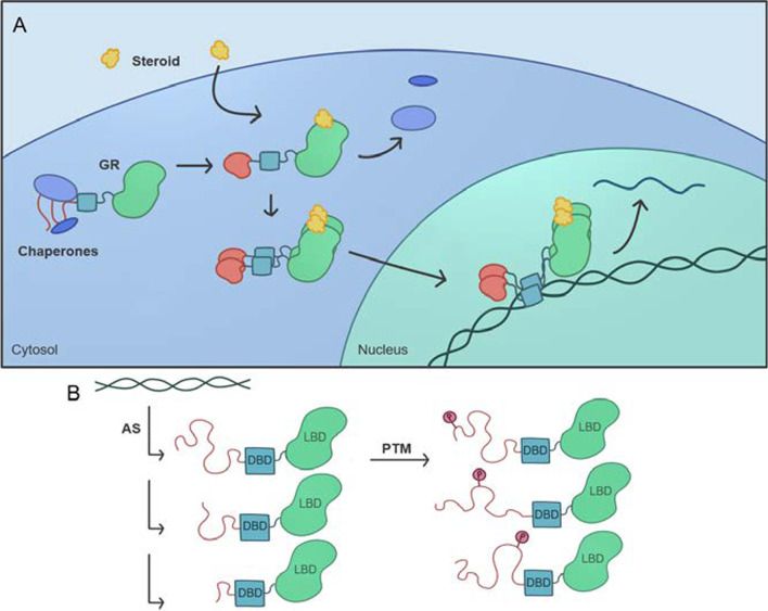
“Intrinsic disorder may be present, and provide regulatory opportunities, for any of the following steps: ligand production, ligand activity, ligand bioavailability, receptor structure, intracellular transmission, termination/intracellular trafficking, and effector proteins (Fig. 4).”
§ Intrinsically disordered proteins play diverse roles in cell signaling > IDRS/IDPS are found in every step of cell signaling pathways
hedgedconditional- Asserted: passes — The authors put it forward as true.
- Checkable: passes — It can be checked against evidence.
- Load-bearing: passes — The paper’s conclusions depend on it.
- Based: passes — The paper gives a reason why it holds.
It may state two separate claims as one.
In the paper
“Laboratory experiments show that a 55 residue segment of axin containing binding sites for both β-catenin and GSK3b has the biophysical properties of an unstructured protein [263], which is in agreement with the predictions of disorder on this protein.”
§ Intrinsically disordered proteins play diverse roles in cell signaling > IDRS/IDPS are found in every category of cell signaling pathways
What the sources say
- Not checked yetcites [263] Noutsou M, Duarte AM, Anvarian Z, Didenko T, Minde DP, Kuper I, et al. Critical scaffolding regions of the tumor suppressor Axin1 are natively unfolded. J Mol Biol. 2011;405:773–86. source
plaincorrelational- Asserted: passes — The authors put it forward as true.
- Checkable: passes — It can be checked against evidence.
- Load-bearing: passes — The paper’s conclusions depend on it.
- Based: passes — The paper gives a reason why it holds.
It may state two separate claims as one.
In the paper
“Heparin binds > 400 proteins, including many involved in cell signaling [279].”
§ Intrinsically disordered proteins play diverse roles in cell signaling > IDRS/IDPS are found in every step of cell signaling pathways
What the sources say
- Not checked yetcites [279] Peysselon F, Richard-Blum S. Heparin-protein interactions: from affinity and kinetics to biological roles. Application to an interaction network regulating angiogenesis. Matrix Biol. 2014;35:73–81. source
plain- Asserted: passes — The authors put it forward as true.
- Checkable: passes — It can be checked against evidence.
- Load-bearing: unclear — It is unclear whether the paper’s conclusions depend on it.
- Based: passes — The paper gives a reason why it holds.
It may state two separate claims as one.
In the paper
“Random movements of the bound proteins enabled by the flexible linkers bring about frequent kinase-substrate collisions, which, in turn, lead to efficient phosphorylation.”
§ Intrinsically disordered proteins play diverse roles in cell signaling > IDRS/IDPS are found in every category of cell signaling pathways
What the sources say
- Not checked yetcites [259] Xue B, Romero PR, Noutsou M, Maurice MM, Rüdiger SG, William AM Jr, et al. Stochastic machines as a colocalization mechanism for scaffold protein function. FEBS Lett. 2013;587:1587–91. source
plaindirect- Asserted: passes — The authors put it forward as true.
- Checkable: passes — It can be checked against evidence.
- Load-bearing: passes — The paper’s conclusions depend on it.
- Based: passes — The paper gives a reason why it holds.
It may state two separate claims as one.
None of the sources cited for this claim have been checked yet.
In the paper
“Since intrinsic disordered regions are highly prevalent in transcription factors [273–277], intrinsic disorder is a major factor in regulating the production of cell signals.”
§ Intrinsically disordered proteins play diverse roles in cell signaling > IDRS/IDPS are found in every step of cell signaling pathways
What the sources say
- Not checked yetcites [273] Brodsky S, Jana T, Mittelman K, Chapal M, Kumar DK, Carmi M, et al. Intrinsically disordered regions direct transcription factor in vivo binding specificity. Mol Cell. 2020;79:459–71. source
- Not checked yetcites [274] Clark S, Myers J, King A, Fiala R, Novacek J, Pearce G, et al. Multivalency regulates activity in an intrinsically disordered transcription factor. eLife. 2018;2018(7):e36258. source
- Not checked yetcites [275] Liu J, Perumal NB, Oldfield CJ, Su EW, Uversky VN, Dunker AK. Intrinsic disorder in transcription factors. Biochemistry. 2006;45:6873–88. source
- Not checked yetcites [276] Tarczewska A, Greb-Markiewicz B. The significance of the intrinsically disordered regions for the functions of the bHLH transcription factors. Int J Mol Sci. 2019;20:5306. source
- Not checked yetcites [277] Vuzman D, Levy Y. Intrisically disordered regions as affinity tuners in protein-DNA interactions. Mol BioSyst. 2012;8:47–57. source
plaindirect- Asserted: passes — The authors put it forward as true.
- Checkable: passes — It can be checked against evidence.
- Load-bearing: passes — The paper’s conclusions depend on it.
- Based: passes — The paper gives a reason why it holds.
It may state two separate claims as one.
The paper states this without a source or data behind it here, so there is nothing to check it against. The quote shows it is in the paper.
In the paper
“The resultant molecular complex Wnt-Fzd-LRP5/6-Dvl forms a structural region for Axin interaction that disrupts Axin-mediated phosphorylation/degradation of the transcriptional co-activator β-catenin, thereby allowing it to stabilize and accumulate in the nucleus where it activates the expression of multiple Wnt-dependent genes.”
§ Intrinsically disordered proteins play diverse roles in cell signaling > IDRS/IDPS are found in every category of cell signaling pathways
plaindirect- Asserted: passes — The authors put it forward as true.
- Checkable: passes — It can be checked against evidence.
- Load-bearing: unclear — It is unclear whether the paper’s conclusions depend on it.
- Based: passes — The paper gives a reason why it holds.
It may state two separate claims as one.
None of the sources cited for this claim have been checked yet.
In the paper
“This binding promotes recruitment of the scaffolding protein disheveled (Dvl), which results in the phosphorylation of the cytoplasmic domain of LRP5/6, a domain whose disorder [264, 265] reflects disorder in the cytoplasmic domains of other single pass membrane proteins [266] and like most other segments that undergo phosphorylation [41, 265].”
§ Intrinsically disordered proteins play diverse roles in cell signaling > IDRS/IDPS are found in every category of cell signaling pathways
What the sources say
- Not checked yetcites [264] Harnoš J, Cañizal MCA, Jurásek M, Kumar J, Holler C, Schambony A, et al. Dishevelled-3 conformation dynamics analyzed by FRET-based biosensors reveals a key role of casein kinase 1. Nat Commun. 2019;10:1804. source
- Not checked yetcites [265] Hanáková K, Bernatík O, Kravec M, Micka M, Kumar J, Harnoš J, et al. Comparative phosphorylation map of Dishevelled 3 links phosphor-signatures to biological outputs. Cell Commun Signal. 2019;17:170. source
- Not checked yetcites [266] Burgi J, Xue B, Uversky VN, van der Groot G. Intrinsic disorder in transmembrane proteins: roles in signaling and topology prediction. PLoS ONE. 2016;11:e0158594. source
plaincorrelational- Asserted: passes — The authors put it forward as true.
- Checkable: passes — It can be checked against evidence.
- Load-bearing: unclear — It is unclear whether the paper’s conclusions depend on it.
- Based: passes — The paper gives a reason why it holds.
It may state two separate claims as one.
In the paper
“Overall, these data show that the phosphorylation rate enhancement occurs by the binding of both the kinase and the substrate to a single flexible tether and not by activation resulting from the binding of axin to the enzyme or the substrate [263].”
§ Intrinsically disordered proteins play diverse roles in cell signaling > IDRS/IDPS are found in every category of cell signaling pathways
What the sources say
- Not checked yetcites [263] Noutsou M, Duarte AM, Anvarian Z, Didenko T, Minde DP, Kuper I, et al. Critical scaffolding regions of the tumor suppressor Axin1 are natively unfolded. J Mol Biol. 2011;405:773–86. source
boosteddirect- Asserted: passes — The authors put it forward as true.
- Checkable: passes — It can be checked against evidence.
- Load-bearing: passes — The paper’s conclusions depend on it.
- Based: passes — The paper gives a reason why it holds.
It may state two separate claims as one.
None of the sources cited for this claim have been checked yet.
In the paper
“Gab2 is a type of LMD protein that operates as part of many signaling pathways [308, 311] and transmits signals from integrins, receptor tyrosine kinases, cytokine receptors, multi-chain immune recognition receptors, and G protein-coupled receptors, and is required to activate Akt, Ras/Raf, Rac, c-fos, Jak/Stat, Rac, and a host of other intracellular transducers [308, 311, 312].”
§ Intrinsically disordered proteins play diverse roles in cell signaling > IDRS/IDPS are found in every step of cell signaling pathways
What the sources say
- Not checked yetcites [308] Simister PC, Feller SM. Order and disorder in large multi-site docking proteins of the Gab family—implications for signaling complex formation and inhibitor design strategies. Mol BioSyst. 2012;8:33–46. source
- Not checked yetcites [311] Hunzicker-DunnME Lopez-Biladeau B, Law NC, Fiedler SE, Carr DW, Maizels ET. PKA and GAB2 play central roles in the FSH signaling pathways to PI3K and AKT in ovarian granulosa cells. Proc Natl Acad Sci USA. 2012;109:E2979–88. source
- Not checked yetcites [312] Wöhrle FU, Daly RJ, Brummer T. Function, regulation and pathological roles of the Gab/DOS docking proteins. Cell Commun Signaling. 2009;7:22. source
plaindirect- Asserted: passes — The authors put it forward as true.
- Checkable: passes — It can be checked against evidence.
- Load-bearing: unclear — It is unclear whether the paper’s conclusions depend on it.
- Based: passes — The paper gives a reason why it holds.
It may state two separate claims as one.
In the paper
“Similar regulatory mechanisms have been established for G-Protein Coupled Receptors (Fig. 5), which were recently reviewed by Zhou et al. [39].”
§ Intrinsically disordered proteins play diverse roles in cell signaling > IDRS/IDPS are found in every step of cell signaling pathways
What the sources say
- Not checked yetcites [39] Zhou J, Zhao S, Dunker AK. Intrinsically disordered proteins link alternative splicing and post-translational modifications to complex cell signaling and regulation. J Mol Biol. 2018;430:2342–59. source
boosted- Asserted: passes — The authors put it forward as true.
- Checkable: passes — It can be checked against evidence.
- Load-bearing: fails — The paper’s conclusions may not depend on it.
- Based: passes — The paper gives a reason why it holds.
Probably not a substantial claim. The paper’s conclusions may not depend on it.
None of the sources cited for this claim have been checked yet.
In the paper
“Thus, LMDs bind to both integrate signals from multiple pathways and coordinate the downstream response [27, 307, 308].”
§ Intrinsically disordered proteins play diverse roles in cell signaling > IDRS/IDPS are found in every step of cell signaling pathways
What the sources say
- Not checked yetcites [27] Kreiger JM, Fusco G, Lewitzky M, Simister PC, Marchant J, Camilloni C, et al. Conformational recognition of an intrinsically disordered protein. Biophys J. 2014;106:1771–9. source
- Not checked yetcites [307] McDonald CB, Seldeen KL, Deegan BJ, Bhat V, Farooq A. Assembly of the Sos1-Grb2-Gab1 ternary signaling complex is under allosteric control. Arch Biochem Biophys. 2010;494:216–25. source
- Not checked yetcites [308] Simister PC, Feller SM. Order and disorder in large multi-site docking proteins of the Gab family—implications for signaling complex formation and inhibitor design strategies. Mol BioSyst. 2012;8:33–46. source
plaindirect- Asserted: passes — The authors put it forward as true.
- Checkable: passes — It can be checked against evidence.
- Load-bearing: passes — The paper’s conclusions depend on it.
- Based: passes — The paper gives a reason why it holds.
It may state two separate claims as one.
In the paper
“The α-helical propensity, dynamics, and affinity in the β tails of integrins strongly suggest that conformational entropy plays an important role in Talin binding, with a preformed helix binding more readily than a disordered one [299].”
§ Intrinsically disordered proteins play diverse roles in cell signaling > IDRS/IDPS are found in every step of cell signaling pathways
What the sources say
- Not checked yetcites [299] Anthis NJ, Wegener KL, Critchley DR, Campbell ID. Structural diversity in integrin/talin interactions. Structure. 2010;18:1654–66. source
boosteddirect- Asserted: passes — The authors put it forward as true.
- Checkable: passes — It can be checked against evidence.
- Load-bearing: unclear — It is unclear whether the paper’s conclusions depend on it.
- Based: passes — The paper gives a reason why it holds.
It may state two separate claims as one.
In the paper
“Integrins not only mediate two-way communication between the cell interior and the extracellular matrix, but they also regulate ion channels, growth factor receptors, and the activity of cytoplasmic kinases [291].”
§ Intrinsically disordered proteins play diverse roles in cell signaling > IDRS/IDPS are found in every step of cell signaling pathways
What the sources say
- Not checked yetcites [291] Giancotti FG, Ruoslahti E. Integrin signaling. Science. 1999;285:1028–33. source
plaindirect- Asserted: passes — The authors put it forward as true.
- Checkable: passes — It can be checked against evidence.
- Load-bearing: unclear — It is unclear whether the paper’s conclusions depend on it.
- Based: passes — The paper gives a reason why it holds.
It may state two separate claims as one.
None of the sources cited for this claim have been checked yet.
In the paper
“The disorder in the tail enables the occurrence of PTMs that add lipids and other groups to the tail, regulating membrane anchoring, domain positioning, auto-inhibition, effector protein binding, and, ultimately, Ras-mediated signaling [320, 321].”
§ Intrinsically disordered proteins play diverse roles in cell signaling > IDRS/IDPS are found in every step of cell signaling pathways
What the sources say
- Not checked yetcites [320] Nussinov R, Jang H, Tsai C-J, Liao T-J, Li S, Fushman D, et al. Intrinsic protein disorder in oncogenic KRAS signaling. Cell Mol Life Sci. 2017;74:3245–61. source
- Not checked yetcites [321] Abdelkarim H, Banerjee A, Grudzien P, Leschinsky N, Abushaer M, Gaponenko V. The hypervariable region of K-Ras4B governs molecular recognition and function. Int J Mol Sci. 2019;20:5718. source
plaindirect- Asserted: passes — The authors put it forward as true.
- Checkable: passes — It can be checked against evidence.
- Load-bearing: unclear — It is unclear whether the paper’s conclusions depend on it.
- Based: passes — The paper gives a reason why it holds.
It may state two separate claims as one.
In the paper
“This conformational change promotes autophosphorylation, and hence activation, of the two tyrosine kinase domains [281].”
§ Intrinsically disordered proteins play diverse roles in cell signaling > IDRS/IDPS are found in every step of cell signaling pathways
What the sources say
- Not checked yetcites [281] Cornish J, Chamerlain SG, Own D, Mott HR. Intrinsically disordered proteins and membranes: a marriage of convenience for cell signaling? Biochem Soc Trans. 2020;48:2669–89. source
plaindirect- Asserted: passes — The authors put it forward as true.
- Checkable: passes — It can be checked against evidence.
- Load-bearing: unclear — It is unclear whether the paper’s conclusions depend on it.
- Based: passes — The paper gives a reason why it holds.
It may state two separate claims as one.
The paper relies on this without support.
The examples reviewed are representative of cell signaling pathways generally.
“IDPs/IDRs are uniquely suited to solving these problems, as demonstrated by several examples detailed in this review (Table 1).”
Claims other papers make about this one
Each is checked against this paper’s own text: does it say what the citing paper says it does?
The source backs part of the claim, not all of it — or backs it for a different population, condition or comparison.
In the citing paper
“Disordered proteins (regions) are characterized by broad and heterogeneous ensembles where the differences in the relative conformational stabilities are small, or even minor and the barriers are low.”
From AlphaFold, Artificial Intelligence (AI), and Allostery (2022) · cites this paper as [132]
Experimental — accuracy not yet measured
Self-check
Percentages, sums and reported differences re-computed from the paper’s own numbers.
Each contradiction between two passages listed here was confirmed by an independent check.
Rated minor: it would not touch a result — a value no result rests on, a p-value that stays on the same side of the significance level, or a conflict that is clerical, resolved elsewhere in the paper, or outside its own findings.
In the paper
§ Intrinsically disordered proteins play diverse roles in cell signaling > IDRS/IDPS pervade pathways that respond to a wide variety of signals
“Figure: Fig. 3 The function of the glucocorticoid receptor is regulated in part by its intrinsically disordered C-terminal tail.”
§ Intrinsically disordered proteins play diverse roles in cell signaling > IDRS/IDPS pervade pathways that respond to a wide variety of signals
“Positions of the N-terminal domain (residues 1–420), DNA binding domain (residues 420–485), a hinge region (residues 486–527), and ligand binding domain (residues 528–777) are shown as red, cyan, blue and lime shaded areas. Clearly, the N-terminal domain and a hinge region are mostly disordered”
Rated minor: it would not touch a result — a value no result rests on, a p-value that stays on the same side of the significance level, or a conflict that is clerical, resolved elsewhere in the paper, or outside its own findings.
In the paper
§ Intrinsically disordered proteins play diverse roles in cell signaling > IDRS/IDPS are found in every category of cell signaling pathways
“Signaling via glutamate is a critical component of long-term potentiation and long-term depression, which alter the strength of downstream signaling in response to glutamine binding to receptor.”
§ Intrinsically disordered proteins play diverse roles in cell signaling > IDRS/IDPS are found in every category of cell signaling pathways
“Glutamate is the primary neurotransmitter for excitatory stimulation.”
Rated minor: it would not touch a result — a value no result rests on, a p-value that stays on the same side of the significance level, or a conflict that is clerical, resolved elsewhere in the paper, or outside its own findings.
In the paper
§ Intrinsically disordered proteins play diverse roles in cell signaling > IDRS/IDPS are found in every category of cell signaling pathways
“The Wnt signaling pathway exemplifies how intrinsic disorder can play multiple roles in a single pathway (Fig. 2).”
§ Intrinsically disordered proteins play diverse roles in cell signaling > IDRS/IDPS pervade pathways that respond to a wide variety of signals
“Figure: Fig. 2 Intrinsic disorder predisposition of human glucocorticoid receptor (UniProt ID: P04150)”
Citations
Cites
Items 201–330 of 330
Order, Disorder, and Everything in Between
DeForte, S., & Uversky, V. N. (2016) · Molecules · doi:10.3390/molecules21081090 · Cited by 93
Self-citationFonin, A. V., Darling, A. L., Kuznetsova, I. M., Turoverov, K. K., & Uversky, V. N. (2019) · Cellular and Molecular Life Sciences · doi:10.1007/s00018-019-03276-1 · Cited by 91
Self-citationZhou, J., Zhao, S., & Dunker, A. K. (2018) · Journal of Molecular Biology · doi:10.1016/j.jmb.2018.03.028 · Cited by 90 · cited 9×
“tissue or cell specific) regulation as discussed previously and above for GPCRs, N-FATs, and Sarc Family kinases [39].”
BackgroundMethodInfluentialSelf-citationCorrelation between posttranslational modification and intrinsic disorder in protein.
Gao, J., & Xu, D. H. (2012) · PubMed · Cited by 88
Prediction of Short Linear Protein Binding Regions
Mooney, C., Pollastri, G., Shields, D. C., & Haslam, N. J. (2011) · Journal of Molecular Biology · doi:10.1016/j.jmb.2011.10.025 · Cited by 87
Prediction of Disordered RNA, DNA, and Protein Binding Regions Using DisoRDPbind
Peng, Z., Wang, C., Uversky, V. N., & Kurgan, L. (2016) · Methods in molecular biology · doi:10.1007/978-1-4939-6406-2_14 · Cited by 87
Self-citationDistinct Binding Modes of Two Epitopes in Gab2 that Interact with the SH3C Domain of Grb2
Harkiolaki, M., Tsirka, T., Lewitzky, M., Simister, P. C., Joshi, D., Bird, L. E., Jones, E. Y., O’Reilly, N., & Feller, S. M. (2009) · Structure · doi:10.1016/j.str.2009.03.017 · Cited by 86
INTRINSIC PROTEIN DISORDER AND PROTEIN-PROTEIN INTERACTIONS
Hsu, W.-L., Oldfield, C. J., Meng, J., Huang, F., Xue, B., Uversky, V. N., Romero, P. R., & Dunker, A. K. (2011) · Pacific Symposium on Biocomputing (PSB) · doi:10.1142/9789814366496_0012 · Cited by 85
Self-citationIdentification of the Cytoplasmic Domains of CXCR4 Involved in Jak2 and STAT3 Phosphorylation
Ahr, B., Denizot, M., Robert-Hebmann, V., Brelot, A., & Biard‐Piechaczyk, M. (2004) · Journal of Biological Chemistry · doi:10.1074/jbc.m408481200 · Cited by 81
Intrinsic Disorder in Transmembrane Proteins: Roles in Signaling and Topology Prediction
Bürgi, J., Xue, B., Uversky, V. N., & van der Goot, F. G. (2016) · PLoS ONE · doi:10.1371/journal.pone.0158594 · Cited by 79
Self-citationIntrinsically disordered proteins in synaptic vesicle trafficking and release
Snead, D., & Eliezer, D. (2019) · Journal of Biological Chemistry · doi:10.1074/jbc.rev118.006493 · Cited by 79
Structural Mechanisms of PTEN RegulationPreprint
Masson, G. R., & Williams, R. L. (2019) · Cold Spring Harbor Perspectives in Medicine · doi:10.1101/cshperspect.a036152 · Cited by 79
Masson, G. R., Perišić, O., Burke, J. E., & Williams, R. L. (2015) · Biochemical Journal · doi:10.1042/bj20150931 · Cited by 78
Fang, Z., Takizawa, N., Wilson, K. A., Smith, T. C., Delprato, A., Davidson, M. W., Lambright, D. G., & Luna, E. J. (2010) · Traffic · doi:10.1111/j.1600-0854.2010.01062.x · Cited by 77
Rajbhandari, P., Finn, G., Solodin, N. M., Singarapu, K. K., Sahu, S. C., Markley, J. L., Kadunc, K., Miner, S. J., Kariagina, A., Haslam, S. Z., et al. (2011) · Molecular and Cellular Biology · doi:10.1128/mcb.06073-11 · Cited by 77
OPAL: prediction of MoRF regions in intrinsically disordered protein sequences
Sharma, R., Raicar, G., Tsunoda, T., Patil, A., & Sharma, A. K. (2018) · Bioinformatics · doi:10.1093/bioinformatics/bty032 · Cited by 77
Sequential Tyrosine Sulfation of CXCR4 by Tyrosylprotein Sulfotransferases
Seibert, C., Veldkamp, C. T., Peterson, F. C., Chait, B. T., Volkman, B. F., & Sakmar, T. P. (2008) · Biochemistry · doi:10.1021/bi800965m · Cited by 75
Intrinsic Disorder in Proteins with Pathogenic Repeat Expansions
Darling, A. L., & Uversky, V. N. (2017) · Molecules · doi:10.3390/molecules22122027 · Cited by 75
Self-citationComputational Prediction of MoRFs, Short Disorder-to-order Transitioning Protein Binding Regions
Katuwawala, A., Peng, Z., Yang, J., & Kurgan, L. (2019) · Computational and Structural Biotechnology Journal · doi:10.1016/j.csbj.2019.03.013 · Cited by 75
BEEUMEN, J. V., Devreese, B. V., Bun, S. M. V., Hoff, W. D., Hellingwerf, K. J., Meyer, T. E., Cusanovich, M. A., & McRee, D. E. (1993) · Protein Science · doi:10.1002/pro.5560020706 · Cited by 73
A Computational Investigation of Allostery in the Catabolite Activator Protein
Li, L., Uversky, V. N., Dunker, A. K., & Meroueh, S. O. (2007) · Journal of the American Chemical Society · doi:10.1021/ja076046a · Cited by 71
Self-citationStructure and function of Gab2 and its role in cancer (Review)
DING, C.-B., YU, W.-N., FENG, J.-H., & LUO, J.-M. (2015) · Molecular Medicine Reports · doi:10.3892/mmr.2015.3951 · Cited by 71
Schramm, A., Bignon, C., Brocca, S., Grandori, R., Santambrogio, C., & Longhi, S. (2019) · Archives of Biochemistry and Biophysics · doi:10.1016/j.abb.2019.07.020 · Cited by 71
CORRELATION BETWEEN POSTTRANSLATIONAL MODIFICATION AND INTRINSIC DISORDER IN PROTEIN
Gao, J., & XU, D. (2011) · Pacific Symposium on Biocomputing (PSB) · doi:10.1142/9789814366496_0010 · Cited by 71
Intrinsically disordered proteins and membranes: a marriage of convenience for cell signalling?
Cornish, J., Chamberlain, S. G., Owen, D., & Mott, H. R. (2020) · Biochemical Society Transactions · doi:10.1042/bst20200467 · Cited by 69
A coactivator code for transcription
Gamble, M. J., & Freedman, L. P. (2002) · Trends in Biochemical Sciences · doi:10.1016/s0968-0004(02)02076-5 · Cited by 68
Fang, C., Noguchi, T., Tominaga, D., & Yamana, H. (2013) · BMC Bioinformatics · doi:10.1186/1471-2105-14-300 · Cited by 67
Heparanase, cell signaling, and viral infections
Koganti, R., Suryawanshi, R., & Shukla, D. (2020) · Cellular and Molecular Life Sciences · doi:10.1007/s00018-020-03559-y · Cited by 67
Love, J. J., Li, X., Chung, J., Dyson, H. J., & Wright, P. E. (2004) · Biochemistry · doi:10.1021/bi049591m · Cited by 66
Early Events in Cell Spreading as a Model for Quantitative Analysis of Biomechanical Events
Wolfenson, H., Iskratsch, T., & Sheetz, M. P. (2014) · Biophysical Journal · doi:10.1016/j.bpj.2014.10.041 · Cited by 65
Anantharaman, V., Abhiman, S., de Souza, R. F., & Aravind, L. (2010) · Gene · doi:10.1016/j.gene.2010.12.001 · Cited by 65
The Sequence Attribute Method for Determining Relationships Between Sequence and Protein Disorder.
Xie, Q., Arnold, Romero, P. R., Zoran Obradovic, A., Garner, E. C., & Dunker, K. (1998) · PubMed · Cited by 64
Structure of metabotropic glutamate receptor C-terminal domains in contact with interacting proteins
Enz, R. (2012) · Frontiers in Molecular Neuroscience · doi:10.3389/fnmol.2012.00052 · Cited by 63
Templated folding of intrinsically disordered proteins
Toto, A., Malagrinò, F., Visconti, L., Troilo, F., Pagano, L., Brunori, M., Jemth, P., & Gianni, S. (2020) · Journal of Biological Chemistry · doi:10.1074/jbc.rev120.012413 · Cited by 60
Critical Scaffolding Regions of the Tumor Suppressor Axin1 Are Natively Unfolded
Noutsou, M., Duarte, A. M. S., Anvarian, Z., Didenko, T., Minde, D. P., Kuper, I., de Ridder, I., Oikonomou, C., Friedler, A., Boelens, R., et al. (2011) · Journal of Molecular Biology · doi:10.1016/j.jmb.2010.11.013 · Cited by 58
Interplay between allostery and intrinsic disorder in an ensemble
Motlagh, H. N., Li, J., Thompson, E. B., & Hilser, V. J. (2012) · Biochemical Society Transactions · doi:10.1042/bst20120163 · Cited by 58
Intrinsic protein disorder in oncogenic KRAS signaling
Nussinov, R., Jang, H., Tsai, C., Liao, T.-J., Li, S., Fushman, D., & Zhang, J. (2017) · Cellular and Molecular Life Sciences · doi:10.1007/s00018-017-2564-3 · Cited by 58
Conformational Recognition of an Intrinsically Disordered Protein
Krieger, J. M., Fusco, G., Lewitzky, M., Simister, P. C., Marchant, J., Camilloni, C., Feller, S. M., & De Simone, A. (2014) · Biophysical Journal · doi:10.1016/j.bpj.2014.03.004 · Cited by 56
Multivalency regulates activity in an intrinsically disordered transcription factor
Clark, S. J., Myers, J. B., King, A., Fiala, R., Nováček, J., Pearce, F. G., Heierhorst, J., Reichow, S., & Barbar, E. J. (2018) · eLife · doi:10.7554/elife.36258 · Cited by 56
The Plasma Membrane as a Competitive Inhibitor and Positive Allosteric Modulator of KRas4B Signaling
Neale, C., & García, A. E. (2020) · Biophysical Journal · doi:10.1016/j.bpj.2019.12.039 · Cited by 56
Heparin: an intervenor in cell communication
Xu, X., & Dai, Y. (2009) · Journal of Cellular and Molecular Medicine · doi:10.1111/j.1582-4934.2009.00871.x · Cited by 55
Sharma, R., Bayarjargal, M., Tsunoda, T., Patil, A., & Sharma, A. K. (2017) · Journal of Theoretical Biology · doi:10.1016/j.jtbi.2017.10.015 · Cited by 55
Accuracy of protein-level disorder predictions
Katuwawala, A., Oldfield, C. J., & Kurgan, L. (2019) · Briefings in Bioinformatics · doi:10.1093/bib/bbz100 · Cited by 55
Native mass spectrometry reveals the conformational diversity of the UVR8 photoreceptor
Camacho, I. S., Theisen, A., Johannissen, L. O., Díaz-Ramos, L. A., Christie, J. M., Jenkins, G. I., Bellina, B., Barran, P., & Jones, A. R. (2019) · Proceedings of the National Academy of Sciences · doi:10.1073/pnas.1813254116 · Cited by 54
Simister, P. C., & Feller, S. M. (2011) · Molecular BioSystems · doi:10.1039/c1mb05272a · Cited by 53
Boudria, A., Abou Faycal, C., Jia, T., Gout, S., Keramidas, M., Didier, C., Lemaître, N., Manet, S., Coll, J.-L., Toffart, A.-C., et al. (2019) · Oncogene · doi:10.1038/s41388-018-0486-7 · Cited by 53
Hanson, J., Litfin, T., Paliwal, K. K., & Zhou, Y. (2019) · Bioinformatics · doi:10.1093/bioinformatics/btz691 · Cited by 53
DeForte, S., & Uversky, V. N. (2017) · Molecular BioSystems · doi:10.1039/c6mb00741d · Cited by 52
Self-citationProtein intrinsic disorder and oligomericity in cell signaling
Sigalov, A. B. (2009) · Molecular BioSystems · doi:10.1039/b916030m · Cited by 51
Grzybowska, E. A. (2018) · Biomolecules · doi:10.3390/biom8020042 · Cited by 51
Paradoxes and wonders of intrinsic disorder: Complexity of simplicity
Uversky, V. (2016) · Intrinsically Disordered Proteins · doi:10.1080/21690707.2015.1135015 · Cited by 50
Self-citationWarnet, X. L., Bakke Krog, H., Sevillano‐Quispe, O. G., Poulsen, H., & Kjaergaard, M. (2021) · European Journal of Neuroscience · doi:10.1111/ejn.14842 · Cited by 50
Hasty, J., & Collins, J. J. (2001) · Nature · doi:10.1038/35075182 · Cited by 49
Malhis, N., Wong, E. T. C., Nassar, R., & Gsponer, J. A. (2015) · PLoS ONE · doi:10.1371/journal.pone.0141603 · Cited by 49
Tarczewska, A., & Greb‐Markiewicz, B. (2019) · International Journal of Molecular Sciences · doi:10.3390/ijms20215306 · Cited by 49
Xue, B., Dunker, A. K., & Uversky, V. N. (2010) · International Journal of Molecular Sciences · doi:10.3390/ijms11103725 · Cited by 48
Self-citationLi, J., Motlagh, H. N., Chakuroff, C., Thompson, E. B., & Hilser, V. J. (2012) · Journal of Biological Chemistry · doi:10.1074/jbc.m112.355651 · Cited by 48
The Roles of Intrinsic Disorder in Orchestrating the Wnt-Pathway
Xue, B., Dunker, A. K., & Uversky, V. N. (2012) · Journal of Biomolecular Structure and Dynamics · doi:10.1080/073911012010525024 · Cited by 48
Self-citationStochastic machines as a colocalization mechanism for scaffold protein function
Xue, B., Romero, P. R., Noutsou, M., Maurice, M. M., Rüdiger, S. G. D., William, A. M., Mizianty, M. J., Kurgan, L., Uversky, V. N., & Dunker, A. K. (2013) · FEBS Letters · doi:10.1016/j.febslet.2013.04.006 · Cited by 47
Self-citationStructural Disorder Provides Increased Adaptability for Vesicle Trafficking Pathways
Pietrosemoli, N., Pancsa, R., & Tompa, P. (2013) · PLoS Computational Biology · doi:10.1371/journal.pcbi.1003144 · Cited by 46
A Metastable Contact and Structural Disorder in the Estrogen Receptor Transactivation Domain
Peng, Y., Cao, S., Kiselar, J., Xiao, X., Du, Z., Hsieh, A. H., Ko, S., Chen, Y., Agrawal, P., Zheng, W., et al. (2018) · Structure · doi:10.1016/j.str.2018.10.026 · Cited by 46
An Overview of Predictors for Intrinsically Disordered Proteins over 2010–2014
Li, J., Yu, F., Wang, X., Li, J., Liu, W., Róng, L., & Bao, J. (2015) · International Journal of Molecular Sciences · doi:10.3390/ijms161023446 · Cited by 45
Hidden intermediates and levinthal paradox in the folding of small proteins
Bai, Y. (2003) · Biochemical and Biophysical Research Communications · doi:10.1016/s0006-291x(03)00800-3 · Cited by 43
OPAL+: Length‐Specific MoRF Prediction in Intrinsically Disordered Protein Sequences
Sharma, R., Sharma, A. K., Raicar, G., Tsunoda, T., & Patil, A. (2018) · PROTEOMICS · doi:10.1002/pmic.201800058 · Cited by 43
Uversky, V. N. (2015) · Advances in experimental medicine and biology · doi:10.1007/978-3-319-20164-1_7 · Cited by 42
Self-citationBush, M., Alhanshali, B. M., Qian, S., Stanley, C. B., Heller, W. T., Matsui, T., Weiss, T. M., Nicholl, I. D., Walz, T., Callaway, D. J. E., et al. (2019) · Proceedings of the National Academy of Sciences · doi:10.1073/pnas.1911489116 · Cited by 42
Comparative phosphorylation map of Dishevelled 3 links phospho-signatures to biological outputs
Hanáková, K., Bernatík, O., Kravec, M., Micka, M., Kumar, J., Harnoš, J., Ovesná, P., Paclíková, P., Rádsetoulal, M., Potěšil, D., et al. (2019) · Cell Communication and Signaling · doi:10.1186/s12964-019-0470-z · Cited by 41
Angiotensin II receptors: Impact for COVID ‐19 severity
Aksoy, H. K., Karadağ, A. S., & Wollina, U. (2020) · Dermatologic Therapy · doi:10.1111/dth.13989 · Cited by 41
Ras assemblies and signaling at the membrane
Nussinov, R., Tsai, C., & Jang, H. (2020) · Current Opinion in Structural Biology · doi:10.1016/j.sbi.2020.01.009 · Cited by 40
Lipid-Protein Interplay in Dimerization of Juxtamembrane Domains of Epidermal Growth Factor Receptor
Maeda, R., Sato, T., Okamoto, K., Yanagawa, M., & Sako, Y. (2018) · Biophysical Journal · doi:10.1016/j.bpj.2017.12.029 · Cited by 39
Fold recognition and accurate query-template alignment by a combination of PSI-BLAST and threading
Shan, Y., Wang, G., & Zhou, H. (2000) · Proteins Structure Function and Bioinformatics · doi:10.1002/1097-0134(20010101)42:1 · Cited by 38
Amplification of signaling via cellular allosteric relay and protein disorder
Ma, B., & Nussinov, R. (2009) · Proceedings of the National Academy of Sciences · doi:10.1073/pnas.0903024106 · Cited by 36
Predicting MoRFs in protein sequences using HMM profiles
Sharma, R., Kumar, S., Tsunoda, T., Patil, A., & Sharma, A. (2016) · BMC Bioinformatics · doi:10.1186/s12859-016-1375-0 · Cited by 36
Khan, S. H., McLaughlin, W. A., & Kumar, P. (2017) · Scientific Reports · doi:10.1038/s41598-017-15549-5 · Cited by 36
Kumar, P., & Thompson, E. A. (2019) · Biomolecules · doi:10.3390/biom9030095 · Cited by 36
Harnoš, J., Cañizal, M. C. A., Jurásek, M., Kumar, J., Holler, C., Schambony, A., Hanáková, K., Bernatík, O., Zdráhal, Z., Gömöryová, K., et al. (2019) · Nature Communications · doi:10.1038/s41467-019-09651-7 · Cited by 35
Does Intrinsic Disorder in Proteins Favor Their Interaction with Lipids?
Deryusheva, E., Nemashkalova, E., Galloux, M., Richard, C., Eléouët, J., Kovacs, D., Van Belle, K., Tompa, P., Uversky, V., & Permyakov, S. (2019) · PROTEOMICS · doi:10.1002/pmic.201800098 · Cited by 34
Self-citationBuchner, S., Schlundt, A., Lassak, J., Sattler, M., & Jung, K. (2015) · Journal of Molecular Biology · doi:10.1016/j.jmb.2015.05.001 · Cited by 32
Disordered RNA-Binding Region Prediction with DisoRDPbind
Oldfield, C. J., Peng, Z., & Kurgan, L. (2019) · Methods in molecular biology · doi:10.1007/978-1-0716-0231-7_14 · Cited by 32
Presence and utility of intrinsically disordered regions in kinases
Kathiriya, J. J., Pathak, R. R., Clayman, E., Xue, B., Uversky, V. N., & Davé, V. (2014) · Molecular BioSystems · doi:10.1039/c4mb00224e · Cited by 30
Self-citationKeppel, T. R., Sarpong, K., Murray, E. M., Monsey, J., Zhu, J., & Bose, R. (2017) · Journal of Biological Chemistry · doi:10.1074/jbc.m116.747485 · Cited by 29
A model for hydrophobic protrusions on peripheral membrane proteins
Fuglebakk, E., & Reuter, N. (2018) · PLOS Computational Biology · doi:10.1371/journal.pcbi.1006325 · Cited by 29
Leventhal, S. M., Lim, D., Green, T. L., Cantrell, A. E., Cho, K., & Greenhalgh, D. G. (2019) · BMC Genetics · doi:10.1186/s12863-019-0718-z · Cited by 28
Yokoyama, T., Ohkubo, T., Kamiya, K., & Hara, M. (2020) · Archives of Biochemistry and Biophysics · doi:10.1016/j.abb.2020.108510 · Cited by 28
Notch Pathway: A Journey from Notching Phenotypes to Cancer Immunotherapy
Monticone, G., & Miele, L. (2020) · Advances in experimental medicine and biology · doi:10.1007/978-3-030-55031-8_13 · Cited by 28
Networks of Networks: An Essay on Multi-Level Biological Organization
Uversky, V. N., & Giuliani, A. (2021) · Frontiers in Genetics · doi:10.3389/fgene.2021.706260 · Cited by 28
Self-citationGab2 mediates hepatocellular carcinogenesis by integrating multiple signaling pathways
Cheng, J., Zhong, Y., Chen, S., Sun, Y., Huang, L., Kang, Y., Chen, B., Chen, G., Wang, F., Tian, Y., et al. (2017) · The FASEB Journal · doi:10.1096/fj.201700120rr · Cited by 27
Protein Activity Sensing in Bacteria in Regulating Metabolism and Motility
Alvarado, A., Behrens, W., & Josenhans, C. (2020) · Frontiers in Microbiology · doi:10.3389/fmicb.2019.03055 · Cited by 26
New technologies to analyse protein function: an intrinsic disorder perspective
Uversky, V. N. (2020) · F1000Research · doi:10.12688/f1000research.20867.1 · Cited by 26
Self-citationConformational landscape of multidomain SMAD proteins
Gomes, T., Martín-Malpartida, P., Ruiz, L., Aragón, E., Cordeiro, T. N., & Macías, M. J. (2021) · Computational and Structural Biotechnology Journal · doi:10.1016/j.csbj.2021.09.009 · Cited by 26
Evolution of a Signaling Nexus Constrained by Protein Interfaces and Conformational States
Temple, B. R. S., Jones, C. D., & Jones, A. M. (2010) · PLoS Computational Biology · doi:10.1371/journal.pcbi.1000962 · Cited by 25
Knr4: a disordered hub protein at the heart of fungal cell wall signalling
Martin‐Yken, H., François, J., & Zerbib, D. (2016) · Cellular Microbiology · doi:10.1111/cmi.12618 · Cited by 25
Hotta, K., Ranganathan, S., Liu, R., Wu, F., Machiyama, H., Gao, R., Hirata, H., Soni, N., Ohe, T., Hogue, C. W. V., et al. (2014) · PLoS Computational Biology · doi:10.1371/journal.pcbi.1003532 · Cited by 24
Sigalov, A. B., & Uversky, V. N. (2011) · Self/Nonself · doi:10.4161/self.2.1.14790 · Cited by 23
Self-citationAssembly of the Sos1–Grb2–Gab1 ternary signaling complex is under allosteric control
McDonald, C. B., Seldeen, K. L., Deegan, B. J., Bhat, V. S., & Farooq, A. (2009) · Archives of Biochemistry and Biophysics · doi:10.1016/j.abb.2009.12.011 · Cited by 23
Fang, C., Moriwaki, Y., Tian, A., Li, C., & Shimizu, K. (2018) · Journal of Bioinformatics and Computational Biology · doi:10.1142/s0219720019500045 · Cited by 23
Precise definition of PTEN C-terminal epitopes and its implications in clinical oncology
Mingo, J., Luna, S., Gaafar, A., Nunes-Xavier, C. E., Torices, L., Mosteiro, L., Ruiz, R., Guerra, I., Llarena, R., Angulo, J. C., et al. (2019) · npj Precision Oncology · doi:10.1038/s41698-019-0083-4 · Cited by 22
Computational prediction of MoRFs based on protein sequences and minimax probability machine
He, H., Zhao, J., & Sun, G. (2019) · BMC Bioinformatics · doi:10.1186/s12859-019-3111-z · Cited by 22
Peptidylprolyl Isomerase Pin1 Directly Enhances the DNA Binding Functions of Estrogen Receptor α
Rajbhandari, P., Ozers, M. S., Solodin, N. M., Warren, C. L., & Alarid, E. T. (2015) · Journal of Biological Chemistry · doi:10.1074/jbc.m114.621698 · Cited by 21
Engelhardt, S. H., & Rochais, F. (2007) · Circulation Research · doi:10.1161/01.res.0000266971.15127.e8 · Cited by 20
Quality assessment for the putative intrinsic disorder in proteins
Hu, G., Wu, Z., Oldfield, C. J., Wang, C., & Kurgan, L. (2018) · Bioinformatics · doi:10.1093/bioinformatics/bty881 · Cited by 20
Zamora‐Briseño, J. A., Pereira‐Santana, A., Reyes-Hernández, S. J., Cerqueda‐García, D., Castaño, E., & Rodríguez‐Zapata, L. C. (2020) · Cell Stress and Chaperones · doi:10.1007/s12192-020-01162-5 · Cited by 19
Malaney, P., Uversky, V. N., & Davé, V. (2015) · Methods · doi:10.1016/j.ymeth.2014.10.005 · Cited by 18
Self-citationThe Hypervariable Region of K-Ras4B Governs Molecular Recognition and Function
Abdelkarim, H., Banerjee, A., Grudzien, P. K., Leschinsky, N., Abushaer, M., & Gaponenko, V. (2019) · International Journal of Molecular Sciences · doi:10.3390/ijms20225718 · Cited by 18
Orchestration of algal metabolism by protein disorder
Launay, H., Receveur-Bréchot, V., Carrière, F., & Gontero, B. (2019) · Archives of Biochemistry and Biophysics · doi:10.1016/j.abb.2019.108070 · Cited by 16
Critical Assessment of Protein Intrinsic Disorder PredictionPreprint
Necci, M., Piovesan, D., Predictors, C., et al. (2020) · bioRxiv (Cold Spring Harbor Laboratory) · doi:10.1101/2020.08.11.245852 · Cited by 16
Understanding the mechanism of binding between Gab2 and the C terminal SH3 domain from Grb2
Toto, A., Bonetti, D., Simone, A. D., & Gianni, S. (2017) · Oncotarget · doi:10.18632/oncotarget.19323 · Cited by 14
de Geus, T. J., Patijn, J., & Joosten, E. A. J. (2020) · Developmental Neurobiology · doi:10.1002/dneu.22789 · Cited by 14
Intrinsic disorder in spondins and some of their interacting partners
Alowolodu, O., Johnson, G., Alashwal, L., Addou, I., Zhdanova, I. V., & Uversky, V. N. (2016) · Intrinsically Disordered Proteins · doi:10.1080/21690707.2016.1255295 · Cited by 13
Self-citationMoRFPred_en: Sequence-based prediction of MoRFs using an ensemble learning strategy
Fang, C., Moriwaki, Y., Li, C., & Shimizu, K. (2019) · Journal of Bioinformatics and Computational Biology · doi:10.1142/s0219720019400158 · Cited by 13
Many-to-one binding by intrinsically disordered protein regions
Alterovitz, W.-L., Faraggi, E., Oldfield, C. J., Meng, J., Xue, B., Huang, F., Romero, P. R., Kloczkowski, A., Uversky, V. N., & Dunker, A. K. (2019) · Pacific Symposium on Biocomputing (PSB) · doi:10.1142/9789811215636_0015 · Cited by 13
Self-citationMurvai, N., Kalmar, L., Szalaine Agoston, B., Szabo, B., Tantos, A., Csikos, G., Micsonai, A., Kardos, J., Vertommen, D., Nguyen, P. N., et al. (2020) · Cells · doi:10.3390/cells9081856 · Cited by 13
Wöhrle, F. U., Daly, R. J., & Brummer, T. (2009) · Structure · doi:10.1016/j.str.2009.05.003 · Cited by 12
Predicting Conformational Disorder
Lieutaud, P., Ferron, F. P., & Longhi, S. (2016) · Methods in molecular biology · doi:10.1007/978-1-4939-3572-7_14 · Cited by 12
Self-regulation of functional pathways by motifs inside the disordered tails of beta-catenin
Zhao, B., & Xue, B. (2016) · BMC Genomics · doi:10.1186/s12864-016-2825-9 · Cited by 12
Turki-Judeh, W., & Courey, A. J. (2012) · PLoS ONE · doi:10.1371/journal.pone.0030610 · Cited by 11
Duma, D., & Cidlowski, J. A. (2010) · Hormone Molecular Biology and Clinical Investigation · doi:10.1515/hmbci.2010.039 · Cited by 11
Role of Intrinsically Disordered Regions in Acceleration of Protein–Protein Association
Misiura, M. M., & Kolomeisky, A. B. (2020) · The Journal of Physical Chemistry B · doi:10.1021/acs.jpcb.9b08793 · Cited by 10
Understanding the Mechanism of Recognition of Gab2 by the N-SH2 Domain of SHP2
Visconti, L., Malagrinò, F., Pagano, L., & Toto, A. (2020) · Life · doi:10.3390/life10060085 · Cited by 9
A disordered encounter complex is central to the yeast Abp1p SH3 domain binding pathway
Gerlach, G., Carrock, R., Stix, R., Stollar, E. J., & Ball, K. A. (2020) · PLoS Computational Biology · doi:10.1371/journal.pcbi.1007815 · Cited by 9
Ligand interactions and the protein order-disorder energetic continuum
Moosa, M. M., Ferreon, J. C., & Ferreon, A. C. M. (2018) · Seminars in Cell and Developmental Biology · doi:10.1016/j.semcdb.2018.05.007 · Cited by 8
Intrinsically Disordered Proteins: Dynamics, Binding, and Function
Salvi, N. (2019) · HAL (Le Centre pour la Communication Scientifique Directe) · Cited by 8
The Transcriptional Repressor Domain of Gli3 Is Intrinsically Disordered
Tsanev, R., Vanatalu, K., Jarvet, J., Tanner, R., Laur, K., Tiigimägi, P., Kragelund, B. B., Østerlund, T., & Kogerman, P. (2013) · PLoS ONE · doi:10.1371/journal.pone.0076972 · Cited by 7
Wang, J., Anastasía, A., Bains, H., Giza, J. I., Clossey, D. G., Deng, J., Neubert, T. A., Rice, W. J., Lee, F. S., Hempstead, B. L., et al. (2020) · Metallomics · doi:10.1039/d0mt00108b · Cited by 6
Sequences and topology: disorder, modularity, and post/pre translation modification
Gough, J., & Dunker, A. K. (2013) · Current Opinion in Structural Biology · doi:10.1016/j.sbi.2013.05.001 · Cited by 5
Self-citationEck, M. J., & Hahn, W. C. (2012) · Cell · doi:10.1016/j.cell.2012.04.015 · Cited by 5
Regulation of Connexin32 by ephrin receptors and T-cell protein-tyrosine phosphatase
Trease, A. J., Li, H., Spagnol, G., Zheng, L., Stauch, K. L., & Sorgen, P. L. (2018) · Journal of Biological Chemistry · doi:10.1074/jbc.ra118.003883 · Cited by 5
Protein hormone fragmentation in intercellular signaling: hormones as nested information systems
Campbell, K. L., Haspel, N., Gath, C., Kurniatash, N., Akkiraju, I., Stuffers, N., & Vadher, U. S. (2021) · Biology of Reproduction · doi:10.1093/biolre/ioaa234 · Cited by 5
Anthony James Pawson (1952–2013)
Bernstein, A., & Rossant, J. (2013) · Nature · doi:10.1038/501168a · Cited by 1
Conformational landscape of full-length Smad proteinsPreprint
Gomes, T., Martín-Malpartida, P., Ruiz, L., Aragón, E., Cordeiro, T. N., & Macías, M. J. (2021) · bioRxiv (Cold Spring Harbor Laboratory) · doi:10.1101/2021.04.13.439655 · Cited by 1
Research works printed but not linked: 4
- Dunker AK, Obradovic Z, Romero P, Kissinger C, Villafranca JE. On the importance of being disordered. Protein Data Bank Q Newsl. 1997;81:3–5.
- Daughdrill GW, Pielak GJ, Uversky VN, Cortese MS, Dunker AK. Natively disordered proteins. In: Buchner J, Kiefhaber T, editors. Handbook of protein folding. Weinheim: Wiley, Verlag GmbH & Co KGaA; 2005. p. 271–353.
- Schulz GE. Nucleotide binding proteins. In: Balaban M, editor. Molecular mechanism of biological recognition. New York: Elsevier/North-Holland Biomedical Press; 1979. p. 79–94.
- Meek DW, Anderson CW. Post-translational modiication of p53” cooperative integrators of function. Cold Spring Harb Perspect Biol. 2009;1:a000950.
Cited by
Sorted by citations
Intrinsically Disordered Proteins: An Overview
Trivedi, R., & Nagarajaram, H. A. (2022) · International Journal of Molecular Sciences · doi:10.3390/ijms232214050 · Cited by 277
Vidović, M., & Rikalović, M. (2022) · Cells · doi:10.3390/cells11111732 · Cited by 214
AlphaFold, Artificial Intelligence (AI), and Allostery
Nussinov, R., Zhang, M., Liu, Y., & Jang, H. (2022) · The Journal of Physical Chemistry B · doi:10.1021/acs.jpcb.2c04346 · Cited by 190
Macromolecular condensation buffers intracellular water potential
Watson, J. L., Seinkmane, E., Styles, C. T., Mihut, A., Krüger, L. K., McNally, K. E., Planelles-Herrero, V. J., Dudek, M., McCall, P. M., Barbiero, S., et al. (2023) · Nature · doi:10.1038/s41586-023-06626-z · Cited by 178
On the roles of intrinsically disordered proteins and regions in cell communication and signaling
Bondos, S. E., Dunker, A. K., & Uversky, V. N. (2021) · Cell Communication and Signaling · doi:10.1186/s12964-021-00774-3 · Cited by 113
Self-citation
Metadata
- Authors
- Bondos, S. E., Dunker, A. K., & Uversky, V. N.
- Affiliations
- Texas A&M Health Science Center (US)
- Indiana University Indianapolis (US)
- Russian Academy of Sciences (RU)
- Corresponding author
- Bondos, Sarah E.
- Venue
- Cell Communication and Signaling
- Published
- 2022-02-17
- Volume
- 20
- Issue
- 1
- Pages
- 20-20
- ISSN
- 1478-811X (ISSN-L)
- PMC
- PMC8851865
- OpenAlex
- W4212940766
- Semantic Scholar
- fe30172eab9a4c8b8f27061cb9011b6e6d6de4c2
- Cited by
- 222
- References
- 330
- Citations by year
- 2026: 50 · 2025: 62 · 2024: 45 · 2023: 46 · 2022: 18
- FWCI
- 4.45
- Type
- journal article
- Language
- en
- Open access
- gold
- doi.org
- Locations
- Cell Communication and Signaling (publishedVersion, cc-by)
- PubMed Central (publishedVersion, cc-by)
- PubMed (publishedVersion)
- IUScholarWorks (Indiana University) (submittedVersion, cc-by)
- Digital Commons - University of South Florida (University of South Florida) (submittedVersion, cc-by)
- DOAJ (DOAJ: Directory of Open Access Journals) (submittedVersion, cc-by-sa)
- Funders
- National Institutes of Health (GM099827-06)
- National Institute on Aging (RF1 AG055088)
- Topics
- Life Sciences › Biochemistry, Genetics and Molecular Biology › Molecular Biology › Protein Structure and Dynamics
- Life Sciences › Biochemistry, Genetics and Molecular Biology › Molecular Biology › Receptor Mechanisms and Signaling
- Life Sciences › Biochemistry, Genetics and Molecular Biology › Molecular Biology › Protein Kinase Regulation and GTPase Signaling
- Keywords
- intrinsically disordered proteins, intrinsically disordered regions, signal transduction, protein-protein interaction, ligand binding, receptor signaling, transducers, signal amplification, signal termination, autocrine signaling, paracrine signaling, hormone signaling, juxtacrine signaling, intracrine signaling, intercellular communication
- MeSH terms
- Animals, Cell Communication, Signal Transduction, Intrinsically Disordered Proteins
About these checks
Registry, retraction, Cites and statistics results are computed, not judged, with one exception: a printed reference that no index lists is read once by a model before it counts as a citation error. Claims are extracted by a model and kept only when their quote appears verbatim in the text; each cited source is then checked, with a model’s help, for whether it says what the claim relies on. Those verdicts can be wrong — read the claim’s quote and the cited source before relying on one.
A language model drew the claims from the paper on 2026-10-09, under version 3 of the claim definition (model deepseek-v4.1-flash, prompt extract-v3).
A second model labelled each claim on 2026-10-09 (model jev-1.13-20260917, definition revision gold-guideline-v0.2).
Cited sources are checked with a language model’s help (gpt-6-sol).
Statistics and arithmetic are recomputed, not judged.
Confidence bands are shown only for fields calibrated on our annotated test set. None is calibrated yet, so no band is shown.
What counts as a claim ›How we measure confidence ›How we check this ›
Are you an author, or do you see a problem with this page? Request a review
