

The DOI matches its Crossref record; 85 of the 85 references it cites were found in a registry or an index.
Microbial Symbionts Accelerate Wound Healing via the Neuropeptide Hormone Oxytocin
Open accessAbstract
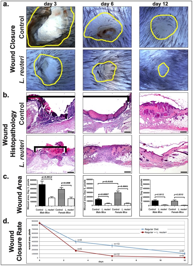
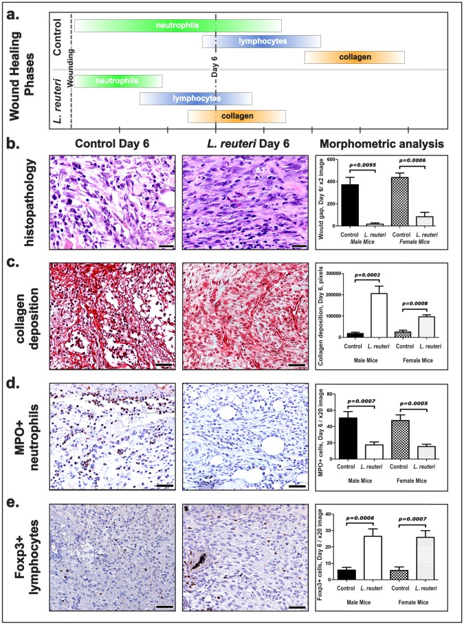
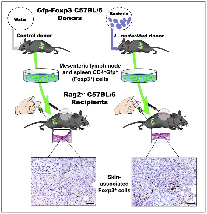
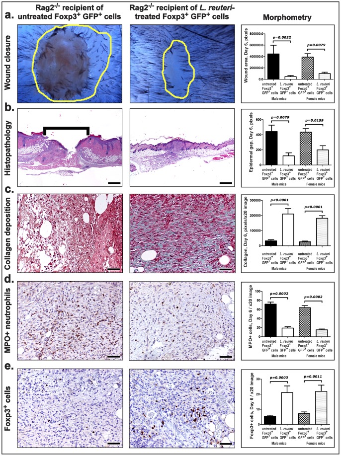
Wound healing capability is inextricably linked with diverse aspects of physical fitness ranging from recovery after minor injuries and surgery to diabetes and some types of cancer. Impact of the microbiome upon the mammalian wound healing process is poorly understood. We discover that supplementing the gut microbiome with lactic acid microbes in drinking water accelerates the wound-healing process to occur in half the time required for matched control animals. Further, we find that Lactobacillus reuteri enhances wound-healing properties through up-regulation of the neuropeptide hormone oxytocin, a factor integral in social bonding and reproduction, by a vagus nerve-mediated pathway. Bacteria-triggered oxytocin serves to activate host CD4+Foxp3+CD25+ immune T regulatory cells conveying transplantable wound healing capacity to naive Rag2-deficient animals. This study determined oxytocin to be a novel component of a multi-directional gut microbe-brain-immune axis, with wound-healing capability as a previously unrecognized output of this axis. We also provide experimental evidence to support long-standing medical traditions associating diet, social practices, and the immune system with efficient recovery after injury, sustained good health, and longevity.
Overview
The claims and statistics of this paper haven’t been checked yet.
Run the check — free, no account needed. It reads the open-access text and checks the sources it cites.
Integrity signals
Registry
Cites
Statistics
Figures
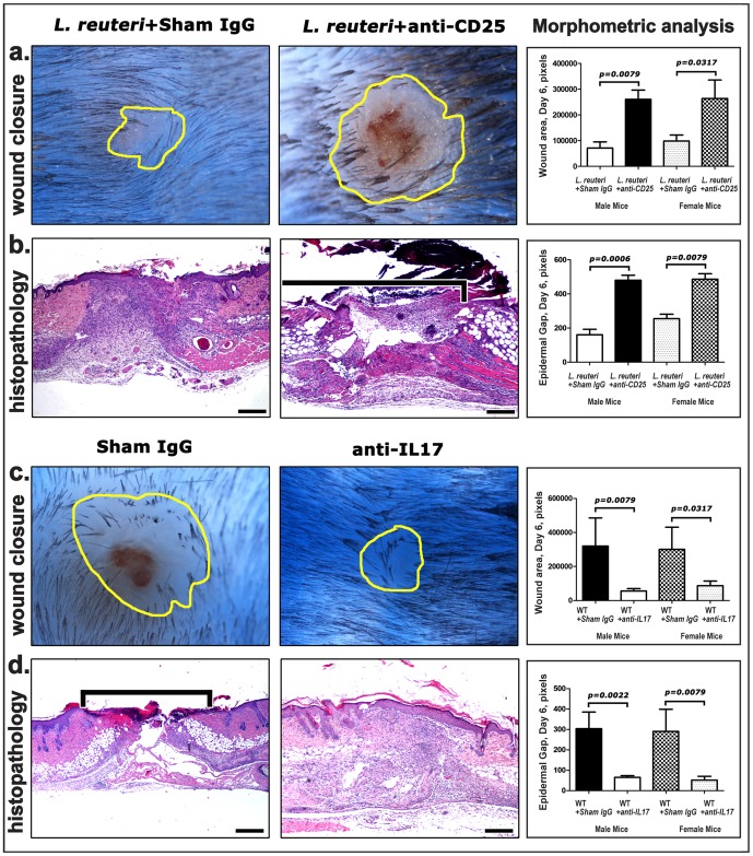
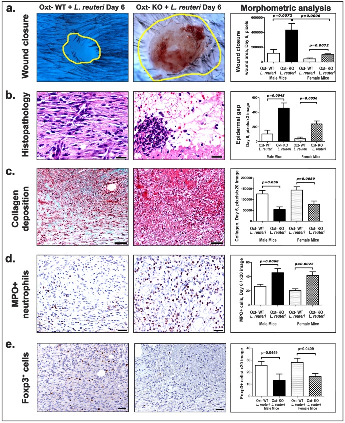
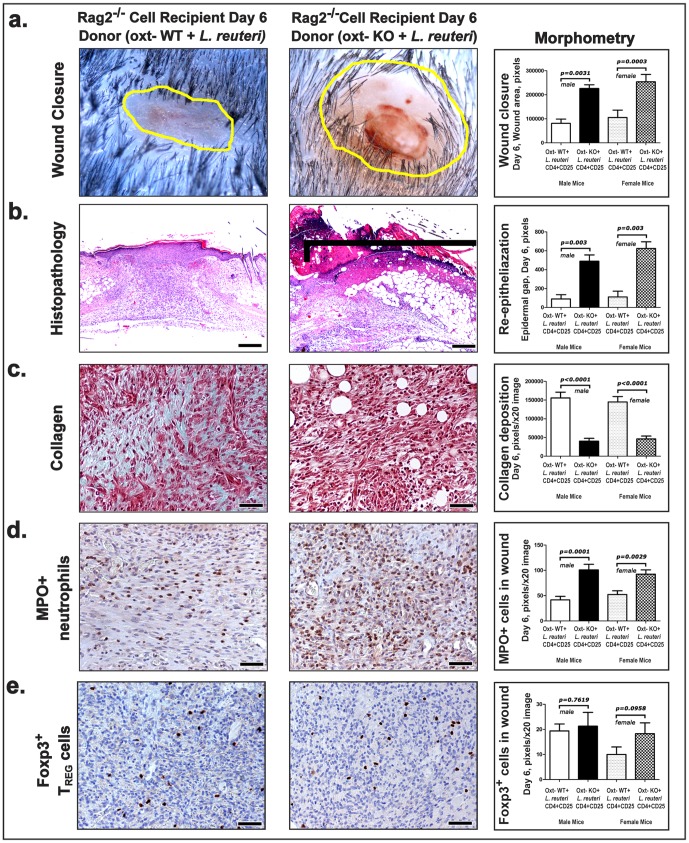
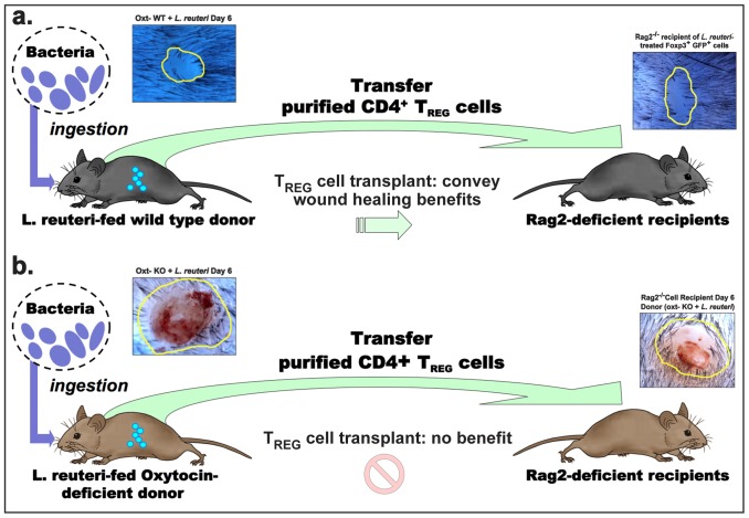
Verification
Claims the paper makes
The claims and statistics of this paper haven’t been checked yet.
Run the check — free, no account needed. It reads the open-access text and checks the sources it cites.
Claims other papers make about this one
Each is checked against this paper’s own text: does it say what the citing paper says it does?
The source backs part of the claim, not all of it — or backs it for a different population, condition or comparison.
In the citing paper
“Indeed, administration of L. reuteri, to mice resulted in an increase in the circulation levels and expression of oxytocin, which was associated with an increase in social behavior (Buffington et al., 2016;Poutahidis et al., 2013).”
From The Microbiota-Gut-Brain Axis (2019) · cites this paper as Poutahidis et al., 2013
Experimental — accuracy not yet measuredIn the citing paper
“Interestingly, the effects observed above are dependent on vagal signaling, which blunted the effects of Lactobacillus on host plasma and hypothalamic oxytocin [132,133].”
From Anxiety, depression, and the microbiome: A role for gut peptides (2018) · cites this paper as [132]
In the citing paper
“We and others have shown that L. reuteri increases oxytocin levels (Buffington et al., 2016; Poutahidis et al., 2013).”
From Mechanisms underlying microbial-mediated changes in social behavior in mouse models of autism spectrum disorder (2019) · cites this paper as Poutahidis et al., 2013
In the citing paper
“We and others have previously provided correlational evidence that L. reuteri increases oxytocin levels (Buffington et al., 2016; Poutahidis et al., 2013)”
From Mechanisms underlying microbial-mediated changes in social behavior in mouse models of autism spectrum disorder (2019) · cites this paper as Poutahidis et al., 2013
Self-check
Percentages, sums and reported differences re-computed from the paper’s own numbers.
The self-check needs the paper’s full text.
Citations
Cites
Bettelli, E., Carrier, Y., Gao, W., Korn, T., Strom, T. B., Oukka, M., Weiner, H. L., & Kuchroo, V. K. (2006) · Nature · doi:10.1038/nature04753 · Cited by 6,992
Gurtner, G. C., Werner, S., Barrandon, Y., & Longaker, M. T. (2008) · Nature · doi:10.1038/nature07039 · Cited by 6,542
Singer, A. J., & Clark, R. A. F. (1999) · New England Journal of Medicine · doi:10.1056/nejm199909023411006 · Cited by 6,106
Wound Healing--Aiming for Perfect Skin Regeneration
Martin, P. B. (1997) · Science · doi:10.1126/science.276.5309.75 · Cited by 4,842
Host-gut microbiota metabolic interactions
Nicholson, J. K., Holmes, E., Kinross, J., Burcelin, R., Gibson, G., Jia, W., & Pettersson, S. (2012) · Science · doi:10.1126/science.1223813 · Cited by 4,760
Mind-altering microorganisms: the impact of the gut microbiota on brain and behaviour
Cryan, J. F., & Dinan, T. G. (2012) · Nature Reviews Neuroscience · doi:10.1038/nrn3346 · Cited by 4,572
Tumors: Wounds That Do Not Heal
Flier, J. S., Underhill, L. H., & Dvorak, H. F. (1986) · New England Journal of Medicine · doi:10.1056/nejm198612253152606 · Cited by 4,391 · cited 1×
“The processes of re-epithelialization and granulation tissue formation with collagen deposition and successive influx of different subsets of immune cells match the classical wound healing timeline previously described in C57BL/6 mice [33,48,56].”
BackgroundVagus nerve stimulation attenuates the systemic inflammatory response to endotoxin
Borovikova, L. V., Ivanova, S., Zhang, M., Yang, H., Botchkina, G. I., Watkins, L. R., Wang, H., Abumrad, N., Eaton, J. W., & Tracey, K. J. (2000) · Nature · doi:10.1038/35013070 · Cited by 4,211
Interactions Between the Microbiota and the Immune System
Hooper, L. V., Littman, D. R., & Macpherson, A. J. (2012) · Science · doi:10.1126/science.1223490 · Cited by 4,173
Bravo, J. A., Forsythe, P., Chew, M. V., Escaravage, E., Savignac, H. M., Dinan, T. G., Bienenstock, J., & Cryan, J. F. (2011) · Proceedings of the National Academy of Sciences · doi:10.1073/pnas.1102999108 · Cited by 3,888
Cited by
Sorted by citations
Cryan, J. F., O'Riordan, K. J., Cowan, C. S. M., Sandhu, K. V., Bastiaanssen, T. F. S., Boehme, M., Codagnone, M. G., Cussotto, S., Fulling, C., Golubeva, A. V., et al. (2019) · Physiological Reviews · doi:10.1152/physrev.00018.2018 · Cited by 5,286
Microbiota in health and diseases
Hou, K., Wu, Z.-X., Chen, X.-Y., Wang, J.-Q., Zhang, D., Xiao, C., Zhu, D., Koya, J. B., Wei, L., Li, J., et al. (2022) · Signal Transduction and Targeted Therapy · doi:10.1038/s41392-022-00974-4 · Cited by 3,171
The gut microbiota–brain axis in behaviour and brain disorders
Morais, L. H., Schreiber, H. L., & Mazmanian, S. K. (2021) · Nature Reviews Microbiology · doi:10.1038/s41579-020-00460-0 · Cited by 2,149
Microbial reconstitution reverses maternal diet-induced social and synaptic deficits in offspring
Buffington, S. A., Di Prisco, G. V., Auchtung, T. A., Ajami, N. J., Petrosino, J. F., & Costa-Mattioli, M. (2016) · Cell · doi:10.1016/j.cell.2016.06.001 · Cited by 1,163
The gut microbiome: Relationships with disease and opportunities for therapy
Durack, J., & Lynch, S. V. (2018) · The Journal of Experimental Medicine · doi:10.1084/jem.20180448 · Cited by 975
Sgritta, M., Dooling, S. W., Buffington, S. A., Momin, E. N., Francis, M. B., Britton, R. A., & Costa-Mattioli, M. (2019) · Neuron · doi:10.1016/j.neuron.2018.11.018 · Cited by 865
Role of Lactobacillus reuteri in Human Health and Diseases
Mu, Q., Tavella, V. J., & Luo, X. M. (2018) · Frontiers in Microbiology · doi:10.3389/fmicb.2018.00757 · Cited by 744
The gut microbiome as a major regulator of the gut-skin axis
Salem, I., Ramser, A., Isham, N., & Ghannoum, M. A. (2018) · Frontiers in Microbiology · doi:10.3389/fmicb.2018.01459 · Cited by 707
Pessemier, B. D., Grine, L., Debaere, M., Maes, A., Paetzold, B., & Callewaert, C. (2021) · Microorganisms · doi:10.3390/microorganisms9020353 · Cited by 684
Microbial endocrinology: the interplay between the microbiota and the endocrine system
Neuman, H., Debelius, J. W., Knight, R. D., & Koren, O. (2015) · FEMS Microbiology Reviews · doi:10.1093/femsre/fuu010 · Cited by 624
Anxiety, depression, and the microbiome: A role for gut peptides
Lach, G., Schellekens, H., Dinan, T. G., & Cryan, J. F. (2018) · Neurotherapeutics · doi:10.1007/s13311-017-0585-0 · Cited by 573
Microbiota and the social brain
Sherwin, E., Bordenstein, S. R., Quinn, J. L., Dinan, T. G., & Cryan, J. F. (2019) · Science · doi:10.1126/science.aar2016 · Cited by 462
The gut-skin axis in health and disease: A paradigm with therapeutic implications
O'Neill, C. A., Monteleone, G., McLaughlin, J. T., & Paus, R. (2016) · BioEssays · doi:10.1002/bies.201600008 · Cited by 449
Is oxytocin “nature’s medicine”
Carter, C. S., Kenkel, W. M., MacLean, E. L., Wilson, S. R., Perkeybile, A. M., Yee, J. R., Ferris, C. F., Nazarloo, H. P., Porges, S. W., Davis, J. M., et al. (2020) · Pharmacological Reviews · doi:10.1124/pr.120.019398 · Cited by 394
Flandroy, L., Poutahidis, T., Berg, G., Clarke, G. A., Dao, M. C., Decaestecker, E., Furman, E., Haahtela, T., Massart, S., Plovier, H., et al. (2018) · The Science of The Total Environment · doi:10.1016/j.scitotenv.2018.01.288 · Cited by 416
The neuroendocrinology of the microbiota-gut-brain axis: a behavioural perspective
Cussotto, S., Sandhu, K. V., Dinan, T. G., & Cryan, J. F. (2018) · Frontiers in Neuroendocrinology · doi:10.1016/j.yfrne.2018.04.002 · Cited by 326
“40 suggests that the gut microbiota is able to directly modulate the levels of host oxytocin (Poutahidis et al., 2013; Varian et al., 2017) and so it is interesting to speculate as to whether”
BackgroundSkin Barrier Function and the Microbiome
Lee, H.-J., & Kim, M. (2022) · International Journal of Molecular Sciences · doi:10.3390/ijms232113071 · Cited by 317
Recent developments in understanding the role of the gut microbiota in brain health and disease
Sherwin, E., Dinan, T. G., & Cryan, J. F. (2018) · Annals of the New York Academy of Sciences · doi:10.1111/nyas.13416 · Cited by 338
Sherwin, E., Sandhu, K. V., Dinan, T. G., & Cryan, J. F. (2016) · CNS Drugs · doi:10.1007/s40263-016-0370-3 · Cited by 308
Neuronal regulation of immunity: why, how and where?
Schiller, M., Ben-Shaanan, T. L., & Rolls, A. (2020) · Nature reviews. Immunology · doi:10.1038/s41577-020-0387-1 · Cited by 280
The gut microbiota–brain axis in neurological disorder
Ullah, H., Arbab, S., Tian, Y., Liu, C., Chen, Y., Li, Q., Khan, M. I. U., Hassan, I. U., & Li, K. (2023) · Frontiers in Neuroscience · doi:10.3389/fnins.2023.1225875 · Cited by 292
Dissecting the contribution of host genetics and the microbiome in complex behaviors
Buffington, S. A., Dooling, S. W., Sgritta, M., Noecker, C. A., Murillo, O. D., Felice, D., Turnbaugh, P. J., & Costa‐Mattioli, M. (2021) · Cell · doi:10.1016/j.cell.2021.02.009 · Cited by 214
Neuroimmune circuits in inter-organ communication
Huh, J. R., & Veiga‐Fernandes, H. (2019) · Nature reviews. Immunology · doi:10.1038/s41577-019-0247-z · Cited by 216
The Cutaneous Microbiome and Wounds: New Molecular Targets to Promote Wound Healing
Johnson, T. R., Gómez, B. I., McIntyre, M. K., Dubick, M. A., Christy, R. J., Nicholson, S. E., & Burmeister, D. M. (2018) · International Journal of Molecular Sciences · doi:10.3390/ijms19092699 · Cited by 219
A gut (microbiome) feeling about the brain
Sherwin, E., Rea, K., Dinan, T. G., & Cryan, J. F. (2016) · Current Opinion in Gastroenterology · doi:10.1097/mog.0000000000000244 · Cited by 206
Sinus microbiota varies among chronic rhinosinusitis phenotypes and predicts surgical outcome
Ramakrishnan, V. R., Hauser, L. J., Feazel, L. M., Ir, D., Robertson, C. E., & Frank, D. N. (2015) · Journal of Allergy and Clinical Immunology · doi:10.1016/j.jaci.2015.02.008 · Cited by 187
Brown, S. L., & Brown, R. M. (2015) · Neuroscience & Biobehavioral Reviews · doi:10.1016/j.neubiorev.2015.04.004 · Cited by 212
Probiotic Microbes Sustain Youthful Serum Testosterone Levels and Testicular Size in Aging Mice
Poutahidis, T., Springer, A., Levkovich, T., Qi, P., Varian, B. J., Lakritz, J. R., Ibrahim, Y. M., Chatzigiagkos, A., Alm, E. J., & Erdman, S. E. (2014) · PLoS ONE · doi:10.1371/journal.pone.0084877 · Cited by 177
Self-citationThe Th17/Treg Cell Balance: A Gut Microbiota-Modulated Story
Cheng, H., Guan, X., Chen, D., & Ma, W. (2019) · Microorganisms · doi:10.3390/microorganisms7120583 · Cited by 191
Probiotics or pro‐healers: the role of beneficial bacteria in tissue repair
Lukić, J., Chen, V., Strahinić, I., Begović, J., Lev‐Tov, H. A., Davis, S. C., Tomic‐Canic, M., & Pastar, I. (2017) · Wound Repair and Regeneration · doi:10.1111/wrr.12607 · Cited by 180
Gut–Skin Axis: Unravelling the Connection between the Gut Microbiome and Psoriasis
Thye, A. Y.-K., Bah, Y.-R., Law, J. W.-F., Tan, L. T.-H., He, Y.-W., Wong, S.-H., Thurairajasingam, S., Chan, K.-G., Lee, L.-H., & Letchumanan, V. (2022) · Biomedicines · doi:10.3390/biomedicines10051037 · Cited by 156
Microbial lysate upregulates host oxytocin
Varian, B. J., Poutahidis, T., DiBenedictis, B. T., Levkovich, T., Ibrahim, Y., Didyk, E., Shikhman, L., Cheung, H. K., Hardas, A., Ricciardi, C. E., et al. (2017) · Brain Behavior and Immunity · doi:10.1016/j.bbi.2016.11.002 · Cited by 153
Self-citationLakritz, J. R., Poutahidis, T., Levkovich, T., Varian, B. J., Ibrahim, Y. M., Chatzigiagkos, A., Mirabal, S., Alm, E. J., & Erdman, S. E. (2013) · International Journal of Cancer · doi:10.1002/ijc.28702 · Cited by 150
Beneficial bacteria inhibit cachexia
Varian, B. J., Goureshetti, S., Poutahidis, T., Lakritz, J. R., Levkovich, T., Kwok, C., Teliousis, K., Ibrahim, Y. M., Mirabal, S., & Erdman, S. E. (2016) · Oncotarget · doi:10.18632/oncotarget.7730 · Cited by 146
McCormack, S. E., Blevins, J. E., & Lawson, E. A. (2019) · Endocrine Reviews · doi:10.1210/endrev/bnz012 · Cited by 148
The role of the microbiome in the neurobiology of social behaviour
Sarkar, A., Harty, S., Johnson, K. V. ‐., Moeller, A. H., Carmody, R. N., Lehto, S. M., Erdman, S. E., Dunbar, R. I. M., & Burnet, P. W. J. (2020) · Biological Reviews · doi:10.1111/brv.12603 · Cited by 135
Exploring the Emerging Role of the Gut Microbiota and Tumor Microenvironment in Cancer Immunotherapy
Qiu, Q., Lin, Y., Ma, Y., Li, X., Liang, J. B., Chen, Z., Liu, K., Huang, Y., Luo, H., Huang, R., et al. (2021) · Frontiers in Immunology · doi:10.3389/fimmu.2020.612202 · Cited by 140
Oxytocin modulation of neural circuits for social behavior
Marlin, B. J., & Froemke, R. C. (2017) · Developmental Neurobiology · doi:10.1002/dneu.22452 · Cited by 130
Re-examining causes of surgical site infections following elective surgery in the era of asepsis
Alverdy, J. C., Hyman, N., & Gilbert, J. A. (2020) · The Lancet Infectious Diseases · doi:10.1016/s1473-3099(19)30756-x · Cited by 127
Gut bacteria require neutrophils to promote mammary tumorigenesis
Lakritz, J. R., Poutahidis, T., Mirabal, S., Varian, B. J., Levkovich, T., Ibrahim, Y. M., Ward, J. M., Teng, E. C., Fisher, B., Parry, N. M., et al. (2015) · Oncotarget · doi:10.18632/oncotarget.3328 · Cited by 120
The gut microbiome and the mechanism of surgical infection
Alverdy, J. C., Hyoju, S. K., Weigerinck, M., & Gilbert, J. A. (2017) · British journal of surgery · doi:10.1002/bjs.10405 · Cited by 119
Khmaladze, I., Butler, É., Fabre, S., & Gillbro, J. M. (2019) · Experimental Dermatology · doi:10.1111/exd.13950 · Cited by 120
Barengolts, E., Green, S. J., Eisenberg, Y., Akbar, A., Reddivari, B., Layden, B. T., Dugas, L., & Chlipala, G. (2018) · PLoS ONE · doi:10.1371/journal.pone.0194171 · Cited by 111
Skin Health from the Inside Out
Woodby, B. L., Penta, K., Pecorelli, A., Lila, M. A., & Valacchi, G. (2020) · Annual Review of Food Science and Technology · doi:10.1146/annurev-food-032519-051722 · Cited by 114
Antwis, R. E., Edwards, K. L., Unwin, B., Walker, S. L., & Shultz, S. (2019) · Microbiome · doi:10.1186/s40168-019-0639-0 · Cited by 109
Mazzone, L., Dooling, S. W., Volpe, E., Uljarević, M., Waters, J. L., Sabatini, A., Arturi, L., Abate, R., Riccioni, A., Siracusano, M., et al. (2023) · Cell Host & Microbe · doi:10.1016/j.chom.2023.11.021 · Cited by 113
Clinical applications of gut microbiota in cancer biology
Wong, S. H., Kwong, T. N. Y., Wu, C., & Yu, J. Q. (2018) · Seminars in Cancer Biology · doi:10.1016/j.semcancer.2018.05.003 · Cited by 102
Novel probiotic-bound oxidized Bletilla striata polysaccharide-chitosan composite hydrogel
Yang, L., Han, Z., Chen, C., Li, Z., Yu, S., Qu, Y., & Zeng, R. (2020) · Materials Science and Engineering C · doi:10.1016/j.msec.2020.111265 · Cited by 101
The Role of Oxytocin in Cardiovascular Protection
Jankowski, M., Broderick, T. L., & Gutkowska, J. (2020) · Frontiers in Psychology · doi:10.3389/fpsyg.2020.02139 · Cited by 101
Role of oxytocin in the control of stress and food intake
Onaka, T., & Takayanagi, Y. (2019) · Journal of Neuroendocrinology · doi:10.1111/jne.12700 · Cited by 102
Patel, B. K., Patel, K. H., Huang, R. Y., Lee, C. N., & Moochhala, S. (2022) · International Journal of Molecular Sciences · doi:10.3390/ijms23042375 · Cited by 97
Oxytocin-secreting system: A major part of the neuroendocrine center regulating immunologic activity
Wang, P., Yang, H.-P., Tian, S., Wang, L., Wang, S. C., Zhang, F., & Wang, Y. (2015) · Journal of Neuroimmunology · doi:10.1016/j.jneuroim.2015.11.001 · Cited by 96
Reciprocal Interactions Between Gut Microbiota and Host Social Behavior
Münger, E., Montiel-Castro, A. J., Langhans, W., & Pacheco‐López, G. (2018) · Frontiers in Integrative Neuroscience · doi:10.3389/fnint.2018.00021 · Cited by 88
Liu, Y., Sanderson, D., Mian, M. F., Neufeld, K. M., & Forsythe, P. (2021) · Neuropharmacology · doi:10.1016/j.neuropharm.2021.108682 · Cited by 87
Recent Advances in Understanding the Structure and Function of the Human Microbiome
Mousa, W. K., Chehadeh, F., & Husband, S. (2022) · Frontiers in Microbiology · doi:10.3389/fmicb.2022.825338 · Cited by 90
Lin, A., Xiong, M., Jiang, A., Huang, L., Wong, H. Z. H., Feng, S., Zhang, C., Yu, H., Chen, L., Chi, H., et al. (2025) · iMeta · doi:10.1002/imt2.70070 · Cited by 94
Gut Microbiota and Bone Health
Quach, D., & Britton, R. A. (2017) · Advances in experimental medicine and biology · doi:10.1007/978-3-319-66653-2_4 · Cited by 85
Narmaki, E., Borazjani, M., Ataie‐Jafari, A., Hariri, N., Doost, A. H., Qorbani, M., & Saidpour, A. (2020) · Nutritional Neuroscience · doi:10.1080/1028415x.2020.1826763 · Cited by 85
Schlagenhauf, U., Jakob, L. M., Eigenthaler, M., Segerer, S. E., Jockel‐Schneider, Y., & Rehn, M. (2016) · Journal Of Clinical Periodontology · doi:10.1111/jcpe.12606 · Cited by 74
McIntyre, M. K., Peacock, T. J., Akers, K. S., & Burmeister, D. M. (2016) · PLoS ONE · doi:10.1371/journal.pone.0166176 · Cited by 71
Potential Impacts of Prebiotics and Probiotics on Cancer Prevention
Samanta, S. (2020) · Anti-Cancer Agents in Medicinal Chemistry · doi:10.2174/1871520621999201210220442 · Cited by 73
Human microflora, probiotics and wound healing
Tsiouris, C. G., & Tsiouri, M. G. (2017) · Wound Medicine · doi:10.1016/j.wndm.2017.09.006 · Cited by 70
Erdman, S. E., & Poutahidis, T. (2016) · International review of neurobiology · doi:10.1016/bs.irn.2016.07.004 · Cited by 69
Laughter Yoga: Benefits of Mixing Laughter and Yoga
Woodbury-Fariña, M. A., & Schwabe, M. M. R. (2015) · Journal of Yoga & Physical Therapy · doi:10.4172/2157-7595.1000209 · Cited by 66
Buemann, B., & Uvnäs-Moberg, K. (2020) · Medical Hypotheses · doi:10.1016/j.mehy.2020.109597 · Cited by 66
Immunomodulation of dendritic cells byLactobacillus reuterisurface components and metabolites
Engevik, M. A., Ruan, W. H., Esparza, M., Fultz, R., Shi, Z., Engevik, K. A., Engevik, A. C., Ihekweazu, F. D., Visuthranukul, C., Venable, S. F., et al. (2021) · Physiological Reports · doi:10.14814/phy2.14719 · Cited by 69
Kingsbury, M. A., & Bilbo, S. D. (2019) · Frontiers in Neuroendocrinology · doi:10.1016/j.yfrne.2019.100794 · Cited by 68
Tsiouris, C. G., Kelesi, Μ., Vasilopoulos, G., Kalemikerakis, I., & Papageorgiou, E. G. (2017) · European Journal of Pharmaceutical Sciences · doi:10.1016/j.ejps.2017.04.002 · Cited by 64
Alesa, D. I., Alshamrani, H. M., Alzahrani, Y. A., Alamssi, D., Alzahrani, N., & Almohammadi, M. (2019) · Journal of Family Medicine and Primary Care · doi:10.4103/jfmpc.jfmpc_709_19 · Cited by 65
RetractedH3K27me3 dynamics dictate evolving uterine states in pregnancy and parturition
Nancy, P., Siewiera, J., Rizzuto, G. A., Tagliani, E., Osokine, I., Manandhar, P., Dolgalev, I., Clementi, C., Tsirigos, A., & Erlebacher, A. (2017) · Journal of Clinical Investigation · doi:10.1172/jci95937 · Cited by 63
The microbiome and disorders of the central nervous system
Felice, V. D., & O'Mahony, S. M. (2017) · Pharmacology Biochemistry and Behavior · doi:10.1016/j.pbb.2017.06.016 · Cited by 60
Wang, J., Li, X., Zhao, X., Yuan, S., Dou, H., Cheng, T., Huang, T., Lv, Z., Tu, Y., Shi, Y., et al. (2024) · Journal of Nanobiotechnology · doi:10.1186/s12951-024-02893-8 · Cited by 59
Intestinal Microbiota in Colorectal Cancer Surgery
Koliarakis, I., Athanasakis, E., Sgantzos, M. N., Mariolis-Sapsakos, T. D., Xynos, E., Chrysos, E., Souglakos, J., & Tsiaoussis, J. (2020) · Cancers · doi:10.3390/cancers12103011 · Cited by 63
Quirós, M., & Nusrat, A. (2018) · Annual Review of Physiology · doi:10.1146/annurev-physiol-020518-114504 · Cited by 59
Lui, P. P., Cho, I., & Ali, N. (2020) · Immunology · doi:10.1111/imm.13208 · Cited by 61
Probiotic ‘glow of health’: it's more than skin deep
Erdman, S. E., & Poutahidis, T. (2014) · Beneficial Microbes · doi:10.3920/bm2013.0042 · Cited by 56
Bekiaridou, Α., Karlafti, E., Oikonomou, I. M., Ioannidis, A., & Papavramidis, T. S. (2021) · Nutrients · doi:10.3390/nu13124265 · Cited by 55
Microbial stimulation of oxytocin release from the intestinal epithelium via secretin signaling
Danhof, H. A., Lee, J., Thapa, A., Britton, R. A., & Rienzi, S. C. D. (2023) · Gut Microbes · doi:10.1080/19490976.2023.2256043 · Cited by 60
Erdman, S. E., & Poutahidis, T. (2015) · Biochimica et Biophysica Acta (BBA) - Reviews on Cancer · doi:10.1016/j.bbcan.2015.05.007 · Cited by 55
Dietary Microbes Modulate Transgenerational Cancer Risk
Poutahidis, T., Varian, B. J., Levkovich, T., Lakritz, J. R., Mirabal, S., Kwok, C., Ibrahim, Y. M., Kearney, S. M., Chatzigiagkos, A., Alm, E. J., et al. (2015) · Cancer Research · doi:10.1158/0008-5472.can-14-2732 · Cited by 53
Leveau, J. H. J. (2024) · Annual Review of Phytopathology · doi:10.1146/annurev-phyto-121423-042021 · Cited by 61
Mohtashami, M., Mohamadi, M., Azimi–Nezhad, M., Saeidi, J., Nia, F. F., & Ghasemi, A. (2020) · Biotechnology and Applied Biochemistry · doi:10.1002/bab.2064 · Cited by 53
Schlagenhauf, U., Rehder, J., Gelbrich, G., & Jockel‐Schneider, Y. (2020) · Journal of Periodontology · doi:10.1002/jper.19-0393 · Cited by 52
Human microbiome science: vision for the future, Bethesda, MD, July 24 to 26, 2013
Ravel, J., Blaser, M. J., Braun, J. G., Brown, E., Bushman, F. D., Chang, E. B., DAVIES, J. E., Dewey, K. G., Dinan, T. G., Domínguez-Bello, M. G., et al. (2014) · Microbiome · doi:10.1186/2049-2618-2-16 · Cited by 58
Bartolini, I., Risaliti, M., Ringressi, M. N., Melli, F., Nannini, G., Amedei, A., Muiesan, P., & Taddei, A. (2020) · World Journal of Gastroenterology · doi:10.3748/wjg.v26.i20.2498 · Cited by 54
Schmitt, L., Smith, E. G., Pedapati, E. V., Horn, P. S., Will, M., Lamy, M., Barber, L., Trebley, J., Meyer, K., Heiman, M. L., et al. (2023) · Scientific Reports · doi:10.1038/s41598-023-30909-0 · Cited by 58
Connection and communication between the nervous and immune systems
Leunig, A., Gianeselli, M., Russo, S. J., & Świrski, F. K. (2025) · Nature reviews. Immunology · doi:10.1038/s41577-025-01199-6 · Cited by 57
Commensal bacteria modulate the tumor microenvironment
Poutahidis, T., & Erdman, S. E. (2015) · Cancer Letters · doi:10.1016/j.canlet.2015.12.028 · Cited by 48
Maternal effects in mammals: Broadening our understanding of offspring programming
Edwards, P. D., Lavergne, S. G., McCaw, L. K., Wijenayake, S., Boonstra, R., McGowan, P. O., & Holmes, M. M. (2021) · Frontiers in Neuroendocrinology · doi:10.1016/j.yfrne.2021.100924 · Cited by 49
Lagraoui, M., Sukumar, G., Latoche, J. R., Maynard, S. K., Dalgard, C. L., & Schaefer, B. C. (2016) · Brain Behavior and Immunity · doi:10.1016/j.bbi.2016.12.005 · Cited by 47
Moreira, C. F., Vieira, P. C., Canesso, M. C. C., Felipetto, M., Ranfley, H., Teixeira, M. M., Nicoli, J. R., Martins, F. S., & Barcelos, L. S. (2021) · Probiotics and Antimicrobial Proteins · doi:10.1007/s12602-020-09713-z · Cited by 47
Does Probiotic Consumption Enhance Wound Healing? A Systematic Review
Togo, C. C. G., Zidório, A. P. C., Gonçalves, V. S. S., Botelho, P. B., de Carvalho, K. M. B., & Dutra, E. S. (2021) · Nutrients · doi:10.3390/nu14010111 · Cited by 47
The Relationship Between Microbial Community and Breast Cancer
Song, X., Wei, C., & Li, X. (2022) · Frontiers in Cellular and Infection Microbiology · doi:10.3389/fcimb.2022.849022 · Cited by 45
Between Dysbiosis, Maternal Immune Activation and Autism: Is There a Common Pathway?
Suprunowicz, M., Tomaszek, N., Urbaniak, A., Zackiewicz, K., Modzelewski, S., & Waszkiewicz, N. (2024) · Nutrients · doi:10.3390/nu16040549 · Cited by 44
Multiorgan‐on‐a‐chip for realization of gut‐skin axis
Lee, H. R., & Sung, J. H. (2022) · Biotechnology and Bioengineering · doi:10.1002/bit.28164 · Cited by 46
Oxytocin and cancer: An emerging link
Lerman, B., Harricharran, T., & Ogunwobi, O. O. (2018) · World Journal of Clinical Oncology · doi:10.5306/wjco.v9.i5.74 · Cited by 47
Prostate cancer: Therapeutic prospect with herbal medicine
Ghosh, S., Hazra, J., Pal, K., Nelson, V. K., & Pal, M. (2021) · Current Research in Pharmacology and Drug Discovery · doi:10.1016/j.crphar.2021.100034 · Cited by 46
Butler, É., Öien, R. F., Lindholm, C., Olofsson, T. C., Nilson, B., & Vásquez, A. (2014) · International Wound Journal · doi:10.1111/iwj.12360 · Cited by 43
Machado, P., Ribeiro, F. N., Giublin, F. C. W., Mieres, N. G., Tonin, F. S., Pontarolo, R., Sari, M. H. M., Lazo, R. E. L., & Ferreira, L. M. (2025) · Pharmaceuticals · doi:10.3390/ph18050704 · Cited by 44
Metadata
- Authors
- Poutahidis, T., Kearney, S. M., Levkovich, T., Qi, P., Varian, B. J., Lakritz, J. R., Ibrahim, Y. M., Chatzigiagkos, A., Alm, E. J., & Erdman, S. E.
- Affiliations
- Aristotle University of Thessaloniki (GR)
- Massachusetts Institute of Technology (US)
- Broad Institute (US)
- Corresponding author
- Erdman, Susan E.
- ORCID
- Venue
- PLoS ONE
- Publisher
- Public Library of Science (PLoS)
- Published
- 2013-10-30
- Volume
- 8
- Issue
- 10
- Pages
- e78898
- ISSN
- 1932-6203 (ISSN-L)
- PMC
- PMC3813596
- OpenAlex
- W2068527256
- Cited by
- 310
- References
- 85
- Citations by year
- 2026: 19 · 2025: 29 · 2024: 33 · 2023: 25 · 2022: 31 · 2021: 32 · 2020: 31 · 2019: 21 · 2018: 22 · 2017: 18 · 2016: 24 · 2015: 10 · 2014: 14 · 2013: 1
- FWCI
- 17.43
- Type
- journal article
- Language
- en
- Open access
- gold
- doi.org
- Locations
- PLoS ONE (publishedVersion, cc-by)
- Europe PMC (PubMed Central) (publishedVersion, cc-by)
- PubMed Central (publishedVersion, cc-by)
- PubMed (publishedVersion)
- DOAJ (DOAJ: Directory of Open Access Journals) (submittedVersion, cc-by-sa)
- DSpace@MIT (Massachusetts Institute of Technology) (submittedVersion, cc-by)
- Figshare (submittedVersion, cc-by)
- Funders
- National Institutes of Health
- National Cancer Institute (U01CA164337, R01CA108854)
- National Institute of Environmental Health Sciences (P30ES002109)
- Topics
- Health Sciences › Medicine › Surgery › Wound Healing and Treatments
- Life Sciences › Biochemistry, Genetics and Molecular Biology › Molecular Biology › Gut microbiota and health
- Life Sciences › Agricultural and Biological Sciences › Food Science › Probiotics and Fermented Foods
- Keywords
- wound healing, gut microbiota, Lactobacillus reuteri, oxytocin, vagus nerve, regulatory T cells, gut-brain-immune axis, lactic acid bacteria, drinking water supplementation, microbial symbionts
- MeSH terms
- Animals, Collagen, DNA-Binding Proteins, Female, Oxytocin, Symbiosis, Time Factors, Wound Healing, CD4-Positive T-Lymphocytes, Up-Regulation, Mice, Limosilactobacillus reuteri, Drinking Water
About these checks
Registry, retraction, Cites and statistics results are computed, not judged, with one exception: a printed reference that no index lists is read once by a model before it counts as a citation error. Claims are extracted by a model and kept only when their quote appears verbatim in the text; each cited source is then checked, with a model’s help, for whether it says what the claim relies on. Those verdicts can be wrong — read the claim’s quote and the cited source before relying on one.
Statistics and arithmetic are recomputed, not judged.
Confidence bands are shown only for fields calibrated on our annotated test set. None is calibrated yet, so no band is shown.
What counts as a claim ›How we measure confidence ›How we check this ›
Are you an author, or do you see a problem with this page? Request a review
