

The DOI matches its Crossref record; 52 of the 52 references it cites were found in a registry or an index.
Protective Microglial Subset in Development, Aging, and Disease: Lessons From Transcriptomic Studies
Overview
The claims and statistics of this paper haven’t been checked yet.
Run the check — free, no account needed. It reads the open-access text and checks the sources it cites.
Integrity signals
Registry
Cites
Statistics
Figures
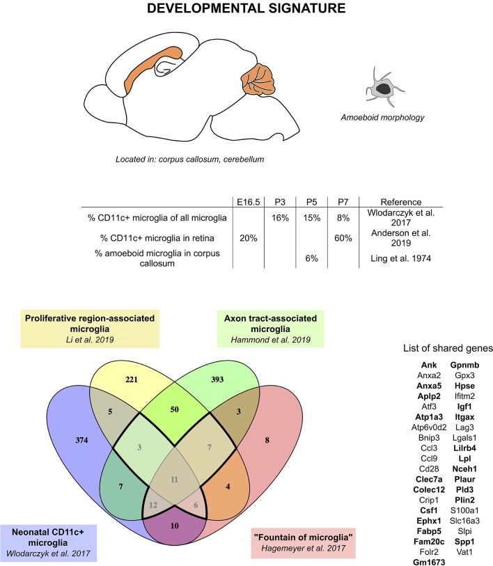
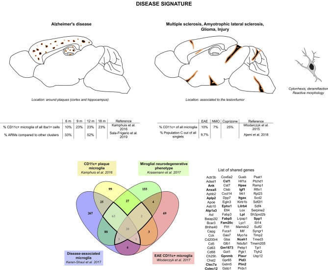
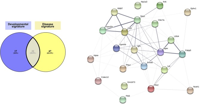
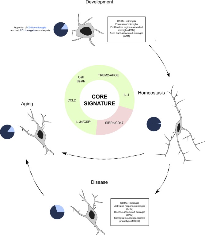
Verification
Claims the paper makes
The claims and statistics of this paper haven’t been checked yet.
Run the check — free, no account needed. It reads the open-access text and checks the sources it cites.
Claims other papers make about this one
Each is checked against this paper’s own text: does it say what the citing paper says it does?
In the citing paper
“Atp1a3 that belongs to the signature of CD11c+ microglia (72)”
From Absence of miRNA-146a Differentially Alters Microglia Function and Proteome (2020) · cites this paper as [72]
Self-check
Percentages, sums and reported differences re-computed from the paper’s own numbers.
The self-check needs the paper’s full text.
Citations
Cites
Resting microglial cells are highly dynamic surveillants of brain parenchyma in vivo
Nimmerjahn, A., Kirchhoff, F., & Helmchen, F. (2005) · Science · doi:10.1126/science.1110647 · Cited by 5,827
A Unique Microglia Type Associated with Restricting Development of Alzheimer’s Disease
Keren-Shaul, H., Spinrad, A., Weiner, A., Matcovitch-Natan, O., Dvir-Szternfeld, R., Ulland, T. K., David, E., Baruch, K., Lara-Astaiso, D., Toth, B., et al. (2017) · Cell · doi:10.1016/j.cell.2017.05.018 · Cited by 5,493
Fate Mapping Analysis Reveals That Adult Microglia Derive from Primitive Macrophages
Ginhoux, F., Greter, M., Leboeuf, M., Nandi, S., See, P., Gokhan, S., Mehler, M. F., Conway, S. J., Ng, L. G., Stanley, E. R., et al. (2010) · Science · doi:10.1126/science.1194637 · Cited by 5,175
Synaptic Pruning by Microglia Is Necessary for Normal Brain Development
Paolicelli, R. C., Bolasco, G., Pagani, F., Maggi, L., Scianni, M., Panzanelli, P., Giustetto, M., Ferreira, T. A., Guiducci, E., Dumas, L., et al. (2011) · Science · doi:10.1126/science.1202529 · Cited by 4,244
Microglia Sculpt Postnatal Neural Circuits in an Activity and Complement-Dependent Manner
Schafer, D. P., Lehrman, E. K., Kautzman, A. G., Koyama, R., Mardinly, A. R., Yamasaki, R., Ransohoff, R. M., Greenberg, M. E., Barres, B. A., & Stevens, B. (2012) · Neuron · doi:10.1016/j.neuron.2012.03.026 · Cited by 4,089
ATP mediates rapid microglial response to local brain injury in vivo
Davalos, D., Grutzendler, J., Yang, G., Kim, J. V., Zuo, Y., Jung, S., Littman, D. R., Dustin, M. L., & Gan, W.-B. (2005) · Nature Neuroscience · doi:10.1038/nn1472 · Cited by 3,913
Krasemann, S., Madore, C., Cialic, R., Baufeld, C., Calcagno, N., El Fatimy, R., Beckers, L., O’Loughlin, E., Xu, Y., Fanek, Z., et al. (2017) · Immunity · doi:10.1016/j.immuni.2017.08.008 · Cited by 2,979
Identification of a unique TGF-β–dependent molecular and functional signature in microglia
Butovsky, O., Jedrychowski, M. P., Moore, C. S., Cialic, R., Lanser, A. J., Gabriely, G., Koeglsperger, T., Dake, B., Wu, P. M., Doykan, C. E., et al. (2014) · Nature Neuroscience · doi:10.1038/nn.3599 · Cited by 2,543
Hammond, T. R., Dufort, C., Dissing-Olesen, L., Giera, S., Young, A., Wysoker, A., Walker, A. J., Segel, M., Nemesh, J., Saunders, A., et al. (2018) · doi:10.1101/406140 · Cited by 2,225
Microglial physiology: unique stimuli, specialized responses
Ransohoff, R. M., & Perry, V. H. (2009) · Annual Review of Immunology · doi:10.1146/annurev.immunol.021908.132528 · Cited by 1,858
Spatial and temporal heterogeneity of mouse and human microglia at single-cell resolution
Masuda, T., Sankowski, R., Staszewski, O., Böttcher, C., Amann, L., Sagar, Scheiwe, C., Nessler, S., Kunz, P., van Loo, G., et al. (2019) · Nature · doi:10.1038/s41586-019-0924-x · Cited by 1,310
The far-reaching scope of neuroinflammation after traumatic brain injury
Simon, D. W., McGeachy, M. J., Bayır, H., Clark, R. S. B., Loane, D. J., & Kochanek, P. M. (2017) · Nature Reviews Neurology · doi:10.1038/nrneurol.2017.13 · Cited by 1,190 · cited 1×
“Studying microglia in context of inflammation can get quite complicated due to massive infiltration of peripheral immune cells, notably monocytes and macrophages, occurring subsequently to TBI (128) , SCI (129) , and stroke (127, 130, 131) .”
BackgroundMicroglia development follows a stepwise program to regulate brain homeostasis
Matcovitch-Natan, O., Winter, D. R., Giladi, A., Vargas Aguilar, S., Spinrad, A., Sarrazin, S., Ben-Yehuda, H., David, E., Zelada González, F., Perrin, P., et al. (2016) · Science · doi:10.1126/science.aad8670 · Cited by 1,180 · cited 1×
“Interestingly, Csf1, one of the genes of the core signature, has been identified as one of the prominent genes characteristic of the pre-microglia homeostatic signature (23).”
BackgroundDectin-1: a signalling non-TLR pattern-recognition receptor
Brown, G. D. (2006) · Nature Reviews Immunology · doi:10.1038/nri1745 · Cited by 1,158
Li, Q., Cheng, Z., Zhou, L., Darmanis, S., Neff, N. F., Okamoto, J., Gulati, G., Bennett, M. L., Sun, L. O., Clarke, L. E., et al. (2019) · Neuron · doi:10.1016/j.neuron.2018.12.006 · Cited by 1,129
Van Hove, H., Martens, L., Scheyltjens, I., De Vlaminck, K., Pombo Antunes, A. R., De Prijck, S., Vandamme, N., De Schepper, S., Van Isterdael, G., Scott, C. L., et al. (2019) · Nature Neuroscience · doi:10.1038/s41593-019-0393-4 · Cited by 1,074 · cited 1×
“This population named “Kolmer’s epiplexus cells” closely resembles microglial cells and was associated with enriched expression of Spp1, Apoe, and Igf1 (82).”
BackgroundEl-Behi, M., Ciric, B., Dai, H., Yan, Y., Cullimore, M., Safavi, F., Zhang, G.-X., Dittel, B. N., & Rostami, A. (2011) · Nature Immunology · doi:10.1038/ni.2031 · Cited by 1,071 · cited 1×
“Interestingly, in contrast to CD11c+ blood-derived cells and CD11c− microglia, CD11c+ microglia completely lacked mRNA expression for IL-23 (34) that is known to induce GM-CSF-producing CD4+ T cells, critical for EAE pathology (141) .”
Sala Frigerio, C., Wolfs, L., Fattorelli, N., Thrupp, N., Voytyuk, I., Schmidt, I., Mancuso, R., Chen, W.-T., Woodbury, M. E., Srivastava, G., et al. (2019) · Cell Reports · doi:10.1016/j.celrep.2019.03.099 · Cited by 926 · cited 4×
“Given that AD risk genes are enriched in this population (80) , mutations in such genes may have an impact on the ability of CD11c+ microglia to cope with Aβ plaque burden, either promoting or limiting AD pathology.”
BackgroundResultInfluentialPerry, V. H., Hume, D. A., & Gordon, S. (1985) · Neuroscience · doi:10.1016/0306-4522(85)90215-5 · Cited by 902
TREM2-mediated early microglial response limits diffusion and toxicity of amyloid plaques
Wang, Y., Ulland, T. K., Ulrich, J. D., Song, W., Tzaferis, J. A., Hole, J. T., Yuan, P., Mahan, T. E., Shi, Y., Gilfillan, S., et al. (2016) · Journal of Experimental Medicine · doi:10.1084/jem.20151948 · Cited by 840
Neuroimmunology of Traumatic Brain Injury: Time for a Paradigm Shift
Jassam, Y. N., Izzy, S., Whalen, M., McGavern, D. B., & El Khoury, J. (2017) · Neuron · doi:10.1016/j.neuron.2017.07.010 · Cited by 799
Layer V cortical neurons require microglial support for survival during postnatal development
Ueno, M., Fujita, Y., Tanaka, T., Nakamura, Y., Kikuta, J., Ishii, M., & Yamashita, T. (2013) · Nature Neuroscience · doi:10.1038/nn.3358 · Cited by 781
TREM2 — a key player in microglial biology and Alzheimer disease
Ulland, T. K., & Colonna, M. (2018) · Nature Reviews Neurology · doi:10.1038/s41582-018-0072-1 · Cited by 735
Jay, T. R., Miller, C. M., Cheng, P. J., Graham, L. C., Bemiller, S., Broihier, M. L., Xu, G., Margevicius, D., Karlo, J. C., Sousa, G. L., et al. (2015) · Journal of Experimental Medicine · doi:10.1084/jem.20142322 · Cited by 682 · cited 2×
“Nonetheless, all these studies demonstrate lack of microglial proliferation and clustering around plaques in TREM2-deficient animals, thus allowing for more dispersed Aβ localization in AD models (75, (112) (113) (114) (115) (116) .”
BackgroundMicroglia contact induces synapse formation in developing somatosensory cortex
Miyamoto, A., Wake, H., Ishikawa, A. W., Eto, K., Shibata, K., Murakoshi, H., Koizumi, S., Moorhouse, A. J., Yoshimura, Y., & Nabekura, J. (2016) · Nature Communications · doi:10.1038/ncomms12540 · Cited by 649
Bruttger, J., Karram, K., Wörtge, S., Regen, T., Marini, F., Hoppmann, N., Klein, M., Blank, T., Yona, S., Wolf, Y., et al. (2015) · Immunity · doi:10.1016/j.immuni.2015.06.012 · Cited by 634
Holtman, I. R., Raj, D. D., Miller, J. A., Schaafsma, W., Yin, Z., Brouwer, N., Wes, P. D., Möller, T., Orre, M., Kamphuis, W., et al. (2015) · Acta Neuropathologica Communications · doi:10.1186/s40478-015-0203-5 · Cited by 619
A new fate mapping system reveals context-dependent random or clonal expansion of microglia
Tay, T. L., Mai, D., Dautzenberg, J., Fernández-Klett, F., Lin, G., Sagar, Datta, M., Drougard, A., Stempfl, T., Ardura-Fabregat, A., et al. (2017) · Nature Neuroscience · doi:10.1038/nn.4547 · Cited by 597
Herbomel, P., Thisse, B., & Thisse, C. (2001) · Developmental Biology · doi:10.1006/dbio.2001.0393 · Cited by 574
Huang, Y., Xu, Z., Xiong, S., Sun, F., Qin, G., Hu, G., Wang, J., Zhao, L., Liang, Y.-X., Wu, T., et al. (2018) · Nature Neuroscience · doi:10.1038/s41593-018-0090-8 · Cited by 560
Hagemeyer, N., Hanft, K.-M., Akriditou, M.-A., Unger, N., Park, E. S., Stanley, E. R., Staszewski, O., Dimou, L., & Prinz, M. (2017) · Acta Neuropathologica · doi:10.1007/s00401-017-1747-1 · Cited by 558
TREM2 sustains microglial expansion during aging and response to demyelination
Poliani, P. L., Wang, Y., Fontana, E., Robinette, M. L., Yamanishi, Y., Gilfillan, S., & Colonna, M. (2015) · Journal of Clinical Investigation · doi:10.1172/jci77983 · Cited by 525
NBB-Psy, Böttcher, C., Schlickeiser, S., Sneeboer, M. A. M., Kunkel, D., Knop, A., Paza, E., Fidzinski, P., Kraus, L., Snijders, G. J. L., et al. (2019) · Nature Neuroscience · doi:10.1038/s41593-018-0290-2 · Cited by 436
Brain microglia constitutively express β-2 integrins
Akiyama, H., & McGeer, P. L. (1990) · Journal of Neuroimmunology · doi:10.1016/0165-5728(90)90055-r · Cited by 430
The pro-remyelination properties of microglia in the central nervous system
Lloyd, A. F., & Miron, V. E. (2019) · Nature Reviews Neurology · doi:10.1038/s41582-019-0184-2 · Cited by 381
Identification of a microglia phenotype supportive of remyelination
Olah, M., Amor, S., Brouwer, N., Vinet, J., Eggen, B., Biber, K., & Boddeke, H. W. G. M. (2012) · Glia · doi:10.1002/glia.21266 · Cited by 358
TREM2 deficiency impairs chemotaxis and microglial responses to neuronal injury
Mazaheri, F., Snaidero, N., Kleinberger, G., Madore, C., Daria, A., Werner, G., Krasemann, S., Capell, A., Trümbach, D., Wurst, W., et al. (2017) · The EMBO Reports · doi:10.15252/embr.201743922 · Cited by 351
Ajami, B., Samusik, N., Wieghofer, P., Ho, P. P., Crotti, A., Bjornson, Z., Prinz, M., Fantl, W. J., Nolan, G. P., & Steinman, L. (2018) · Nature Neuroscience · doi:10.1038/s41593-018-0100-x · Cited by 338
Central nervous system regeneration is driven by microglia necroptosis and repopulation
Lloyd, A. F., Davies, C. L., Holloway, R. K., Labrak, Y., Ireland, G., Carradori, D., Dillenburg, A., Borger, E., Soong, D., Richardson, J. C., et al. (2019) · Nature Neuroscience · doi:10.1038/s41593-019-0418-z · Cited by 316 · cited 2×
“Interestingly, microglial necroptosis in demyelination models leads to brain repopulation with CD11c+ microglia from nestin+ resident microglia (148) .”
BackgroundTREM2 regulates microglial cell activation in response to demyelination in vivo
Cantoni, C., Bollman, B., Licastro, D., Xie, M., Mikesell, R., Schmidt, R., Yuede, C. M., Galimberti, D., Olivecrona, G., Klein, R. S., et al. (2015) · Acta Neuropathologica · doi:10.1007/s00401-015-1388-1 · Cited by 296
Blockade of TREM‐2 exacerbates experimental autoimmune encephalomyelitis
Piccio, L., Buonsanti, C., Mariani, M., Cella, M., Gilfillan, S., Cross, A. H., Colonna, M., & Panina‐Bordignon, P. (2007) · European Journal of Immunology · doi:10.1002/eji.200636837 · Cited by 284 · cited 1×
“Moreover, decreasing CD11c+ microglia by blocking of TREM2 signaling (as discussed below) led to increased severity of EAE and exacerbated demyelinating lesions in the spinal cord (142) , further supporting protective roles of CD11c+ microglia.”
BackgroundMicroglial translational profiling reveals a convergent APOE pathway from aging, amyloid, and tau
Kang, S. S., Ebbert, M. T. W., Baker, K. E., Cook, C., Wang, X., Sens, J. P., Kocher, J.-P., Petrucelli, L., & Fryer, J. D. (2018) · Journal of Experimental Medicine · doi:10.1084/jem.20180653 · Cited by 260 · cited 2×
“Importantly, none of the studies that have investigated induction of inflammation by means of lipopolysaccharide, poly(I:C), or other immune challenges could recapitulate the robust CD11c+ signature found in steady state and disease and injury contexts (86, 88, 90, 124, 155, 156) .”
BackgroundLund, H., Pieber, M., Parsa, R., Han, J., Grommisch, D., Ewing, E., Kular, L., Needhamsen, M., Espinosa, A., Nilsson, E., et al. (2018) · Nature Communications · doi:10.1038/s41467-018-07295-7 · Cited by 249
Elmore, M. R. P., Lee, R. J., West, B. L., & Green, K. N. (2015) · PLoS ONE · doi:10.1371/journal.pone.0122912 · Cited by 228
Microglial repopulation resolves inflammation and promotes brain recovery after injury: RICE et al.
Rice, R. A., Pham, J., Lee, R. J., Najafi, A. R., West, B. L., & Green, K. N. (2017) · Glia · doi:10.1002/glia.23135 · Cited by 228
Kamphuis, W., Kooijman, L., Schetters, S., Orre, M., & Hol, E. M. (2016) · Biochimica et Biophysica Acta (BBA) - Molecular Basis of Disease · doi:10.1016/j.bbadis.2016.07.007 · Cited by 209
Increased White Matter Inflammation in Aging- and Alzheimer’s Disease Brain
Raj, D., Yin, Z., Breur, M., Doorduin, J., Holtman, I. R., Olah, M., Mantingh-Otter, I. J., Van Dam, D., De Deyn, P. P., den Dunnen, W., et al. (2017) · Frontiers in Molecular Neuroscience · doi:10.3389/fnmol.2017.00206 · Cited by 180 · cited 1×
“Although reports agree on the changes in morphology and general phenotype of microglia toward dystrophic microglia (deramification, cytorrhexis, and fragmentation) in aging [reviewed in (83)], genomic studies have given discrepant results, with some arguing for shift toward neuroprotection (84) and others highlighting…”
ResultLandel, V., Baranger, K., Virard, I., Loriod, B., Khrestchatisky, M., Rivera, S., Benech, P., & Féron, F. (2014) · Molecular Neurodegeneration · doi:10.1186/1750-1326-9-33 · Cited by 176
Zhan, L., Krabbe, G., Du, F., Jones, I., Reichert, M. C., Telpoukhovskaia, M., Kodama, L., Wang, C., Cho, S.-H., Sayed, F., et al. (2019) · PLOS Biology · doi:10.1371/journal.pbio.3000134 · Cited by 163
Wlodarczyk, A., Løbner, M., Cédile, O., & Owens, T. (2014) · Journal of Neuroinflammation · doi:10.1186/1742-2094-11-57 · Cited by 149
Self-citationCSF1R Stimulation Promotes Increased Neuroprotection by CD11c+ Microglia in EAE
Wlodarczyk, A., Benmamar-Badel, A., Cédile, O., Jensen, K. N., Kramer, I., Elsborg, N. B., & Owens, T. (2019) · Frontiers in Cellular Neuroscience · doi:10.3389/fncel.2018.00523 · Cited by 81
Self-citationImplications of Microglia in Amyotrophic Lateral Sclerosis and Frontotemporal Dementia
Haukedal, H., & Freude, K. (2019) · Journal of Molecular Biology · doi:10.1016/j.jmb.2019.02.004 · Cited by 68
Research works printed but not linked: 104
- del, El “tercer elemento” de los centros nerviosos. I. La microglía en estado normal. II. Intervención de la microglía en los procesos patológicos (células en bastoncito y cuerpos gránulo-adiposos). III. Naturaleza probable de la microglía, Bol Soc Esp Biol, 69
- Alliot, 1999, Microglia derive from progenitors, originating from the yolk sac, and which proliferate in the brain, Dev Brain Res, 117, 145, 10.1016/S0165-3806(99)00113-3 · DOI · Paper page
- Monier, 2007, Entry and distribution of microglial cells in human embryonic and fetal cerebral cortex, J Neuropathol Exp Neurol, 66, 372, 10.1097/nen.0b013e3180517b46 · DOI · Paper page
- Menassa, 2018, Microglial dynamics during human brain development, Front Immunol, 9, 1014, 10.3389/fimmu.2018.01014 · DOI
- Parkhurst, 2013, Microglia promote learning-dependent synapse formation through brain-derived neurotrophic factor, Cell, 155, 1596, 10.1016/j.cell.2013.11.030 · DOI · Paper page
- Weinhard, 2018, Microglia remodel synapses by presynaptic trogocytosis and spine head filopodia induction, Nat Commun, 9, 1228, 10.1038/s41467-018-03566-5 · DOI · Paper page
- Squarzoni, 2014, Microglia modulate wiring of the embryonic forebrain, Cell Rep, 8, 1271, 10.1016/j.celrep.2014.07.042 · DOI
- Pont-Lezica, 2014, Microglia shape corpus callosum axon tract fasciculation: functional impact of prenatal inflammation, Eur J Neurosci, 39, 1551, 10.1111/ejn.12508 · DOI
- Arnò, 2014, Neural progenitor cells orchestrate microglia migration and positioning into the developing cortex, Nat Commun, 5, 1, 10.1038/ncomms6611 · DOI
- Cunningham, 2013, Microglia regulate the number of neural precursor cells in the developing cerebral cortex, J Neurosci, 33, 4216, 10.1523/JNEUROSCI.3441-12.2013 · DOI · Paper page
- Liu, 2019, Neuronal network activity controls microglial process surveillance in awake mice via norepinephrine signaling, Nat Neurosci, 22, 1771, 10.1038/s41593-019-0511-3 · DOI
- Torres, 2016, Dynamic microglial modulation of spatial learning and social behavior, Brain Behav Immun, 55, 6, 10.1016/j.bbi.2015.09.001 · DOI
- Greenhalgh, 2020, Immune cell regulation of glia during CNS injury and disease, Nat Rev Neurosci, 21, 139, 10.1038/s41583-020-0263-9 · DOI
- Ransohoff, 2016, A polarizing question: do M1 and M2 microglia exist?, Nat Neurosci, 19, 987, 10.1038/nn.4338 · DOI · Paper page
- Stratoulias, 2019, Microglial subtypes: diversity within the microglial community, EMBO J, 38, e101997, 10.15252/embj.2019101997 · DOI
- Amici, 2017, Molecular mechanisms modulating the phenotype of macrophages and microglia, Front Immunol, 8, 1520, 10.3389/fimmu.2017.01520 · DOI
- Bennett, 2016, New tools for studying microglia in the mouse and human CNS, Proc Natl Acad Sci USA, 113, E1738, 10.1073/pnas.1525528113 · DOI · Paper page
- Haage, 2019, Comprehensive gene expression meta-analysis identifies signature genes that distinguish microglia from peripheral monocytes/macrophages in health and glioma, Acta Neuropathol Commun, 7, 1, 10.1186/s40478-019-0665-y · DOI
- Ulvestad, 1994, Phenotypic differences between human monocytes/macrophages and microglial cells studied in situ and in vitro, J Neuropathol Exp Neurol, 53, 492, 10.1097/00005072-199409000-00008 · DOI
- Butovsky, 2006, Glatiramer acetate fights against Alzheimer's disease by inducing dendritic-like microglia expressing insulin-like growth factor 1, Proc Natl Acad Sci USA, 103, 11784, 10.1073/pnas.0604681103 · DOI · Paper page
- Ford, 1995, Normal adult ramified microglia separated from other central nervous system macrophages by flow cytometric sorting. Phenotypic differences defined and direct ex vivo antigen presentation to myelin basic protein-reactive CD4+ T cells compared, J Immunol, 154, 4309, 10.4049/jimmunol.154.9.4309 · DOI · Paper page
- Remington, 2007, Microglial recruitment, activation, and proliferation in response to primary demyelination, Am J Pathol, 170, 1713, 10.2353/ajpath.2007.060783 · DOI · Paper page
- Wlodarczyk, 2015, Pathologic and protective roles for microglial subsets and bone marrow- and blood-derived myeloid cells in central nervous system inflammation, Front Immunol, 6, 463, 10.3389/fimmu.2015.00463 · DOI · Paper page
- Wlodarczyk, 2017, A novel microglial subset plays a key role in myelinogenesis in developing brain, EMBO J, 36, 3292, 10.15252/embj.201696056 · DOI · Paper page
- Hristova, 2010, Activation and deactivation of periventricular white matter phagocytes during postnatal mouse development, Glia, 58, 11, 10.1002/glia.20896 · DOI
- Fischer, 2001, Brain dendritic cells and macrophages/microglia in central nervous system inflammation, J Immunol, 166, 2717, 10.4049/jimmunol.166.4.2717 · DOI
- Reichmann, 2002, Dendritic cells and dendritic-like microglia in focal cortical ischemia of the mouse brain, J Neuroimmunol, 129, 125, 10.1016/S0165-5728(02)00184-4 · DOI
- Bulloch, 2008, CD11c/EYFP transgene illuminates a discrete network of dendritic cells within the embryonic, neonatal, adult, and injured mouse brain, J Comp Neurol, 508, 687, 10.1002/cne.21668 · DOI
- Prodinger, 2011, CD11c-expressing cells reside in the juxtavascular parenchyma and extend processes into the glia limitans of the mouse nervous system, Acta Neuropathol, 121, 445, 10.1007/s00401-010-0774-y · DOI
- Immig, 2015, CD11c-positive cells from brain, spleen, lung, and liver exhibit site-specific immune phenotypes and plastically adapt to new environments, Glia, 63, 611, 10.1002/glia.22771 · DOI
- Saunders, 2010, Modulation of immune cell signalling by the leukocyte common tyrosine phosphatase, CD45, Cell Signal, 22, 339, 10.1016/j.cellsig.2009.10.003 · DOI
- Arnoux, 2015, Fractalkine signaling and microglia functions in the developing brain, Neural Plast, 2015, 1, 10.1155/2015/689404 · DOI
- Ohsawa, 2004, Microglia/macrophage-specific protein Iba1 binds to fimbrin and enhances its actin-bundling activity, J Neurochem, 88, 844, 10.1046/j.1471-4159.2003.02213.x · DOI
- Erdei, 2019, Non-identical twins: different faces of CR3 and CR4 in myeloid and lymphoid cells of mice and men, Semin Cell Dev Biol, 85, 110, 10.1016/j.semcdb.2017.11.025 · DOI
- Icer, 2018, The multiple functions and mechanisms of osteopontin, Clin Biochem, 59, 17, 10.1016/j.clinbiochem.2018.07.003 · DOI · Paper page
- Hayem, 1868, Études Sur Les Diverses Formes D'encéphalite: (Anatomie Et Physiologie Pathologiques)
- Jastrowitz, 1870, Studien über die encephalitis und myelitis des ersten kindesalters, Arch Für Psychiatr Nervenkrankh, 2, 389, 10.1007/BF02046645 · DOI
- Merzbacher, 1909, Untersuchungen über die Morphologie und Biologie der Abraümzellen im Zentralnervesystem.
- Parrot, 1868, Étude de la stéatose interstitielle diffuse de l'encéphale chez le nouveau-né, Arch Physiol Norm Pathol., 1, 530
- Boll, 1874, Die histologie und histogenese der nervösen centralorgane, Arch Psychiatr, 4, 1, 10.1007/BF02346085 · DOI
- Eichhorst, 1875, Über die entwicklung des menschlichen rückenmarks und seiner Formelemente, Virchows Arch., 64, 425, 10.1007/BF01991422 · DOI
- Fleichsig, 1876, Die Leitungsbahnen im Gehirn und Rückenmark des Menschen auf Grund Entwicklungsgeschichtlicher Untersuchungen Dargestellt
- Penfield, 1932, Cytology & Cellular Pathology of the Nervous System.
- Penfield, 1925, Microglia and the process of phagocytosis in gliomas, Am J Pathol, 1, 77
- Stensaas, 1971, Round and amoeboid microglial cells in the neonatal rabbit brain, Z Für Zellforsch Mikrosk Anat, 119, 147, 10.1007/BF00324517 · DOI
- Ling, 1974, Amoeboid microglial cells in the corpus callosum of neonatal rats, Arch Histol Jpn Nihon Soshikigaku Kiroku, 36, 265, 10.1679/aohc1950.36.265 · DOI
- Cuadros, 1993, First appearance, distribution, and origin of macrophages in the early development of the avian central nervous system, J Comp Neurol, 330, 113, 10.1002/cne.903300110 · DOI · Paper page
- Rezaie, 1999, Colonisation of the developing human brain and spinal cord by microglia: a review, Microsc Res Tech, 45, 359, 10.1002/(SICI)1097-0029(19990615)45:6<359::AID-JEMT4>3.0.CO;2-D · DOI
- Imamoto, 1978, Radioautographic investigation of gliogenesis in the corpus callosum of young rats II. Origin of microglial cells, J Comp Neurol, 180, 139, 10.1002/cne.901800109 · DOI
- Ling, 1979, Transformation of monocytes into amoeboid microglia in the corpus callosum of postnatal rats, as shown by labelling monocytes by carbon particles, J Anat, 128, 847
- Milligan, 1991, Differential immunochemical markers reveal the normal distribution of brain macrophages and microglia in the developing rat brain, J Comp Neurol, 314, 125, 10.1002/cne.903140112 · DOI
- Valentino, 1981, Morphological and immunocytochemical identification of macrophages in the developing corpus callosum, Anat Embryol., 163, 157, 10.1007/BF00320673 · DOI
- Myhre, 2019, Microglia express insulin-like growth factor-1 in the hippocampus of aged APPswe/PS1ΔE9 transgenic mice, Front Cell Neurosci, 13, 308, 10.3389/fncel.2019.00308 · DOI
- Anderson, 2019, Developmental apoptosis promotes a disease-related gene signature and independence from CSF1R signaling in retinal microglia, Cell Rep, 27, 2002, 10.1016/j.celrep.2019.04.062 · DOI
- Hirbec, 2017, Microglia responses in acute and chronic neurological diseases: what microglia-specific transcriptomic studies taught (and did not teach) us, Front Aging Neurosci., 9, 227, 10.3389/fnagi.2017.00227 · DOI
- Mrdjen, 2018, High-dimensional single-cell mapping of central nervous system immune cells reveals distinct myeloid subsets in health, aging, and disease, Immunity, 48, 380, 10.1016/j.immuni.2018.01.011 · DOI · Paper page
- Dando, 2016, A case of mistaken identity: CD11c-eYFP+ cells in the normal mouse brain parenchyma and neural retina display the phenotype of microglia, not dendritic cells, Glia, 64, 1331, 10.1002/glia.23005 · DOI
- Verkhratsky, 2017, Neuroglia in ageing, 181
- Hickman, 2013, The microglial sensome revealed by direct RNA sequencing, Nat Neurosci, 16, 1896, 10.1038/nn.3554 · DOI · Paper page
- Orre, 2014, Acute isolation and transcriptome characterization of cortical astrocytes and microglia from young and aged mice, Neurobiol Aging, 35, 1, 10.1016/j.neurobiolaging.2013.07.008 · DOI
- Sato-Hashimoto, 2019, Microglial SIRPα regulates the emergence of CD11c+ microglia and demyelination damage in white matter, eLife, 8, e42025, 10.7554/eLife.42025 · DOI
- Hart, 2012, Age related changes in microglial phenotype vary between CNS regions: grey versus white matter differences, Brain Behav Immun, 26, 754, 10.1016/j.bbi.2011.11.006 · DOI · Paper page
- Füger, 2017, Microglia turnover with aging and in an Alzheimer's model via long-term in vivo single-cell imaging, Nat Neurosci, 20, 1371, 10.1038/nn.4631 · DOI · Paper page
- Askew, 2017, Coupled proliferation and apoptosis maintain the rapid turnover of microglia in the adult brain, Cell Rep, 18, 391, 10.1016/j.celrep.2016.12.041 · DOI · Paper page
- Réu, 2017, The lifespan and turnover of microglia in the human brain, Cell Rep, 20, 779, 10.1016/j.celrep.2017.07.004 · DOI · Paper page
- Wolf, 2017, Microglia in physiology and disease, Annu Rev Physiol, 79, 619, 10.1146/annurev-physiol-022516-034406 · DOI · Paper page
- Feng, 2017, Microglia mediate postoperative hippocampal inflammation and cognitive decline in mice, JCI Insight, 2, e91229, 10.1172/jci.insight.91229 · DOI
- Varvel, 2012, Microglial repopulation model reveals a robust homeostatic process for replacing CNS myeloid cells, Proc Natl Acad Sci USA, 109, 18150, 10.1073/pnas.1210150109 · DOI
- Han, 2019, Enforced microglial depletion and repopulation as a promising strategy for the treatment of neurological disorders, Glia, 67, 217, 10.1002/glia.23529 · DOI
- McQuade, 2019, Microglia in alzheimer's disease: exploring how genetics and phenotype influence risk, J Mol Biol, 431, 1805, 10.1016/j.jmb.2019.01.045 · DOI · Paper page
- Manczak, 2009, Neutralization of granulocyte macrophage colony-stimulating factor decreases amyloid beta 1-42 and suppresses microglial activity in a transgenic mouse model of Alzheimer's disease, Hum Mol Genet, 18, 3876, 10.1093/hmg/ddp331 · DOI · Paper page
- Mathys, 2017, Temporal tracking of microglia activation in neurodegeneration at single-cell resolution, Cell Rep, 21, 366, 10.1016/j.celrep.2017.09.039 · DOI · Paper page
- O'Koren, 2019, Microglial function is distinct in different anatomical locations during retinal homeostasis and degeneration, Immunity, 50, 723, 10.1016/j.immuni.2019.02.007 · DOI · Paper page
- Kan, 2015, Arginine deprivation and immune suppression in a mouse model of Alzheimer's disease, J Neurosci, 35, 5969, 10.1523/JNEUROSCI.4668-14.2015 · DOI
- Wang, 2015, TREM2 lipid sensing sustains the microglial response in an Alzheimer's disease model, Cell, 160, 1061, 10.1016/j.cell.2015.01.049 · DOI · Paper page
- Yuan, 2016, TREM2 haplodeficiency in mice and humans impairs the microglia barrier function leading to decreased amyloid compaction and severe axonal dystrophy, Neuron, 90, 724, 10.1016/j.neuron.2016.05.003 · DOI · Paper page
- Jay, 2017, Disease progression-dependent effects of TREM2 deficiency in a mouse model of Alzheimer's disease, J Neurosci, 37, 637, 10.1523/JNEUROSCI.2110-16.2016 · DOI · Paper page
- Hall, 1998, Relationship of microglial and astrocytic activation to disease onset and progression in a transgenic model of familial ALS, Glia, 23, 249, 10.1002/(SICI)1098-1136(199807)23:3<249::AID-GLIA7>3.0.CO;2-# · DOI
- McGeer, 2002, Inflammatory processes in amyotrophic lateral sclerosis, Muscle Nerve, 26, 459, 10.1002/mus.10191 · DOI · Paper page
- Henkel, 2009, Microglia in ALS: the good, the bad, and the resting, J Neuroimmune Pharmacol, 4, 389, 10.1007/s11481-009-9171-5 · DOI · Paper page
- Gowing, 2008, Ablation of proliferating microglia does not affect motor neuron degeneration in amyotrophic lateral sclerosis caused by mutant superoxide dismutase, J Neurosci, 28, 10234, 10.1523/JNEUROSCI.3494-08.2008 · DOI
- Chiu, 2013, A neurodegeneration-specific gene-expression signature of acutely isolated microglia from an amyotrophic lateral sclerosis mouse model, Cell Rep, 4, 385, 10.1016/j.celrep.2013.06.018 · DOI · Paper page
- David, 2011, Repertoire of microglial and macrophage responses after spinal cord injury, Nat Rev Neurosci, 12, 388, 10.1038/nrn3053 · DOI · Paper page
- Denes, 2010, Inflammation and brain injury: acute cerebral ischaemia, peripheral and central inflammation, Brain Behav Immun, 24, 708, 10.1016/j.bbi.2009.09.010 · DOI
- Shechter, 2013, Recruitment of beneficial M2 macrophages to injured spinal cord is orchestrated by remote brain choroid plexus, Immunity, 38, 555, 10.1016/j.immuni.2013.02.012 · DOI · Paper page
- Miró-Mur, 2016, Immature monocytes recruited to the ischemic mouse brain differentiate into macrophages with features of alternative activation, Brain Behav Immun, 53, 18, 10.1016/j.bbi.2015.08.010 · DOI
- Wattananit, 2016, Monocyte-derived macrophages contribute to spontaneous long-term functional recovery after stroke in mice, J Neurosci, 36, 4182, 10.1523/JNEUROSCI.4317-15.2016 · DOI
- Rajan, 2018, Dissecting functional phenotypes of microglia and macrophages in the rat brain after transient cerebral ischemia, Glia, 67, 232, 10.1002/glia.23536 · DOI
- Kaiser, 2019, The spinal transcriptome after cortical stroke: in search of molecular factors regulating spontaneous recovery in the spinal cord, J Neurosci, 39, 4714, 10.1523/JNEUROSCI.2571-18.2019 · DOI
- Tay, 2018, Unique microglia recovery population revealed by single-cell RNAseq following neurodegeneration, Acta Neuropathol Commun, 6, 87, 10.1186/s40478-018-0584-3 · DOI
- Noristani, 2017, RNA-seq analysis of microglia reveals time-dependent activation of specific genetic programs following spinal cord injury, Front Mol Neurosci, 10, 90, 10.3389/fnmol.2017.00090 · DOI
- Izzy, 2019, Time-dependent changes in microglia transcriptional networks following traumatic brain injury, Front Cell Neurosci, 13, 307, 10.3389/fncel.2019.00307 · DOI · Paper page
- Almolda, 2011, Antigen presentation in EAE: role of microglia, macrophages and dendritic cells, Front Biosci, 16, 1157, 10.2741/3781 · DOI
- Lewis, 2014, RNA sequencing of microglia and monocyte-derived macrophages from mice with experimental autoimmune encephalomyelitis illustrates a changing phenotype with disease course, J Neuroimmunol, 277, 26, 10.1016/j.jneuroim.2014.09.014 · DOI
- Laflamme, 2018, mCSF-Induced microglial activation prevents myelin loss and promotes its repair in a mouse model of multiple sclerosis, Front Cell Neurosci, 12, 178, 10.3389/fncel.2018.00178 · DOI · Paper page
- Elliott, 2013, Analysis of the host transcriptome from demyelinating spinal cord of murine coronavirus-infected mice, PLoS ONE, 8, e75346, 10.1371/journal.pone.0075346 · DOI
- Charles, 2011, The brain tumor microenvironment, Glia, 59, 1169, 10.1002/glia.21136 · DOI · Paper page
- Li, 2012, The molecular profile of microglia under the influence of glioma, Neuro-Oncol, 14, 958, 10.1093/neuonc/nos116 · DOI
- Szulzewsky, 2015, Glioma-Associated microglia/macrophages display an expression profile different from M1 and M2 polarization and highly express Gpnmb and spp1, PLoS ONE, 10, e0116644, 10.1371/journal.pone.0116644 · DOI
- Kucukural, 2019, DEBrowser: interactive differential expression analysis and visualization tool for count data, BMC Genomics, 20, 6, 10.1186/s12864-018-5362-x · DOI
- Dubbelaar, 2018, The kaleidoscope of microglial phenotypes, Front Immunol, 9, 1753, 10.3389/fimmu.2018.01753 · DOI · Paper page
- Gonzalez-Pena, 2016, Microglia transcriptome changes in a model of depressive behavior after immune challenge, PLoS ONE, 11, e0150858, 10.1371/journal.pone.0150858 · DOI
- Sousa, 2018, Single-cell transcriptomics reveals distinct inflammation-induced microglia signatures, EMBO Rep, 19, e46171, 10.15252/embr.201846171 · DOI · Paper page
- Butovsky, 2007, Microglia can be induced by IFN-γ or IL-4 to express neural or dendritic-like markers, Mol Cell Neurosci, 35, 490, 10.1016/j.mcn.2007.04.009 · DOI
Cited by
Sorted by citations
Overview of General and Discriminating Markers of Differential Microglia Phenotypes
Jurga, A. M., Paleczna, M., & Kuter, K. Z. (2020) · Frontiers in Cellular Neuroscience · doi:10.3389/fncel.2020.00198 · Cited by 1,000
Absence of miRNA-146a Differentially Alters Microglia Function and Proteome
Martin, N. A., Hyrlov, K. H., Elkjaer, M. L., Thygesen, E. K., Wlodarczyk, A., Elbaek, K. J., Aboo, C., Okarmus, J., Benedikz, E., Reynolds, R., et al. (2020) · Frontiers in Immunology · doi:10.3389/fimmu.2020.01110 · Cited by 36
Self-citation
Metadata
- Authors
- Benmamar-Badel, A., Owens, T., & Wlodarczyk, A.
- Venue
- Frontiers in Immunology
- Publisher
- Frontiers Media SA
- Published
- 2020-04-03
- Volume
- 11
- ISSN
- 1664-3224
- PMC
- PMC7147523
- Cited by
- 124
- References
- 52
- Funders
- Lundbeckfonden (R209-2015-2724)
- Gangstedfonden
About these checks
Registry, retraction, Cites and statistics results are computed, not judged, with one exception: a printed reference that no index lists is read once by a model before it counts as a citation error. Claims are extracted by a model and kept only when their quote appears verbatim in the text; each cited source is then checked, with a model’s help, for whether it says what the claim relies on. Those verdicts can be wrong — read the claim’s quote and the cited source before relying on one.
Statistics and arithmetic are recomputed, not judged.
Confidence bands are shown only for fields calibrated on our annotated test set. None is calibrated yet, so no band is shown.
What counts as a claim ›How we measure confidence ›How we check this ›
Are you an author, or do you see a problem with this page? Request a review
