

The DOI matches its Crossref record; 205 of the 209 references it cites were found in a registry or an index; 1 of the 111 sources its claims cite were checked for support.
Living on the Edge of the CNS: Meninges Cell Diversity in Health and Disease
Open accessAbstract
The meninges are the fibrous covering of the central nervous system (CNS) which contain vastly heterogeneous cell types within its three layers (dura, arachnoid, and pia). The dural compartment of the meninges, closest to the skull, is predominantly composed of fibroblasts, but also includes fenestrated blood vasculature, an elaborate lymphatic system, as well as immune cells which are distinct from the CNS. Segregating the outer and inner meningeal compartments is the epithelial-like arachnoid barrier cells, connected by tight and adherens junctions, which regulate the movement of pathogens, molecules, and cells into and out of the cerebral spinal fluid (CSF) and brain parenchyma. Most proximate to the brain is the collagen and basement membrane-rich pia matter that abuts the glial limitans and has recently be shown to have regional heterogeneity within the developing mouse brain. While the meninges were historically seen as a purely structural support for the CNS and protection from trauma, the emerging view of the meninges is as an essential interface between the CNS and the periphery, critical to brain development, required for brain homeostasis, and involved in a variety of diseases. In this review, we will summarize what is known regarding the development, specification, and maturation of the meninges during homeostatic conditions and discuss the rapidly emerging evidence that specific meningeal cell compartments play differential and important roles in the pathophysiology of a myriad of diseases including: multiple sclerosis, dementia, stroke, viral/bacterial meningitis, traumatic brain injury, and cancer. We will conclude with a list of major questions and mechanisms that remain unknown, the study of which represent new, future directions for the field of meninges biology.
Overview
AI snapshot
Written from the paper’s full text.
TL;DR
This review summarizes the cellular diversity of the meninges—dura, arachnoid, and pia—and argues that meningeal fibroblasts, barrier cells, vasculature, lymphatics, and immune cells are active interfaces between the CNS and periphery in development, homeostasis, injury, and disease. It highlights emerging roles in multiple sclerosis, Alzheimer’s disease, stroke/TBI, meningitis, and cancer, and lists major open questions.
Key findings
- Meningeal fibroblasts are molecularly distinct by layer: pial fibroblasts are enriched in ECM genes, arachnoid fibroblasts express RALDH2 and CRABP2 and produce retinoic acid that regulates brain/neurovascular development, and dural fibroblasts include periosteal and dural border populations. The primary meninx appears by E10.5 in mouse, layers are identifiable by E13.5 in ventral forebrain, and arachnoid barrier cells are first seen around E13; perivascular fibroblasts can extend over 200 μm on cerebral penetrating arterioles but only short distances on ascending venules.
- The arachnoid barrier is epithelial-like, with tight junctions (Claudin 11) and E-cadherin, and is enriched in transporters including ABCB1/Pgp, ABCG2/BCRP, ABCC4, OAT1, and OAT3. Pial vessels are barrier-like, lack PLVAP, express GLUT-1, and are impermeable to 44 kD horseradish peroxidase, whereas dural vessels are fenestrated and permeable to 44 kD HRP; dural lymphatics develop postnatally and become fully functional by approximately postnatal day 20 in mouse, draining CNS content to cervical lymph nodes.
- Leptomeninges primarily harbor macrophages and non-migratory dendritic cells, while dura contains macrophages, mast cells, B cells, T cells, neutrophils, innate lymphoid cells, and the largest CNS dendritic cell population. Border-associated macrophages originate from yolk sac erythro-myeloid progenitors; leptomeningeal BAMs express CD206 and Lyve1 and are long-lived, whereas dural BAMs lack Lyve1 and are continuously renewed by peripheral monocytes.
- In disease, meningeal and perivascular fibroblasts drive fibrotic scar formation after acute CNS injury; in EAE/MS, fibroblast expansion is deleterious for oligodendrocyte precursor cell differentiation and migration; meningeal lymphatic dysfunction is implicated in Alzheimer’s disease and TBI; meningitis involves immune infiltration and barrier disruption; and the leptomeninges are a common site for cancer metastasis.
Methods
- Narrative review; no primary data are reported.
- It synthesizes published literature on meningeal development, homeostasis, injury, and disease, with figures and a table of future directions.
- Main analysis is thematic synthesis across meningeal cell types and disease contexts; no systematic search or meta-analysis is described.
Limitations
- Stated: many unknowns and future directions are listed, including whether embryonic regionalization persists in adult meninges, layer-specific meningeal functions, arachnoid barrier development/function and leakiness, dural vasculature development, and CNS fibroblast identity/nomenclature.
- Stated: there is a lack of consensus on fibrotic scar cell nomenclature, with cells called stromal cells, mesenchymal cells, Type A pericytes, Type 2 pericytes, and fibroblasts.
- Evident: as a narrative review, it does not report primary experimental results, systematic search criteria, or quantitative synthesis; conclusions depend on the cited literature.
Model-written summary. Check it against the paper before relying on it.
Integrity signals
Registry
Cites
Statistics
Figures
Verification
Claims the paper makes
61 Not checked yet · 21 Own interpretation · 1 Partly supported · 1 No basis given · accuracy not yet measured
In the paper
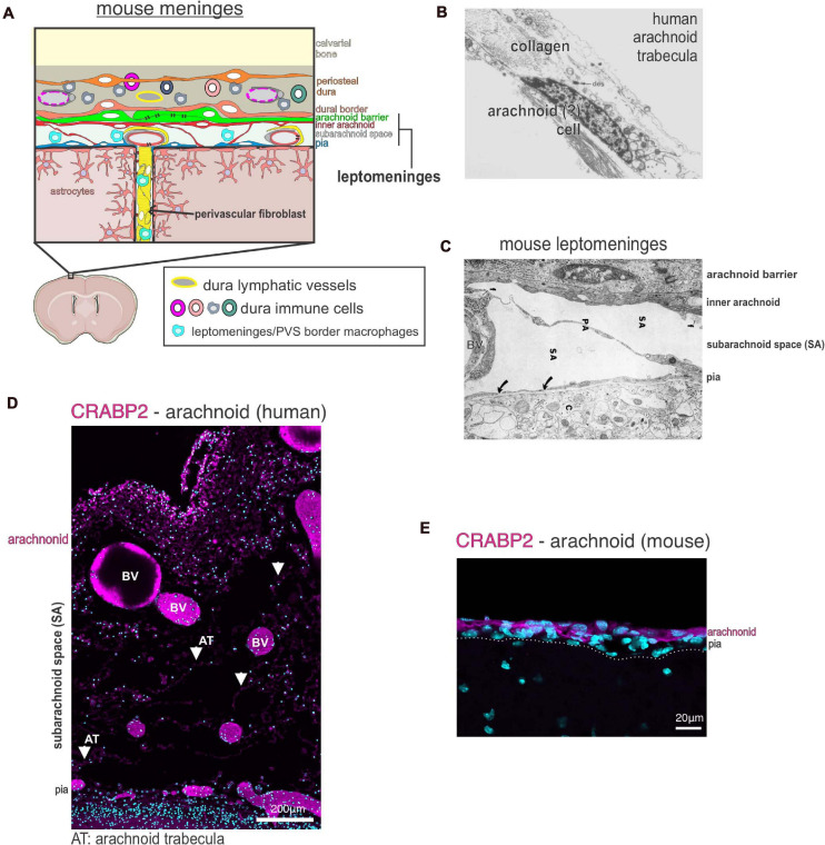
“Arachnoid fibroblasts express RALDH2 and CRABP2, a retinoic acid (RA) synthesizing enzyme and an RA binding protein, respectively (Figures 1D,E; DeSisto et al., 2020).”
§ Living on the Edge of the CNS: Meninges Cell Diversity in Health and Disease > Meningeal Cell Types > Fibroblasts
What the sources say
- Not checked yetcites DeSisto et al., 2020 source
plain- Asserted: passes — The authors put it forward as true.
- Checkable: passes — It can be checked against evidence.
- Load-bearing: passes — The paper’s conclusions depend on it.
- Based: passes — The paper gives a reason why it holds.
In the paper
“disruption of glymphatic function results in lower levels of TBI-associated biomarkers in the cervical lymph nodes and bloodstream”
§ Living on the Edge of the CNS: Meninges Cell Diversity in Health and Disease > Meningeal Response to Injury and Disease > Meningeal Lymphatics in Acute CNS Injury
What the sources say
- Not checked yetcites Plog et al., 2015 source
plaindirect- Asserted: passes — The authors put it forward as true.
- Checkable: passes — It can be checked against evidence.
- Load-bearing: passes — The paper’s conclusions depend on it.
- Based: passes — The paper gives a reason why it holds.
None of the sources cited for this claim have been checked yet.
In the paper
“Production of RA by the arachnoid layers critically regulates brain and neurovascular development (Siegenthaler et al., 2009; Choi et al., 2014; Bonney et al., 2016; Mishra et al., 2016; Haushalter et al., 2017).”
§ Living on the Edge of the CNS: Meninges Cell Diversity in Health and Disease > Meningeal Cell Types > Fibroblasts
What the sources say
plaindirect- Asserted: passes — The authors put it forward as true.
- Checkable: passes — It can be checked against evidence.
- Load-bearing: unclear — It is unclear whether the paper’s conclusions depend on it.
- Based: passes — The paper gives a reason why it holds.
None of the sources cited for this claim have been checked yet.
In the paper
“numerous studies show that meningeal immune cells play an important role in the brain injury response”
§ Living on the Edge of the CNS: Meninges Cell Diversity in Health and Disease > Meningeal Response to Injury and Disease > Meningeal Immune Cells in Acute CNS Injury
What the sources say
boostedcorrelational- Asserted: passes — The authors put it forward as true.
- Checkable: passes — It can be checked against evidence.
- Load-bearing: passes — The paper’s conclusions depend on it.
- Based: passes — The paper gives a reason why it holds.
- Rests on an assumption: The degree of neuroinflammation following acute CNS injury is a major predictor of clinical outcomes.
A conclusion the paper draws from its own results — it cites no other source for it, so there is none to check. The quote shows it is in the paper.
In the paper
“Resident meningeal macrophages regulate the acute injury response by influencing the severity of neuroinflammation and fibrosis”
§ Living on the Edge of the CNS: Meninges Cell Diversity in Health and Disease > Meningeal Response to Injury and Disease > Meningeal Immune Cells in Acute CNS Injury
plaindirect- Asserted: passes — The authors put it forward as true.
- Checkable: passes — It can be checked against evidence.
- Load-bearing: passes — The paper’s conclusions depend on it.
- Based: passes — The paper gives a reason why it holds.
None of the sources cited for this claim have been checked yet.
In the paper
“The brain glymphatic and meningeal lymphatic systems work together to clear waste and macromolecules from the brain parenchyma in homeostasis and following injury”
§ Living on the Edge of the CNS: Meninges Cell Diversity in Health and Disease > Meningeal Response to Injury and Disease > Meningeal Lymphatics in Acute CNS Injury
What the sources say
plaindirect- Asserted: passes — The authors put it forward as true.
- Checkable: passes — It can be checked against evidence.
- Load-bearing: passes — The paper’s conclusions depend on it.
- Based: passes — The paper gives a reason why it holds.
A conclusion the paper draws from its own results — it cites no other source for it, so there is none to check. The quote shows it is in the paper.
In the paper
“While not done in an acute injury model, this evidence further supports that CNS fibroblasts, not pericytes or vSMCs, are the main reactive fibrotic cell type in CNS injuries and disease.”
§ Living on the Edge of the CNS: Meninges Cell Diversity in Health and Disease > Meningeal Response to Injury and Disease > CNS Fibroblasts in Acute CNS Injury
hedgeddirect- Asserted: passes — The authors put it forward as true.
- Checkable: passes — It can be checked against evidence.
- Load-bearing: passes — The paper’s conclusions depend on it.
- Based: passes — The paper gives a reason why it holds.
None of the sources cited for this claim have been checked yet.
In the paper
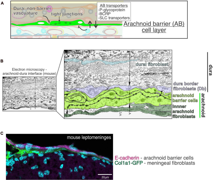
“Segregating the outer and inner meningeal compartments is the epithelial-like arachnoid barrier cells, connected by tight and adherens junctions, which regulate the movement of pathogens, molecules, and cells into and out of the cerebral spinal fluid (CSF) and brain parenchyma.”
§ Living on the Edge of the CNS: Meninges Cell Diversity in Health and Disease > Abstract
What the sources say
plaindirect- Asserted: passes — The authors put it forward as true.
- Checkable: passes — It can be checked against evidence.
- Load-bearing: passes — The paper’s conclusions depend on it.
- Based: passes — The paper gives a reason why it holds.
None of the sources cited for this claim have been checked yet.
In the paper
“The dural lymphatics are now recognized as a critical transport system of macromolecules, interstitial fluid, and CSF from the CNS into the cervical lymph nodes (Aspelund et al., 2015; Absinta et al., 2017; Louveau et al., 2018; Ahn et al., 2019).”
§ Living on the Edge of the CNS: Meninges Cell Diversity in Health and Disease > Meningeal Cell Types > Lymphatic Vessels: Dura
What the sources say
boosteddirect- Asserted: passes — The authors put it forward as true.
- Checkable: passes — It can be checked against evidence.
- Load-bearing: unclear — It is unclear whether the paper’s conclusions depend on it.
- Based: passes — The paper gives a reason why it holds.
A conclusion the paper draws from its own results — it cites no other source for it, so there is none to check. The quote shows it is in the paper.
In the paper
“In summary, fibroblasts from the meninges and perivascular space are major drivers of fibrotic scar formation following CNS injury however establishing a standard nomenclature would be beneficial.”
§ Living on the Edge of the CNS: Meninges Cell Diversity in Health and Disease > Meningeal Response to Injury and Disease > CNS Fibroblasts in Acute CNS Injury
plaindirect- Asserted: passes — The authors put it forward as true.
- Checkable: passes — It can be checked against evidence.
- Load-bearing: passes — The paper’s conclusions depend on it.
- Based: passes — The paper gives a reason why it holds.
A conclusion the paper draws from its own results — it cites no other source for it, so there is none to check. The quote shows it is in the paper.
In the paper
“selective ablation of meningeal lymphatic vessels worsens pathophysiology”
§ Living on the Edge of the CNS: Meninges Cell Diversity in Health and Disease > Meningeal Response to Injury and Disease > Meningeal Lymphatics in Acute CNS Injury
plaindirect- Asserted: passes — The authors put it forward as true.
- Checkable: passes — It can be checked against evidence.
- Load-bearing: passes — The paper’s conclusions depend on it.
- Based: passes — The paper gives a reason why it holds.
In the paper
“Retinoic acid signaling was elevated in neurons and astrocytes in the peri-infarct region”
§ Living on the Edge of the CNS: Meninges Cell Diversity in Health and Disease > Meningeal Response to Injury and Disease > CNS Fibroblasts in Acute CNS Injury
What the sources say
- Not checked yetcites Kelly et al., 2016 source
plaincorrelational- Asserted: passes — The authors put it forward as true.
- Checkable: passes — It can be checked against evidence.
- Load-bearing: passes — The paper’s conclusions depend on it.
- Based: passes — The paper gives a reason why it holds.
In the paper
“depletion of macrophages prior to SCI leads to depletion of pro-fibrotic cytokines, such as tumor necrosis factor superfamily members Tnfsf8 and Tnfsf13”
§ Living on the Edge of the CNS: Meninges Cell Diversity in Health and Disease > Meningeal Response to Injury and Disease > Meningeal Immune Cells in Acute CNS Injury
What the sources say
- Not checked yetcites Zhu et al., 2015 source
plaindirect- Asserted: passes — The authors put it forward as true.
- Checkable: passes — It can be checked against evidence.
- Load-bearing: passes — The paper’s conclusions depend on it.
- Based: passes — The paper gives a reason why it holds.
In the paper
“suggesting that scar fibroblasts signal to the surrounding CNS cells via retinoic acid release”
§ Living on the Edge of the CNS: Meninges Cell Diversity in Health and Disease > Meningeal Response to Injury and Disease > CNS Fibroblasts in Acute CNS Injury
What the sources say
- Not checked yetcites Kelly et al., 2016 source
hedgeddirect- Asserted: passes — The authors put it forward as true.
- Checkable: passes — It can be checked against evidence.
- Load-bearing: passes — The paper’s conclusions depend on it.
- Based: passes — The paper gives a reason why it holds.
None of the sources cited for this claim have been checked yet.
In the paper
“Meningitis induced inflammation is driven by the response of resident meningeal immune cells, meningeal fibroblasts and infiltrating leukocytes to the infectious agent”
§ Living on the Edge of the CNS: Meninges Cell Diversity in Health and Disease > Meningeal Response to Injury and Disease > Response of the Meninges to Meningitis
What the sources say
- Not checked yetcites Coles et al., 2017
- Not checked yetcites Manglani and McGavern, 2018
- Not checked yetcites Rua and McGavern, 2018
plaindirect- Asserted: passes — The authors put it forward as true.
- Checkable: passes — It can be checked against evidence.
- Load-bearing: unclear — It is unclear whether the paper’s conclusions depend on it.
- Based: passes — The paper gives a reason why it holds.
A conclusion the paper draws from its own results — it cites no other source for it, so there is none to check. The quote shows it is in the paper.
In the paper
“Overall, the pathology of meningitis is driven by a complex interplay between cells of the meninges and the peripheral immune system.”
§ Living on the Edge of the CNS: Meninges Cell Diversity in Health and Disease > Meningeal Response to Injury and Disease > Response of the Meninges to Meningitis
plaindirect- Asserted: passes — The authors put it forward as true.
- Checkable: passes — It can be checked against evidence.
- Load-bearing: passes — The paper’s conclusions depend on it.
- Based: passes — The paper gives a reason why it holds.
A conclusion the paper draws from its own results — it cites no other source for it, so there is none to check. The quote shows it is in the paper.
In the paper
“further supporting fibroblast driven fibrosis as a feature of MS”
§ Living on the Edge of the CNS: Meninges Cell Diversity in Health and Disease > Meningeal Response to Injury and Disease > Meninges and Perivascular Fibroblasts in Multiple Sclerosis
hedgeddirect- Asserted: passes — The authors put it forward as true.
- Checkable: passes — It can be checked against evidence.
- Load-bearing: passes — The paper’s conclusions depend on it.
- Based: passes — The paper gives a reason why it holds.
A conclusion the paper draws from its own results — it cites no other source for it, so there is none to check. The quote shows it is in the paper.
In the paper
“Overall, the meninges play a critical role in MS”
§ Living on the Edge of the CNS: Meninges Cell Diversity in Health and Disease > Meningeal Response to Injury and Disease > Meninges and Perivascular Fibroblasts in Multiple Sclerosis
plaindirect- Asserted: passes — The authors put it forward as true.
- Checkable: passes — It can be checked against evidence.
- Load-bearing: passes — The paper’s conclusions depend on it.
- Based: passes — The paper gives a reason why it holds.
None of the sources cited for this claim have been checked yet.
In the paper
“Several lines of evidence implicate meningeal-located structures in the genesis and progression of Alzheimer’s disease (AD)”
§ Living on the Edge of the CNS: Meninges Cell Diversity in Health and Disease > Meningeal Response to Injury and Disease > The Meninges and Alzheimer’s Disease
What the sources say
plaincorrelational- Asserted: passes — The authors put it forward as true.
- Checkable: passes — It can be checked against evidence.
- Load-bearing: passes — The paper’s conclusions depend on it.
- Based: passes — The paper gives a reason why it holds.
In the paper
“intracisternal injection of IL1-β, highly upregulated in meningitis, induces rapid meningeal barrier leakage”
§ Living on the Edge of the CNS: Meninges Cell Diversity in Health and Disease > Meningeal Response to Injury and Disease > Response of the Meninges to Meningitis
What the sources say
- Not checked yetcites Ichikawa and Itoh, 2011
plaindirect- Asserted: passes — The authors put it forward as true.
- Checkable: passes — It can be checked against evidence.
- Load-bearing: unclear — It is unclear whether the paper’s conclusions depend on it.
- Based: passes — The paper gives a reason why it holds.
None of the sources cited for this claim have been checked yet.
In the paper
“during murine CNS infections, including mouse hepatitis virus, a murine coronavirus (strains Srr7, cl-2, and A59), the Armstrong strain of lymphocytic choriomeningitis virus (LCMV), all show there is an influx into the brain and subarachnoid space of CD8+ T cells, monocytes and neutrophils into the meninges quickly after infection”
§ Living on the Edge of the CNS: Meninges Cell Diversity in Health and Disease > Meningeal Response to Injury and Disease > Response of the Meninges to Meningitis
What the sources say
plaindirect- Asserted: passes — The authors put it forward as true.
- Checkable: passes — It can be checked against evidence.
- Load-bearing: unclear — It is unclear whether the paper’s conclusions depend on it.
- Based: passes — The paper gives a reason why it holds.
In the paper
“lymphoid follicle-like structures in the cerebral leptomeninges contained CD20+ B-cells, CD8+, CD4+, and CD3+ T-cells, CD138+ plasma cells, and a network of CD21+ and CD35+ follicular dendritic cells”
§ Living on the Edge of the CNS: Meninges Cell Diversity in Health and Disease > Meningeal Response to Injury and Disease > Meninges and Perivascular Fibroblasts in Multiple Sclerosis
What the sources say
- Not checked yetcites Serafini et al., 2004 source
plain- Asserted: passes — The authors put it forward as true.
- Checkable: passes — It can be checked against evidence.
- Load-bearing: passes — The paper’s conclusions depend on it.
- Based: passes — The paper gives a reason why it holds.
A conclusion the paper draws from its own results — it cites no other source for it, so there is none to check. The quote shows it is in the paper.
In the paper
“the meninges are clearly an important player in MS induced neuroinflammation”
§ Living on the Edge of the CNS: Meninges Cell Diversity in Health and Disease > Meningeal Response to Injury and Disease > Meninges and Perivascular Fibroblasts in Multiple Sclerosis
boostedcorrelational- Asserted: passes — The authors put it forward as true.
- Checkable: passes — It can be checked against evidence.
- Load-bearing: passes — The paper’s conclusions depend on it.
- Based: passes — The paper gives a reason why it holds.
A conclusion the paper draws from its own results — it cites no other source for it, so there is none to check. The quote shows it is in the paper.
In the paper
“suggesting that fibrosis may be deleterious for remyelination”
§ Living on the Edge of the CNS: Meninges Cell Diversity in Health and Disease > Meningeal Response to Injury and Disease > Meninges and Perivascular Fibroblasts in Multiple Sclerosis
hedgedconditional- Asserted: passes — The authors put it forward as true.
- Checkable: passes — It can be checked against evidence.
- Load-bearing: passes — The paper’s conclusions depend on it.
- Based: passes — The paper gives a reason why it holds.
None of the sources cited for this claim have been checked yet.
In the paper
“The transport of interstitial fluids and macromolecules (including amyloid-β peptides) out of the brain occurs via a complex transport network that utilizes the perivascular glymphatic system of the brain and lymphatic vessels of the meninges”
§ Living on the Edge of the CNS: Meninges Cell Diversity in Health and Disease > Meningeal Response to Injury and Disease > The Meninges and Alzheimer’s Disease
What the sources say
plaindirect- Asserted: passes — The authors put it forward as true.
- Checkable: passes — It can be checked against evidence.
- Load-bearing: passes — The paper’s conclusions depend on it.
- Based: passes — The paper gives a reason why it holds.
A conclusion the paper draws from its own results — it cites no other source for it, so there is none to check. The quote shows it is in the paper.
In the paper
“Altogether, meningeal lymphatics play a key role in regulating the post-injury response”
§ Living on the Edge of the CNS: Meninges Cell Diversity in Health and Disease > Meningeal Response to Injury and Disease > Meningeal Lymphatics in Acute CNS Injury
plaindirect- Asserted: passes — The authors put it forward as true.
- Checkable: passes — It can be checked against evidence.
- Load-bearing: passes — The paper’s conclusions depend on it.
- Based: passes — The paper gives a reason why it holds.
In the paper
“in the mouse model of corona virus MHV A59 infection, production of cytokines CCL19 and CCL21 by a meningeal fibroblasts subpopulation, marked by ER-TR7 antigen, and podoplanin, work to recruit anti-viral CD8+ T cells”
§ Living on the Edge of the CNS: Meninges Cell Diversity in Health and Disease > Meningeal Response to Injury and Disease > Response of the Meninges to Meningitis
What the sources say
- Not checked yetcites Cupovic et al., 2016 source
plaindirect- Asserted: passes — The authors put it forward as true.
- Checkable: passes — It can be checked against evidence.
- Load-bearing: unclear — It is unclear whether the paper’s conclusions depend on it.
- Based: passes — The paper gives a reason why it holds.
A conclusion the paper draws from its own results — it cites no other source for it, so there is none to check. The quote shows it is in the paper.
In the paper
“Like acute CNS injury, CNS fibroblast activation and expansion contribute to MS induced lesions.”
§ Living on the Edge of the CNS: Meninges Cell Diversity in Health and Disease > Meningeal Response to Injury and Disease > Meninges and Perivascular Fibroblasts in Multiple Sclerosis
plaindirect- Asserted: passes — The authors put it forward as true.
- Checkable: passes — It can be checked against evidence.
- Load-bearing: passes — The paper’s conclusions depend on it.
- Based: passes — The paper gives a reason why it holds.
In the paper
“the ablation of meningeal lymphatics impedes anti-amyloid-β therapy by exacerbating microgliosis, neurovascular dysfunction, and behavior defects in the 5XFAD model of Alzheimer’s disease”
§ Living on the Edge of the CNS: Meninges Cell Diversity in Health and Disease > Meningeal Response to Injury and Disease > The Meninges and Alzheimer’s Disease
What the sources say
- Not checked yetcites Da Mesquita et al., 2021 source
plaindirect- Asserted: passes — The authors put it forward as true.
- Checkable: passes — It can be checked against evidence.
- Load-bearing: passes — The paper’s conclusions depend on it.
- Based: passes — The paper gives a reason why it holds.
In the paper
“recent scRNAseq profiling of human brain vasculature from healthy and AD cerebral cortex showed that perivascular fibroblasts were significantly under-represented in the AD brain single cell data set”
§ Living on the Edge of the CNS: Meninges Cell Diversity in Health and Disease > Meningeal Response to Injury and Disease > The Meninges and Alzheimer’s Disease
What the sources say
- Not checked yetcites Yang et al., 2021 source
plaincorrelational- Asserted: passes — The authors put it forward as true.
- Checkable: passes — It can be checked against evidence.
- Load-bearing: passes — The paper’s conclusions depend on it.
- Based: passes — The paper gives a reason why it holds.
In the paper
“perivascular and barrier macrophages, found both in the brain parenchyma and leptomeninges, play a role in amyloid-β removal as their depletion causes increased amyloid deposition”
§ Living on the Edge of the CNS: Meninges Cell Diversity in Health and Disease > Meningeal Response to Injury and Disease > The Meninges and Alzheimer’s Disease
What the sources say
- Not checked yetcites Hawkes and McLaurin, 2009 source
plaindirect- Asserted: passes — The authors put it forward as true.
- Checkable: passes — It can be checked against evidence.
- Load-bearing: unclear — It is unclear whether the paper’s conclusions depend on it.
- Based: passes — The paper gives a reason why it holds.
A conclusion the paper draws from its own results — it cites no other source for it, so there is none to check. The quote shows it is in the paper.
In the paper
“how the meninges serves as a key interface between the CNS and periphery”
§ Living on the Edge of the CNS: Meninges Cell Diversity in Health and Disease > Conclusion
plaincorrelational- Asserted: passes — The authors put it forward as true.
- Checkable: passes — It can be checked against evidence.
- Load-bearing: passes — The paper’s conclusions depend on it.
- Based: passes — The paper gives a reason why it holds.
A conclusion the paper draws from its own results — it cites no other source for it, so there is none to check. The quote shows it is in the paper.
In the paper
“This is in contrast to large increases in CNS fibroblast numbers after acute CNS injuries and in neuroinflammation, demonstrating a different response of perivascular fibroblasts in AD.”
§ Living on the Edge of the CNS: Meninges Cell Diversity in Health and Disease > Meningeal Response to Injury and Disease > The Meninges and Alzheimer’s Disease
boostedcorrelational- Asserted: passes — The authors put it forward as true.
- Checkable: passes — It can be checked against evidence.
- Load-bearing: passes — The paper’s conclusions depend on it.
- Based: passes — The paper gives a reason why it holds.
A conclusion the paper draws from its own results — it cites no other source for it, so there is none to check. The quote shows it is in the paper.
In the paper
“Thus, tumor cells can effectively alter the leptomeninges microenvironment”
§ Living on the Edge of the CNS: Meninges Cell Diversity in Health and Disease > Meningeal Response to Injury and Disease > Meninges as a Site of Cancer Metastasis
plaindirect- Asserted: passes — The authors put it forward as true.
- Checkable: passes — It can be checked against evidence.
- Load-bearing: passes — The paper’s conclusions depend on it.
- Based: passes — The paper gives a reason why it holds.
A conclusion the paper draws from its own results — it cites no other source for it, so there is none to check. The quote shows it is in the paper.
In the paper
“Once in the leptomeninges, cancer cells can modulate the CSF content in the subarachnoid space content to support cancer growth.”
§ Living on the Edge of the CNS: Meninges Cell Diversity in Health and Disease > Meningeal Response to Injury and Disease > Meninges as a Site of Cancer Metastasis
plaindirect- Asserted: passes — The authors put it forward as true.
- Checkable: passes — It can be checked against evidence.
- Load-bearing: passes — The paper’s conclusions depend on it.
- Based: passes — The paper gives a reason why it holds.
- Rests on an assumption: CSF is acellular, poor in protein, glucose and cytokine content, which is not conducive to cancer cell proliferation.
- It may state two separate claims as one. 49
- Probably not a substantial claim. 2
- The quote only partly states this claim. 1
The cited sources support only part of this claim — read each source’s verdict below.
In the paper
“Primary tumors of the meninges are quite rare, however, the leptomeninges is a relatively common site for by contiguous extension of primary tumors of the central nervous system, paranasal sinuses and skull base origin or tumor metastasis which can lead to dissemination into the CNS parenchyma and poor prognosis”
§ Living on the Edge of the CNS: Meninges Cell Diversity in Health and Disease > Meningeal Response to Injury and Disease > Meninges as a Site of Cancer Metastasis
What the sources say
- Not checked yetcites Mahendru and Chong, 2009
- Not checked yetcites Waki et al., 2009 source
- Partly supportedcites Oechsle et al., 2010 sourceExperimental — accuracy not yet measured
Split into 2 atoms — 0 confirmed, 1 partially supported, 0 disputed, 0 misattributed, 1 unverifiable. Expand the claim to see per-atom verdicts.
plaindirect- Asserted: passes — The authors put it forward as true.
- Checkable: passes — It can be checked against evidence.
- Load-bearing: unclear — It is unclear whether the paper’s conclusions depend on it.
- Based: passes — The paper gives a reason why it holds.
It may state two separate claims as one.
In the paper
“Our recent single cell RNA sequencing (scRNAseq) of developing mouse meninges showed pia, arachnoid and dura fibroblasts are molecularly distinct and likely have layer specific functions (DeSisto et al., 2020).”
§ Living on the Edge of the CNS: Meninges Cell Diversity in Health and Disease > Meningeal Cell Types > Fibroblasts
What the sources say
- Not checked yetcites DeSisto et al., 2020 source
hedgedcorrelational- Asserted: passes — The authors put it forward as true.
- Checkable: passes — It can be checked against evidence.
- Load-bearing: passes — The paper’s conclusions depend on it.
- Based: passes — The paper gives a reason why it holds.
It may state two separate claims as one.
In the paper
“We recently found that arachnoid barrier cells can be first seen in the mouse forebrain around E13 and they differentiate from mesenchymal cells of the primary meninx (DeSisto et al., 2020).”
§ Living on the Edge of the CNS: Meninges Cell Diversity in Health and Disease > Meningeal Cell Types > Arachnoid Barrier Cells
What the sources say
- Not checked yetcites DeSisto et al., 2020 source
plaindirect- Asserted: passes — The authors put it forward as true.
- Checkable: passes — It can be checked against evidence.
- Load-bearing: unclear — It is unclear whether the paper’s conclusions depend on it.
- Based: passes — The paper gives a reason why it holds.
It may state two separate claims as one.
None of the sources cited for this claim have been checked yet.
In the paper
“Meningeal fibroblasts in different CNS regions have different origins; forebrain meningeal fibroblasts are neural crest derived whereas mid-, hindbrain and spinal cord meninges are derived from the mesoderm (Jiang et al., 2002; Yoshida et al., 2008).”
§ Living on the Edge of the CNS: Meninges Cell Diversity in Health and Disease > Meningeal Cell Types > Fibroblasts
What the sources say
plaincorrelational- Asserted: passes — The authors put it forward as true.
- Checkable: passes — It can be checked against evidence.
- Load-bearing: unclear — It is unclear whether the paper’s conclusions depend on it.
- Based: passes — The paper gives a reason why it holds.
It may state two separate claims as one.
None of the sources cited for this claim have been checked yet.
In the paper
“The dural compartment of the meninges, closest to the skull, is predominantly composed of fibroblasts, but also includes fenestrated blood vasculature, an elaborate lymphatic system, as well as immune cells which are distinct from the CNS.”
§ Living on the Edge of the CNS: Meninges Cell Diversity in Health and Disease > Abstract
What the sources say
plain- Asserted: passes — The authors put it forward as true.
- Checkable: passes — It can be checked against evidence.
- Load-bearing: unclear — It is unclear whether the paper’s conclusions depend on it.
- Based: passes — The paper gives a reason why it holds.
It may state two separate claims as one.
In the paper
“Cx3cr1+ meningeal macrophages are observed dying within 30 min of injury induction, leading to formation of a “honeycomb” network of microglia at the glial limitans, likely preventing the spread of reactive oxygen species from the leptomeninges into the brain parenchyma”
§ Living on the Edge of the CNS: Meninges Cell Diversity in Health and Disease > Meningeal Response to Injury and Disease > Meningeal Immune Cells in Acute CNS Injury
What the sources say
- Not checked yetcites Roth et al., 2013 source
hedgeddirect- Asserted: passes — The authors put it forward as true.
- Checkable: passes — It can be checked against evidence.
- Load-bearing: passes — The paper’s conclusions depend on it.
- Based: passes — The paper gives a reason why it holds.
It may state two separate claims as one.
A conclusion the paper draws from its own results — it cites no other source for it, so there is none to check. The quote shows it is in the paper.
In the paper
“Thus, fibroblast production of retinoic acid seems to impair CNS recovery while stimulating fibrosis.”
§ Living on the Edge of the CNS: Meninges Cell Diversity in Health and Disease > Meningeal Response to Injury and Disease > CNS Fibroblasts in Acute CNS Injury
hedgeddirect- Asserted: passes — The authors put it forward as true.
- Checkable: passes — It can be checked against evidence.
- Load-bearing: passes — The paper’s conclusions depend on it.
- Based: passes — The paper gives a reason why it holds.
It may state two separate claims as one.
None of the sources cited for this claim have been checked yet.
In the paper
“In both MCAO and SCI models, macrophages are observed in the fibrotic scar and may associate with fibroblasts”
- Also stated in: § Meningeal Immune Cells in Acute CNS Injury · plain
“CD11b+ macrophages are partially responsible for recruitment of fibroblasts to the fibrotic scar following SCI”
§ Living on the Edge of the CNS: Meninges Cell Diversity in Health and Disease > Meningeal Response to Injury and Disease > Meningeal Immune Cells in Acute CNS Injury
What the sources say
plainconditional- Asserted: passes — The authors put it forward as true.
- Checkable: passes — It can be checked against evidence.
- Load-bearing: passes — The paper’s conclusions depend on it.
- Based: passes — The paper gives a reason why it holds.
It may state two separate claims as one.
- Also stated in: § Meningeal Immune Cells in Acute CNS Injury · plain
A conclusion the paper draws from its own results — it cites no other source for it, so there is none to check. The quote shows it is in the paper.
In the paper
“While the meninges were historically seen as a purely structural support for the CNS and protection from trauma, the emerging view of the meninges is as an essential interface between the CNS and the periphery, critical to brain development, required for brain homeostasis, and involved in a variety of diseases.”
§ Living on the Edge of the CNS: Meninges Cell Diversity in Health and Disease > Abstract
plaindirect- Asserted: passes — The authors put it forward as true.
- Checkable: passes — It can be checked against evidence.
- Load-bearing: passes — The paper’s conclusions depend on it.
- Based: unclear — It is unclear why the paper says it holds.
It may state two separate claims as one.
None of the sources cited for this claim have been checked yet.
In the paper
“Several studies show this vasculature has barrier properties and are therefore part of the meninges blood-CSF barrier.”
§ Living on the Edge of the CNS: Meninges Cell Diversity in Health and Disease > Meningeal Cell Types > Blood Vessels: Leptomeninges and Dura
What the sources say
plaincorrelational- Asserted: passes — The authors put it forward as true.
- Checkable: passes — It can be checked against evidence.
- Load-bearing: unclear — It is unclear whether the paper’s conclusions depend on it.
- Based: passes — The paper gives a reason why it holds.
It may state two separate claims as one.
A conclusion the paper draws from its own results — it cites no other source for it, so there is none to check. The quote shows it is in the paper.
In the paper
“These results highlight the important function of the glymphatic system in clearance of molecular substances from the brain and potential role in facilitating signaling between the brain and periphery.”
§ Living on the Edge of the CNS: Meninges Cell Diversity in Health and Disease > Meningeal Response to Injury and Disease > Meningeal Lymphatics in Acute CNS Injury
hedgedconditional- Asserted: passes — The authors put it forward as true.
- Checkable: passes — It can be checked against evidence.
- Load-bearing: passes — The paper’s conclusions depend on it.
- Based: passes — The paper gives a reason why it holds.
It may state two separate claims as one.
In the paper
“retinoic acid treatment following stroke in rodents reduces neurogenesis while also reducing angiogenesis and gliogenesis in the peri-infarct region”
§ Living on the Edge of the CNS: Meninges Cell Diversity in Health and Disease > Meningeal Response to Injury and Disease > CNS Fibroblasts in Acute CNS Injury
What the sources say
- Not checked yetcites Jung et al., 2007 source
plaindirect- Asserted: passes — The authors put it forward as true.
- Checkable: passes — It can be checked against evidence.
- Load-bearing: passes — The paper’s conclusions depend on it.
- Based: passes — The paper gives a reason why it holds.
It may state two separate claims as one.
In the paper
“Most proximate to the brain is the collagen and basement membrane-rich pia matter that abuts the glial limitans and has recently be shown to have regional heterogeneity within the developing mouse brain.”
§ Living on the Edge of the CNS: Meninges Cell Diversity in Health and Disease > Abstract
What the sources say
- Not checked yetcites DeSisto et al., 2020 source
plain- Asserted: passes — The authors put it forward as true.
- Checkable: passes — It can be checked against evidence.
- Load-bearing: unclear — It is unclear whether the paper’s conclusions depend on it.
- Based: passes — The paper gives a reason why it holds.
It may state two separate claims as one.
In the paper
“Following TBI, glymphatic fluid flow is disrupted and greater amounts of tau protein is observed accumulating in the parenchyma”
- Also stated in: § Meningeal Lymphatics in Acute CNS Injury · plain
“knock-out of Aqp4, a water transport channel necessary for interstitial solute clearance and glymphatic function, following TBI, exacerbates the accumulation of tau in the parenchyma”
§ Living on the Edge of the CNS: Meninges Cell Diversity in Health and Disease > Meningeal Response to Injury and Disease > Meningeal Lymphatics in Acute CNS Injury
What the sources say
- Not checked yetcites Iliff et al., 2014 source
plaindirect- Asserted: passes — The authors put it forward as true.
- Checkable: passes — It can be checked against evidence.
- Load-bearing: passes — The paper’s conclusions depend on it.
- Based: passes — The paper gives a reason why it holds.
It may state two separate claims as one.
- Also stated in: § Meningeal Lymphatics in Acute CNS Injury · plain
In the paper
“depletion of mast cells resulted in decreased levels of neuroinflammation, brain swelling, and infarct size following stroke and this was partially mediated by mast-cell derived IL-6”
§ Living on the Edge of the CNS: Meninges Cell Diversity in Health and Disease > Meningeal Response to Injury and Disease > Meningeal Immune Cells in Acute CNS Injury
What the sources say
- Not checked yetcites Arac et al., 2019 source
plaindirect- Asserted: passes — The authors put it forward as true.
- Checkable: passes — It can be checked against evidence.
- Load-bearing: passes — The paper’s conclusions depend on it.
- Based: passes — The paper gives a reason why it holds.
It may state two separate claims as one.
In the paper
“lymphangiogenesis was seen following photothrombotic stroke lesion in the mouse cortex, but this effect was not seen following MCAO”
§ Living on the Edge of the CNS: Meninges Cell Diversity in Health and Disease > Meningeal Response to Injury and Disease > Meningeal Lymphatics in Acute CNS Injury
What the sources say
- Not checked yetcites Yanev et al., 2020 source
plain- Asserted: passes — The authors put it forward as true.
- Checkable: passes — It can be checked against evidence.
- Load-bearing: passes — The paper’s conclusions depend on it.
- Based: passes — The paper gives a reason why it holds.
It may state two separate claims as one.
In the paper
“depletion of macrophages in the leptomeninges and perivascular spaces surrounding large arterioles prior to injury results in improved neurological scoring and reduced levels of inflammation and neuronal cell death”
§ Living on the Edge of the CNS: Meninges Cell Diversity in Health and Disease > Meningeal Response to Injury and Disease > Meningeal Immune Cells in Acute CNS Injury
What the sources say
- Not checked yetcites Wan et al., 2021 source
plaindirect- Asserted: passes — The authors put it forward as true.
- Checkable: passes — It can be checked against evidence.
- Load-bearing: passes — The paper’s conclusions depend on it.
- Based: passes — The paper gives a reason why it holds.
It may state two separate claims as one.
In the paper
“In a mild closed-skull TBI model, meningeal lymphatic drainage is impaired likely due to increases in intracranial pressure, accompanied by lymphatic morphological changes”
- Also stated in: § Meningeal Lymphatics in Acute CNS Injury · plain
“lymphangiogenesis was observed up to 2 weeks following TBI, though this did not correspond with increased lymphatic function”
§ Living on the Edge of the CNS: Meninges Cell Diversity in Health and Disease > Meningeal Response to Injury and Disease > Meningeal Lymphatics in Acute CNS Injury
What the sources say
- Not checked yetcites Bolte et al., 2020 source
plainconditional- Asserted: passes — The authors put it forward as true.
- Checkable: passes — It can be checked against evidence.
- Load-bearing: passes — The paper’s conclusions depend on it.
- Based: passes — The paper gives a reason why it holds.
It may state two separate claims as one.
- Also stated in: § Meningeal Lymphatics in Acute CNS Injury · plain
A conclusion the paper draws from its own results — it cites no other source for it, so there is none to check. The quote shows it is in the paper.
In the paper
“The dural lymphatic function is also disrupted following acute CNS injury.”
- Also stated in: § Meningeal Lymphatics in Acute CNS Injury · plain
“Thus, the degree of lymphatic impairment and response is dependent on the injury mechanism”
§ Living on the Edge of the CNS: Meninges Cell Diversity in Health and Disease > Meningeal Response to Injury and Disease > Meningeal Lymphatics in Acute CNS Injury
plaindirect- Asserted: passes — The authors put it forward as true.
- Checkable: passes — It can be checked against evidence.
- Load-bearing: passes — The paper’s conclusions depend on it.
- Based: passes — The paper gives a reason why it holds.
It may state two separate claims as one.
- Also stated in: § Meningeal Lymphatics in Acute CNS Injury · plain
None of the sources cited for this claim have been checked yet.
In the paper
“BBB breakdown is a well-documented feature of MS”
- Also stated in: § Meninges and Perivascular Fibroblasts in Multiple Sclerosis · hedged
“there is some evidence that MS can also induce disruption of the arachnoid barrier in the meninges, part of the B-CSF barrier”
- Also stated in: § Meninges and Perivascular Fibroblasts in Multiple Sclerosis · plain
“Claudin 11 is a tight junction protein enriched in arachnoid barrier cells and it is downregulated in late stages of an EAE mouse model”
§ Living on the Edge of the CNS: Meninges Cell Diversity in Health and Disease > Meningeal Response to Injury and Disease > Meninges and Perivascular Fibroblasts in Multiple Sclerosis
What the sources say
- Not checked yetcites Russi and Brown, 2015
- Not checked yetcites Uchida et al., 2019b
boostedconditional- Asserted: passes — The authors put it forward as true.
- Checkable: passes — It can be checked against evidence.
- Load-bearing: unclear — It is unclear whether the paper’s conclusions depend on it.
- Based: passes — The paper gives a reason why it holds.
It may state two separate claims as one.
- Also stated in: § Meninges and Perivascular Fibroblasts in Multiple Sclerosis · hedged
None of the sources cited for this claim have been checked yet.
In the paper
“The leptomeninges are now recognized as a key player in multiple sclerosis (MS), an inflammatory CNS autoimmune disorder characterized by axon demyelination”
- Also stated in: § Meninges and Perivascular Fibroblasts in Multiple Sclerosis · plain
“In both MS patients and MS mouse models, immune cells infiltrate the leptomeninges and pathology is commonly seen in cortical areas adjacent to the meninges”
§ Living on the Edge of the CNS: Meninges Cell Diversity in Health and Disease > Meningeal Response to Injury and Disease > Meninges and Perivascular Fibroblasts in Multiple Sclerosis
What the sources say
- Not checked yetcites Russi and Brown, 2015
- Not checked yetcites Lucchinetti et al., 2011 source
plaincorrelational- Asserted: passes — The authors put it forward as true.
- Checkable: passes — It can be checked against evidence.
- Load-bearing: passes — The paper’s conclusions depend on it.
- Based: passes — The paper gives a reason why it holds.
It may state two separate claims as one.
- Also stated in: § Meninges and Perivascular Fibroblasts in Multiple Sclerosis · plain
A conclusion the paper draws from its own results — it cites no other source for it, so there is none to check. The quote shows it is in the paper.
In the paper
“A disruption to the arachnoid barrier could permit dural and peripheral immune cells to migrate into the leptomeninges, and potentially contribute to leptomeningeal immune infiltrate that is common in MS pathology.”
§ Living on the Edge of the CNS: Meninges Cell Diversity in Health and Disease > Meningeal Response to Injury and Disease > Meninges and Perivascular Fibroblasts in Multiple Sclerosis
hedgedconditional- Asserted: passes — The authors put it forward as true.
- Checkable: passes — It can be checked against evidence.
- Load-bearing: passes — The paper’s conclusions depend on it.
- Based: passes — The paper gives a reason why it holds.
It may state two separate claims as one.
None of the sources cited for this claim have been checked yet.
In the paper
“cytokine administration into the cisterna magna of mice is sufficient to drive arachnoid barrier and blood-cerebral spinal fluid barrier functional breakdown”
- Also stated in: § Meninges and Perivascular Fibroblasts in Multiple Sclerosis · boosted
“Activated T cells in the leptomeninges produce cytokines known to perturb tight junction integrity of brain endothelial cells, including IFNγ which is known to drive BBB breakdown in viral encephalitis”
§ Living on the Edge of the CNS: Meninges Cell Diversity in Health and Disease > Meningeal Response to Injury and Disease > Meninges and Perivascular Fibroblasts in Multiple Sclerosis
What the sources say
- Not checked yetcites Ichikawa and Itoh, 2011
- Not checked yetcites Bonney et al., 2019 source
boosteddirect- Asserted: passes — The authors put it forward as true.
- Checkable: passes — It can be checked against evidence.
- Load-bearing: unclear — It is unclear whether the paper’s conclusions depend on it.
- Based: passes — The paper gives a reason why it holds.
It may state two separate claims as one.
- Also stated in: § Meninges and Perivascular Fibroblasts in Multiple Sclerosis · boosted
None of the sources cited for this claim have been checked yet.
In the paper
“In the experimental autoimmune encephalomyelitis (EAE) mouse model of MS, autoreactive effector T cells first infiltrate the leptomeninges via the pial vasculature, are activated by antigen presenting meningeal/perivascular macrophages, and subsequently enter the CNS parenchyma, triggering lesion formation”
§ Living on the Edge of the CNS: Meninges Cell Diversity in Health and Disease > Meningeal Response to Injury and Disease > Meninges and Perivascular Fibroblasts in Multiple Sclerosis
What the sources say
plaindirect- Asserted: passes — The authors put it forward as true.
- Checkable: passes — It can be checked against evidence.
- Load-bearing: unclear — It is unclear whether the paper’s conclusions depend on it.
- Based: passes — The paper gives a reason why it holds.
It may state two separate claims as one.
None of the sources cited for this claim have been checked yet.
In the paper
“In cases of bacterial meningitis, infections usually start as skin, gastrointestinal or respiratory infections that spread to the blood”
- Also stated in: § Response of the Meninges to Meningitis · plain
“Once in the blood, the meningitis causing bacteria accumulate in the leptomeningeal vasculature”
- Also stated in: § Response of the Meninges to Meningitis · plain
“move into the meninges by inducing BBB breakdown through multiple mechanisms”
§ Living on the Edge of the CNS: Meninges Cell Diversity in Health and Disease > Meningeal Response to Injury and Disease > Response of the Meninges to Meningitis
What the sources say
plaindirect- Asserted: passes — The authors put it forward as true.
- Checkable: passes — It can be checked against evidence.
- Load-bearing: unclear — It is unclear whether the paper’s conclusions depend on it.
- Based: passes — The paper gives a reason why it holds.
It may state two separate claims as one.
- Also stated in: § Response of the Meninges to Meningitis · plain
In the paper
“pre-existing defects in meningeal lymphatics causes worsened neuroinflammation following TBI”
- Also stated in: § Meningeal Lymphatics in Acute CNS Injury · plain
“stimulating meningeal lymphatic vessel outgrowth via VEGF-C administration had decreased levels of gliosis”
§ Living on the Edge of the CNS: Meninges Cell Diversity in Health and Disease > Meningeal Response to Injury and Disease > Meningeal Lymphatics in Acute CNS Injury
What the sources say
- Not checked yetcites Bolte et al., 2020 source
plaindirect- Asserted: passes — The authors put it forward as true.
- Checkable: passes — It can be checked against evidence.
- Load-bearing: passes — The paper’s conclusions depend on it.
- Based: passes — The paper gives a reason why it holds.
It may state two separate claims as one.
- Also stated in: § Meningeal Lymphatics in Acute CNS Injury · plain
None of the sources cited for this claim have been checked yet.
In the paper
“Blockage of VEGFR3 (expressed by lymphatic endothelial cells) in mice following focal cerebral ischemia resulted in reduced inflammatory response and infarct”
- Also stated in: § Meningeal Lymphatics in Acute CNS Injury · plain
“VEGFR3 mutant mice subjected to MCAO develop larger stroke volumes, thus worsened edema”
§ Living on the Edge of the CNS: Meninges Cell Diversity in Health and Disease > Meningeal Response to Injury and Disease > Meningeal Lymphatics in Acute CNS Injury
What the sources say
plaindirect- Asserted: passes — The authors put it forward as true.
- Checkable: passes — It can be checked against evidence.
- Load-bearing: passes — The paper’s conclusions depend on it.
- Based: passes — The paper gives a reason why it holds.
It may state two separate claims as one.
- Also stated in: § Meningeal Lymphatics in Acute CNS Injury · plain
None of the sources cited for this claim have been checked yet.
In the paper
“In the LCMV Armstrong mouse model, it was shown that the recruitment of antiviral CD8+ T cells, while necessary to defeat the infection, can also be deleterious.”
- Also stated in: § Response of the Meninges to Meningitis · hedged
“These CD8+ T cells also produce cytokines that can lead to over infiltration of monocytes and neutrophils that drive breakdown of vascular integrity and induce severe edema and brainstem herniation”
§ Living on the Edge of the CNS: Meninges Cell Diversity in Health and Disease > Meningeal Response to Injury and Disease > Response of the Meninges to Meningitis
What the sources say
- Not checked yetcites Kim et al., 2009 source
- Not checked yetcites Mastorakos and McGavern, 2019
plaindirect- Asserted: passes — The authors put it forward as true.
- Checkable: passes — It can be checked against evidence.
- Load-bearing: unclear — It is unclear whether the paper’s conclusions depend on it.
- Based: passes — The paper gives a reason why it holds.
It may state two separate claims as one.
- Also stated in: § Response of the Meninges to Meningitis · hedged
None of the sources cited for this claim have been checked yet.
In the paper
“AD is a dementia inducing neurodegenerative disorder characterized by the accumulation of amyloid-β containing plaques, which were initially isolated from homogenates of AD patient meningeal tissue”
§ Living on the Edge of the CNS: Meninges Cell Diversity in Health and Disease > Meningeal Response to Injury and Disease > The Meninges and Alzheimer’s Disease
What the sources say
- Not checked yetcites Joachim et al., 1988 source
- Not checked yetcites Da Mesquita et al., 2018b
plain- Asserted: passes — The authors put it forward as true.
- Checkable: passes — It can be checked against evidence.
- Load-bearing: fails — The paper’s conclusions may not depend on it.
- Based: passes — The paper gives a reason why it holds.
Probably not a substantial claim. The paper’s conclusions may not depend on it. It may state two separate claims as one.
None of the sources cited for this claim have been checked yet.
In the paper
“The presentation of MS induced leptomeningeal inflammation can be variable, ranging from disorganized collections of immune cells to organized ectopic lymphoid follicle-like structures”
- Also stated in: § Meninges and Perivascular Fibroblasts in Multiple Sclerosis · plain
“the ectopic lymph structures seen in MS are potentiated by leptomeningeal fibroblasts which form a reticular cell network consisting of activated fibroblasts in a scaffold-like structure”
§ Living on the Edge of the CNS: Meninges Cell Diversity in Health and Disease > Meningeal Response to Injury and Disease > Meninges and Perivascular Fibroblasts in Multiple Sclerosis
What the sources say
plaindirect- Asserted: passes — The authors put it forward as true.
- Checkable: passes — It can be checked against evidence.
- Load-bearing: passes — The paper’s conclusions depend on it.
- Based: passes — The paper gives a reason why it holds.
It may state two separate claims as one.
- Also stated in: § Meninges and Perivascular Fibroblasts in Multiple Sclerosis · plain
None of the sources cited for this claim have been checked yet.
In the paper
“Increased number of PDGFRβ-expressing cells are detected in human MS pathology samples”
- Also stated in: § Meninges and Perivascular Fibroblasts in Multiple Sclerosis · plain
“increased ECM protein deposition is a common pathological feature of MS lesions”
§ Living on the Edge of the CNS: Meninges Cell Diversity in Health and Disease > Meningeal Response to Injury and Disease > Meninges and Perivascular Fibroblasts in Multiple Sclerosis
What the sources say
plaincorrelational- Asserted: passes — The authors put it forward as true.
- Checkable: passes — It can be checked against evidence.
- Load-bearing: passes — The paper’s conclusions depend on it.
- Based: passes — The paper gives a reason why it holds.
It may state two separate claims as one.
- Also stated in: § Meninges and Perivascular Fibroblasts in Multiple Sclerosis · plain
None of the sources cited for this claim have been checked yet.
In the paper
“both the brain glymphatic and meningeal lymphatic systems deteriorate with age”
- Also stated in: § The Meninges and Alzheimer’s Disease · plain
“their dysfunction can potentiate AD pathology and dementia”
§ Living on the Edge of the CNS: Meninges Cell Diversity in Health and Disease > Meningeal Response to Injury and Disease > The Meninges and Alzheimer’s Disease
What the sources say
plaindirect- Asserted: passes — The authors put it forward as true.
- Checkable: passes — It can be checked against evidence.
- Load-bearing: passes — The paper’s conclusions depend on it.
- Based: passes — The paper gives a reason why it holds.
It may state two separate claims as one.
- Also stated in: § The Meninges and Alzheimer’s Disease · plain
In the paper
“a scRNAseq analysis showed that fibroblasts from EAE lesions upregulate interferon γ (IFNγ) signaling, and conditional deletion of IFN receptor γ from fibroblasts partially blocked fibroblast expansion”
- Also stated in: § Meninges and Perivascular Fibroblasts in Multiple Sclerosis · hedged
“IFNγ is produced by spinal cord T cells in EAE mice, implicating immune cells in EAE lesions as a source of fibroblast activation signals”
§ Living on the Edge of the CNS: Meninges Cell Diversity in Health and Disease > Meningeal Response to Injury and Disease > Meninges and Perivascular Fibroblasts in Multiple Sclerosis
What the sources say
- Not checked yetcites Dorrier et al., 2021 source
plainconditional- Asserted: passes — The authors put it forward as true.
- Checkable: passes — It can be checked against evidence.
- Load-bearing: passes — The paper’s conclusions depend on it.
- Based: passes — The paper gives a reason why it holds.
It may state two separate claims as one.
- Also stated in: § Meninges and Perivascular Fibroblasts in Multiple Sclerosis · hedged
None of the sources cited for this claim have been checked yet.
In the paper
“Bacterial presence in the meninges induces a strong inflammatory response, including production of multiple cytokines (tumor necrosis factor (TNF)-alpha, interleukin (IL)-1-beta, and IL-6), and a massive infiltration of neutrophils”
- Also stated in: § Response of the Meninges to Meningitis · plain
“The inflammatory cytokines and neutrophils work to quell the infection but also drive the breakdown of endothelial cell tight junctions”
§ Living on the Edge of the CNS: Meninges Cell Diversity in Health and Disease > Meningeal Response to Injury and Disease > Response of the Meninges to Meningitis
What the sources say
boosteddirect- Asserted: passes — The authors put it forward as true.
- Checkable: passes — It can be checked against evidence.
- Load-bearing: unclear — It is unclear whether the paper’s conclusions depend on it.
- Based: passes — The paper gives a reason why it holds.
It may state two separate claims as one.
- Also stated in: § Response of the Meninges to Meningitis · plain
In the paper
“erythrocytes accrue in the cervical lymph nodes via meningeal lymphatics following injury, and ablation of meningeal lymphatic vessels leads to less erythrocyte aggregation and greater neuroinflammation and neurological defects”
§ Living on the Edge of the CNS: Meninges Cell Diversity in Health and Disease > Meningeal Response to Injury and Disease > Meningeal Lymphatics in Acute CNS Injury
What the sources say
- Not checked yetcites Chen et al., 2020 source
plaindirect- Asserted: passes — The authors put it forward as true.
- Checkable: passes — It can be checked against evidence.
- Load-bearing: passes — The paper’s conclusions depend on it.
- Based: passes — The paper gives a reason why it holds.
It may state two separate claims as one.
None of the sources cited for this claim have been checked yet.
In the paper
“Using a Col1a1-GFP mouse line in conjunction with an EAE model, two independent groups identified substantial increases in fibroblasts within spinal cord lesion sites”
- Also stated in: § Meninges and Perivascular Fibroblasts in Multiple Sclerosis · plain
“Fibroblast expansion was deleterious for oligodendrocyte precursor cell (OPC) differentiation into mature oligodendrocytes and OPC migration into the lesion”
§ Living on the Edge of the CNS: Meninges Cell Diversity in Health and Disease > Meningeal Response to Injury and Disease > Meninges and Perivascular Fibroblasts in Multiple Sclerosis
What the sources say
plaindirect- Asserted: passes — The authors put it forward as true.
- Checkable: passes — It can be checked against evidence.
- Load-bearing: passes — The paper’s conclusions depend on it.
- Based: passes — The paper gives a reason why it holds.
It may state two separate claims as one.
- Also stated in: § Meninges and Perivascular Fibroblasts in Multiple Sclerosis · plain
In the paper
“Biopsies taken from early MS patients with cortical demyelination were more likely to have leptomeningeal inflammation, consisting of effector T cells and IgA B cells infiltrates, than those without cortical demyelination.”
- Also stated in: § Meninges and Perivascular Fibroblasts in Multiple Sclerosis · plain
“in human cases of MS the severity of cortical lesions is correlated to the extent of meningeal inflammation”
§ Living on the Edge of the CNS: Meninges Cell Diversity in Health and Disease > Meningeal Response to Injury and Disease > Meninges and Perivascular Fibroblasts in Multiple Sclerosis
What the sources say
- Not checked yetcites Lucchinetti et al., 2011 source
plaincorrelational- Asserted: passes — The authors put it forward as true.
- Checkable: passes — It can be checked against evidence.
- Load-bearing: passes — The paper’s conclusions depend on it.
- Based: passes — The paper gives a reason why it holds.
It may state two separate claims as one. The quote only partly states this claim.
- Also stated in: § Meninges and Perivascular Fibroblasts in Multiple Sclerosis · plain
None of the sources cited for this claim have been checked yet.
In the paper
“All major meningitis causing bacteria can adhere strongly to meningeal fibroblasts and arachnoid barrier-like cells”
- Also stated in: § Response of the Meninges to Meningitis · plain
“Group B Streptococcus bacteria cross the blood brain barrier by binding to vimentin on endothelial cells”
- Also stated in: § Response of the Meninges to Meningitis · plain
“Vimentin is also highly expressed by arachnoid barrier cells”
§ Living on the Edge of the CNS: Meninges Cell Diversity in Health and Disease > Meningeal Response to Injury and Disease > Response of the Meninges to Meningitis
What the sources say
plaindirect- Asserted: passes — The authors put it forward as true.
- Checkable: passes — It can be checked against evidence.
- Load-bearing: fails — The paper’s conclusions may not depend on it.
- Based: passes — The paper gives a reason why it holds.
Probably not a substantial claim. The paper’s conclusions may not depend on it. It may state two separate claims as one.
- Also stated in: § Response of the Meninges to Meningitis · plain
None of the sources cited for this claim have been checked yet.
In the paper
“Cancer cells may enter the meninges via the choroid plexus, the brain, by crossing pial blood vessels or by vascular channels that connect the bone marrow and meninges”
§ Living on the Edge of the CNS: Meninges Cell Diversity in Health and Disease > Meningeal Response to Injury and Disease > Meninges as a Site of Cancer Metastasis
What the sources say
hedgedconditional- Asserted: passes — The authors put it forward as true.
- Checkable: passes — It can be checked against evidence.
- Load-bearing: unclear — It is unclear whether the paper’s conclusions depend on it.
- Based: passes — The paper gives a reason why it holds.
It may state two separate claims as one.
In the paper
“cancer cells are able to grow in the nutrient deficient CSF filled leptomeninges using several notable mechanisms”
- Also stated in: § Meninges as a Site of Cancer Metastasis · plain
“tumor cells present in the CSF secrete complement component 3”
- Also stated in: § Meninges as a Site of Cancer Metastasis · plain
“which disrupts the barrier functions of choroid plexus epithelial cells, allowing nutrients and macromolecules to enter the CSF”
§ Living on the Edge of the CNS: Meninges Cell Diversity in Health and Disease > Meningeal Response to Injury and Disease > Meninges as a Site of Cancer Metastasis
What the sources say
- Not checked yetcites Boire et al., 2017 source
plaindirect- Asserted: passes — The authors put it forward as true.
- Checkable: passes — It can be checked against evidence.
- Load-bearing: unclear — It is unclear whether the paper’s conclusions depend on it.
- Based: passes — The paper gives a reason why it holds.
It may state two separate claims as one.
- Also stated in: § Meninges as a Site of Cancer Metastasis · plain
None of the sources cited for this claim have been checked yet.
In the paper
“breast cancer cells express ST6GALNAC5, which is normally exclusively expressed in the brain, allowing for increased adhesion to brain endothelial cells to pass through the BBB”
- Also stated in: § Meninges as a Site of Cancer Metastasis · plain
“acute lymphoblastic leukemia cells access the CNS via vascular channels that exist between bone marrow located in the vertebral and calvarium bone and the meninges”
§ Living on the Edge of the CNS: Meninges Cell Diversity in Health and Disease > Meningeal Response to Injury and Disease > Meninges as a Site of Cancer Metastasis
What the sources say
plaindirect- Asserted: passes — The authors put it forward as true.
- Checkable: passes — It can be checked against evidence.
- Load-bearing: unclear — It is unclear whether the paper’s conclusions depend on it.
- Based: passes — The paper gives a reason why it holds.
It may state two separate claims as one.
- Also stated in: § Meninges as a Site of Cancer Metastasis · plain
None of the sources cited for this claim have been checked yet.
In the paper
“proteolytic enzymes such as heparanase and seprase are important for the capacity of metastatic cells to traverse the BBB and occupy the brain”
- Also stated in: § Meninges as a Site of Cancer Metastasis · plain
“micrometastases give rise to macrometastases through proliferation along brain microvessels”
§ Living on the Edge of the CNS: Meninges Cell Diversity in Health and Disease > Meningeal Response to Injury and Disease > Meninges as a Site of Cancer Metastasis
What the sources say
plaindirect- Asserted: passes — The authors put it forward as true.
- Checkable: passes — It can be checked against evidence.
- Load-bearing: unclear — It is unclear whether the paper’s conclusions depend on it.
- Based: passes — The paper gives a reason why it holds.
It may state two separate claims as one.
- Also stated in: § Meninges as a Site of Cancer Metastasis · plain
A conclusion the paper draws from its own results — it cites no other source for it, so there is none to check. The quote shows it is in the paper.
In the paper
“Together this supports that impairment of meningeal lymphatics is a feature of AD and a potential therapeutic target.”
§ Living on the Edge of the CNS: Meninges Cell Diversity in Health and Disease > Meningeal Response to Injury and Disease > The Meninges and Alzheimer’s Disease
plaincorrelational- Asserted: passes — The authors put it forward as true.
- Checkable: passes — It can be checked against evidence.
- Load-bearing: passes — The paper’s conclusions depend on it.
- Based: passes — The paper gives a reason why it holds.
It may state two separate claims as one.
In the paper
“scRNAseq analysis done on tumor cells isolated from the leptomeninges of patients with metastatic growth showed high expression of the iron-binding protein lipocalin-2 (Lcn2) its receptor SLC22A17”
- Also stated in: § Meninges as a Site of Cancer Metastasis · plain
“Lcn2 expression, which is induced by macrophage cytokine release, allows tumor cells to grow more effectively in the low nutrient CSF environment.”
§ Living on the Edge of the CNS: Meninges Cell Diversity in Health and Disease > Meningeal Response to Injury and Disease > Meninges as a Site of Cancer Metastasis
What the sources say
- Not checked yetcites Chi et al., 2020 source
plaindirect- Asserted: passes — The authors put it forward as true.
- Checkable: passes — It can be checked against evidence.
- Load-bearing: passes — The paper’s conclusions depend on it.
- Based: passes — The paper gives a reason why it holds.
It may state two separate claims as one.
- Also stated in: § Meninges as a Site of Cancer Metastasis · plain
The paper states this without a source or data behind it here, so there is nothing to check it against. The quote shows it is in the paper.
In the paper
“CSF is acellular, poor in protein, glucose and cytokine content, which is not conducive to cancer cell proliferation”
§ Living on the Edge of the CNS: Meninges Cell Diversity in Health and Disease > Meningeal Response to Injury and Disease > Meninges as a Site of Cancer Metastasis
plaindirect- Asserted: passes — The authors put it forward as true.
- Checkable: passes — It can be checked against evidence.
- Load-bearing: passes — The paper’s conclusions depend on it.
- Based: unclear — It is unclear why the paper says it holds.
It may state two separate claims as one.
- Rests on an assumption: CSF is acellular, poor in protein, glucose and cytokine content, which is not conducive to cancer cell proliferation.
None of the sources cited for this claim have been checked yet.
In the paper
“To cross the BBB, tumor cells bind endothelial cells and disrupt their tight junctions”
- Also stated in: § Meninges as a Site of Cancer Metastasis · plain
“Melanoma cells adhere to and disturb the interaction of brain endothelial cells, which maintain the integrity of the BBB, through a disruption of tight and adherence junction proteins such as Claudin 5 and ZO-1.”
§ Living on the Edge of the CNS: Meninges Cell Diversity in Health and Disease > Meningeal Response to Injury and Disease > Meninges as a Site of Cancer Metastasis
What the sources say
plaindirect- Asserted: passes — The authors put it forward as true.
- Checkable: passes — It can be checked against evidence.
- Load-bearing: unclear — It is unclear whether the paper’s conclusions depend on it.
- Based: passes — The paper gives a reason why it holds.
It may state two separate claims as one.
- Also stated in: § Meninges as a Site of Cancer Metastasis · plain
None of the sources cited for this claim have been checked yet.
In the paper
“In the APP/PS1 mouse model of AD, glymphatic dysfunction as measured by CSF clearance rates was impaired”
- Also stated in: § The Meninges and Alzheimer’s Disease · plain
“injection of amyloid-β into the CSF reduced glymphatic activity”
§ Living on the Edge of the CNS: Meninges Cell Diversity in Health and Disease > Meningeal Response to Injury and Disease > The Meninges and Alzheimer’s Disease
What the sources say
- Not checked yetcites Peng et al., 2016 source
- Not checked yetcites Wang L. et al., 2019
plaindirect- Asserted: passes — The authors put it forward as true.
- Checkable: passes — It can be checked against evidence.
- Load-bearing: passes — The paper’s conclusions depend on it.
- Based: passes — The paper gives a reason why it holds.
It may state two separate claims as one.
- Also stated in: § The Meninges and Alzheimer’s Disease · plain
In the paper
“There is overwhelming evidence for cerebrovascular alterations in AD pathology and amyloid-β deposition around leptomeningeal vessels is a hall mark of cerebral amyloid angiopathy, occurring in almost all AD patients”
§ Living on the Edge of the CNS: Meninges Cell Diversity in Health and Disease > Meningeal Response to Injury and Disease > The Meninges and Alzheimer’s Disease
What the sources say
- Not checked yetcites Greenberg et al., 2020 source
boostedcorrelational- Asserted: passes — The authors put it forward as true.
- Checkable: passes — It can be checked against evidence.
- Load-bearing: unclear — It is unclear whether the paper’s conclusions depend on it.
- Based: passes — The paper gives a reason why it holds.
It may state two separate claims as one.
None of the sources cited for this claim have been checked yet.
In the paper
“disruption of meningeal lymphatic vessels promotes amyloid-β deposition, in both the brain and meninges”
- Also stated in: § The Meninges and Alzheimer’s Disease · plain
“the rescue of age-induced meningeal lymphatic defects improved cognitive performance”
§ Living on the Edge of the CNS: Meninges Cell Diversity in Health and Disease > Meningeal Response to Injury and Disease > The Meninges and Alzheimer’s Disease
What the sources say
- Not checked yetcites Da Mesquita et al., 2018b
- Not checked yetcites Wang L. et al., 2019
plaindirect- Asserted: passes — The authors put it forward as true.
- Checkable: passes — It can be checked against evidence.
- Load-bearing: passes — The paper’s conclusions depend on it.
- Based: passes — The paper gives a reason why it holds.
It may state two separate claims as one.
- Also stated in: § The Meninges and Alzheimer’s Disease · plain
The paper relies on this without support.
The degree of neuroinflammation following acute CNS injury is a major predictor of clinical outcomes.
“The degree of neuroinflammation following acute CNS injury is a major predictor of clinical outcomes”
The paper relies on this without support.
CSF is acellular, poor in protein, glucose and cytokine content, which is not conducive to cancer cell proliferation.
“CSF is acellular, poor in protein, glucose and cytokine content, which is not conducive to cancer cell proliferation”
Claims other papers make about this one
Each is checked against this paper’s own text: does it say what the citing paper says it does?
The source backs part of the claim, not all of it — or backs it for a different population, condition or comparison.
In the citing paper
“VLMC types66,82 also show highly specific spatial and colocalization patterns.”
From A high-resolution transcriptomic and spatial atlas of cell types in the whole mouse brain (2023) · cites this paper as [82]
Experimental — accuracy not yet measuredThe source backs part of the claim, not all of it — or backs it for a different population, condition or comparison.
In the citing paper
“Interactions between various VLMC and ependymal cell clusters, together with ABCs, likely regulate the movement of molecules and cells across the barriers between brain and blood or CSF82.”
From A high-resolution transcriptomic and spatial atlas of cell types in the whole mouse brain (2023) · cites this paper as [82]
Experimental — accuracy not yet measured
Citations
Cites
Iliff, J. J., Wang, M., Liao, Y., Plogg, B. A., Peng, W., Gundersen, G. A., Benveniste, H., Vates, G. E., Deane, R., Goldman, S. A., et al. (2012) · Science Translational Medicine · doi:10.1126/scitranslmed.3003748 · Cited by 5,824
Structural and functional features of central nervous system lymphatic vessels
Louveau, A., Smirnov, I., Keyes, T. J., Eccles, J. D., Rouhani, S. J., Peske, J. D., Derecki, N. C., Castle, D., Mandell, J. W., Lee, K. S., et al. (2015) · Nature · doi:10.1038/nature14432 · Cited by 4,413
Blood-Brain Barrier: From Physiology to Disease and Back
Sweeney, M. D., Zhao, Z., Montagne, A., Nelson, A. R., & Zlokovic, B. V. (2019) · Physiological Reviews · doi:10.1152/physrev.00050.2017 · Cited by 2,312
A dural lymphatic vascular system that drains brain interstitial fluid and macromolecules
Aspelund, A., Antila, S., Proulx, S. T., Karlsen, T. V., Karaman, S., Detmar, M., Wiig, H., & Alitalo, K. (2015) · Journal of Experimental Medicine · doi:10.1084/jem.20142290 · Cited by 2,198
Pericytes are required for blood–brain barrier integrity during embryogenesis
Daneman, R., Zhou, L., Kebede, A. A., & Barres, B. A. (2010) · Nature · doi:10.1038/nature09513 · Cited by 2,128
A molecular atlas of cell types and zonation in the brain vasculature
Vanlandewijck, M., He, L., Mäe, M. A., Andrae, J., Ando, K., Del Gaudio, F., Nahar, K., Lebouvier, T., Laviña, B., Gouveia, L., et al. (2018) · Nature · doi:10.1038/nature25739 · Cited by 1,892
Genes that mediate breast cancer metastasis to the brain
Bos, P. D., Zhang, X. H.-F., Nadal, C., Shu, W., Gomis, R. R., Nguyen, D. X., Minn, A. J., van de Vijver, M. J., Gerald, W. L., Foekens, J. A., et al. (2009) · Nature · doi:10.1038/nature08021 · Cited by 1,863
The glymphatic pathway in neurological disorders.
Rasmussen, M. K., Mestre, H., & Nedergaard, M. (2018) · The Lancet Neurology · doi:10.1016/s1474-4422(18)30318-1 · Cited by 1,560
Tight junctions: from simple barriers to multifunctional molecular gates
Zihni, C., Mills, C., Matter, K., & Balda, M. S. (2016) · Nature Reviews Molecular Cell Biology · doi:10.1038/nrm.2016.80 · Cited by 1,544 · cited 1×
“Structural studies show that pial blood vessels have tight junctions, including expression of occludin and claudin proteins, as well as adherens junctions, that link to actin filaments (Nabeshima et al., 1975; Nakao, 1979; Cassella et al., 1997; Mazaud-Guittot et al., 2010; Zihni et al., 2016; Liebner et al., 2018; Ru…”
BackgroundMolecular diversity and specializations among the cells of the adult mouse brain
Saunders, A., Macosko, E. Z., Wysoker, A., Goldman, M., Krienen, F. M., de Rivera, H., Bien, E., Baum, M., Bortolin, L., Wang, S., et al. (2018) · Cell · doi:10.1016/j.cell.2018.07.028 · Cited by 1,531
Cited by
Sorted by citations
A high-resolution transcriptomic and spatial atlas of cell types in the whole mouse brain
Yao, Z., van Velthoven, C. T. J., Kunst, M., Zhang, M., McMillen, D., Lee, C., Jung, W., Goldy, J., Abdelhak, A., Aitken, M., et al. (2023) · Nature · doi:10.1038/s41586-023-06812-z · Cited by 1,203
A mesothelium divides the subarachnoid space into functional compartments
Møllgård, K., Beinlich, F. R. M., Kusk, P., Miyakoshi, L. M., Delle, C., Plá, V., Hauglund, N. L., Esmail, T., Rasmussen, M. K., Gomolka, R. S., et al. (2023) · Science · doi:10.1126/science.adc8810 · Cited by 281
Molecular anatomy of adult mouse leptomeninges
Pietilä, R., Del Gaudio, F., He, L., Vázquez-Liébanas, E., Vanlandewijck, M., Muhl, L., Mocci, G., Bjørnholm, K. D., Lindblad, C., Fletcher-Sandersjöö, A., et al. (2023) · Neuron · doi:10.1016/j.neuron.2023.09.002 · Cited by 145
“48,79 These transcriptomes have been named vascular leptomeningeal cells ( VLMC ) -1 , VLMC-2 , and ABCs , 29 FL1–5 , 43 and Pia , FB arachnoid , AB , and Dura1–3 .”
A high-resolution transcriptomic and spatial atlas of cell types in the whole mouse brainPreprint
Yao, Z., van Velthoven, C. T. J., Kunst, M., et al. (2023) · bioRxiv (Cold Spring Harbor Laboratory) · doi:10.1101/2023.03.06.531121 · Cited by 138
Border-associated macrophages in the central nervous system
Sun, R., & Jiang, H. (2024) · Journal of Neuroinflammation · doi:10.1186/s12974-024-03059-x · Cited by 120
“Moreover, during the chronic phase of MS, while the population of sd Μ Φ decreases, PVMs continue to proliferate [154].”
Background
Metadata
- Authors
- Derk, J., Jones, H. E., Como, C., Pawlikowski, B., & Siegenthaler, J. A.
- Affiliations
- University of Colorado Anschutz (US)
- Corresponding author
- Derk, Julia
- Siegenthaler, Julie A.
- Venue
- Frontiers in Cellular Neuroscience
- Publisher
- Frontiers Media SA
- Published
- 2021-07-01
- Volume
- 15
- Pages
- 703944-703944
- ISSN
- 1662-5102 (ISSN-L)
- PMC
- PMC8281977
- OpenAlex
- W3183553035
- Semantic Scholar
- 2966a8fcf8ac4fde0aeecca0729e94168bb52f39
- Cited by
- 120
- References
- 209
- Citations by year
- 2026: 11 · 2025: 23 · 2024: 28 · 2023: 36 · 2022: 22
- FWCI
- 2.51
- Type
- journal article
- Language
- en
- Update notices
- corrigendum (2021-10-08)
- Open access
- gold
- doi.org
- Licence
- Locations
- Frontiers in Cellular Neuroscience (publishedVersion, cc-by)
- PubMed Central (publishedVersion, cc-by)
- PubMed (publishedVersion)
- DOAJ (DOAJ: Directory of Open Access Journals) (submittedVersion, cc-by-sa)
- Funders
- National Institute of Neurological Disorders and Stroke
- Topics
- Life Sciences › Neuroscience › Neurology › Barrier Structure and Function Studies
- Health Sciences › Medicine › Clinical Neurology › Cerebrospinal fluid and hydrocephalus
- Life Sciences › Neuroscience › Neurology › Neuroinflammation and Neurodegeneration Mechanisms
- Keywords
- meninges, dura mater, pia mater, meningeal lymphatic system, meningeal immune cells, blood-CSF barrier, multiple sclerosis, dementia, stroke, bacterial meningitis, viral meningitis, traumatic brain injury
About these checks
Registry, retraction, Cites and statistics results are computed, not judged, with one exception: a printed reference that no index lists is read once by a model before it counts as a citation error. Claims are extracted by a model and kept only when their quote appears verbatim in the text; each cited source is then checked, with a model’s help, for whether it says what the claim relies on. Those verdicts can be wrong — read the claim’s quote and the cited source before relying on one.
A language model drew the claims from the paper on 2026-10-07, under version 3 of the claim definition (model deepseek-v4.1-flash, prompt extract-v3).
A second model labelled each claim on 2026-10-07 (model jev-1.13-20260917, definition revision gold-guideline-v0.2).
Cited sources are checked with a language model’s help (gpt-6-sol).
Statistics and arithmetic are recomputed, not judged.
Confidence bands are shown only for fields calibrated on our annotated test set. None is calibrated yet, so no band is shown.
What counts as a claim ›How we measure confidence ›How we check this ›
Are you an author, or do you see a problem with this page? Request a review
