

The DOI matches its Crossref record; 205 of the 209 references it cites were found in a registry or an index; 2 of the 111 sources its claims cite were checked for support.
Living on the Edge of the CNS: Meninges Cell Diversity in Health and Disease
Open accessAbstract
The meninges are the fibrous covering of the central nervous system (CNS) which contain vastly heterogeneous cell types within its three layers (dura, arachnoid, and pia). The dural compartment of the meninges, closest to the skull, is predominantly composed of fibroblasts, but also includes fenestrated blood vasculature, an elaborate lymphatic system, as well as immune cells which are distinct from the CNS. Segregating the outer and inner meningeal compartments is the epithelial-like arachnoid barrier cells, connected by tight and adherens junctions, which regulate the movement of pathogens, molecules, and cells into and out of the cerebral spinal fluid (CSF) and brain parenchyma. Most proximate to the brain is the collagen and basement membrane-rich pia matter that abuts the glial limitans and has recently be shown to have regional heterogeneity within the developing mouse brain. While the meninges were historically seen as a purely structural support for the CNS and protection from trauma, the emerging view of the meninges is as an essential interface between the CNS and the periphery, critical to brain development, required for brain homeostasis, and involved in a variety of diseases. In this review, we will summarize what is known regarding the development, specification, and maturation of the meninges during homeostatic conditions and discuss the rapidly emerging evidence that specific meningeal cell compartments play differential and important roles in the pathophysiology of a myriad of diseases including: multiple sclerosis, dementia, stroke, viral/bacterial meningitis, traumatic brain injury, and cancer. We will conclude with a list of major questions and mechanisms that remain unknown, the study of which represent new, future directions for the field of meninges biology.
Overview
AI snapshot
Written from the paper’s full text.
TL;DR
This review summarizes the cellular diversity of the meninges—dura, arachnoid, and pia—and argues that meningeal fibroblasts, barrier cells, vasculature, lymphatics, and immune cells are active interfaces between the CNS and periphery in development, homeostasis, injury, and disease. It highlights emerging roles in multiple sclerosis, Alzheimer’s disease, stroke/TBI, meningitis, and cancer, and lists major open questions.
Key findings
- Meningeal fibroblasts are molecularly distinct by layer: pial fibroblasts are enriched in ECM genes, arachnoid fibroblasts express RALDH2 and CRABP2 and produce retinoic acid that regulates brain/neurovascular development, and dural fibroblasts include periosteal and dural border populations. The primary meninx appears by E10.5 in mouse, layers are identifiable by E13.5 in ventral forebrain, and arachnoid barrier cells are first seen around E13; perivascular fibroblasts can extend over 200 μm on cerebral penetrating arterioles but only short distances on ascending venules.
- The arachnoid barrier is epithelial-like, with tight junctions (Claudin 11) and E-cadherin, and is enriched in transporters including ABCB1/Pgp, ABCG2/BCRP, ABCC4, OAT1, and OAT3. Pial vessels are barrier-like, lack PLVAP, express GLUT-1, and are impermeable to 44 kD horseradish peroxidase, whereas dural vessels are fenestrated and permeable to 44 kD HRP; dural lymphatics develop postnatally and become fully functional by approximately postnatal day 20 in mouse, draining CNS content to cervical lymph nodes.
- Leptomeninges primarily harbor macrophages and non-migratory dendritic cells, while dura contains macrophages, mast cells, B cells, T cells, neutrophils, innate lymphoid cells, and the largest CNS dendritic cell population. Border-associated macrophages originate from yolk sac erythro-myeloid progenitors; leptomeningeal BAMs express CD206 and Lyve1 and are long-lived, whereas dural BAMs lack Lyve1 and are continuously renewed by peripheral monocytes.
- In disease, meningeal and perivascular fibroblasts drive fibrotic scar formation after acute CNS injury; in EAE/MS, fibroblast expansion is deleterious for oligodendrocyte precursor cell differentiation and migration; meningeal lymphatic dysfunction is implicated in Alzheimer’s disease and TBI; meningitis involves immune infiltration and barrier disruption; and the leptomeninges are a common site for cancer metastasis.
Methods
- Narrative review; no primary data are reported.
- It synthesizes published literature on meningeal development, homeostasis, injury, and disease, with figures and a table of future directions.
- Main analysis is thematic synthesis across meningeal cell types and disease contexts; no systematic search or meta-analysis is described.
Limitations
- Stated: many unknowns and future directions are listed, including whether embryonic regionalization persists in adult meninges, layer-specific meningeal functions, arachnoid barrier development/function and leakiness, dural vasculature development, and CNS fibroblast identity/nomenclature.
- Stated: there is a lack of consensus on fibrotic scar cell nomenclature, with cells called stromal cells, mesenchymal cells, Type A pericytes, Type 2 pericytes, and fibroblasts.
- Evident: as a narrative review, it does not report primary experimental results, systematic search criteria, or quantitative synthesis; conclusions depend on the cited literature.
Model-written summary. Check it against the paper before relying on it.
Integrity signals
Registry
Cites
Statistics
Figures
Verification
Claims the paper makes
60 Not checked yet · 21 Own interpretation · 1 Supported · 1 Partly supported · 1 No basis given · accuracy not yet measured
In the paper
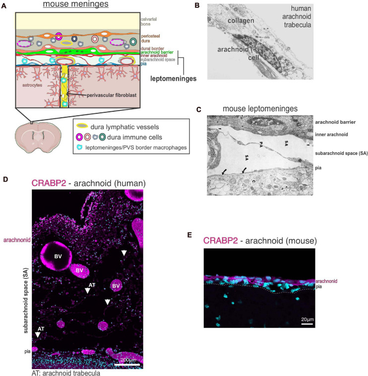
“Arachnoid fibroblasts express RALDH2 and CRABP2, a retinoic acid (RA) synthesizing enzyme and an RA binding protein, respectively (Figures 1D,E; DeSisto et al., 2020).”
§ Living on the Edge of the CNS: Meninges Cell Diversity in Health and Disease > Meningeal Cell Types > Fibroblasts
What the sources say
- Not checked yetcites DeSisto et al., 2020 source
plain- Asserted: passes — The authors put it forward as true.
- Checkable: passes — It can be checked against evidence.
- Load-bearing: passes — The paper’s conclusions depend on it.
- Based: passes — The paper gives a reason why it holds.
In the paper
“disruption of glymphatic function results in lower levels of TBI-associated biomarkers in the cervical lymph nodes and bloodstream”
§ Living on the Edge of the CNS: Meninges Cell Diversity in Health and Disease > Meningeal Response to Injury and Disease > Meningeal Lymphatics in Acute CNS Injury
What the sources say
- Not checked yetcites Plog et al., 2015 source
plaindirect- Asserted: passes — The authors put it forward as true.
- Checkable: passes — It can be checked against evidence.
- Load-bearing: passes — The paper’s conclusions depend on it.
- Based: passes — The paper gives a reason why it holds.
None of the sources cited for this claim have been checked yet.
In the paper
“Production of RA by the arachnoid layers critically regulates brain and neurovascular development (Siegenthaler et al., 2009; Choi et al., 2014; Bonney et al., 2016; Mishra et al., 2016; Haushalter et al., 2017).”
§ Living on the Edge of the CNS: Meninges Cell Diversity in Health and Disease > Meningeal Cell Types > Fibroblasts
What the sources say
plaindirect- Asserted: passes — The authors put it forward as true.
- Checkable: passes — It can be checked against evidence.
- Load-bearing: unclear — It is unclear whether the paper’s conclusions depend on it.
- Based: passes — The paper gives a reason why it holds.
None of the sources cited for this claim have been checked yet.
In the paper
“numerous studies show that meningeal immune cells play an important role in the brain injury response”
§ Living on the Edge of the CNS: Meninges Cell Diversity in Health and Disease > Meningeal Response to Injury and Disease > Meningeal Immune Cells in Acute CNS Injury
What the sources say
boostedcorrelational- Asserted: passes — The authors put it forward as true.
- Checkable: passes — It can be checked against evidence.
- Load-bearing: passes — The paper’s conclusions depend on it.
- Based: passes — The paper gives a reason why it holds.
- Rests on an assumption: The degree of neuroinflammation following acute CNS injury is a major predictor of clinical outcomes.
A conclusion the paper draws from its own results — it cites no other source for it, so there is none to check. The quote shows it is in the paper.
In the paper
“Resident meningeal macrophages regulate the acute injury response by influencing the severity of neuroinflammation and fibrosis”
§ Living on the Edge of the CNS: Meninges Cell Diversity in Health and Disease > Meningeal Response to Injury and Disease > Meningeal Immune Cells in Acute CNS Injury
plaindirect- Asserted: passes — The authors put it forward as true.
- Checkable: passes — It can be checked against evidence.
- Load-bearing: passes — The paper’s conclusions depend on it.
- Based: passes — The paper gives a reason why it holds.
None of the sources cited for this claim have been checked yet.
In the paper
“The brain glymphatic and meningeal lymphatic systems work together to clear waste and macromolecules from the brain parenchyma in homeostasis and following injury”
§ Living on the Edge of the CNS: Meninges Cell Diversity in Health and Disease > Meningeal Response to Injury and Disease > Meningeal Lymphatics in Acute CNS Injury
What the sources say
plaindirect- Asserted: passes — The authors put it forward as true.
- Checkable: passes — It can be checked against evidence.
- Load-bearing: passes — The paper’s conclusions depend on it.
- Based: passes — The paper gives a reason why it holds.
A conclusion the paper draws from its own results — it cites no other source for it, so there is none to check. The quote shows it is in the paper.
In the paper
“While not done in an acute injury model, this evidence further supports that CNS fibroblasts, not pericytes or vSMCs, are the main reactive fibrotic cell type in CNS injuries and disease.”
§ Living on the Edge of the CNS: Meninges Cell Diversity in Health and Disease > Meningeal Response to Injury and Disease > CNS Fibroblasts in Acute CNS Injury
hedgeddirect- Asserted: passes — The authors put it forward as true.
- Checkable: passes — It can be checked against evidence.
- Load-bearing: passes — The paper’s conclusions depend on it.
- Based: passes — The paper gives a reason why it holds.
None of the sources cited for this claim have been checked yet.
In the paper
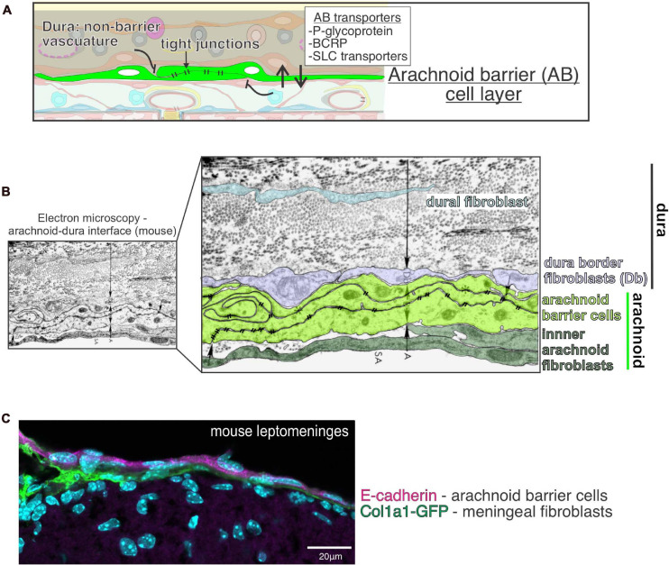
“Segregating the outer and inner meningeal compartments is the epithelial-like arachnoid barrier cells, connected by tight and adherens junctions, which regulate the movement of pathogens, molecules, and cells into and out of the cerebral spinal fluid (CSF) and brain parenchyma.”
§ Living on the Edge of the CNS: Meninges Cell Diversity in Health and Disease > Abstract
What the sources say
plaindirect- Asserted: passes — The authors put it forward as true.
- Checkable: passes — It can be checked against evidence.
- Load-bearing: passes — The paper’s conclusions depend on it.
- Based: passes — The paper gives a reason why it holds.
None of the sources cited for this claim have been checked yet.
In the paper
“The dural lymphatics are now recognized as a critical transport system of macromolecules, interstitial fluid, and CSF from the CNS into the cervical lymph nodes (Aspelund et al., 2015; Absinta et al., 2017; Louveau et al., 2018; Ahn et al., 2019).”
§ Living on the Edge of the CNS: Meninges Cell Diversity in Health and Disease > Meningeal Cell Types > Lymphatic Vessels: Dura
What the sources say
boosteddirect- Asserted: passes — The authors put it forward as true.
- Checkable: passes — It can be checked against evidence.
- Load-bearing: unclear — It is unclear whether the paper’s conclusions depend on it.
- Based: passes — The paper gives a reason why it holds.
A conclusion the paper draws from its own results — it cites no other source for it, so there is none to check. The quote shows it is in the paper.
In the paper
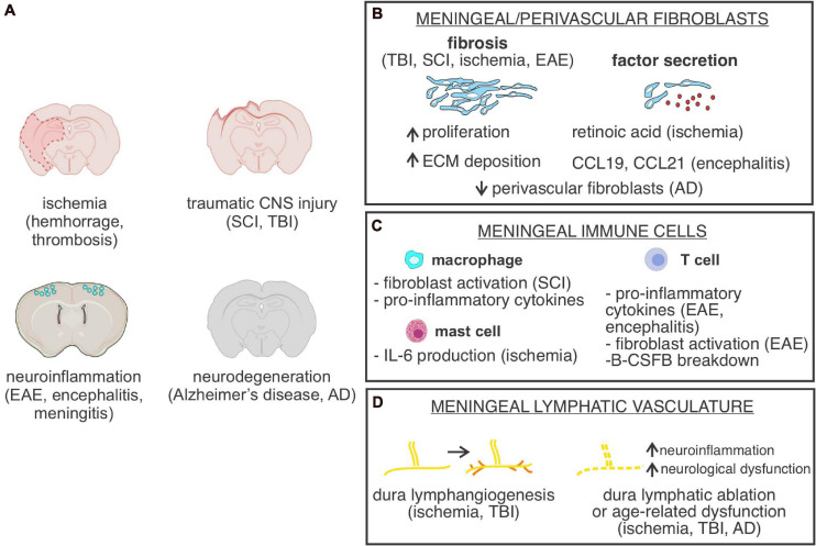
“In summary, fibroblasts from the meninges and perivascular space are major drivers of fibrotic scar formation following CNS injury however establishing a standard nomenclature would be beneficial.”
§ Living on the Edge of the CNS: Meninges Cell Diversity in Health and Disease > Meningeal Response to Injury and Disease > CNS Fibroblasts in Acute CNS Injury
plaindirect- Asserted: passes — The authors put it forward as true.
- Checkable: passes — It can be checked against evidence.
- Load-bearing: passes — The paper’s conclusions depend on it.
- Based: passes — The paper gives a reason why it holds.
- It may state two separate claims as one. 49
- Probably not a substantial claim. 2
- The quote only partly states this claim. 1
Every source the paper cites for this claim was checked and supports it.
In the paper
“In a mild closed-skull TBI model, meningeal lymphatic drainage is impaired likely due to increases in intracranial pressure, accompanied by lymphatic morphological changes”
- Also stated in: § Meningeal Lymphatics in Acute CNS Injury · plain
“lymphangiogenesis was observed up to 2 weeks following TBI, though this did not correspond with increased lymphatic function”
§ Living on the Edge of the CNS: Meninges Cell Diversity in Health and Disease > Meningeal Response to Injury and Disease > Meningeal Lymphatics in Acute CNS Injury
What the sources say
- Supportedcites Bolte et al., 2020 source
Split into 2 atoms — 2 confirmed, 0 partially supported, 0 disputed, 0 misattributed, 0 unverifiable. Expand the claim to see per-atom verdicts.
plainconditional- Asserted: passes — The authors put it forward as true.
- Checkable: passes — It can be checked against evidence.
- Load-bearing: passes — The paper’s conclusions depend on it.
- Based: passes — The paper gives a reason why it holds.
It may state two separate claims as one.
- Also stated in: § Meningeal Lymphatics in Acute CNS Injury · plain
The cited sources support only part of this claim — read each source’s verdict below.
In the paper
“Primary tumors of the meninges are quite rare, however, the leptomeninges is a relatively common site for by contiguous extension of primary tumors of the central nervous system, paranasal sinuses and skull base origin or tumor metastasis which can lead to dissemination into the CNS parenchyma and poor prognosis”
§ Living on the Edge of the CNS: Meninges Cell Diversity in Health and Disease > Meningeal Response to Injury and Disease > Meninges as a Site of Cancer Metastasis
What the sources say
- Not checked yetcites Mahendru and Chong, 2009
- Not checked yetcites Waki et al., 2009 source
- Partly supportedcites Oechsle et al., 2010 sourceExperimental — accuracy not yet measured
Split into 2 atoms — 0 confirmed, 1 partially supported, 0 disputed, 0 misattributed, 1 unverifiable. Expand the claim to see per-atom verdicts.
plaindirect- Asserted: passes — The authors put it forward as true.
- Checkable: passes — It can be checked against evidence.
- Load-bearing: unclear — It is unclear whether the paper’s conclusions depend on it.
- Based: passes — The paper gives a reason why it holds.
It may state two separate claims as one.
In the paper
“Our recent single cell RNA sequencing (scRNAseq) of developing mouse meninges showed pia, arachnoid and dura fibroblasts are molecularly distinct and likely have layer specific functions (DeSisto et al., 2020).”
§ Living on the Edge of the CNS: Meninges Cell Diversity in Health and Disease > Meningeal Cell Types > Fibroblasts
What the sources say
- Not checked yetcites DeSisto et al., 2020 source
hedgedcorrelational- Asserted: passes — The authors put it forward as true.
- Checkable: passes — It can be checked against evidence.
- Load-bearing: passes — The paper’s conclusions depend on it.
- Based: passes — The paper gives a reason why it holds.
It may state two separate claims as one.
In the paper
“We recently found that arachnoid barrier cells can be first seen in the mouse forebrain around E13 and they differentiate from mesenchymal cells of the primary meninx (DeSisto et al., 2020).”
§ Living on the Edge of the CNS: Meninges Cell Diversity in Health and Disease > Meningeal Cell Types > Arachnoid Barrier Cells
What the sources say
- Not checked yetcites DeSisto et al., 2020 source
plaindirect- Asserted: passes — The authors put it forward as true.
- Checkable: passes — It can be checked against evidence.
- Load-bearing: unclear — It is unclear whether the paper’s conclusions depend on it.
- Based: passes — The paper gives a reason why it holds.
It may state two separate claims as one.
None of the sources cited for this claim have been checked yet.
In the paper
“Meningeal fibroblasts in different CNS regions have different origins; forebrain meningeal fibroblasts are neural crest derived whereas mid-, hindbrain and spinal cord meninges are derived from the mesoderm (Jiang et al., 2002; Yoshida et al., 2008).”
§ Living on the Edge of the CNS: Meninges Cell Diversity in Health and Disease > Meningeal Cell Types > Fibroblasts
What the sources say
plaincorrelational- Asserted: passes — The authors put it forward as true.
- Checkable: passes — It can be checked against evidence.
- Load-bearing: unclear — It is unclear whether the paper’s conclusions depend on it.
- Based: passes — The paper gives a reason why it holds.
It may state two separate claims as one.
None of the sources cited for this claim have been checked yet.
In the paper
“The dural compartment of the meninges, closest to the skull, is predominantly composed of fibroblasts, but also includes fenestrated blood vasculature, an elaborate lymphatic system, as well as immune cells which are distinct from the CNS.”
§ Living on the Edge of the CNS: Meninges Cell Diversity in Health and Disease > Abstract
What the sources say
plain- Asserted: passes — The authors put it forward as true.
- Checkable: passes — It can be checked against evidence.
- Load-bearing: unclear — It is unclear whether the paper’s conclusions depend on it.
- Based: passes — The paper gives a reason why it holds.
It may state two separate claims as one.
In the paper
“Cx3cr1+ meningeal macrophages are observed dying within 30 min of injury induction, leading to formation of a “honeycomb” network of microglia at the glial limitans, likely preventing the spread of reactive oxygen species from the leptomeninges into the brain parenchyma”
§ Living on the Edge of the CNS: Meninges Cell Diversity in Health and Disease > Meningeal Response to Injury and Disease > Meningeal Immune Cells in Acute CNS Injury
What the sources say
- Not checked yetcites Roth et al., 2013 source
hedgeddirect- Asserted: passes — The authors put it forward as true.
- Checkable: passes — It can be checked against evidence.
- Load-bearing: passes — The paper’s conclusions depend on it.
- Based: passes — The paper gives a reason why it holds.
It may state two separate claims as one.
A conclusion the paper draws from its own results — it cites no other source for it, so there is none to check. The quote shows it is in the paper.
In the paper
“Thus, fibroblast production of retinoic acid seems to impair CNS recovery while stimulating fibrosis.”
§ Living on the Edge of the CNS: Meninges Cell Diversity in Health and Disease > Meningeal Response to Injury and Disease > CNS Fibroblasts in Acute CNS Injury
hedgeddirect- Asserted: passes — The authors put it forward as true.
- Checkable: passes — It can be checked against evidence.
- Load-bearing: passes — The paper’s conclusions depend on it.
- Based: passes — The paper gives a reason why it holds.
It may state two separate claims as one.
None of the sources cited for this claim have been checked yet.
In the paper
“In both MCAO and SCI models, macrophages are observed in the fibrotic scar and may associate with fibroblasts”
- Also stated in: § Meningeal Immune Cells in Acute CNS Injury · plain
“CD11b+ macrophages are partially responsible for recruitment of fibroblasts to the fibrotic scar following SCI”
§ Living on the Edge of the CNS: Meninges Cell Diversity in Health and Disease > Meningeal Response to Injury and Disease > Meningeal Immune Cells in Acute CNS Injury
What the sources say
plainconditional- Asserted: passes — The authors put it forward as true.
- Checkable: passes — It can be checked against evidence.
- Load-bearing: passes — The paper’s conclusions depend on it.
- Based: passes — The paper gives a reason why it holds.
It may state two separate claims as one.
- Also stated in: § Meningeal Immune Cells in Acute CNS Injury · plain
A conclusion the paper draws from its own results — it cites no other source for it, so there is none to check. The quote shows it is in the paper.
In the paper
“While the meninges were historically seen as a purely structural support for the CNS and protection from trauma, the emerging view of the meninges is as an essential interface between the CNS and the periphery, critical to brain development, required for brain homeostasis, and involved in a variety of diseases.”
§ Living on the Edge of the CNS: Meninges Cell Diversity in Health and Disease > Abstract
plaindirect- Asserted: passes — The authors put it forward as true.
- Checkable: passes — It can be checked against evidence.
- Load-bearing: passes — The paper’s conclusions depend on it.
- Based: unclear — It is unclear why the paper says it holds.
It may state two separate claims as one.
None of the sources cited for this claim have been checked yet.
In the paper
“Several studies show this vasculature has barrier properties and are therefore part of the meninges blood-CSF barrier.”
§ Living on the Edge of the CNS: Meninges Cell Diversity in Health and Disease > Meningeal Cell Types > Blood Vessels: Leptomeninges and Dura
What the sources say
plaincorrelational- Asserted: passes — The authors put it forward as true.
- Checkable: passes — It can be checked against evidence.
- Load-bearing: unclear — It is unclear whether the paper’s conclusions depend on it.
- Based: passes — The paper gives a reason why it holds.
It may state two separate claims as one.
A conclusion the paper draws from its own results — it cites no other source for it, so there is none to check. The quote shows it is in the paper.
In the paper
“These results highlight the important function of the glymphatic system in clearance of molecular substances from the brain and potential role in facilitating signaling between the brain and periphery.”
§ Living on the Edge of the CNS: Meninges Cell Diversity in Health and Disease > Meningeal Response to Injury and Disease > Meningeal Lymphatics in Acute CNS Injury
hedgedconditional- Asserted: passes — The authors put it forward as true.
- Checkable: passes — It can be checked against evidence.
- Load-bearing: passes — The paper’s conclusions depend on it.
- Based: passes — The paper gives a reason why it holds.
It may state two separate claims as one.
In the paper
“retinoic acid treatment following stroke in rodents reduces neurogenesis while also reducing angiogenesis and gliogenesis in the peri-infarct region”
§ Living on the Edge of the CNS: Meninges Cell Diversity in Health and Disease > Meningeal Response to Injury and Disease > CNS Fibroblasts in Acute CNS Injury
What the sources say
- Not checked yetcites Jung et al., 2007 source
plaindirect- Asserted: passes — The authors put it forward as true.
- Checkable: passes — It can be checked against evidence.
- Load-bearing: passes — The paper’s conclusions depend on it.
- Based: passes — The paper gives a reason why it holds.
It may state two separate claims as one.
In the paper
“Most proximate to the brain is the collagen and basement membrane-rich pia matter that abuts the glial limitans and has recently be shown to have regional heterogeneity within the developing mouse brain.”
§ Living on the Edge of the CNS: Meninges Cell Diversity in Health and Disease > Abstract
What the sources say
- Not checked yetcites DeSisto et al., 2020 source
plain- Asserted: passes — The authors put it forward as true.
- Checkable: passes — It can be checked against evidence.
- Load-bearing: unclear — It is unclear whether the paper’s conclusions depend on it.
- Based: passes — The paper gives a reason why it holds.
It may state two separate claims as one.
In the paper
“Following TBI, glymphatic fluid flow is disrupted and greater amounts of tau protein is observed accumulating in the parenchyma”
- Also stated in: § Meningeal Lymphatics in Acute CNS Injury · plain
“knock-out of Aqp4, a water transport channel necessary for interstitial solute clearance and glymphatic function, following TBI, exacerbates the accumulation of tau in the parenchyma”
§ Living on the Edge of the CNS: Meninges Cell Diversity in Health and Disease > Meningeal Response to Injury and Disease > Meningeal Lymphatics in Acute CNS Injury
What the sources say
- Not checked yetcites Iliff et al., 2014 source
plaindirect- Asserted: passes — The authors put it forward as true.
- Checkable: passes — It can be checked against evidence.
- Load-bearing: passes — The paper’s conclusions depend on it.
- Based: passes — The paper gives a reason why it holds.
It may state two separate claims as one.
- Also stated in: § Meningeal Lymphatics in Acute CNS Injury · plain
In the paper
“depletion of mast cells resulted in decreased levels of neuroinflammation, brain swelling, and infarct size following stroke and this was partially mediated by mast-cell derived IL-6”
§ Living on the Edge of the CNS: Meninges Cell Diversity in Health and Disease > Meningeal Response to Injury and Disease > Meningeal Immune Cells in Acute CNS Injury
What the sources say
- Not checked yetcites Arac et al., 2019 source
plaindirect- Asserted: passes — The authors put it forward as true.
- Checkable: passes — It can be checked against evidence.
- Load-bearing: passes — The paper’s conclusions depend on it.
- Based: passes — The paper gives a reason why it holds.
It may state two separate claims as one.
In the paper
“lymphangiogenesis was seen following photothrombotic stroke lesion in the mouse cortex, but this effect was not seen following MCAO”
§ Living on the Edge of the CNS: Meninges Cell Diversity in Health and Disease > Meningeal Response to Injury and Disease > Meningeal Lymphatics in Acute CNS Injury
What the sources say
- Not checked yetcites Yanev et al., 2020 source
plain- Asserted: passes — The authors put it forward as true.
- Checkable: passes — It can be checked against evidence.
- Load-bearing: passes — The paper’s conclusions depend on it.
- Based: passes — The paper gives a reason why it holds.
It may state two separate claims as one.
In the paper
“depletion of macrophages in the leptomeninges and perivascular spaces surrounding large arterioles prior to injury results in improved neurological scoring and reduced levels of inflammation and neuronal cell death”
§ Living on the Edge of the CNS: Meninges Cell Diversity in Health and Disease > Meningeal Response to Injury and Disease > Meningeal Immune Cells in Acute CNS Injury
What the sources say
- Not checked yetcites Wan et al., 2021 source
plaindirect- Asserted: passes — The authors put it forward as true.
- Checkable: passes — It can be checked against evidence.
- Load-bearing: passes — The paper’s conclusions depend on it.
- Based: passes — The paper gives a reason why it holds.
It may state two separate claims as one.
A conclusion the paper draws from its own results — it cites no other source for it, so there is none to check. The quote shows it is in the paper.
In the paper
“The dural lymphatic function is also disrupted following acute CNS injury.”
- Also stated in: § Meningeal Lymphatics in Acute CNS Injury · plain
“Thus, the degree of lymphatic impairment and response is dependent on the injury mechanism”
§ Living on the Edge of the CNS: Meninges Cell Diversity in Health and Disease > Meningeal Response to Injury and Disease > Meningeal Lymphatics in Acute CNS Injury
plaindirect- Asserted: passes — The authors put it forward as true.
- Checkable: passes — It can be checked against evidence.
- Load-bearing: passes — The paper’s conclusions depend on it.
- Based: passes — The paper gives a reason why it holds.
It may state two separate claims as one.
- Also stated in: § Meningeal Lymphatics in Acute CNS Injury · plain
None of the sources cited for this claim have been checked yet.
In the paper
“BBB breakdown is a well-documented feature of MS”
- Also stated in: § Meninges and Perivascular Fibroblasts in Multiple Sclerosis · hedged
“there is some evidence that MS can also induce disruption of the arachnoid barrier in the meninges, part of the B-CSF barrier”
- Also stated in: § Meninges and Perivascular Fibroblasts in Multiple Sclerosis · plain
“Claudin 11 is a tight junction protein enriched in arachnoid barrier cells and it is downregulated in late stages of an EAE mouse model”
§ Living on the Edge of the CNS: Meninges Cell Diversity in Health and Disease > Meningeal Response to Injury and Disease > Meninges and Perivascular Fibroblasts in Multiple Sclerosis
What the sources say
- Not checked yetcites Russi and Brown, 2015
- Not checked yetcites Uchida et al., 2019b
boostedconditional- Asserted: passes — The authors put it forward as true.
- Checkable: passes — It can be checked against evidence.
- Load-bearing: unclear — It is unclear whether the paper’s conclusions depend on it.
- Based: passes — The paper gives a reason why it holds.
It may state two separate claims as one.
- Also stated in: § Meninges and Perivascular Fibroblasts in Multiple Sclerosis · hedged
None of the sources cited for this claim have been checked yet.
In the paper
“The leptomeninges are now recognized as a key player in multiple sclerosis (MS), an inflammatory CNS autoimmune disorder characterized by axon demyelination”
- Also stated in: § Meninges and Perivascular Fibroblasts in Multiple Sclerosis · plain
“In both MS patients and MS mouse models, immune cells infiltrate the leptomeninges and pathology is commonly seen in cortical areas adjacent to the meninges”
§ Living on the Edge of the CNS: Meninges Cell Diversity in Health and Disease > Meningeal Response to Injury and Disease > Meninges and Perivascular Fibroblasts in Multiple Sclerosis
What the sources say
- Not checked yetcites Russi and Brown, 2015
- Not checked yetcites Lucchinetti et al., 2011 source
plaincorrelational- Asserted: passes — The authors put it forward as true.
- Checkable: passes — It can be checked against evidence.
- Load-bearing: passes — The paper’s conclusions depend on it.
- Based: passes — The paper gives a reason why it holds.
It may state two separate claims as one.
- Also stated in: § Meninges and Perivascular Fibroblasts in Multiple Sclerosis · plain
A conclusion the paper draws from its own results — it cites no other source for it, so there is none to check. The quote shows it is in the paper.
In the paper
“A disruption to the arachnoid barrier could permit dural and peripheral immune cells to migrate into the leptomeninges, and potentially contribute to leptomeningeal immune infiltrate that is common in MS pathology.”
§ Living on the Edge of the CNS: Meninges Cell Diversity in Health and Disease > Meningeal Response to Injury and Disease > Meninges and Perivascular Fibroblasts in Multiple Sclerosis
hedgedconditional- Asserted: passes — The authors put it forward as true.
- Checkable: passes — It can be checked against evidence.
- Load-bearing: passes — The paper’s conclusions depend on it.
- Based: passes — The paper gives a reason why it holds.
It may state two separate claims as one.
None of the sources cited for this claim have been checked yet.
In the paper
“cytokine administration into the cisterna magna of mice is sufficient to drive arachnoid barrier and blood-cerebral spinal fluid barrier functional breakdown”
- Also stated in: § Meninges and Perivascular Fibroblasts in Multiple Sclerosis · boosted
“Activated T cells in the leptomeninges produce cytokines known to perturb tight junction integrity of brain endothelial cells, including IFNγ which is known to drive BBB breakdown in viral encephalitis”
§ Living on the Edge of the CNS: Meninges Cell Diversity in Health and Disease > Meningeal Response to Injury and Disease > Meninges and Perivascular Fibroblasts in Multiple Sclerosis
What the sources say
- Not checked yetcites Ichikawa and Itoh, 2011
- Not checked yetcites Bonney et al., 2019 source
boosteddirect- Asserted: passes — The authors put it forward as true.
- Checkable: passes — It can be checked against evidence.
- Load-bearing: unclear — It is unclear whether the paper’s conclusions depend on it.
- Based: passes — The paper gives a reason why it holds.
It may state two separate claims as one.
- Also stated in: § Meninges and Perivascular Fibroblasts in Multiple Sclerosis · boosted
None of the sources cited for this claim have been checked yet.
In the paper
“In the experimental autoimmune encephalomyelitis (EAE) mouse model of MS, autoreactive effector T cells first infiltrate the leptomeninges via the pial vasculature, are activated by antigen presenting meningeal/perivascular macrophages, and subsequently enter the CNS parenchyma, triggering lesion formation”
§ Living on the Edge of the CNS: Meninges Cell Diversity in Health and Disease > Meningeal Response to Injury and Disease > Meninges and Perivascular Fibroblasts in Multiple Sclerosis
What the sources say
plaindirect- Asserted: passes — The authors put it forward as true.
- Checkable: passes — It can be checked against evidence.
- Load-bearing: unclear — It is unclear whether the paper’s conclusions depend on it.
- Based: passes — The paper gives a reason why it holds.
It may state two separate claims as one.
None of the sources cited for this claim have been checked yet.
In the paper
“In cases of bacterial meningitis, infections usually start as skin, gastrointestinal or respiratory infections that spread to the blood”
- Also stated in: § Response of the Meninges to Meningitis · plain
“Once in the blood, the meningitis causing bacteria accumulate in the leptomeningeal vasculature”
- Also stated in: § Response of the Meninges to Meningitis · plain
“move into the meninges by inducing BBB breakdown through multiple mechanisms”
§ Living on the Edge of the CNS: Meninges Cell Diversity in Health and Disease > Meningeal Response to Injury and Disease > Response of the Meninges to Meningitis
What the sources say
plaindirect- Asserted: passes — The authors put it forward as true.
- Checkable: passes — It can be checked against evidence.
- Load-bearing: unclear — It is unclear whether the paper’s conclusions depend on it.
- Based: passes — The paper gives a reason why it holds.
It may state two separate claims as one.
- Also stated in: § Response of the Meninges to Meningitis · plain
In the paper
“pre-existing defects in meningeal lymphatics causes worsened neuroinflammation following TBI”
- Also stated in: § Meningeal Lymphatics in Acute CNS Injury · plain
“stimulating meningeal lymphatic vessel outgrowth via VEGF-C administration had decreased levels of gliosis”
§ Living on the Edge of the CNS: Meninges Cell Diversity in Health and Disease > Meningeal Response to Injury and Disease > Meningeal Lymphatics in Acute CNS Injury
What the sources say
- Not checked yetcites Bolte et al., 2020 source
plaindirect- Asserted: passes — The authors put it forward as true.
- Checkable: passes — It can be checked against evidence.
- Load-bearing: passes — The paper’s conclusions depend on it.
- Based: passes — The paper gives a reason why it holds.
It may state two separate claims as one.
- Also stated in: § Meningeal Lymphatics in Acute CNS Injury · plain
None of the sources cited for this claim have been checked yet.
In the paper
“Blockage of VEGFR3 (expressed by lymphatic endothelial cells) in mice following focal cerebral ischemia resulted in reduced inflammatory response and infarct”
- Also stated in: § Meningeal Lymphatics in Acute CNS Injury · plain
“VEGFR3 mutant mice subjected to MCAO develop larger stroke volumes, thus worsened edema”
§ Living on the Edge of the CNS: Meninges Cell Diversity in Health and Disease > Meningeal Response to Injury and Disease > Meningeal Lymphatics in Acute CNS Injury
What the sources say
plaindirect- Asserted: passes — The authors put it forward as true.
- Checkable: passes — It can be checked against evidence.
- Load-bearing: passes — The paper’s conclusions depend on it.
- Based: passes — The paper gives a reason why it holds.
It may state two separate claims as one.
- Also stated in: § Meningeal Lymphatics in Acute CNS Injury · plain
None of the sources cited for this claim have been checked yet.
In the paper
“In the LCMV Armstrong mouse model, it was shown that the recruitment of antiviral CD8+ T cells, while necessary to defeat the infection, can also be deleterious.”
- Also stated in: § Response of the Meninges to Meningitis · hedged
“These CD8+ T cells also produce cytokines that can lead to over infiltration of monocytes and neutrophils that drive breakdown of vascular integrity and induce severe edema and brainstem herniation”
§ Living on the Edge of the CNS: Meninges Cell Diversity in Health and Disease > Meningeal Response to Injury and Disease > Response of the Meninges to Meningitis
What the sources say
- Not checked yetcites Kim et al., 2009 source
- Not checked yetcites Mastorakos and McGavern, 2019
plaindirect- Asserted: passes — The authors put it forward as true.
- Checkable: passes — It can be checked against evidence.
- Load-bearing: unclear — It is unclear whether the paper’s conclusions depend on it.
- Based: passes — The paper gives a reason why it holds.
It may state two separate claims as one.
- Also stated in: § Response of the Meninges to Meningitis · hedged
None of the sources cited for this claim have been checked yet.
In the paper
“AD is a dementia inducing neurodegenerative disorder characterized by the accumulation of amyloid-β containing plaques, which were initially isolated from homogenates of AD patient meningeal tissue”
§ Living on the Edge of the CNS: Meninges Cell Diversity in Health and Disease > Meningeal Response to Injury and Disease > The Meninges and Alzheimer’s Disease
What the sources say
- Not checked yetcites Joachim et al., 1988 source
- Not checked yetcites Da Mesquita et al., 2018b
plain- Asserted: passes — The authors put it forward as true.
- Checkable: passes — It can be checked against evidence.
- Load-bearing: fails — The paper’s conclusions may not depend on it.
- Based: passes — The paper gives a reason why it holds.
Probably not a substantial claim. The paper’s conclusions may not depend on it. It may state two separate claims as one.
None of the sources cited for this claim have been checked yet.
In the paper
“The presentation of MS induced leptomeningeal inflammation can be variable, ranging from disorganized collections of immune cells to organized ectopic lymphoid follicle-like structures”
- Also stated in: § Meninges and Perivascular Fibroblasts in Multiple Sclerosis · plain
“the ectopic lymph structures seen in MS are potentiated by leptomeningeal fibroblasts which form a reticular cell network consisting of activated fibroblasts in a scaffold-like structure”
§ Living on the Edge of the CNS: Meninges Cell Diversity in Health and Disease > Meningeal Response to Injury and Disease > Meninges and Perivascular Fibroblasts in Multiple Sclerosis
What the sources say
plaindirect- Asserted: passes — The authors put it forward as true.
- Checkable: passes — It can be checked against evidence.
- Load-bearing: passes — The paper’s conclusions depend on it.
- Based: passes — The paper gives a reason why it holds.
It may state two separate claims as one.
- Also stated in: § Meninges and Perivascular Fibroblasts in Multiple Sclerosis · plain
None of the sources cited for this claim have been checked yet.
In the paper
“Increased number of PDGFRβ-expressing cells are detected in human MS pathology samples”
- Also stated in: § Meninges and Perivascular Fibroblasts in Multiple Sclerosis · plain
“increased ECM protein deposition is a common pathological feature of MS lesions”
§ Living on the Edge of the CNS: Meninges Cell Diversity in Health and Disease > Meningeal Response to Injury and Disease > Meninges and Perivascular Fibroblasts in Multiple Sclerosis
What the sources say
plaincorrelational- Asserted: passes — The authors put it forward as true.
- Checkable: passes — It can be checked against evidence.
- Load-bearing: passes — The paper’s conclusions depend on it.
- Based: passes — The paper gives a reason why it holds.
It may state two separate claims as one.
- Also stated in: § Meninges and Perivascular Fibroblasts in Multiple Sclerosis · plain
None of the sources cited for this claim have been checked yet.
In the paper
“both the brain glymphatic and meningeal lymphatic systems deteriorate with age”
- Also stated in: § The Meninges and Alzheimer’s Disease · plain
“their dysfunction can potentiate AD pathology and dementia”
§ Living on the Edge of the CNS: Meninges Cell Diversity in Health and Disease > Meningeal Response to Injury and Disease > The Meninges and Alzheimer’s Disease
What the sources say
plaindirect- Asserted: passes — The authors put it forward as true.
- Checkable: passes — It can be checked against evidence.
- Load-bearing: passes — The paper’s conclusions depend on it.
- Based: passes — The paper gives a reason why it holds.
It may state two separate claims as one.
- Also stated in: § The Meninges and Alzheimer’s Disease · plain
In the paper
“a scRNAseq analysis showed that fibroblasts from EAE lesions upregulate interferon γ (IFNγ) signaling, and conditional deletion of IFN receptor γ from fibroblasts partially blocked fibroblast expansion”
- Also stated in: § Meninges and Perivascular Fibroblasts in Multiple Sclerosis · hedged
“IFNγ is produced by spinal cord T cells in EAE mice, implicating immune cells in EAE lesions as a source of fibroblast activation signals”
§ Living on the Edge of the CNS: Meninges Cell Diversity in Health and Disease > Meningeal Response to Injury and Disease > Meninges and Perivascular Fibroblasts in Multiple Sclerosis
What the sources say
- Not checked yetcites Dorrier et al., 2021 source
plainconditional- Asserted: passes — The authors put it forward as true.
- Checkable: passes — It can be checked against evidence.
- Load-bearing: passes — The paper’s conclusions depend on it.
- Based: passes — The paper gives a reason why it holds.
It may state two separate claims as one.
- Also stated in: § Meninges and Perivascular Fibroblasts in Multiple Sclerosis · hedged
None of the sources cited for this claim have been checked yet.
In the paper
“Bacterial presence in the meninges induces a strong inflammatory response, including production of multiple cytokines (tumor necrosis factor (TNF)-alpha, interleukin (IL)-1-beta, and IL-6), and a massive infiltration of neutrophils”
- Also stated in: § Response of the Meninges to Meningitis · plain
“The inflammatory cytokines and neutrophils work to quell the infection but also drive the breakdown of endothelial cell tight junctions”
§ Living on the Edge of the CNS: Meninges Cell Diversity in Health and Disease > Meningeal Response to Injury and Disease > Response of the Meninges to Meningitis
What the sources say
boosteddirect- Asserted: passes — The authors put it forward as true.
- Checkable: passes — It can be checked against evidence.
- Load-bearing: unclear — It is unclear whether the paper’s conclusions depend on it.
- Based: passes — The paper gives a reason why it holds.
It may state two separate claims as one.
- Also stated in: § Response of the Meninges to Meningitis · plain
In the paper
“erythrocytes accrue in the cervical lymph nodes via meningeal lymphatics following injury, and ablation of meningeal lymphatic vessels leads to less erythrocyte aggregation and greater neuroinflammation and neurological defects”
§ Living on the Edge of the CNS: Meninges Cell Diversity in Health and Disease > Meningeal Response to Injury and Disease > Meningeal Lymphatics in Acute CNS Injury
What the sources say
- Not checked yetcites Chen et al., 2020 source
plaindirect- Asserted: passes — The authors put it forward as true.
- Checkable: passes — It can be checked against evidence.
- Load-bearing: passes — The paper’s conclusions depend on it.
- Based: passes — The paper gives a reason why it holds.
It may state two separate claims as one.
None of the sources cited for this claim have been checked yet.
In the paper
“Using a Col1a1-GFP mouse line in conjunction with an EAE model, two independent groups identified substantial increases in fibroblasts within spinal cord lesion sites”
- Also stated in: § Meninges and Perivascular Fibroblasts in Multiple Sclerosis · plain
“Fibroblast expansion was deleterious for oligodendrocyte precursor cell (OPC) differentiation into mature oligodendrocytes and OPC migration into the lesion”
§ Living on the Edge of the CNS: Meninges Cell Diversity in Health and Disease > Meningeal Response to Injury and Disease > Meninges and Perivascular Fibroblasts in Multiple Sclerosis
What the sources say
plaindirect- Asserted: passes — The authors put it forward as true.
- Checkable: passes — It can be checked against evidence.
- Load-bearing: passes — The paper’s conclusions depend on it.
- Based: passes — The paper gives a reason why it holds.
It may state two separate claims as one.
- Also stated in: § Meninges and Perivascular Fibroblasts in Multiple Sclerosis · plain
In the paper
“Biopsies taken from early MS patients with cortical demyelination were more likely to have leptomeningeal inflammation, consisting of effector T cells and IgA B cells infiltrates, than those without cortical demyelination.”
- Also stated in: § Meninges and Perivascular Fibroblasts in Multiple Sclerosis · plain
“in human cases of MS the severity of cortical lesions is correlated to the extent of meningeal inflammation”
§ Living on the Edge of the CNS: Meninges Cell Diversity in Health and Disease > Meningeal Response to Injury and Disease > Meninges and Perivascular Fibroblasts in Multiple Sclerosis
What the sources say
- Not checked yetcites Lucchinetti et al., 2011 source
plaincorrelational- Asserted: passes — The authors put it forward as true.
- Checkable: passes — It can be checked against evidence.
- Load-bearing: passes — The paper’s conclusions depend on it.
- Based: passes — The paper gives a reason why it holds.
It may state two separate claims as one. The quote only partly states this claim.
- Also stated in: § Meninges and Perivascular Fibroblasts in Multiple Sclerosis · plain
None of the sources cited for this claim have been checked yet.
In the paper
“All major meningitis causing bacteria can adhere strongly to meningeal fibroblasts and arachnoid barrier-like cells”
- Also stated in: § Response of the Meninges to Meningitis · plain
“Group B Streptococcus bacteria cross the blood brain barrier by binding to vimentin on endothelial cells”
- Also stated in: § Response of the Meninges to Meningitis · plain
“Vimentin is also highly expressed by arachnoid barrier cells”
§ Living on the Edge of the CNS: Meninges Cell Diversity in Health and Disease > Meningeal Response to Injury and Disease > Response of the Meninges to Meningitis
What the sources say
plaindirect- Asserted: passes — The authors put it forward as true.
- Checkable: passes — It can be checked against evidence.
- Load-bearing: fails — The paper’s conclusions may not depend on it.
- Based: passes — The paper gives a reason why it holds.
Probably not a substantial claim. The paper’s conclusions may not depend on it. It may state two separate claims as one.
- Also stated in: § Response of the Meninges to Meningitis · plain
None of the sources cited for this claim have been checked yet.
In the paper
“Cancer cells may enter the meninges via the choroid plexus, the brain, by crossing pial blood vessels or by vascular channels that connect the bone marrow and meninges”
§ Living on the Edge of the CNS: Meninges Cell Diversity in Health and Disease > Meningeal Response to Injury and Disease > Meninges as a Site of Cancer Metastasis
What the sources say
hedgedconditional- Asserted: passes — The authors put it forward as true.
- Checkable: passes — It can be checked against evidence.
- Load-bearing: unclear — It is unclear whether the paper’s conclusions depend on it.
- Based: passes — The paper gives a reason why it holds.
It may state two separate claims as one.
In the paper
“cancer cells are able to grow in the nutrient deficient CSF filled leptomeninges using several notable mechanisms”
- Also stated in: § Meninges as a Site of Cancer Metastasis · plain
“tumor cells present in the CSF secrete complement component 3”
- Also stated in: § Meninges as a Site of Cancer Metastasis · plain
“which disrupts the barrier functions of choroid plexus epithelial cells, allowing nutrients and macromolecules to enter the CSF”
§ Living on the Edge of the CNS: Meninges Cell Diversity in Health and Disease > Meningeal Response to Injury and Disease > Meninges as a Site of Cancer Metastasis
What the sources say
- Not checked yetcites Boire et al., 2017 source
plaindirect- Asserted: passes — The authors put it forward as true.
- Checkable: passes — It can be checked against evidence.
- Load-bearing: unclear — It is unclear whether the paper’s conclusions depend on it.
- Based: passes — The paper gives a reason why it holds.
It may state two separate claims as one.
- Also stated in: § Meninges as a Site of Cancer Metastasis · plain
None of the sources cited for this claim have been checked yet.
In the paper
“breast cancer cells express ST6GALNAC5, which is normally exclusively expressed in the brain, allowing for increased adhesion to brain endothelial cells to pass through the BBB”
- Also stated in: § Meninges as a Site of Cancer Metastasis · plain
“acute lymphoblastic leukemia cells access the CNS via vascular channels that exist between bone marrow located in the vertebral and calvarium bone and the meninges”
§ Living on the Edge of the CNS: Meninges Cell Diversity in Health and Disease > Meningeal Response to Injury and Disease > Meninges as a Site of Cancer Metastasis
What the sources say
plaindirect- Asserted: passes — The authors put it forward as true.
- Checkable: passes — It can be checked against evidence.
- Load-bearing: unclear — It is unclear whether the paper’s conclusions depend on it.
- Based: passes — The paper gives a reason why it holds.
It may state two separate claims as one.
- Also stated in: § Meninges as a Site of Cancer Metastasis · plain
None of the sources cited for this claim have been checked yet.
In the paper
“proteolytic enzymes such as heparanase and seprase are important for the capacity of metastatic cells to traverse the BBB and occupy the brain”
- Also stated in: § Meninges as a Site of Cancer Metastasis · plain
“micrometastases give rise to macrometastases through proliferation along brain microvessels”
§ Living on the Edge of the CNS: Meninges Cell Diversity in Health and Disease > Meningeal Response to Injury and Disease > Meninges as a Site of Cancer Metastasis
What the sources say
plaindirect- Asserted: passes — The authors put it forward as true.
- Checkable: passes — It can be checked against evidence.
- Load-bearing: unclear — It is unclear whether the paper’s conclusions depend on it.
- Based: passes — The paper gives a reason why it holds.
It may state two separate claims as one.
- Also stated in: § Meninges as a Site of Cancer Metastasis · plain
A conclusion the paper draws from its own results — it cites no other source for it, so there is none to check. The quote shows it is in the paper.
In the paper
“Together this supports that impairment of meningeal lymphatics is a feature of AD and a potential therapeutic target.”
§ Living on the Edge of the CNS: Meninges Cell Diversity in Health and Disease > Meningeal Response to Injury and Disease > The Meninges and Alzheimer’s Disease
plaincorrelational- Asserted: passes — The authors put it forward as true.
- Checkable: passes — It can be checked against evidence.
- Load-bearing: passes — The paper’s conclusions depend on it.
- Based: passes — The paper gives a reason why it holds.
It may state two separate claims as one.
In the paper
“scRNAseq analysis done on tumor cells isolated from the leptomeninges of patients with metastatic growth showed high expression of the iron-binding protein lipocalin-2 (Lcn2) its receptor SLC22A17”
- Also stated in: § Meninges as a Site of Cancer Metastasis · plain
“Lcn2 expression, which is induced by macrophage cytokine release, allows tumor cells to grow more effectively in the low nutrient CSF environment.”
§ Living on the Edge of the CNS: Meninges Cell Diversity in Health and Disease > Meningeal Response to Injury and Disease > Meninges as a Site of Cancer Metastasis
What the sources say
- Not checked yetcites Chi et al., 2020 source
plaindirect- Asserted: passes — The authors put it forward as true.
- Checkable: passes — It can be checked against evidence.
- Load-bearing: passes — The paper’s conclusions depend on it.
- Based: passes — The paper gives a reason why it holds.
It may state two separate claims as one.
- Also stated in: § Meninges as a Site of Cancer Metastasis · plain
The paper states this without a source or data behind it here, so there is nothing to check it against. The quote shows it is in the paper.
In the paper
“CSF is acellular, poor in protein, glucose and cytokine content, which is not conducive to cancer cell proliferation”
§ Living on the Edge of the CNS: Meninges Cell Diversity in Health and Disease > Meningeal Response to Injury and Disease > Meninges as a Site of Cancer Metastasis
plaindirect- Asserted: passes — The authors put it forward as true.
- Checkable: passes — It can be checked against evidence.
- Load-bearing: passes — The paper’s conclusions depend on it.
- Based: unclear — It is unclear why the paper says it holds.
It may state two separate claims as one.
- Rests on an assumption: CSF is acellular, poor in protein, glucose and cytokine content, which is not conducive to cancer cell proliferation.
None of the sources cited for this claim have been checked yet.
In the paper
“To cross the BBB, tumor cells bind endothelial cells and disrupt their tight junctions”
- Also stated in: § Meninges as a Site of Cancer Metastasis · plain
“Melanoma cells adhere to and disturb the interaction of brain endothelial cells, which maintain the integrity of the BBB, through a disruption of tight and adherence junction proteins such as Claudin 5 and ZO-1.”
§ Living on the Edge of the CNS: Meninges Cell Diversity in Health and Disease > Meningeal Response to Injury and Disease > Meninges as a Site of Cancer Metastasis
What the sources say
plaindirect- Asserted: passes — The authors put it forward as true.
- Checkable: passes — It can be checked against evidence.
- Load-bearing: unclear — It is unclear whether the paper’s conclusions depend on it.
- Based: passes — The paper gives a reason why it holds.
It may state two separate claims as one.
- Also stated in: § Meninges as a Site of Cancer Metastasis · plain
None of the sources cited for this claim have been checked yet.
In the paper
“In the APP/PS1 mouse model of AD, glymphatic dysfunction as measured by CSF clearance rates was impaired”
- Also stated in: § The Meninges and Alzheimer’s Disease · plain
“injection of amyloid-β into the CSF reduced glymphatic activity”
§ Living on the Edge of the CNS: Meninges Cell Diversity in Health and Disease > Meningeal Response to Injury and Disease > The Meninges and Alzheimer’s Disease
What the sources say
- Not checked yetcites Peng et al., 2016 source
- Not checked yetcites Wang L. et al., 2019
plaindirect- Asserted: passes — The authors put it forward as true.
- Checkable: passes — It can be checked against evidence.
- Load-bearing: passes — The paper’s conclusions depend on it.
- Based: passes — The paper gives a reason why it holds.
It may state two separate claims as one.
- Also stated in: § The Meninges and Alzheimer’s Disease · plain
In the paper
“There is overwhelming evidence for cerebrovascular alterations in AD pathology and amyloid-β deposition around leptomeningeal vessels is a hall mark of cerebral amyloid angiopathy, occurring in almost all AD patients”
§ Living on the Edge of the CNS: Meninges Cell Diversity in Health and Disease > Meningeal Response to Injury and Disease > The Meninges and Alzheimer’s Disease
What the sources say
- Not checked yetcites Greenberg et al., 2020 source
boostedcorrelational- Asserted: passes — The authors put it forward as true.
- Checkable: passes — It can be checked against evidence.
- Load-bearing: unclear — It is unclear whether the paper’s conclusions depend on it.
- Based: passes — The paper gives a reason why it holds.
It may state two separate claims as one.
None of the sources cited for this claim have been checked yet.
In the paper
“disruption of meningeal lymphatic vessels promotes amyloid-β deposition, in both the brain and meninges”
- Also stated in: § The Meninges and Alzheimer’s Disease · plain
“the rescue of age-induced meningeal lymphatic defects improved cognitive performance”
§ Living on the Edge of the CNS: Meninges Cell Diversity in Health and Disease > Meningeal Response to Injury and Disease > The Meninges and Alzheimer’s Disease
What the sources say
- Not checked yetcites Da Mesquita et al., 2018b
- Not checked yetcites Wang L. et al., 2019
plaindirect- Asserted: passes — The authors put it forward as true.
- Checkable: passes — It can be checked against evidence.
- Load-bearing: passes — The paper’s conclusions depend on it.
- Based: passes — The paper gives a reason why it holds.
It may state two separate claims as one.
- Also stated in: § The Meninges and Alzheimer’s Disease · plain
The paper relies on this without support.
The degree of neuroinflammation following acute CNS injury is a major predictor of clinical outcomes.
“The degree of neuroinflammation following acute CNS injury is a major predictor of clinical outcomes”
The paper relies on this without support.
CSF is acellular, poor in protein, glucose and cytokine content, which is not conducive to cancer cell proliferation.
“CSF is acellular, poor in protein, glucose and cytokine content, which is not conducive to cancer cell proliferation”
Claims other papers make about this one
Each is checked against this paper’s own text: does it say what the citing paper says it does?
The source backs part of the claim, not all of it — or backs it for a different population, condition or comparison.
In the citing paper
“VLMC types66,82 also show highly specific spatial and colocalization patterns.”
From A high-resolution transcriptomic and spatial atlas of cell types in the whole mouse brain (2023) · cites this paper as [82]
Experimental — accuracy not yet measuredThe source backs part of the claim, not all of it — or backs it for a different population, condition or comparison.
In the citing paper
“Interactions between various VLMC and ependymal cell clusters, together with ABCs, likely regulate the movement of molecules and cells across the barriers between brain and blood or CSF82.”
From A high-resolution transcriptomic and spatial atlas of cell types in the whole mouse brain (2023) · cites this paper as [82]
Experimental — accuracy not yet measured
Citations
Cites
Iliff, J. J., Wang, M., Liao, Y., Plogg, B. A., Peng, W., Gundersen, G. A., Benveniste, H., Vates, G. E., Deane, R., Goldman, S. A., et al. (2012) · Science Translational Medicine · doi:10.1126/scitranslmed.3003748 · Cited by 5,824
Structural and functional features of central nervous system lymphatic vessels
Louveau, A., Smirnov, I., Keyes, T. J., Eccles, J. D., Rouhani, S. J., Peske, J. D., Derecki, N. C., Castle, D., Mandell, J. W., Lee, K. S., et al. (2015) · Nature · doi:10.1038/nature14432 · Cited by 4,413
Blood-Brain Barrier: From Physiology to Disease and Back
Sweeney, M. D., Zhao, Z., Montagne, A., Nelson, A. R., & Zlokovic, B. V. (2019) · Physiological Reviews · doi:10.1152/physrev.00050.2017 · Cited by 2,312
A dural lymphatic vascular system that drains brain interstitial fluid and macromolecules
Aspelund, A., Antila, S., Proulx, S. T., Karlsen, T. V., Karaman, S., Detmar, M., Wiig, H., & Alitalo, K. (2015) · Journal of Experimental Medicine · doi:10.1084/jem.20142290 · Cited by 2,198
Pericytes are required for blood–brain barrier integrity during embryogenesis
Daneman, R., Zhou, L., Kebede, A. A., & Barres, B. A. (2010) · Nature · doi:10.1038/nature09513 · Cited by 2,128
A molecular atlas of cell types and zonation in the brain vasculature
Vanlandewijck, M., He, L., Mäe, M. A., Andrae, J., Ando, K., Del Gaudio, F., Nahar, K., Lebouvier, T., Laviña, B., Gouveia, L., et al. (2018) · Nature · doi:10.1038/nature25739 · Cited by 1,892
Genes that mediate breast cancer metastasis to the brain
Bos, P. D., Zhang, X. H.-F., Nadal, C., Shu, W., Gomis, R. R., Nguyen, D. X., Minn, A. J., van de Vijver, M. J., Gerald, W. L., Foekens, J. A., et al. (2009) · Nature · doi:10.1038/nature08021 · Cited by 1,863
The glymphatic pathway in neurological disorders.
Rasmussen, M. K., Mestre, H., & Nedergaard, M. (2018) · The Lancet Neurology · doi:10.1016/s1474-4422(18)30318-1 · Cited by 1,560
Tight junctions: from simple barriers to multifunctional molecular gates
Zihni, C., Mills, C., Matter, K., & Balda, M. S. (2016) · Nature Reviews Molecular Cell Biology · doi:10.1038/nrm.2016.80 · Cited by 1,544 · cited 1×
“Structural studies show that pial blood vessels have tight junctions, including expression of occludin and claudin proteins, as well as adherens junctions, that link to actin filaments (Nabeshima et al., 1975; Nakao, 1979; Cassella et al., 1997; Mazaud-Guittot et al., 2010; Zihni et al., 2016; Liebner et al., 2018; Ru…”
BackgroundMolecular diversity and specializations among the cells of the adult mouse brain
Saunders, A., Macosko, E. Z., Wysoker, A., Goldman, M., Krienen, F. M., de Rivera, H., Bien, E., Baum, M., Bortolin, L., Wang, S., et al. (2018) · Cell · doi:10.1016/j.cell.2018.07.028 · Cited by 1,531
Impairment of paravascular clearance pathways in the aging brain
Kress, B. T., Iliff, J. J., Xia, M., Wang, M., Wei, H. S., Zeppenfeld, D., Xie, L., Kang, H., Xu, Q., Liew, J. A., et al. (2014) · Annals of Neurology · doi:10.1002/ana.24271 · Cited by 1,521
Functional aspects of meningeal lymphatics in ageing and Alzheimer’s disease
Da Mesquita, S., Louveau, A., Vaccari, A., Smirnov, I., Cornelison, R. C., Kingsmore, K. M., Contarino, C., Onengut-Gumuscu, S., Farber, E., Raper, D., et al. (2018) · Nature · doi:10.1038/s41586-018-0368-8 · Cited by 1,445
Origin, fate and dynamics of macrophages at central nervous system interfaces
Goldmann, T., Wieghofer, P., Jordão, M. J. C., Prutek, F., Hagemeyer, N., Frenzel, K., Amann, L., Staszewski, O., Kierdorf, K., Krueger, M., et al. (2016) · Nature Immunology · doi:10.1038/ni.3423 · Cited by 1,209
Serafini, B., Rosicarelli, B., Magliozzi, R., Stigliano, E., & Aloisi, F. (2004) · Brain Pathology · doi:10.1111/j.1750-3639.2004.tb00049.x · Cited by 1,208
Impairment of Glymphatic Pathway Function Promotes Tau Pathology after Traumatic Brain Injury
Iliff, J. J., Chen, M. J., Plog, B. A., Zeppenfeld, D. M., Soltero, M., Yang, L., Singh, I., Deane, R., & Nedergaard, M. (2014) · The Journal of Neuroscience · doi:10.1523/jneurosci.3020-14.2014 · Cited by 1,159
Glymphatic failure as a final common pathway to dementia
Nedergaard, M., & Goldman, S. A. (2020) · Science · doi:10.1126/science.abb8739 · Cited by 1,145 · cited 1×
“…contiguous extension of primary tumors of the central nervous system, paranasal sinuses and skull base origin or tumor metastasis which can lead to dissemination into the CNS parenchyma and poor prognosis (Mahendru and Chong, 2009; Waki et al., 2009; Oechsle et al., 2010; Scott and Kesari, 2013).”
BackgroundReal-time imaging reveals the single steps of brain metastasis formation.
Kienast, Y., von Baumgarten, L., Fuhrmann, M., Klinkert, W. E. F., Goldbrunner, R., Herms, J., & Winkler, F. (2010) · Nature Medicine · doi:10.1038/nm.2072 · Cited by 1,138
Inflammatory cortical demyelination in early multiple sclerosis.
Lucchinetti, C. F., Popescu, B. F. G., Bunyan, R. F., Moll, N. M., Roemer, S. F., Lassmann, H., Brück, W., Parisi, J. E., Scheithauer, B. W., Giannini, C., et al. (2011) · New England Journal of Medicine · doi:10.1056/nejmoa1100648 · Cited by 1,103
Van Hove, H., Martens, L., Scheyltjens, I., De Vlaminck, K., Pombo Antunes, A. R., De Prijck, S., Vandamme, N., De Schepper, S., Van Isterdael, G., Scott, C. L., et al. (2019) · Nature Neuroscience · doi:10.1038/s41593-019-0393-4 · Cited by 1,074
Mrdjen, D., Pavlovic, A., Hartmann, F. J., Schreiner, B., Utz, S. G., Leung, B. P., Lelios, I., Heppner, F. L., Kipnis, J., Merkler, D., et al. (2018) · Immunity · doi:10.1016/j.immuni.2018.01.011 · Cited by 1,010 · cited 1×
“…immune cells, and lymphatic vessels, through the production of growth factors, cytokines, and extracellular matrix remolding (Liu et al., 2008; Mueller and Germain, 2009; Barron et al., 2016; Chapman et al., 2016; Furtado, 2016; Roulis and Flavell, 2016; Pikor et al., 2017; Buechler and…”
BackgroundCNS lymphatic drainage and neuroinflammation are regulated by meningeal lymphatic vasculature.
Louveau, A., Herz, J., Alme, M. N., Salvador, A. F., Dong, M. Q., Viar, K. E., Herod, S. G., Knopp, J., Setliff, J. C., Lupi, A. L., et al. (2018) · Nature Neuroscience · doi:10.1038/s41593-018-0227-9 · Cited by 956
Cerebral amyloid angiopathy and Alzheimer disease — one peptide, two pathways
Greenberg, S. M., Bacskai, B. J., Hernandez-Guillamon, M., Pruzin, J., Sperling, R., & van Veluw, S. J. (2020) · Nature Reviews Neurology · doi:10.1038/s41582-019-0281-2 · Cited by 947
Functional morphology of the blood–brain barrier in health and disease
Liebner, S., Dijkhuizen, R. M., Reiss, Y., Plate, K. H., Agalliu, D., & Constantin, G. (2018) · Acta Neuropathologica · doi:10.1007/s00401-018-1815-1 · Cited by 872
A Pericyte Origin of Spinal Cord Scar Tissue
Göritz, C., Dias, D. O., Tomilin, N., Barbacid, M., Shupliakov, O., & Frisén, J. (2011) · Science · doi:10.1126/science.1203165 · Cited by 858
Inflammation and neuroprotection in traumatic brain injury.
Corps, K. N., Roth, T. L., & McGavern, D. B. (2015) · JAMA Neurology · doi:10.1001/jamaneurol.2014.3558 · Cited by 853
Immune responses to stroke: mechanisms, modulation, and therapeutic potential.
Iadecola, C., Buckwalter, M. S., & Anrather, J. (2020) · Journal of Clinical Investigation · doi:10.1172/jci135530 · Cited by 853
Single-cell profiling identifies myeloid cell subsets with distinct fates during neuroinflammation
Jordão, M. J. C., Sankowski, R., Brendecke, S. M., Sagar, Locatelli, G., Tai, Y.-H., Tay, T. L., Schramm, E., Armbruster, S., Hagemeyer, N., et al. (2019) · Science · doi:10.1126/science.aat7554 · Cited by 843
Meningeal lymphatic vessels at the skull base drain cerebrospinal fluid
Ahn, J. H., Cho, H., Kim, J.-H., Kim, S. H., Ham, J.-S., Park, I., Suh, S. H., Hong, S. P., Song, J.-H., Hong, Y.-K., et al. (2019) · Nature · doi:10.1038/s41586-019-1419-5 · Cited by 811 · cited 3×
“Multiple publications show that both the brain glymphatic and meningeal lymphatic systems deteriorate with age (Kress et al., 2014; Ma et al., 2017; Da Mesquita et al., 2018b; Ahn et al., 2019) and their dysfunction can potentiate AD pathology and dementia (Peng et al.”
BackgroundA human brain vascular atlas reveals diverse mediators of Alzheimer’s risk
Yang, A. C., Vest, R. T., Kern, F., Lee, D. P., Agam, M., Maat, C. A., Losada, P. M., Chen, M. B., Schaum, N., Khoury, N., et al. (2022) · Nature · doi:10.1038/s41586-021-04369-3 · Cited by 797
Self-citationTissue origins and interactions in the mammalian skull vault.
Jiang, X., Iseki, S., Maxson, R. E., Sucov, H. M., & Morriss-Kay, G. M. (2002) · Developmental Biology · doi:10.1006/dbio.2001.0487 · Cited by 782
Electrical resistance across the blood-brain barrier in anaesthetized rats: a developmental study.
Butt, A. M., Jones, H. C., & Abbott, N. J. (1990) · The Journal of Physiology · doi:10.1113/jphysiol.1990.sp018243 · Cited by 775
Self-citationTsukui, T., Sun, K.-H., Wetter, J. B., Wilson-Kanamori, J. R., Hazelwood, L. A., Henderson, N. C., Adams, T. S., Schupp, J. C., Poli, S. D., Rosas, I. O., et al. (2020) · Nature Communications · doi:10.1038/s41467-020-15647-5 · Cited by 765
Single-cell transcriptomic profiling of the aging mouse brain
Ximerakis, M., Lipnick, S. L., Innes, B. T., Simmons, S. K., Adiconis, X., Dionne, D., Mayweather, B. A., Nguyen, L., Niziolek, Z., Ozek, C., et al. (2019) · Nature Neuroscience · doi:10.1038/s41593-019-0491-3 · Cited by 736
Aberrant Wnt/β-Catenin Pathway Activation in Idiopathic Pulmonary Fibrosis
Chilosi, M., Poletti, V., Zamò, A., Lestani, M., Montagna, L., Piccoli, P., Pedron, S., Bertaso, M., Scarpa, A., Murer, B., et al. (2003) · The American Journal of Pathology · doi:10.1016/s0002-9440(10)64282-4 · Cited by 708
Ma, Q., Ineichen, B. V., Detmar, M., & Proulx, S. T. (2017) · Nature Communications · doi:10.1038/s41467-017-01484-6 · Cited by 690
Effector T cell interactions with meningeal vascular structures in nascent autoimmune CNS lesions.
Bartholomäus, I., Kawakami, N., Odoardi, F., Schläger, C., Miljkovic, D., Ellwart, J. W., Klinkert, W. E. F., Flügel-Koch, C., Issekutz, T. B., Wekerle, H., et al. (2009) · Nature · doi:10.1038/nature08478 · Cited by 685
Functional characterization of the dural sinuses as a neuroimmune interface
Rustenhoven, J., Drieu, A., Mamuladze, T., de Lima, K. A., Dykstra, T., Wall, M., Papadopoulos, Z., Kanamori, M., Salvador, A. F., Baker, W., et al. (2021) · Cell · doi:10.1016/j.cell.2020.12.040 · Cited by 685
Skull and vertebral bone marrow are myeloid cell reservoirs for the meninges and CNS parenchyma
Cugurra, A., Mamuladze, T., Rustenhoven, J., Dykstra, T., Beroshvili, G., Greenberg, Z. J., Baker, W., Papadopoulos, Z., Drieu, A., Blackburn, S., et al. (2021) · Science · doi:10.1126/science.abf7844 · Cited by 681
Absinta, M., Ha, S.-K., Nair, G., Sati, P., Luciano, N. J., Palisoc, M., Louveau, A., Zaghloul, K. A., Pittaluga, S., Kipnis, J., et al. (2017) · eLife · doi:10.7554/elife.29738 · Cited by 643 · cited 1×
“The dural lymphatics are now recognized as a critical transport system of macromolecules, interstitial fluid, and CSF from the CNS into the cervical lymph nodes (Aspelund et al., 2015; Absinta et al., 2017; Louveau et al., 2018; Ahn et al., 2019).”
BackgroundUnderstanding the functions and relationships of the glymphatic system and meningeal lymphatics
Louveau, A., Plog, B. A., Antila, S., Alitalo, K., Nedergaard, M., & Kipnis, J. (2017) · Journal of Clinical Investigation · doi:10.1172/jci90603 · Cited by 640
Herisson, F., Frodermann, V., Courties, G., Rohde, D., Sun, Y., Vandoorne, K., Wojtkiewicz, G. R., Masson, G. S., Vinegoni, C., Kim, J., et al. (2018) · Nature Neuroscience · doi:10.1038/s41593-018-0213-2 · Cited by 610
Suppression of glymphatic fluid transport in a mouse model of Alzheimer's disease
Peng, W., Achariyar, T. M., Li, B., Liao, Y., Mestre, H., Hitomi, E., Regan, S., Kasper, T., Peng, S., Ding, F., et al. (2016) · Neurobiology of Disease · doi:10.1016/j.nbd.2016.05.015 · Cited by 580
Transcranial amelioration of inflammation and cell death after brain injury.
Roth, T. L., Nayak, D., Atanasijevic, T., Koretsky, A. P., Latour, L. L., & McGavern, D. B. (2014) · Nature · doi:10.1038/nature12808 · Cited by 574
Stromal cell contributions to the homeostasis and functionality of the immune system
Mueller, S. N., & Germain, R. N. (2009) · Nature reviews. Immunology · doi:10.1038/nri2588 · Cited by 547 · cited 5×
“Tight junction containing arachnoid barrier cells are observed in the meniges of multiple species from jawless fish to humans, demonstrating evolutionary conservation (Nabeshima et al., 1975; Nakao et al., 1988; Vandenabeele et al., 1996; Rascher and Wolburg, 1997; Brøchner et al., 2015).”
BackgroundInfluentialBiomarkers of traumatic injury are transported from brain to blood via the glymphatic system.
Plog, B. A., Dashnaw, M. L., Hitomi, E., Peng, W., Liao, Y., Lou, N., Deane, R., & Nedergaard, M. (2015) · The Journal of Neuroscience · doi:10.1523/jneurosci.3742-14.2015 · Cited by 525
Pathogenesis and Pathophysiology of Pneumococcal Meningitis
Mook-Kanamori, B. B., Geldhoff, M., van der Poll, T., & van de Beek, D. (2011) · Clinical Microbiology Reviews · doi:10.1128/cmr.00008-11 · Cited by 492
The Meningeal Lymphatic System: A New Player in Neurophysiology
Da Mesquita, S., Fu, Z., & Kipnis, J. (2018) · Neuron · doi:10.1016/j.neuron.2018.09.022 · Cited by 479
Cai, R., Pan, C., Ghasemigharagoz, A., Todorov, M. I., Förstera, B., Zhao, S., Bhatia, H. S., Parra-Damas, A., Mrowka, L., Theodorou, D., et al. (2019) · Nature Neuroscience · doi:10.1038/s41593-018-0301-3 · Cited by 470
Development and plasticity of meningeal lymphatic vessels
Antila, S., Karaman, S., Nurmi, H. J., Airavaara, M., Voutilainen, M. H., Mathivet, T., Chilov, D., Li, Z., Koppinen, T. K., Park, J., et al. (2017) · The Journal of Experimental Medicine · doi:10.1084/jem.20170391 · Cited by 469 · cited 2×
“Dural lymphatic vessels are composed of specialized endothelial cells that express VEGFR-3, LYVE-1, SLC, PDPN, and Prox1 and require VEGF-C signaling and lymphatic flow to properly mature (Antila et al., 2017; Izen et al., 2018; Bálint et al., 2019).”
Background(1990) · Europe PMC (PubMed Central) · Cited by 463
Junctions in the meninges and marginal glia.
Nabeshima, S., Reese, T. S., Landis, D. M. D., & Brightman, M. W. (1975) · Journal of Comparative Neurology · doi:10.1002/cne.901640202 · Cited by 444 · cited 1×
“…have tight junctions, including expression of occludin and claudin proteins, as well as adherens junctions, that link to actin filaments (Nabeshima et al., 1975; Nakao, 1979; Cassella et al., 1997; Mazaud-Guittot et al., 2010; Zihni et al., 2016; Liebner et al., 2018; Rua and McGavern, 2018).”
BackgroundEarly Fate Defines Microglia and Non-parenchymal Brain Macrophage Development
Utz, S. G., See, P., Mildenberger, W., Thion, M. S., Silvin, A., Lutz, M., Ingelfinger, F., Rayan, N. A., Lelios, I., Buttgereit, A., et al. (2020) · Cell · doi:10.1016/j.cell.2020.03.021 · Cited by 436
Perivascular Fibroblasts Form the Fibrotic Scar after Contusive Spinal Cord Injury
Soderblom, C., Luo, X., Blumenthal, E., Bray, E. R., Lyapichev, K. A., Ramos, J., Krishnan, V., Lai-Hsu, C., Park, K. K., Tsoulfas, P., et al. (2013) · Journal of Neuroscience · doi:10.1523/jneurosci.2524-13.2013 · Cited by 433
Meningeal lymphatics affect microglia responses and anti-Aβ immunotherapy
Da Mesquita, S., Papadopoulos, Z., Dykstra, T., Brase, L., Farias, F. G., Wall, M., Jiang, H., Kodira, C. D., de Lima, K. A., Herz, J., et al. (2021) · Nature · doi:10.1038/s41586-021-03489-0 · Cited by 431
Single-cell transcriptomics uncovers zonation of function in the mesenchyme during liver fibrosis
Dobie, R., Wilson-Kanamori, J. R., Henderson, B. E. P., Smith, J. R., Matchett, K. P., Portman, J. R., Wallenborg, K., Picelli, S., Zagorska, A., Pendem, S. V., et al. (2019) · Cell Reports · doi:10.1016/j.celrep.2019.10.024 · Cited by 420
Macrophages at CNS interfaces: Ontogeny and function in health and disease
Kierdorf, K., Masuda, T., Jordão, M. J. C., & Prinz, M. (2019) · Nature Reviews Neuroscience · doi:10.1038/s41583-019-0201-x · Cited by 416
Reducing Pericyte-Derived Scarring Promotes Recovery after Spinal Cord Injury
Dias, D. O., Kim, H., Holl, D., Solnestam, B. W., Lundeberg, J., Carlén, M., Göritz, C., & Frisén, J. (2018) · Cell · doi:10.1016/j.cell.2018.02.004 · Cited by 406
Retinoic acid from the meninges regulates cortical neuron generation.
Siegenthaler, J. A., Ashique, A. M., Zarbalis, K., Patterson, K. P., Hecht, J. H., Kane, M. A., Folias, A. E., Choe, Y., May, S. R., Kume, T., et al. (2009) · Cell · doi:10.1016/j.cell.2009.10.004 · Cited by 402
Self-citationHawkes, C. A., & McLaurin, J. (2009) · Proceedings of the National Academy of Sciences · doi:10.1073/pnas.0805453106 · Cited by 397
Meningeal inflammation plays a role in the pathology of primary progressive multiple sclerosis.
Choi, S. R., Howell, O. W., Carassiti, D., Magliozzi, R., Gveric, D., Muraro, P. A., Nicholas, R., Roncaroli, F., & Reynolds, R. (2012) · Brain · doi:10.1093/brain/aws189 · Cited by 386
Cell lineage in mammalian craniofacial mesenchyme.
Yoshida, T., Vivatbutsiri, P., Morriss-Kay, G., Saga, Y., & Iseki, S. (2008) · Mechanisms of Development · doi:10.1016/j.mod.2008.06.007 · Cited by 384 · cited 1×
“Meningeal fibroblasts in different CNS regions have different origins; forebrain meningeal fibroblasts are neural crest derived whereas mid-, hindbrain and spinal cord meninges are derived from the mesoderm (Jiang et al., 2002; Yoshida et al., 2008).”
BackgroundEffector T-cell trafficking between the leptomeninges and the cerebrospinal fluid
Schläger, C., Körner, H., Krueger, M., Vidoli, S., Haberl, M., Mielke, D., Brylla, E., Issekutz, T., Cabañas, C., Nelson, P. J., et al. (2016) · Nature · doi:10.1038/nature16939 · Cited by 372
Complement Component 3 adapts the cerebrospinal fluid for leptomeningeal metastasis.
Boire, A., Zou, Y., Shieh, J., Macalinao, D. G., Pentsova, E., & Massagué, J. (2017) · Cell · doi:10.1016/j.cell.2017.02.025 · Cited by 366
Balin, B. J., Broadwell, R. D., Salcman, M., & El‐Kalliny, M. (1986) · Journal of Comparative Neurology · doi:10.1002/cne.902510209 · Cited by 358 · cited 3×
“The nonbarrier, fenestrated blood vessels of the dura, are permeable to horseradish peroxidase (MW 44 kD) administered intravenously (Balin et al., 1986) and allow non-selective movement of cells and molecules from the peripheral circulatory system into the dura.”
BackgroundInfluentialMeningeal lymphatic dysfunction exacerbates traumatic brain injury pathogenesis.
Bolte, A. C., Dutta, A. B., Hurt, M. E., Smirnov, I., Kovacs, M. A., McKee, C. A., Ennerfelt, H. E., Shapiro, D., Nguyen, B. H., Frost, E. L., et al. (2020) · Nature Communications · doi:10.1038/s41467-020-18113-4 · Cited by 357
Canonical WNT signaling components in vascular development and barrier formation
Zhou, Y., Wang, Y., Tischfield, M. A., Williams, J., Smallwood, P. M., Rattner, A., Taketo, M. M., & Nathans, J. (2014) · Journal of Clinical Investigation · doi:10.1172/jci76431 · Cited by 356 · cited 1×
“Endothelial Wnt-ß-catenin signaling likely controls the acquisition of barrier properties in the PNVP, as it does in parenchymal vasculature (Zhou et al., 2014; Mishra et al., 2016).”
BackgroundBundesen, L. Q., Scheel, T. A., Bregman, B. S., & Kromer, L. F. (2003) · Journal of Neuroscience · doi:10.1523/jneurosci.23-21-07789.2003 · Cited by 352 · cited 2×
“For example, in a rat spinal cord transection model of SCI, direct signaling between fibrotic cells and astrocytes mediated the glial/fibrotic scar border formation (Bundesen et al., 2003).”
BackgroundAlves de Lima, K., Rustenhoven, J., & Kipnis, J. (2020) · Annual Review of Immunology · doi:10.1146/annurev-immunol-102319-103410 · Cited by 348 · cited 3×
“The immune cells present in the leptomeninges differ from those present in the dura and several recent reviews detail the transcriptional and functional diversity of meningeal immune cells (Rua and McGavern, 2018; Kierdorf et al., 2019; Alves de Lima et al., 2020).”
BackgroundResultThe anatomy and immunology of vasculature in the central nervous system
Mastorakos, P., & McGavern, D. (2019) · Science Immunology · doi:10.1126/sciimmunol.aav0492 · Cited by 333
Myelomonocytic cell recruitment causes fatal CNS vascular injury during acute viral meningitis
Kim, J. V., Kang, S. S., Dustin, M. L., & McGavern, D. B. (2009) · Nature · doi:10.1038/nature07591 · Cited by 326
Advances in Meningeal Immunity
Rua, R., & McGavern, D. B. (2018) · Trends in Molecular Medicine · doi:10.1016/j.molmed.2018.04.003 · Cited by 326
Cancer cells deploy lipocalin-2 to collect limiting iron in leptomeningeal metastasis.
Chi, Y., Remsik, J., Kiseliovas, V., Derderian, C., Sener, U., Alghader, M., Saadeh, F., Nikishina, K., Bale, T., Iacobuzio-Donahue, C., et al. (2020) · Science · doi:10.1126/science.aaz2193 · Cited by 320
Beggs, H. E., Schahin-Reed, D., Zang, K., Goebbels, S., Nave, K., Gorski, J. A., Jones, K. R., Sretavan, D. W., & Reichardt, L. F. (2003) · Neuron · doi:10.1016/s0896-6273(03)00666-4 · Cited by 305
A journey into the brain: insight into how bacterial pathogens cross blood–brain barriers
Coureuil, M., Lécuyer, H., Bourdoulous, S., & Nassif, X. (2017) · Nature Reviews Microbiology · doi:10.1038/nrmicro.2016.178 · Cited by 305
Gut-educated IgA plasma cells defend the meningeal venous sinuses
Fitzpatrick, Z., Frazer, G. L., Ferro, A., Clare, S., Bouladoux, N. J., Ferdinand, J. R., Tuong, Z. K., Negro‐Demontel, M. L., Kumar, N., Suchánek, O., et al. (2020) · Nature · doi:10.1038/s41586-020-2886-4 · Cited by 305
Tallquist, M. D. (2020) · Annual Review of Physiology · doi:10.1146/annurev-physiol-021119-034527 · Cited by 276 · cited 1×
“…the production of growth factors, cytokines, and extracellular matrix remolding (Liu et al., 2008; Mueller and Germain, 2009; Barron et al., 2016; Chapman et al., 2016; Furtado, 2016; Roulis and Flavell, 2016; Pikor et al., 2017; Buechler and Turley, 2018; Tallquist, 2020; Wang et al., 2020).”
BackgroundNeurological sequelae of bacterial meningitis
Lucas, M. J., Brouwer, M. C., & van de Beek, D. (2016) · Journal of Infection · doi:10.1016/j.jinf.2016.04.009 · Cited by 266
Leukaemia hijacks a neural mechanism to invade the central nervous system.
Yao, H., Price, T. T., Cantelli, G., Ngo, B., Warner, M. J., Olivere, L., Ridge, S. M., Jablonski, E. M., Therrien, J., Tannheimer, S., et al. (2018) · Nature · doi:10.1038/s41586-018-0342-5 · Cited by 264 · cited 4×
“Cancer cells may enter the meninges via the choroid plexus, the brain, by crossing pial blood vessels or by vascular channels that connect the bone marrow and meninges (Redmer, 2018; Yao et al., 2018).”
BackgroundInfluentialFormation and maintenance of the BBB
Blanchette, M., & Daneman, R. (2015) · Mechanisms of Development · doi:10.1016/j.mod.2015.07.007 · Cited by 251 · cited 1×
“Of note, the pial vascular plexus lacks proximate astrocytic end feet, which the BBB possesses pervasively (Blanchette and Daneman, 2015).”
BackgroundThe Cranial Arachnoid and Pia Mater in Man: Anatomical and Ultrastructural Observations
Alcolado, R., Weller, R. O., Parrish, E. P., & Garrod, D. R. (1988) · Neuropathology and Applied Neurobiology · doi:10.1111/j.1365-2990.1988.tb00862.x · Cited by 251 · cited 2×
“Fibroblasts associated with human arachnoid trabeculae have been previously described in EM studies as un-specialized leptomeningeal fibroblasts (Alcolado et al., 1988).”
BackgroundTranscriptional and epigenomic landscapes of CNS and non-CNS vascular endothelial cells
Sabbagh, M. F., Heng, J. S., Luo, C., Castanon, R. G., Nery, J. R., Rattner, A., Goff, L. A., Ecker, J. R., & Nathans, J. (2018) · eLife · doi:10.7554/elife.36187 · Cited by 245
DeSisto, J., O’Rourke, R., Jones, H. E., Pawlikowski, B., Malek, A. D., Bonney, S., Guimiot, F., Jones, K. L., & Siegenthaler, J. A. (2020) · Developmental Cell · doi:10.1016/j.devcel.2020.06.009 · Cited by 241
Self-citationEarly Loss of Pericytes and Perivascular Stromal Cell-Induced Scar Formation after Stroke
Fernández‐Klett, F., Potas, J. R., Hilpert, D., Blazej, K., Radke, J., Huck, J. H. J., Engel, O., Stenzel, W., Genové, G., & Priller, J. (2012) · Journal of Cerebral Blood Flow & Metabolism · doi:10.1038/jcbfm.2012.187 · Cited by 231
Barriers in the brain: a renaissance?
Saunders, N. R., Ek, C. J., Habgood, M. D., & Dzięgielewska, K. M. (2008) · Trends in Neurosciences · doi:10.1016/j.tins.2008.03.003 · Cited by 231
Pikor, N. B., Astarita, J. L., Summers-Deluca, L., Galicia, G., Qu, J., Ward, L. A., Armstrong, S., Dominguez, C. X., Malhotra, D., Heiden, B., et al. (2015) · Immunity · doi:10.1016/j.immuni.2015.11.010 · Cited by 229 · cited 1×
“…in MS are potentiated by leptomeningeal fibroblasts which form a reticular cell network consisting of activated fibroblasts in a scaffold-like structure, and that these fibroblasts “scaffolds” might be driven by a subset of meningeal fibroblast that express podoplanin (Pikor et al., 2015, 2017).”
BackgroundWeller, R. O., Sharp, M. M., Christodoulides, M., Carare, R. O., & Møllgård, K. (2018) · Acta Neuropathologica · doi:10.1007/s00401-018-1809-z · Cited by 222
Pericyte-derived fibrotic scarring is conserved across diverse central nervous system lesions
Dias, D. O., Kalkitsas, J., Kelahmetoglu, Y., Estrada, C. P., Tatarishvili, J., Holl, D., Jansson, L., Banitalebi, S., Amiry-Moghaddam, M. R., Ernst, A., et al. (2021) · Nature Communications · doi:10.1038/s41467-021-25585-5 · Cited by 214
Fibroblasts and myofibroblasts of the intestinal lamina propria in physiology and disease
Roulis, M., & Flavell, R. A. (2016) · Differentiation · doi:10.1016/j.diff.2016.05.002 · Cited by 211
Host–pathogen interactions in bacterial meningitis.
Doran, K. S., Fulde, M., Gratz, N., Kim, B. J., Nau, R., Prasadarao, N., Schubert-Unkmeir, A., Tuomanen, E. I., & Valentin-Weigand, P. (2016) · Acta Neuropathologica · doi:10.1007/s00401-015-1531-z · Cited by 208
Meningeal lymphatics clear erythrocytes that arise from subarachnoid hemorrhage.
Chen, J., Wang, L., Xu, H., Xing, L., Zhuang, Z., Zheng, Y., Li, X., Wang, C., Chen, S., Guo, Z., et al. (2020) · Nature Communications · doi:10.1038/s41467-020-16851-z · Cited by 208
Molecular characterization of perivascular drainage pathways in the murine brain
Hannocks, M.-J., Pizzo, M. E., Huppert, J., Deshpande, T., Abbott, N. J., Thorne, R. G., & Sorokin, L. (2018) · Journal of Cerebral Blood Flow & Metabolism · doi:10.1177/0271678x17749689 · Cited by 201
Zhu, Y., Soderblom, C., Krishnan, V., Ashbaugh, J., Bethea, J. R., & Lee, J. K. (2015) · Neurobiology of Disease · doi:10.1016/j.nbd.2014.10.024 · Cited by 199 · cited 7×
“For example, in a rat spinal cord transection model of SCI, direct signaling between fibrotic cells and astrocytes mediated the glial/fibrotic scar border formation (Bundesen et al., 2003).”
BackgroundInfluentialDeep cervical lymph node ligation aggravates AD‐like pathology of APP/PS1 mice
Wang, L., Zhang, Y., Zhao, Y., Marshall, C., Wu, T., & Xiao, M. (2019) · Brain Pathology · doi:10.1111/bpa.12656 · Cited by 199
CNS fibroblasts form a fibrotic scar in response to immune cell infiltration.
Dorrier, C. E., Aran, D., Haenelt, E. A., Sheehy, R. N., Hoi, K. K., Pintarić, L., Chen, Y., Lizama, C. O., Cautivo, K. M., Weiner, G. A., et al. (2021) · Nature Neuroscience · doi:10.1038/s41593-020-00770-9 · Cited by 199
McGill, F., Griffiths, M. J., Bonnett, L. J., Geretti, A. M., Michael, B. D., Beeching, N. J., McKee, D., Scarlett, P., Hart, I. J., Mutton, K. J., et al. (2018) · The Lancet Infectious Diseases · doi:10.1016/s1473-3099(18)30245-7 · Cited by 193
Mazaud‐Guittot, S., Meugnier, E., Pesenti, S., Wu, X., Vidal, H., Gow, A., & Magueresse‐Battistoni, B. L. (2009) · Biology of Reproduction · doi:10.1095/biolreprod.109.078907 · Cited by 191
Joachim, C. L., Duffy, L. K., Morris, J. H., & Selkoe, D. J. (1988) · Brain Research · doi:10.1016/0006-8993(88)90673-7 · Cited by 180
The neural tube patterns vessels developmentally using the VEGF signaling pathway
Hogan, K. A., Ambler, C. A., Chapman, D. L., & Bautch, V. L. (2004) · Development · doi:10.1242/dev.01039 · Cited by 172
Ultrastructure of the human spinal arachnoid mater and dura mater.
Vandenabeele, F., Creemers, J., & Lambrichts, I. (1996) · PubMed · Cited by 167
Cranial Suture Regeneration Mitigates Skull and Neurocognitive Defects in Craniosynostosis
Yu, M., Ma, L., Yuan, Y., Ye, X., Montagne, A., He, J., Ho, T., Wu, Y., Zhao, Z., Maria, N. S. S., et al. (2021) · Cell · doi:10.1016/j.cell.2020.11.037 · Cited by 159 · cited 1×
“Additionally, dural fibroblasts have been shown to play a role in suture patency during calvarium expansion to accommodate brain growth (Cooper et al., 2012; Yu et al., 2021).”
BackgroundSievers, J., Pehlemann, F. W., Gude, S., & Berry, M. (1994) · Journal of Neurocytology · doi:10.1007/bf01183867 · Cited by 153
Postnatal development of cerebrovascular structure and the neurogliovascular unit
Coelho‐Santos, V., & Shih, A. Y. (2019) · Wiley Interdisciplinary Reviews Developmental Biology · doi:10.1002/wdev.363 · Cited by 151
Hesp, Z. C., Yoseph, R., Suzuki, R., Jukkola, P. I., Wilson, C., Nishiyama, A., & McTigue, D. M. (2017) · Journal of Neuroscience · doi:10.1523/jneurosci.3953-16.2017 · Cited by 148
Fibrotic scarring following lesions to the central nervous system
Dias, D. O., & Göritz, C. (2018) · Matrix Biology · doi:10.1016/j.matbio.2018.02.009 · Cited by 148 · cited 2×
“…detachment from the vasculature, increased expression of ECM molecules (vimentin, fibronectin, type I collagens), and upregulation of smooth muscle actin (SMA) (Fernández-Klett et al., 2013; Soderblom et al., 2013; Kelly et al., 2016; Dias and Göritz, 2018; Riew et al., 2018; Figure 3B).”
BackgroundFazakas, C., Wilhelm, I., Nagyőszi, P., Farkas, A. E., Haskó, J., Molnár, J., Bauer, H., Bauer, H.-C., Ayaydin, F., Dung, N. T. K., et al. (2011) · PLoS ONE · doi:10.1371/journal.pone.0020758 · Cited by 147
Bacterial induction of Snail1 contributes to blood-brain barrier disruption
Kim, B. J., Hancock, B. M., Bermudez, A., Cid, N. D., Reyes, E., van Sorge, N. M., Lauth, X., Smurthwaite, C. A., Hilton, B. J., Stotland, A., et al. (2015) · Journal of Clinical Investigation · doi:10.1172/jci74159 · Cited by 145
Waki, F., Ando, M., Takashima, A., Yonemori, K., Nokihara, H., Miyake, M., Tateishi, U., Tsuta, K., Shimada, Y., Fujiwara, Y., et al. (2009) · Journal of Neuro-Oncology · doi:10.1007/s11060-008-9758-3 · Cited by 145
Skeletal muscle fibroblasts in health and disease
Chapman, M., Meza, R. C., & Lieber, R. L. (2016) · Differentiation · doi:10.1016/j.diff.2016.05.007 · Cited by 143
Banerjee, A., Kim, B. J., Carmona, E. M., Cutting, A. S., Gurney, M. A., Carlos, C., Feuer, R., Prasadarao, N. V., & Doran, K. S. (2011) · Nature Communications · doi:10.1038/ncomms1474 · Cited by 142
Developmental Biology of the Meninges
Dasgupta, K., & Jeong, J. (2019) · Genesis · doi:10.1002/dvg.23288 · Cited by 141
Mechanisms of Pathogen Invasion into the Central Nervous System
Cain, M. D., Salimi, H., Diamond, M., & Klein, R. S. (2019) · Neuron · doi:10.1016/j.neuron.2019.07.015 · Cited by 137
Transporters of the blood–brain and blood–CSF interfaces in development and in the adult
Saunders, N. R., Daneman, R., Dzięgielewska, K. M., & Liddelow, S. A. (2013) · Molecular Aspects of Medicine · doi:10.1016/j.mam.2012.11.006 · Cited by 133
Lung Pericytes and Resident Fibroblasts
Barron, L., Gharib, S. A., & Duffield, J. S. (2016) · American Journal Of Pathology · doi:10.1016/j.ajpath.2016.07.004 · Cited by 132
Drug transporters on arachnoid barrier cells contribute to the blood-cerebrospinal fluid barrier
Yasuda, K., Cline, C., Vogel, P., Onciu, M., Fatima, S., Sorrentino, B. P., Thirumaran, R. K., Ekins, S., Urade, Y., Fujimori, K., et al. (2013) · Drug Metabolism and Disposition · doi:10.1124/dmd.112.050344 · Cited by 128 · cited 1×
“Functional studies show that arachnoid barrier OAT1 and OAT3 participate in solute clearance out of the CSF (Yasuda et al., 2013; Uchida et al., 2019a).”
BackgroundImpaired meningeal lymphatic vessel development worsens stroke outcome.
Yanev, P., Poinsatte, K., Hominick, D., Khurana, N., Zuurbier, K. R., Berndt, M., Plautz, E. J., Dellinger, M. T., & Stowe, A. M. (2020) · Journal of Cerebral Blood Flow & Metabolism · doi:10.1177/0271678x18822921 · Cited by 126 · cited 1×
“However, VEGFR3 mutant mice subjected to MCAO develop larger stroke volumes, thus worsened edema (Yanev et al., 2020).”
BackgroundColes, J. A., Myburgh, E., Brewer, J. M., & McMenamin, P. G. (2017) · Progress in Neurobiology · doi:10.1016/j.pneurobio.2017.05.002 · Cited by 123 · cited 2×
“A unique feature of the dura vasculature is the presence of multiple large veins called dural venous sinuses which serve as the main exit for blood from the brain via the cerebral veins (Shukla et al., 2003; Coles, 2017; Mecheri et al., 2018).”
BackgroundBrain-to-cervical lymph node signaling after stroke.
Esposito, E., Ahn, B. J., Shi, J., Nakamura, Y., Park, J. H., Mandeville, E. T., Yu, Z., Chan, S. J., Desai, R., Hayakawa, A., et al. (2019) · Nature Communications · doi:10.1038/s41467-019-13324-w · Cited by 123
Wang, Y., Sabbagh, M. F., Gu, X., Rattner, A., Williams, J., & Nathans, J. (2019) · eLife · doi:10.7554/elife.43257 · Cited by 122
Efflux mechanisms at the developing brain barriers: ABC-transporters in the fetal and postnatal rat
Ek, C. J., Wong, A., Liddelow, S. A., Johansson, P. A., Dzięgielewska, K. M., & Saunders, N. R. (2010) · Toxicology Letters · doi:10.1016/j.toxlet.2010.04.025 · Cited by 121
A short field guide to fibroblast function in immunity
Buechler, M. B., & Turley, S. J. (2018) · Seminars in Immunology · doi:10.1016/j.smim.2017.11.001 · Cited by 121 · cited 2×
“These functions, which are often organ and tissue specific, include the regulation of neighboring blood vessels, immune cells, and lymphatic vessels, through the production of growth factors, cytokines, and extracellular matrix remolding (Liu et al., 2008; Mueller and Germain, 2009; Barron et al., 2016; Chapman et al.…”
BackgroundExtensive extracellular matrix depositions in active multiple sclerosis lesions.
van Horssen, J., Bö, L., Dijkstra, C. D., & de Vries, H. E. (2006) · Neurobiology of Disease · doi:10.1016/j.nbd.2006.08.005 · Cited by 119 · cited 1×
“…are detected in human MS pathology samples (Dias et al., 2020) and increased ECM protein deposition is a common pathological feature of MS lesions (van Horssen et al., 2006), further supporting fibroblast driven fibrosis as a feature of MS. Continued studies on the mechanisms and consequences of…”
BackgroundOechsle, K., Lange-Brock, V., Kruell, A., Bokemeyer, C., & de Wit, M. (2010) · Journal of Cancer Research and Clinical Oncology · doi:10.1007/s00432-010-0831-x · Cited by 119
Viral (aseptic) meningitis: A review.
Wright, W. F., Pinto, C. N., Palisoc, K., & Baghli, S. (2019) · Journal of the Neurological Sciences · doi:10.1016/j.jns.2019.01.050 · Cited by 116
Defense at the Border: The Blood–Brain Barrier Versus Bacterial Foreigners
van Sorge, N. M., & Doran, K. S. (2012) · Future Microbiology · doi:10.2217/fmb.12.1 · Cited by 113 · cited 2×
“Once in the blood, the meningitis causing bacteria accumulate in the leptomeningeal vasculature (Mook-Kanamori et al., 2011; Iovino et al., 2013) and move into the meninges by inducing BBB breakdown through multiple mechanisms (van Sorge and Doran, 2012; Kim et al., 2015; Coureuil et al., 2017).”
BackgroundFibroblastic reticular cells at the nexus of innate and adaptive immune responses
Perez‐Shibayama, C., Gil‐Cruz, C., & Ludewig, B. (2019) · Immunological Reviews · doi:10.1111/imr.12748 · Cited by 111
Outer brain barriers in rat and human development
Brøchner, C. B., Holst, C. B., & Møllgård, K. (2015) · Frontiers in Neuroscience · doi:10.3389/fnins.2015.00075 · Cited by 110
Tertiary Lymphoid Organs in Central Nervous System Autoimmunity
Mitsdoerffer, M., & Peters, A. (2016) · Frontiers in Immunology · doi:10.3389/fimmu.2016.00451 · Cited by 110
The developing mouse coronal suture at single-cell resolution
Farmer, D. T., Mlčochová, H., Zhou, Y., Koelling, N., Wang, G., Ashley, N., Bugacov, H., Chen, H.-J., Parvez, R. K., Tseng, K.-C., et al. (2021) · Nature Communications · doi:10.1038/s41467-021-24917-9 · Cited by 107 · cited 1×
“Blockage of VEGFR3 (expressed by lymphatic endothelial cells) in mice following focal cerebral ischemia resulted in reduced inflammatory response and infarct (Esposito et al., 2019).”
BackgroundDevelopmental morphology of the subarachnoid space and contiguous structures in the mouse
McLone, D. G., & Bondareff, W. (1975) · American Journal of Anatomy · doi:10.1002/aja.1001420302 · Cited by 104
Iovino, F., Orihuela, C. J., Moorlag, H. E., Molema, G., & Bijlsma, J. J. E. (2013) · PLoS ONE · doi:10.1371/journal.pone.0068408 · Cited by 102
Viral meningitis: current issues in diagnosis and treatment
McGill, F., Griffiths, M. J., & Solomon, T. (2017) · Current Opinion in Infectious Diseases · doi:10.1097/qco.0000000000000355 · Cited by 101
Fibroblasts potentiate blood vessel formation partially through secreted factor TIMP-1
Liu, H., Chen, B., & Lilly, B. (2008) · Angiogenesis · doi:10.1007/s10456-008-9102-8 · Cited by 100
Differential Expression of Wnts after Spinal Cord Contusion Injury in Adult Rats
Fernández-Martos, C. M., González‐Fernández, C., González, P., Maqueda, A., Arenas, E., & Rodríguez, F. J. (2011) · PLoS ONE · doi:10.1371/journal.pone.0027000 · Cited by 100
Cupovic, J., Onder, L., Gil-Cruz, C., Weiler, E., Caviezel-Firner, S., Perez-Shibayama, C., Rülicke, T., Bechmann, I., & Ludewig, B. (2016) · Immunity · doi:10.1016/j.immuni.2015.12.022 · Cited by 98 · cited 3×
“…strain of lymphocytic choriomeningitis virus (LCMV), all show there is an influx into the brain and subarachnoid space of CD8+ T cells, monocytes and neutrophils into the meninges quickly after infection (Kim et al., 2009; Takatsuki et al., 2009; Cupovic et al., 2016; Watanabe et al., 2016).”
BackgroundUchida, Y., Sumiya, T., Tachikawa, M., Yamakawa, T., Murata, S., Yagi, Y., Sato, K., Stephan, A., Ito, K., Ohtsuki, S., et al. (2018) · Molecular Neurobiology · doi:10.1007/s12035-018-1207-5 · Cited by 97
The meninges: new therapeutic targets for multiple sclerosis
Russi, A. E., & Brown, M. A. (2014) · Translational research · doi:10.1016/j.trsl.2014.08.005 · Cited by 90
Benz, F., Wichitnaowarat, V., Lehmann, M., Germano, R. F. V., Mihova, D., Macas, J., Adams, R. H., Taketo, M. M., Plate, K.-H., Guérit, S., et al. (2019) · eLife · doi:10.7554/elife.43818 · Cited by 89
Lymphatic vessels of the dura mater: a new discovery?
Bucchieri, F., Farina, F., Zummo, G., & Cappello, F. (2015) · Journal of Anatomy · doi:10.1111/joa.12381 · Cited by 86
Wang, G., Muhl, L., Padberg, Y., Dupont, L., Peterson-Maduro, J., Stehling, M., le Noble, F. A. C., Colige, A., Betsholtz, C., Schulte‐Merker, S., et al. (2020) · Nature Communications · doi:10.1038/s41467-020-16552-7 · Cited by 82
Bonney, S., Seitz, S., Ryan, C. A., Jones, K. L., Clarke, P., Tyler, K. L., & Siegenthaler, J. A. (2019) · mBio · doi:10.1128/mbio.01675-19 · Cited by 79
Self-citationInteractions of Neisseria meningitidis with cells of the human meninges
Hardy, S. J., Christodoulides, M., Weller, R. O., & Heckels, J. E. (2000) · Molecular Microbiology · doi:10.1046/j.1365-2958.2000.01923.x · Cited by 78
Histology and Morphology of the Brain Subarachnoid Trabeculae
Saboori, P., & Sadegh, A. M. (2015) · Anatomy Research International · doi:10.1155/2015/279814 · Cited by 75
Col1a1+ perivascular cells in the brain are a source of retinoic acid following stroke.
Kelly, K. K., MacPherson, A. M., Grewal, H., Strnad, F., Jones, J. W., Yu, J., Pierzchalski, K., Kane, M. A., Herson, P. S., & Siegenthaler, J. A. (2016) · BMC Neuroscience · doi:10.1186/s12868-016-0284-5 · Cited by 75 · cited 10×
“…changes in meninges located lymphatic vasculature in response to different CNS injuries and disease states. synthesizing enzymes RALDH1 and RALDH2 in uninjured brain, and the number of RALDH-expressing fibroblasts increase in the fibrotic scar following stroke injury in mice (Kelly et al., 2016).”
BackgroundInfluentialSelf-citationEvidence that Meningeal mast cells can worsen stroke pathology in mice.
Arac, A., Grimbaldeston, M. A., Nepomuceno, A. R. B., Olayiwola, O., Pereira, M. P., Nishiyama, Y., Tsykin, A., Goodall, G. J., Schlecht, U., Vogel, H., et al. (2014) · The American Journal of Pathology · doi:10.1016/j.ajpath.2014.06.003 · Cited by 74
Mortazavi, M. M., Quadri, S. A., Khan, M. A., Gustin, A. J., Suriya, S. S., Hassanzadeh, T., Fahimdanesh, K. M., Adl, F. H., Fard, S. A., Taqi, M. A., et al. (2017) · World Neurosurgery · doi:10.1016/j.wneu.2017.12.041 · Cited by 74 · cited 3×
“The adult dura contains macrophages, mast cells, B cells, T cells, neutrophils, innate lymphoid cells, and the largest population of dendritic cells, including migratory dendritic cells, in the CNS (Mrdjen et al., 2018; Jordão et al., 2019; Van Hove et al., 2019).”
BackgroundDeng, L., Spencer, B. L., Holmes, J. A., Mu, R., Rego, S., Weston, T. A., Hu, Y., Sanches, G. F., Yoon, S., Park, N., et al. (2019) · PLOS Pathogens · doi:10.1371/journal.ppat.1007848 · Cited by 74
Experiments on the histologic locus of the hemato‐encephalic barrier
Rodriguez, L. A. (1955) · The Journal of Comparative Neurology · doi:10.1002/cne.901020103 · Cited by 70 · cited 1×
“These findings were preceded by tracer studies demonstrating a functional barrier between the dura and subarachnoid space (Rodriguez, 1955), later confirmed using horseradish peroxidase (HRP) tracer (Balin et al.”
BackgroundBonney, S. K., Sullivan, L. T., Cherry, T. J., Daneman, R., & Shih, A. Y. (2021) · Journal of Cerebral Blood Flow & Metabolism · doi:10.1177/0271678x211068528 · Cited by 70 · cited 3×
“Perivascular fibroblasts are also found around penetrating arterioles and pre-capillary arterioles but not capillaries in the CNS parenchyma of human (Zhang et al., 1990) and rodent (Soderblom et al., 2013; Kelly et al., 2016; Hannocks et al., 2018; Bonney et al., 2021; Dorrier et al., 2021).”
BackgroundWnts Are Expressed in the Spinal Cord of Adult Mice and Are Differentially Induced after Injury
González‐Fernández, C., Fernández-Martos, C. M., Shields, S. D., Arenas, E., & Rodríguez, F. J. (2013) · Journal of Neurotrauma · doi:10.1089/neu.2013.3067 · Cited by 69
Neuroimmune cleanup crews in brain injury
Bolte, A. C., & Lukens, J. R. (2021) · Trends in Immunology · doi:10.1016/j.it.2021.04.003 · Cited by 69
Leptomeningeal metastases in breast cancer.
Scott, B. J., & Kesari, S. (2013) · PubMed · Cited by 67
Signaling and Mechanical Roles of E-cadherin
Bhatt, T., Rizvi, A., Batta, S. P. R., Kataria, S., & Jamora, C. (2013) · Cell Communication & Adhesion · doi:10.3109/15419061.2013.854778 · Cited by 67
Deciphering mechanisms of brain metastasis in melanoma - the gist of the matter
Redmer, T. (2018) · Molecular Cancer · doi:10.1186/s12943-018-0854-5 · Cited by 67 · cited 4×
“To cross Frontiers in Cellular Neuroscience | www.frontiersin.org 11 July 2021 | Volume 15 | Article 703944 the BBB, tumor cells bind endothelial cells and disrupt their tight junctions (Bos et al., 2009; Kienast et al., 2010; Fazakas et al., 2011; Redmer, 2018).”
BackgroundInfluentialMøllgård, K., Dzięgielewska, K. M., Holst, C. B., Habgood, M. D., & Saunders, N. R. (2017) · Scientific Reports · doi:10.1038/s41598-017-11596-0 · Cited by 66
Wan, H., Brathwaite, S., Ai, J., Hynynen, K., & Macdonald, R. L. (2021) · Journal of Cerebral Blood Flow & Metabolism · doi:10.1177/0271678x20980296 · Cited by 65
Postnatal development of lymphatic vasculature in the brain meninges
Izen, R. M., Yamazaki, T., Nishinaka‐Arai, Y., Hong, Y., & Mukouyama, Y. (2018) · Developmental Dynamics · doi:10.1002/dvdy.24624 · Cited by 64
Leptomeningeal metastases: breast cancer
Greif, E. (2013) · Radiopaedia.org · doi:10.53347/rid-26548 · Cited by 56
Retinoic acid controls early neurogenesis in the developing mouse cerebral cortex
Haushalter, C., Asselin, L., Fraulob, V., Dollé, P., & Rhinn, M. (2017) · Developmental Biology · doi:10.1016/j.ydbio.2017.08.006 · Cited by 52
Vivatbutsiri, P., Ichinose, S., Hytönen, M. K., Sainio, K., Eto, K., & Iseki, S. (2008) · Journal of Anatomy · doi:10.1111/j.1469-7580.2008.00893.x · Cited by 48 · cited 1×
“Maturation and differentiation of the meningeal layers continues in a ventral to dorsal pattern over the forebrain (Vivatbutsiri et al., 2008; Siegenthaler et al., 2009; DeSisto et al., 2020).”
BackgroundA human brain vascular atlas reveals diverse cell mediators of Alzheimer’s disease risk
Yang, A. C., Vest, R. T., Kern, F., Lee, D. P., Maat, C. A., Losada, P. M., Chen, M. B., Agam, M., Schaum, N., Khoury, N., et al. (2021) · doi:10.1101/2021.04.26.441262 · Cited by 48 · cited 1×
“Of interest, recent scRNAseq profiling of human brain vasculature from healthy and AD cerebral cortex showed that perivascular fibroblasts were significantly under-represented in the AD brain single cell data set (Yang et al., 2021; Figure 3B).”
BackgroundSelf-citationNakao, T., Ishizawa, A., & Ogawa, R. (1988) · The Anatomical Record · doi:10.1002/ar.1092210212 · Cited by 47 · cited 3×
“Several lines of evidence implicate meningeal-located structures in the genesis and progression of Alzheimer’s disease (AD) (for recent reviews see Da Mesquita et al., 2018a; Rasmussen et al., 2018; Nedergaard and Goldman, 2020).”
BackgroundTischfield, M. A., Robson, C. D., Gilette, N. M., Chim, S. M., Sofela, F. A., DeLisle, M. M., Gelber, A., Barry, B. J., MacKinnon, S. E., Dagi, L. R., et al. (2017) · Developmental Cell · doi:10.1016/j.devcel.2017.07.027 · Cited by 47
Mishra, S., Choe, Y., Pleasure, S. J., & Siegenthaler, J. A. (2016) · Developmental Biology · doi:10.1016/j.ydbio.2016.09.019 · Cited by 46 · cited 3×
“For example, retinoic acid produced by arachnoid fibroblasts regulates endothelial Wnt-ß-catenin signaling to promote PNVP growth (Mishra et al., 2016).”
BackgroundSelf-citationYahn, S. L., Li, J., Goo, I., Gao, H., Brambilla, R., & Lee, J. K. (2020) · Neurobiology of Disease · doi:10.1016/j.nbd.2019.104674 · Cited by 45 · cited 2×
“Using a Col1a1-GFP mouse line in conjunction with an EAE model, two independent groups identified substantial increases in fibroblasts within spinal cord lesion sites (Yahn et al., 2020; Dorrier et al., 2021).”
BackgroundMethodUchida, Y., Goto, R., Takeuchi, H., et al. (2019) · Drug Metabolism and Disposition · doi:10.1124/dmd.119.089516 · Cited by 45
New advances in CNS immunity against viral infection
Manglani, M., & McGavern, D. B. (2017) · Current Opinion in Virology · doi:10.1016/j.coviro.2017.12.003 · Cited by 43
Fowler, M. I., Weller, R. O., Heckels, J. E., & Christodoulides, M. (2004) · Cellular Microbiology · doi:10.1111/j.1462-5822.2004.00382.x · Cited by 42
Mast cell inflammasome activity in the meninges regulates EAE disease severity
Russi, A. E., Caulfield, M. E., & Brown, M. A. (2016) · Clinical Immunology · doi:10.1016/j.clim.2016.04.009 · Cited by 41
Diverse functions of retinoic acid in brain vascular development.
Bonney, S., Harrison-Uy, S., Mishra, S., MacPherson, A. M., Choe, Y., Li, D., Jaminet, S.-C., Fruttiger, M., Pleasure, S. J., & Siegenthaler, J. A. (2016) · Journal of Neuroscience · doi:10.1523/jneurosci.3952-15.2016 · Cited by 40
Coles, J. A., Stewart-Hutchinson, P. J., Myburgh, E., & Brewer, J. M. (2017) · Methods · doi:10.1016/j.ymeth.2017.03.020 · Cited by 40 · cited 2×
“A unique feature of the dura vasculature is the presence of multiple large veins called dural venous sinuses which serve as the main exit for blood from the brain via the cerebral veins (Shukla et al., 2003; Coles, 2017; Mecheri et al., 2018).”
BackgroundLeptomeningeal inflammation in multiple sclerosis: Insights from animal and human studies
Wicken, C., Nguyen, J. V., Karna, R., & Bhargava, P. (2018) · Multiple Sclerosis and Related Disorders · doi:10.1016/j.msard.2018.09.025 · Cited by 40
Sleep disorders are long-term sequelae of both bacterial and viral meningitis
Schmidt, H. (2006) · Journal of Neurology Neurosurgery & Psychiatry · doi:10.1136/jnnp.2005.071142 · Cited by 39 · cited 1×
“Meningitis induces a variety of acute complications (severe headache, fever, photophobia, and neck pain) and regularly induces a range of long term neurological sequelae and sleep defects (Schmidt et al., 2006; Lucas et al., 2016; McGill et al., 2018).”
BackgroundThe tight junctions of the leptomeningeal blood-cerebrospinal fluid barrier during development.
Rascher, G. B., & Wolburg, H. (1997) · PubMed · Cited by 38
Riew, T., Choi, J., Kim, H. L., Jin, X., & Lee, M. (2018) · Frontiers in Molecular Neuroscience · doi:10.3389/fnmol.2018.00402 · Cited by 38
Meningeal mast cells as key effectors of stroke pathology.
Arac, A., Grimbaldeston, M. A., Galli, S. J., Bliss, T. M., & Steinberg, G. K. (2019) · Frontiers in Cellular Neuroscience · doi:10.3389/fncel.2019.00126 · Cited by 37
Cha, J., Wee, H.-J., Seo, J. H., Ahn, B. J., Park, J., Yang, J.-M., Lee, S.-W., Lee, O.-H., Lee, H., Gelman, I. H., et al. (2014) · Nature Communications · doi:10.1038/ncomms5952 · Cited by 36 · cited 1×
“In the photothrombotic stroke injury model, activation of TGF-β1 and retinoic acid signaling pathways in meningeal fibroblasts stimulated arachnoid barrier cells and facilitated reconstruction of the blood-CSF barrier (Cha et al., 2014).”
MethodBarichello, T., Pereira, J. S., Savi, G. D., Generoso, J. S., Cipriano, A. L., Silvestre, C., Petronilho, F. C., Dal‐Pizzol, F., Vilela, M. C., & Teixeira, A. L. (2010) · Journal of Neuroimmunology · doi:10.1016/j.jneuroim.2010.10.035 · Cited by 35
Cassella, J. P., Lawrenson, J. G., & Firth, J. A. (1997) · Journal of Neurocytology · doi:10.1023/a:1015438624949 · Cited by 34
Lymph Flow Induces the Postnatal Formation of Mature and Functional Meningeal Lymphatic Vessels
Bálint, L., Ocskay, Z., Deák, B. A., Aradi, P., & Jakus, Z. (2020) · Frontiers in Immunology · doi:10.3389/fimmu.2019.03043 · Cited by 34
Stromal cell niches in the inflamed central nervous system.
Pikor, N. B., Cupovic, J., Onder, L., Gommerman, J. L., & Ludewig, B. (2017) · The Journal of Immunology · doi:10.4049/jimmunol.1601566 · Cited by 33
Revest, P., Jones, H. C., & Abbott, N. J. (1994) · Brain Research · doi:10.1016/0006-8993(94)90319-0 · Cited by 32
Self-citationGroup B Streptococcus Interactions with Human Meningeal Cells and Astrocytes In Vitro
Alkuwaity, K. K., Taylor, A., Heckels, J. E., Doran, K. S., & Christodoulides, M. (2012) · PLoS ONE · doi:10.1371/journal.pone.0042660 · Cited by 32 · cited 1×
“All major meningitis causing bacteria can adhere strongly to meningeal fibroblasts and arachnoid barrier-like cells (Hardy et al., 2000; Fowler et al., 2004; Alkuwaity et al., 2012; Auger et al., 2015).”
BackgroundMorphology, localization, and postnatal development of dural macrophages
Sato, T., Konishi, H., Tamada, H., Nishiwaki, K., & Kiyama, H. (2021) · Cell and Tissue Research · doi:10.1007/s00441-020-03346-y · Cited by 30
Electron microscopic studies on the lamprey meninges
Nakao, T. (1979) · The Journal of Comparative Neurology · doi:10.1002/cne.901830213 · Cited by 29 · cited 2×
“Structural studies show that pial blood vessels have tight junctions, including expression of occludin and claudin proteins, as well as adherens junctions, that link to actin filaments (Nabeshima et al., 1975; Nakao, 1979; Cassella et al., 1997; Mazaud-Guittot et al., 2010; Zihni et al., 2016; Liebner et al., 2018; Ru…”
BackgroundAdult cranial Dura I: intrinsic vessels.
Shukla, V., Hayman, L. A., Ly, C., Fuller, G., & Taber, K. H. (2002) · Journal of Computer Assisted Tomography · doi:10.1097/00004728-200211000-00038 · Cited by 26
Ichikawa, H., & Itoh, K. (2011) · Brain Research · doi:10.1016/j.brainres.2011.03.035 · Cited by 26 · cited 2×
“Previous studies have indicated that cytokine administration into the cisterna magna of mice is sufficient to drive arachnoid barrier and blood-cerebral spinal fluid barrier functional breakdown, however this has not been shown definitively in the EAE models or MS patients (Ichikawa and Itoh, 2011).”
BackgroundDasgupta, K., Chung, J. U., Asam, K. R., & Jeong, J. (2019) · Developmental Biology · doi:10.1016/j.ydbio.2019.07.015 · Cited by 25
Infectious Diseases and the Lymphoid Extracellular Matrix Remodeling: A Focus on Conduit System
Morgado, F. N., da Silva, A. V. A., & Porrozzi, R. (2020) · Cells · doi:10.3390/cells9030725 · Cited by 25 · cited 1×
“Studies using transmission and scanning EM in rat and human show arachnoid trabeculae are pillars of collagen fibrils surrounded by fibroblasts (Alcolado et al., 1988; Saboori and Sadegh, 2015; Mortazavi et al., 2018; Figure 1B).”
BackgroundMahendru, G. (2009) · Cancer Imaging · doi:10.1102/1470-7330.2009.9004 · Cited by 24 · cited 1×
“Meninges as a Site of Cancer Metastasis Primary tumors of the meninges are quite rare, however, the leptomeninges is a relatively common site for by contiguous extension of primary tumors of the central nervous system, paranasal sinuses and skull base origin or tumor metastasis which can lead to dissemination into the…”
BackgroundHistological investigations on the dura mater vascular system of mice
Mecheri, B., Paris, F., & Lübbert, H. (2018) · Acta Histochemica · doi:10.1016/j.acthis.2018.09.009 · Cited by 24
Yaguchi, Y., Tachikawa, M., Zhang, Z., & Terasaki, T. (2019) · Molecular Pharmaceutics · doi:10.1021/acs.molpharmaceut.9b00005 · Cited by 24 · cited 1×
“Like other CNS barrier cells, arachnoid barrier cells are enriched in a variety of transporters, including: ABCB1 (Pglycoprotein or Pgp), ABCG2 (BCRP), ABCC4 (MRP4), and SLC transporters OAT1 (Slc22a6) and OAT3 (Slc22a8) (Figure 2A; Ek et al., 2010; Møllgård et al., 2017; Yaguchi et al., 2019).”
BackgroundInteractions of Streptococcus suis serotype 2 with human meningeal cells and astrocytes
Auger, J.-P., Christodoulides, M., Segura, M., Xu, J., & Gottschalk, M. G. (2015) · BMC Research Notes · doi:10.1186/s13104-015-1581-2 · Cited by 23 · cited 1×
“All major meningitis causing bacteria can adhere strongly to meningeal fibroblasts and arachnoid barrier-like cells (Hardy et al., 2000; Fowler et al., 2004; Alkuwaity et al., 2012; Auger et al., 2015).”
BackgroundTissue Interactions Between Craniosynostotic Dura Mater and Bone
Cooper, G. M., Durham, E. L., Cray, J., Siegel, M. I., Losee, J. E., & Mooney, M. P. (2012) · Journal of Craniofacial Surgery · doi:10.1097/scs.0b013e31824e645f · Cited by 23
Bacterial meningitis: new treatment options to reduce the risk of brain damage
Principi, N., & Esposito, S. (2019) · Expert Opinion on Pharmacotherapy · doi:10.1080/14656566.2019.1685497 · Cited by 22 · cited 1×
“…of multiple cytokines (tumor necrosis factor (TNF)-alpha, Frontiers in Cellular Neuroscience | www.frontiersin.org 9 July 2021 | Volume 15 | Article 703944 interleukin (IL)-1-beta, and IL-6), and a massive infiltration of neutrophils (Mook-Kanamori et al., 2011; Principi and Esposito, 2020).”
BackgroundYamagami, T., Pleasure, D., Lam, K. S., & Zhou, C. J. (2018) · Biochemical and Biophysical Research Communications · doi:10.1016/j.bbrc.2018.02.004 · Cited by 21 · cited 1×
“In an SCI compression injury model, fibrotic scar fibroblasts transiently increased Wnt/β-catenin signaling (Yamagami et al., 2018), and since Wnt signaling is known to drive fibrosis in other organs (Chilosi et al.”
BackgroundChoi, J., Park, S., & Sockanathan, S. (2014) · Development · doi:10.1242/dev.104505 · Cited by 19
Adult Cranial Dura II: Venous Sinuses and Their Extrameningeal Contributions
Shukla, V., Hayman, L. A., & Taber, K. H. (2003) · Journal of Computer Assisted Tomography · doi:10.1097/00004728-200301000-00018 · Cited by 17
Watanabe, R., Kakizaki, M., Ikehara, Y., & Togayachi, A. (2016) · Neuropathology · doi:10.1111/neup.12302 · Cited by 16
Riew, T., Jin, X., Kim, H. L., Kim, S., & Lee, M. (2019) · Molecular Neurobiology · doi:10.1007/s12035-019-01793-5 · Cited by 16
Takatsuki, H., Taguchi, F., Nomura, R., Kashiwazaki, H., Watanabe, M., Ikehara, Y., & Watanabe, R. (2009) · Neuropathology · doi:10.1111/j.1440-1789.2009.01082.x · Cited by 14
Cited by
Sorted by citations
A high-resolution transcriptomic and spatial atlas of cell types in the whole mouse brain
Yao, Z., van Velthoven, C. T. J., Kunst, M., Zhang, M., McMillen, D., Lee, C., Jung, W., Goldy, J., Abdelhak, A., Aitken, M., et al. (2023) · Nature · doi:10.1038/s41586-023-06812-z · Cited by 1,203
A mesothelium divides the subarachnoid space into functional compartments
Møllgård, K., Beinlich, F. R. M., Kusk, P., Miyakoshi, L. M., Delle, C., Plá, V., Hauglund, N. L., Esmail, T., Rasmussen, M. K., Gomolka, R. S., et al. (2023) · Science · doi:10.1126/science.adc8810 · Cited by 281
Molecular anatomy of adult mouse leptomeninges
Pietilä, R., Del Gaudio, F., He, L., Vázquez-Liébanas, E., Vanlandewijck, M., Muhl, L., Mocci, G., Bjørnholm, K. D., Lindblad, C., Fletcher-Sandersjöö, A., et al. (2023) · Neuron · doi:10.1016/j.neuron.2023.09.002 · Cited by 145
“48,79 These transcriptomes have been named vascular leptomeningeal cells ( VLMC ) -1 , VLMC-2 , and ABCs , 29 FL1–5 , 43 and Pia , FB arachnoid , AB , and Dura1–3 .”
A high-resolution transcriptomic and spatial atlas of cell types in the whole mouse brainPreprint
Yao, Z., van Velthoven, C. T. J., Kunst, M., et al. (2023) · bioRxiv (Cold Spring Harbor Laboratory) · doi:10.1101/2023.03.06.531121 · Cited by 138
Border-associated macrophages in the central nervous system
Sun, R., & Jiang, H. (2024) · Journal of Neuroinflammation · doi:10.1186/s12974-024-03059-x · Cited by 120
“Moreover, during the chronic phase of MS, while the population of sd Μ Φ decreases, PVMs continue to proliferate [154].”
Background
Metadata
- Authors
- Derk, J., Jones, H. E., Como, C., Pawlikowski, B., & Siegenthaler, J. A.
- Affiliations
- University of Colorado Anschutz (US)
- Corresponding author
- Derk, Julia
- Siegenthaler, Julie A.
- Venue
- Frontiers in Cellular Neuroscience
- Publisher
- Frontiers Media SA
- Published
- 2021-07-01
- Volume
- 15
- Pages
- 703944-703944
- ISSN
- 1662-5102 (ISSN-L)
- PMC
- PMC8281977
- OpenAlex
- W3183553035
- Semantic Scholar
- 2966a8fcf8ac4fde0aeecca0729e94168bb52f39
- Cited by
- 120
- References
- 209
- Citations by year
- 2026: 11 · 2025: 23 · 2024: 28 · 2023: 36 · 2022: 22
- FWCI
- 2.51
- Type
- journal article
- Language
- en
- Update notices
- corrigendum (2021-10-08)
- Open access
- gold
- doi.org
- Licence
- Locations
- Frontiers in Cellular Neuroscience (publishedVersion, cc-by)
- PubMed Central (publishedVersion, cc-by)
- PubMed (publishedVersion)
- DOAJ (DOAJ: Directory of Open Access Journals) (submittedVersion, cc-by-sa)
- Funders
- National Institute of Neurological Disorders and Stroke
- Topics
- Life Sciences › Neuroscience › Neurology › Barrier Structure and Function Studies
- Health Sciences › Medicine › Clinical Neurology › Cerebrospinal fluid and hydrocephalus
- Life Sciences › Neuroscience › Neurology › Neuroinflammation and Neurodegeneration Mechanisms
- Keywords
- meninges, dura mater, pia mater, meningeal lymphatic system, meningeal immune cells, blood-CSF barrier, multiple sclerosis, dementia, stroke, bacterial meningitis, viral meningitis, traumatic brain injury
About these checks
Registry, retraction, Cites and statistics results are computed, not judged, with one exception: a printed reference that no index lists is read once by a model before it counts as a citation error. Claims are extracted by a model and kept only when their quote appears verbatim in the text; each cited source is then checked, with a model’s help, for whether it says what the claim relies on. Those verdicts can be wrong — read the claim’s quote and the cited source before relying on one.
A language model drew the claims from the paper on 2026-10-07, under version 3 of the claim definition (model deepseek-v4.1-flash, prompt extract-v3).
A second model labelled each claim on 2026-10-07 (model jev-1.13-20260917, definition revision gold-guideline-v0.2).
Cited sources are checked with a language model’s help (gpt-6-sol).
Statistics and arithmetic are recomputed, not judged.
Confidence bands are shown only for fields calibrated on our annotated test set. None is calibrated yet, so no band is shown.
What counts as a claim ›How we measure confidence ›How we check this ›
Are you an author, or do you see a problem with this page? Request a review
